Center for Vaccinology, Ghent University and Hospital, Ghent, Belgium2
Received 9 January 2011/Accepted 7 June 2011
To facilitate genotype-specific high-throughput studies of hepatitis C virus (HCV), we have developed reporter viruses using JFH1-based recombinants expressing core-nonstructural protein 2 (NS2) of genotype 1 to 7 prototype isolates. We introduced enhanced green fluorescent protein (EGFP) into NS5A domain III of the genotype 2a virus J6/JFH1 [2a(J6)]. During Huh7.5 cell culture adaptation, 2a(J6)-EGFP acquired a 40-amino-acid (aa) (⌬40) or 25-aa (⌬25) deletion in NS5A domain II, rescuing the impairment of viral assembly caused by the EGFP insertion.⌬40 conferred efficient growth characteristics to 2a(J6) tagged with EGFP, DsRed-Express2, mCherry, orRenillaluciferase (RLuc), yielding peak supernatant infectivity titers of 4 to 5 log10focus-forming units (FFU)/ml. 2a(J6) with⌬40 or⌬25 was fully viable in Huh7.5 cells. In human liver
chimeric mice, 2a(J6)-EGFP⌬40 acquired various deletions in EGFP, while 2a(J6)⌬40 did not show an impaired viability. We further developed panels of JFH1-based genotype 1 to 7 core-NS2 recombinants expressing EGFP- or RLuc-NS5A⌬40 fusion proteins. In cell culture, the different EGFP recombinants showed growth characteristics comparable to those of the nontagged recombinants, with peak infectivity titers of 4 to 5 log10FFU/ml. RLuc recombinants showed slightly less efficient growth characteristics, with peak infectivity
titers up to 10-fold lower. Overall, the EGFP and RLuc recombinants were genetically stable after one viral passage. The usefulness of these reporter viruses for high-throughput fluorescence- and luminescence-based studies of HCV-receptor interactions and serum-neutralizing antibodies was demonstrated. Finally, using RLuc viruses, we showed that the genotype-specific core-NS2 sequence did not influence the response to alfa-2b interferon (IFN-alfa-2b) and that genotype 1 to 7 viruses all responded to treatment with p7 ion channel inhibitors.
Hepatitis C virus (HCV) is a small, enveloped virus classified as a member of the family Flaviviridae. Its single-stranded RNA genome contains one long open reading frame (ORF) encoding structural proteins (core and envelope glycoproteins E1 and E2), p7, forming a putative ion channel, and nonstruc-tural protein 2 (NS2), NS3, NS4A, NS4B, NS5A, and NS5B (8). The NS5A phosphoprotein consists of 3 domains (47). While NS5A domains I and II are important for replication (46, 47), domain III has been shown to be important for viral assembly (5, 28, 45). Due to significant genetic heterogeneity, HCV was classified into 7 major genotypes and numerous subtypes that differ in⬃30% and⬃20% of their sequences, respectively (42). Genotypes also differ biologically (34) and in
their rates of response to interferon (IFN)-based combination therapy and specific antivirals (27, 36).
HCV poses a major socioeconomic burden, with approxi-mately 180 million chronically infected individuals worldwide who are at risk of developing liver cirrhosis and hepatocellular carcinoma (15, 41). Treatment options are limited; combina-tion therapy with IFN-alfa-2 and ribavirin has limited efficacy, depending on the HCV genotype and host factors. Sustained viral response rates are 40 to 50% for genotype 1 and⬃80% for genotypes 2 and 3, while intermediate response rates are achieved for patients infected with genotypes 4 to 6 (3, 27). Sequence variations in certain HCV proteins, including core and E2, were suggested previously to influence the response to IFN (1, 2, 8). Therefore, the development of directly acting antivirals is of paramount importance (22, 32, 36). While the development of several compounds directly acting on non-structural proteins, such as the NS3/NS4A protease and the NS5B polymerase, is the most advanced, the fast development of viral resistance mutations necessitates a broad repertoire of antivirals. The p7 protein has been identified as a promising target for novel therapeutics, and several classes of p7 inhibi-tors are being evaluated in the preclinical and in the clinical settings (12). Antibodies with immunotherapeutic potential
* Corresponding author. Mailing address: Copenhagen Hepatitis C Program (CO-HEP), Department of Infectious Diseases and Clinical Research Centre, Copenhagen University Hospital, Hvidovre, Kette-gaard Alle 30, DK-2650 Hvidovre, Denmark. Phone: 45 38 62 63 80. Fax: 45 36 47 49 79. E-mail: [email protected].
† Supplemental material for this article may be found at http://jvi .asm.org/.
䌤Published ahead of print on 22 June 2011.
8913
on November 7, 2019 by guest
and vaccine candidates are being studied mostly in preclinical settings (8, 32).
Research on the HCV viral life cycle and new therapeutics was facilitated by the development of HCV cell culture systems (8). However, only genotype 2a strain JFH1 supports the com-plete viral life cycle in cultures of the hepatoma cell line Huh7 or derived cell lines with efficient growth characteristics (23, 49, 53). We and others have exploited the replication capacity of JFH1 to develop JFH1-based recombinants with core-NS2 spe-cific for genotypes 1 to 7 (10, 11, 16, 33, 38, 39, 52), which produce viral particles representing various genotypes in cell culture. These recombinants enabled genotype-specific func-tional studies of core-NS2, including studies of HCV-receptor interactions, neutralizing antibodies, and therapeutics interfer-ing with HCV entry or assembly (10, 11, 16, 34, 39).
The performance of such assays in a high-throughput man-ner is of major interest; thus, we aimed at establishing efficient reporter HCV culture systems. Previous studies focused pre-dominantly on the development of culture systems yielding infectious genotype 2a reporter viral particles based on mono-cistronic or bimono-cistronic JFH1 (49), J6/JFH1 (23), and Jc1 (33) reporter recombinants (see Table S1 in the supplemental ma-terial). Since Moradpour et al. showed previously that green fluorescent protein (GFP) could be inserted into NS5A do-main III (downstream of amino acid [aa] 2356 or 2390 [all nucleotide and amino acid positions are given as H77 {GenBank accession no. AF009606} absolute reference numbers unless otherwise indicated]) of the Con-1 (genotype 1b) repli-con (31), most of the developed monocistronic reporter viruses exploited insertion sites in NS5A domain III. Thus, fluorescent proteins orRenillaluciferase (RLuc) was inserted into NS5A domain III of JFH1 (14, 18, 26, 28, 50, 51) or of Jc1 (37). Alternative insertion sites such as core (48) or the p7/NS2 junction (17) were used for J6/JFH1. Efficient bicistronic re-porter recombinants expressed (i) firefly luciferase or GFP variants under the control of the HCV internal ribosome entry site (IRES) and (ii) HCV recombinant JFH1, J6/JFH1, Jc1, or Con1/JFH1 under the control of an encephalomyocarditis vi-rus (EMCV) IRES (19, 37).
In this study we have focused on the development of culture systems yielding infectious reporter viral particles of all major HCV genotypes and important subtypes based on previously developed JFH1-based recombinants expressing core-NS2 spe-cific to genotypes 1 to 7 (10, 11, 16, 38, 39). We succeeded in generating monocistronic reporter viruses with enhanced GFP (EGFP) or RLuc inserted into C-terminal domain III of JFH1 NS5A at a site described previously by Moradpour et al. (downstream of aa 2390) (31). We showed the applicability of the developed viruses in fluorescence- and luminescence-based studies of HCV entry and neutralization. In high-throughput treatment assays we investigated the responses of genotype 1 to 7 core-NS2 RLuc viruses to IFN-alfa-2b and to p7 ion channel inhibitors.
MATERIALS AND METHODS
Plasmids.To generate marker viruses, we used previously developed JFH1-based intra- and intergenotypic recombinants with core-NS2 of genotype 1 to 7 reference isolates with cell culture-adaptive mutations (6, 10, 11, 16, 39). These recombinants were genotype 1a H77/JFH1(T2701C,A4081T) (39) and TN/JFH1 (T2701C,A4081T) (38), genotype 1b J4/JFH1(T2997C,A4828T) (11), genotype 2a
J6/JFH1 (23), genotype 2b J8/JFH1 (11), genotype 3a S52/JFH1(T2701G,A4533C) (10), genotype 4a ED43/JFH1(A2820G,A3270T) (39), genotype 5a SA13/ JFH1(C3403G,A3694G) (16), genotype 6a HK6a/JFH1(T1387C,A1591C) (11), and genotype 7a QC69/JFH1(T2975C,C8356T) (11). In this paper, for ease of presentation, recombinants are termed according to the genotype (isolate) of core-NS2: 1a(H77), 1a(TN), 1b(J4), 2a(J6), 2b(J8), 3a(S52), 4a(ED43), 5a(SA13), 6a(HK6a), and 7a(QC69), respectively. Reporter genes introduced into these re-combinants were amplified from pEGFP-N1 (Clontech), pmCherry-C1 (Clontech), pDsRed-Express2-C1 (Clontech), or the pGL4.75[hRluc/CMV] vector (Promega). Reporter plasmids and deletion mutants were constructed by using fusion PCR with
Pfupolymerase (Stratagene) and restriction enzyme-based cloning. The complete
HCV sequence of final DNA preparations (Qiagen Plasmid Maxikit) was confirmed by DNA sequencing (Macrogen) and analysis using Sequencher (Gene Codes Cor-poration). HCV sequences and number tools for determinations of H77 reference numbers were obtained from the European and Los Alamos HCV databases (7, 20, 21).
Transfection, viral passage, and evaluation of cell cultures.Overall, the gen-eration of RNA transcripts and RNA transfection of Huh7.5 cells were done as described previously (10). In brief, transfection complexes were generated by the
incubation of 3.5g RNA with 5l Lipofectamine 2000 (Invitrogen) in 500l
Opti-MEM (Invitrogen) for 20 min at room temperature. A total of 4⫻105
Huh7.5 cells, plated the previous day in 6-well dishes in growth medium, were incubated with transfection complexes for 16 to 24 h.
For determinations of intra- and extracellular infectivity titers in CD81-defi-cient S29 cells (35), transfection was carried out as described above for Huh7.5
cells, except that 2.5g RNA was transfected and growth medium was
ex-changed with Opti-MEM during a 15-h transfection incubation. To release in-tracellular HCV particles from S29 cells after 48 h, cells were trypsinized,
cen-trifuged at 1,000⫻gfor 5 min at 4°C, and resuspended in 100l cold growth
medium. Cells were lysed by four freeze-thaw cycles in liquid nitrogen and a 37°C water bath. Cell lysates containing the intracellular virus population were
clari-fied by two centrifugations at 1,500⫻gfor 5 min at 4°C. For the determination
of extracellular infectivity, supernatants were saved at 48 h posttransfection. Infectivity titers were determined as described below. For determinations of
intracellular core levels, 2⫻105
S29 cells, plated the previous day in 12-well dishes in growth medium, were incubated for 4 h with transfection complexes
(0.9g RNA and 1.8l Lipofectamine 2000, incubated for 20 min in
Opti-MEM). To release the intracellular core protein from S29 cells after 4 or 48 h,
cells were trypsinized, centrifuged at 1,000⫻gfor 5 min at 4°C, and washed in
cold phosphate-buffered saline (PBS) (Invitrogen). Cells were lysed in cold radioimmunoprecipitation assay (RIPA) buffer supplemented with protease in-hibitor cocktail set III (Calbiochem). Cell lysates containing the intracellular
core protein were clarified at 20,000⫻gfor 15 min at 4°C before HCV core
levels were measured by using an Ortho HCV antigen enzyme-linked immu-nosorbent assay (ELISA) kit (Ortho Clinical Diagnostics). The transfection efficacies of 14 replicate experiments, as measured by HCV core ELISA at 4 h posttransfection, varied less than 1.3-fold between replicates and less than 1.2-fold compared to the J6/JFH1 reference virus. This confirms the low intra-assay variability of the transfection experiments.
Viral passage in Huh7.5 cells was done by the inoculation of naïve cells with virus-containing supernatants for 6 to 16 h. Supernatants used for inoculation
were typically derived from the antecedent experiment, whenⱖ80% of cultured
cells were infected. Thereby, first-passage cultures were inoculated with super-natants from antecedent transfection cultures, and second-passage cultures were inoculated with supernatants from antecedent first-passage cultures.
Superna-tants collected during experiments were sterile filtered and stored at⫺80°C.
Fluorescence microscopy was used to evaluate the percentage of cells express-ing EGFP or HCV NS5A antigen. HCV NS5A antigen was visualized by immu-nostaining with the primary antibody anti-NS5A-9E10 (provided by C. M. Rice)
and the secondary antibody Alexa Fluor 594 goat anti-mouse IgG(H⫹L)
(Invi-trogen); cell nuclei were counterstained with Hoechst 33342 dye (Invitrogen) (10).
In addition, flow cytometry was used to evaluate the percentages of cells expressing EGFP. For this, cells trypsinized and pelleted by centrifugation for 5
min at 520⫻gwere washed with PBS supplemented with 1% fetal bovine serum
(FBS; Sigma) at 4°C and then pelleted again by centrifugation for 5 min at 520⫻
g. Cells were resuspended in CellFix (BD Biosciences) and stored at 4°C prior to
analysis on a FACSCalibur flow cytometer (BD Biosciences). At least 6⫻103
cells were gated on forward scatter/side scatter and analyzed for EGFP expres-sion by using Cell Quest Pro software (BD Biosciences). In addition, EGFP expression was monitored by staining with anti-EGFP primary antibody (GFP antibody [LGB-1] [ab291]; Abcam) and phycoerythrin (PE) rat anti-mouse IgG1 secondary antibody (BD Pharmingen), both used at 1:250 dilutions. Intracellular
on November 7, 2019 by guest
http://jvi.asm.org/
were subtracted from luminescence measurements.
Culture supernatant infectivity titers were determined as focus-forming units
(FFU)/ml. For this, 6⫻103cells per well, plated onto poly-D-lysine-coated
96-well plates (Nunc) the day before, were infected with serially diluted super-natant (lowest dilution, 1:2). Forty-eight hours after infection cells were fixed, and HCV NS5A was immunostained with anti-NS5A-9E10 primary antibody and secondary ECL anti-mouse IgG horseradish peroxidase (HRP)-linked whole antibody (GE Healthcare Amersham) and visualized with a DAB substrate kit (Dako) (10). FFU were counted, and infectivity titers were calculated as de-scribed previously (9, 40).
Supernatant HCV RNA titers were measured by a quantitative real-time reverse transcription (RT)-PCR assay (10).
ORF sequencing of cell culture-produced HCV.Methods for RNA extraction from culture supernatant, RT-nested PCR, and direct sequence analysis were described previously (10). Primers used for J6/JFH1 recombinants with marker genes are specified in Table S2 in the supplemental material. Primers used for the amplification of the 1a(H77)-specific (39), 1a(TN)-specific (38), 1b(J4)-spe-cific (11), 2b(J8)-spe1b(J4)-spe-cific (11), 3a(S52)-spe1b(J4)-spe-cific (10), 4a(ED43)-spe1b(J4)-spe-cific (39), 5a(SA13)-specific (16), 6a(HK6a)-specific (11), and 7a(QC69)-specific (11) core-NS2 sequences of JFH1-based recombinants are specified in the indicated ref-erences. Diagnostic RT-PCR amplicons comprising nucleotides corresponding
to nucleotides (nt) 7220 to 8455 of 2a(J6)-EGFP⌬40 were generated for EGFP
marker viruses, and diagnostic amplicons comprising nucleotides corresponding
to nt 7220 to 8671 of 2a(J6)-RLuc⌬40 were generated for RLuc marker viruses
with primers of amplicon 9b (Table S2).
Fluorescence-based receptor-blocking assay.A total of 6⫻103
Huh7.5 cells
per well, plated the previous day onto poly-D-lysine-coated 96-well plates (Nunc),
were preincubated for 1 h with dilution series of endotoxin-free anti-CD81 antibody (JS-81; BD Biosciences Pharmingen) or isotype-matched control anti-body (mouse IgG1; BD Biosciences Pharmingen), followed by infection with EGFP-tagged reporter virus (multiplicity of infection [MOI] of 0.6 to 1). After 48 h, cells were trypsinized, transferred onto noncoated V-bottom 96-well plates (Nunc), and prepared for flow cytometry as described above. Cells were analyzed as described above by gating at least 1,500 cells on forward scatter/side scatter.
Background values measured in noninfected cultures (⬃0.5%) were subtracted
from the presented percentages of EGFP-positive cells. In pilot experiments it was established that the plateau of EGFP fluorescence was reached at 48 h postinfection with EGFP-tagged reporter viruses.
Luminescence-based neutralization assay.Specified doses of viral particles were preincubated for 1 h at 37°C with 5-fold dilution series of heat-inactivated (30 min at 56°C) H06 serum from persistently infected patient H (2006, year 29
after infection, and genotype 1a; provided by H. Alter) (39). Thereafter, 6⫻103
Huh7.5 cells, plated the previous day onto noncoated optical-bottom 96-well plates (Nunc), were incubated for 3 h at 37°C with virus-serum mix. After 48 h of incubation, cells were lysed and prepared for luminescence analysis, as de-scribed above, directly on 96-well plates. The background measured in at least 6 noninfected cultures was subtracted from experimental luminescence values. In
control experiments, cells cultured on poly-D-lysine-coated 96-well plates (Nunc)
were fixed and stained for HCV NS5A antigen as described above, followed by the counting of the number of FFU/well (9). Means of counts obtained with 6 noninfected cultures were subtracted from the obtained FFU counts.
“Per-cent neutralization” was calculated as follows: [1⫺(mean FFU/well of 3 wells
infected with preincubated virus divided by mean FFU/well of 6 wells infected
with nonpreincubated virus)]⫻100. Reciprocal 50% neutralization titers
were derived from the highest serum dilution showing a reduction in FFU of at least 50%.
alfa-2b treatments, median effective concentrations (EC50) were calculated from
sigmoidal dose-response curves,Y⫽top/[1⫹10(logEC50⫺X)⫻HillSlope], after the
logarithmic transformation ofXvalues. For rimantadine and NN-DNJ, in control
experiments, cells infected with selected core-NS2 nonreporter recombinants
(11) were cultured and treated on poly-D-lysine-coated 96-well plates (Nunc),
fixed and stained for HCV NS5A antigen, and evaluated by the automated counting of the number of single HCV NS5A-positive cells (40).
Human liver chimeric mouse infections.Animal studies using uPA-SCID mice engrafted with human hepatocytes were carried out at the Ghent University Hospital, with protocols approved by the local Animals Ethics Committee.
An-imals were infected by intraperitoneal injection with 3.8 to 4.0 log10FFU of cell
culture-derived viral particles. Animal care and sampling were done as described previously (24, 30). Mouse liver repopulation by human hepatocytes was con-firmed at 2 days preinfection by human albumin plasma levels of 7.2 mg/ml (mouse K881), 4.5 mg/ml (mouse K959), and 8.7 mg/ml (mouse K1011). Except for two serum HCV RNA titers that were measured by Roche Cobas TaqMan48 (Vilvoorde, Belgium) (mouse K959 serum at week 1 and mouse K881 serum at week 3), serum HCV RNA titers were measured by an in-house RT-PCR assay
(10). Five microliters of mouse serum or plasma was used to infect 4⫻105
Huh7.5 cells, plated the previous day onto 6-well plates, for 16 h. Infected cultures were evaluated for the expression of EGFP and HCV NS5A antigen by fluorescence microscopy as described above. Diagnostic RT-PCR amplicons
comprising nt 7220 to 8455 of 2a(J6)-EGFP⌬40 were generated from mouse
plasma or serum or cell culture supernatants by PCR with primers of amplicon 9b (see Table S2 in the supplemental material) following RNA extraction and reverse transcription as described previously (10). Amplicons were subcloned by
using the TOPO TA cloning kit (Invitrogen). The ORF sequence of 2a(J6)⌬40
from mouse serum was obtained by using procedures performed to obtain the ORF sequences from cell culture-derived viruses (described above) with primers specific for J6/JFH1 (10).
RESULTS
Deletions in NS5A domain II conferred efficient growth characteristics to 2a(J6) with EGFP inserted into NS5A do-main III and did not impair the viability of nontagged 2a(J6)
in vitro.EGFP was inserted into domain III of NS5A (imme-diately downstream of sequences encoding aa 2390) (31) of the genotype 2a intragenotypic recombinant J6/JFH1 (23), here termed 2a(J6), according to the genotype (isolate) of core-NS2. Following two transfections of Huh7.5 cells with RNA transcripts, 2a(J6)-EGFP spread with a delay (⬎10 days) com-pared to the 2a(J6) reference virus, as determined by an esti-mation of the percentage of HCV NS5A-expressing cells by immunostaining and fluorescence microscopy. In addition, EGFP fluorescence was readily detectable in cultures trans-fected with 2a(J6)-EGFP by fluorescence microscopy (Fig. 1A). Supernatants from the peak of infection from both 2a(J6)-EGFP transfection cultures were used to infect naïve Huh7.5 cells. Sequence analysis of the complete ORF of viral genomes recovered from transfection and viral passage
on November 7, 2019 by guest
natants collected at the peak of infection showed that 2a(J6)-EGFP had acquired either a 40-aa deletion (⌬40) (aa 2222 to 2265) or a 25-aa deletion (⌬25) (aa 2242 to 2266) in domain II of NS5A (see Table S3 in the supplemental material).
In order to achieve the cell culture adaptation of 2a(J6)-EGFP and to examine the effect of the observed deletions on the viability of 2a(J6), we engineered 2a(J6)-EGFP and 2a(J6) recombinants with ⌬40 or ⌬25 in NS5A domain II. After the transfection of 3 replicate Huh7.5 cultures, 2a(J6)⌬40, 2a(J6)⌬25, and 2a(J6) spread to ⱖ80% of cultured cells on day 3 posttransfection, while 2a(J6)-EGFP⌬40 and 2a(J6)-EGFP⌬25 spread to ⱖ80% of cultured cells on day 5 post-transfection, as determined by anti-NS5A immunostaining and fluorescence microscopy. Supernatant HCV infectivity titers were comparable, with peak titers of 3.6 to 4.1 log10FFU/ml (Fig. 1B). In a first-viral-passage kinetic experiment with
inoc-ulation at an MOI of 0.01, we also observed comparable levels of spread, with ⱖ80% of cultured cells being HCV NS5A positive on day 5 postinfection for all recombinants, while peak infectivity titers of EGFP-tagged viruses were⬃0.5 log10lower
than those of the 2a(J6) virus and the 2a(J6) deletion mutants (Fig. 1C). In an additional first-passage experiment, we showed that the percentage of cells infected with 2a(J6)-EGFP⌬40 could be determined by the direct detection of EGFP fluores-cence as well as the detection of NS5A following intracellular staining by flow cytometry. Thus, on day 7 postinfection, 35% of cultured cells were double positive by flow cytometry (Fig. 1D). In addition, cells in which EGFP fluorescence was de-tected were also positive by intracellular anti-EGFP staining. Direct sequencing of overlapping RT-PCR amplicons ning the complete ORF, including a diagnostic amplicon span-ning the EGFP insert, revealed that 2a(J6)-EGFP⌬40,
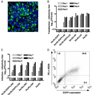
2a(J6)-FIG. 1. Deletions in NS5A domain II conferred efficient growth kinetics to 2a(J6)-EGFP and did not impair the viability of 2a(J6). (A) Huh7.5 cell cultures were transfected with RNA transcripts of 2a(J6)-EGFP. The image was obtained from the peak of infection at day 13 posttransfection using a TCS-SP2 Leica confocal microscope with a PL APO 40⫻objective. Green, EGFP; blue, cell nuclei counterstained with Hoechst dye. (B) Huh7.5 cell cultures were transfected in triplicates with RNA transcripts of the indicated recombinants. HCV infectivity titers in culture supernatants were determined on the indicated days posttransfection as FFU/ml. Titers are the means of 3 replicate determinations from the 3 replicate cultures (9 values total) with standard errors of the means (SEM); the lower cutoff was 2.3 log10FFU/ml, indicated by the axis break. (C) Huh7.5 cells were infected at an MOI of 0.01 with supernatants of cultures transfected with the indicated recombinants, obtained whenⱖ80% of the transfection culture was infected. HCV infectivity titers in culture supernatant were determined on the indicated days postinfection as FFU/ml. Titers are means of 3 replicates with SEM; the lower cutoff was 2.0 log10FFU/ml, indicated by the axis break. #, ORF sequences of viruses obtained fromⱖ80%-infected first-passage cultures are shown in Table S3 in the supplemental material. (D) Representative dot plot of cells infected with 2a(J6)-EGFP⌬40 at an MOI of 0.01, obtained on day 7 postinfection. Cells were prepared as described in Materials and Methods, and at least 104cells were gated on forward scatter/side scatter and analyzed for EGFP expression (xaxis) and intracellular NS5A (yaxis) by staining with a primary anti-NS5A antibody and a PE-conjugated secondary antibody. Numbers represent percentages of gated cells in each quadrant.
on November 7, 2019 by guest
http://jvi.asm.org/
[image:4.585.131.447.69.396.2]EGFP⌬25, 2a(J6)⌬40, and 2a(J6)⌬25 had not acquired nucleotide changes compared to the transfected recombinant (see Table S3 in the supplemental material). Also, 2a(J6)-EGFP⌬40 was genetically stable in four first passages, and one consecutive second passage, following four independent trans-fection experiments (Table S3).
We next investigated at which step of the viral life cycle the NS5A-EGFP insertion impaired the virus and how the identi-fied deletions rescued this impairment. To investigate the rep-lication efficacy and intracellular assembly of infectious viral particles independent of viral spread, we transfected CD81-deficient S29 cells (35) with 2a(J6) recombinants with the var-ious modifications. By comparing the amounts of intracellular core antigen at 48 h posttransfection, we found that⌬40 or⌬25 did not lead to a reduction of intracellular core amounts and, thus, replication efficacy, while the insertion of EGFP with or without the deletion led to a minor (2.5- to 4.5-fold) reduction of the replication efficacy (Fig. 2A). By comparing intracellular infectivity titers, we found that⌬40 or⌬25 did not impair the assembly of intracellular infectious particles (Fig. 2B). How-ever, the insertion of EGFP led to a 70-fold reduction in the level of intracellular infectious virus (Fig. 2B). This impair-ment of the assembly of intracellular infectious viral particles was almost completely rescued by⌬40 or⌬25, since 2a(J6)-EGFP⌬40 and 2a(J6)-EGFP⌬25 showed only 2.5-fold and 1.9-fold reductions in levels of intracellular infectivity (Fig. 2B). Extracellular infectivity titers varied, corresponding to intra-cellular infectivity titers (Fig. 2B).
Thus, either ⌬40 or ⌬25 in NS5A domain II allowed the insertion of EGFP C terminally in NS5A domain III of the 2a(J6) virus in vitro, resulting in reporter viruses with only minor impairments of viability compared to 2a(J6). The⌬40 or ⌬25 deletion rescued an impairment of intracellular virus as-sembly caused by the insertion of EGFP. Furthermore,⌬40 or ⌬25 did not affect the viability of nontagged 2a(J6).
In vivo, 2a(J6)-EGFP⌬40 acquired various deletions in the EGFP insert, while 2a(J6)⌬40 did not show impaired viability.
In order to investigate if 2a(J6)-EGFP⌬40 could be usedin vivo, we carried out infection studies of uPA-SCID mice
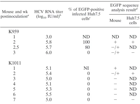
en-grafted with human hepatocytes. Following the inoculation of a first mouse (mouse K959) with 4 log10FFU of cell culture-derived, sequence-confirmed 2a(J6)-EGFP⌬40, serum HCV RNA titers of 3 to 6 log10IU/ml were detected between weeks 1 and 3 (Table 1). The inoculation of Huh7.5 cultures with serum indicated that EGFP was expressed at weeks 2 and 2.5 but not at week 3 (Table 1). Diagnostic RT-PCR, generating amplicons spanning the EGFP insert, and clonal analysis of the obtained amplicons indicated the presence of full-length EGFP in week 2 serum and derived cell cultures, while in week 3 serum and derived cultures, most genomes had acquired deletions in the EGFP insert (Table 1 and Fig. S1 and S2 in the supplemental material). Four different in-frame deletions ranging from 357 to 573 bp of the 717-bp EGFP insert were identified (Fig. S2). At week 3, when the animal was sacrificed, no intrahepatic EGFP expression was recorded by histological examination. In a second mouse (mouse K1011) inoculated with 4 log10 FFU of culture-derived, sequence-confirmed 2a(J6)-EGFP⌬40, serum HCV RNA titers of 5 to 5.5 log10
IU/ml were detected between weeks 1 and 7 (Table 1). Diag-nostic RT-PCR and clonal analysis indicated the presence of full-length EGFP in week 1 and week 2 serum and in the cell culture inoculated with week 2 serum; surprisingly, in this culture no green fluorescence was detected, possibly due to a coding point mutation in the EGFP sequence (Table 1 and Fig. S1). However, in week 3 to week 7 samples, apparently the majority of circulating viruses had acquired a 630-bp deletion in the EGFP insert (Table 1 and Fig. S1). At week 7 no intrahepatic EGFP expression was recorded by histological examination. Thus, even though 2a(J6)-EGFP⌬40 was able to initiate infectionin vivo, the EGFP insert was not maintained. In order to investigate the impact of⌬40 on viral viabilityin vivo, we inoculated one mouse (mouse K881) with 3.8 log10
[image:5.585.135.451.70.220.2]FFU of 2a(J6)⌬40. Serum HCV RNA titers of⬃6 log10IU/ml were measured at weeks 3 and 4. Sequence analysis of the complete ORF from week 3 serum showed that⌬40 was pre-served, even though several coding nucleotide changes were acquired in E2, NS2, NS5A, and NS5B (see Table S4 in the supplemental material). In conclusion, 2a(J6)-EGFP⌬40 was
FIG. 2. Deletions in NS5A domain II rescued the impairment of intracellular virus assembly caused by the insertion of EGFP. (A) HCV core levels were measured by ELISA after 48 h in CD81-deficient S29 cells transfected with the indicated recombinants. Values were normalized for transfection efficacy using 4-h core measurements and are means of single determinations from duplicate experiments with SEM. (B) Intra- and extracellular infectivity titers of S29 cells were determined at 48 h posttransfection. Intracellular titers are given per well of 4⫻105S29 cells. Titers are means of triple determinations of triplicate transfection experiments with SEM; the lower cutoff for intracellular infectivity titers was 1.7 log10 FFU/ml, and the lower cutoff for extracellular infectivity titers was 2.4 log10FFU/ml, both indicated by the axis break.
on November 7, 2019 by guest
not genetically stablein vivoin the uPA-SCID mouse model, where deletions in the EGFP insert were acquired. However, this was most probably not due to ⌬40, because 2a(J6)⌬40 was viable and did not acquire further deletions or insertionsin vivo.
In vitro,⌬40 in JFH1 NS5A domain II allowed the insertion of red fluorescent proteins or RLuc into NS5A domain III of 2a(J6) virus.In order to investigate if⌬40 allowed the engi-neering of viruses with different marker genesin vitro, we first inserted genes encoding the red fluorescent protein mCherry or DsRed-Express2 into NS5A domain III of 2a(J6)⌬40. Following the transfection of Huh7.5 cells, these recombi-nants spread as fast as 2a(J6) and infectedⱖ80% of cultured cells on day 4 posttransfection. In a subsequent first-passage kinetic experiment with infection at an MOI of 0.01, 2a(J6)-mCherry⌬40 spread in a manner comparable to that of 2a(J6), infecting ⱖ80% of cultured cells on day 5, with peak HCV RNA and infectivity titers of⬃7.5 log10IU/ml (not shown) and ⬃5 log10FFU/ml, respectively (Fig. 3A). The spread of
2a(J6)-DSRedExpress⌬40 was slightly delayed, with ⱖ80% of cul-tured cells infected on day 9, with peak HCV RNA and
infec-tivity titers of ⬃7.5 log10 IU/ml (not shown) and ⬃5 log10 FFU/ml, respectively (Fig. 3A). Sequence analysis of the ORF, including the sequencing of a diagnostic RT-PCR amplicon spanning the reporter insert, revealed that at the peak of infection, first-passage 2a(J6)-mCherry⌬40 and 2a(J6)-DSRedExpress⌬40 were genetically stable, without ev-idence of nucleotide changes compared to the transfected re-combinant.
Next, we inserted RLuc in NS5A domain III of 2a(J6)⌬40. After transfection, 2a(J6)-RLuc⌬40 spread as fast as 2a(J6), infecting ⱖ80% of cultured cells on day 4 posttransfection. Following infection at two different MOIs, 2a(J6)-RLuc⌬40 spread comparably to 2a(J6), infectingⱖ80% of cultured cells on day 9 following inoculation at an MOI of 0.001 and on day 5 following inoculation at an MOI of 0.01. Peak HCV RNA and HCV infectivity titers were 6.8 to 7.7 log10 IU/ml (not
shown) and 3.8 to 4.3 log10 FFU/ml, respectively (Fig. 3B). Luminescence signals above 3 log10RLU were detectable on
day 1 postinfection and gradually increased, reflecting the course of infection, up to nearly 7 log10RLU in cultures with
close to 100% HCV antigen-positive cells (Fig. 3C). No signal above the background was detected in cultures infected with nontagged 2a(J6) virus. In a total of three first-viral-passage experiments derived from independent transfections, 2a(J6)-RLuc⌬40 did not acquire coding changes in 2 experiments, while 2 coding changes occurring as a quasispecies together with the original sequence (50%/50%) were observed in one experiment (see Table S5 in the supplemental material). In addition, in a second viral passage, the presence of the com-plete RLuc gene was confirmed by the generation and se-quencing of a diagnostic RT-PCR amplicon spanning the RLuc insert. Thus,⌬40 in NS5A domain II, originally identi-fied after the insertion of EGFP in NS5A domain III of 2a(J6), allowed the insertion of alternative marker genes like red flu-orescent proteins and RLuc in NS5A domain III of 2a(J6).
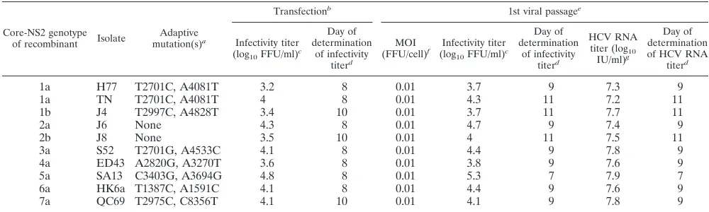
Development of a panel of JFH1-based EGFP marker vi-ruses with core-NS2 of HCV genotypes 1 to 7.Using a previ-ously developed panel of JFH1-based recombinants with core-NS2 of HCV genotype 1 to 7 reference isolates (6, 10, 11, 16, 38, 39), we aimed to develop an equivalent marker virus panel. Thus, we introduced⌬40 into domain II and EGFP into domain III of JFH1 NS5A of each of the core-NS2 recombinants of the following genotypes (isolates): 1a(H77), 1a(TN), 1b(J4), 2b(J8), 3a(S52), 4a(ED43), 5a(SA13), 6a(HK6a), and 7a(QC69). These recombinants had cell culture-adaptive mu-tations previously found to be required for efficient growth (see Materials and Methods) (Table 2) (10, 11, 16, 38, 39). Follow-ing transfection, EGFP-tagged recombinants of isolates of all genotypes spread toⱖ80% of cultured cells in 3 to 6 days. Peak infectivity titers were 3.2 to 4.8 log10 FFU/ml [lowest for
[image:6.585.42.283.89.268.2]1a(H77)-EGFP⌬40 and highest for 5a(SA13)-EGFP⌬40] (Ta-ble 2). In a viral passage kinetic experiment we infected cul-tures at an MOI of 0.01 with virus obtained from the transfec-tion experiment at the peak of infectransfec-tion (Fig. 4). When the percentages of HCV NS5A antigen-positive cells were evalu-ated by fluorescence microscopy, all marker virus recombi-nants spread toⱖ80% of cells between day 5 and day 9 [fastest for 5a(SA13)-EGFP⌬40 and slowest for 1a(H77)-EGFP⌬40 and 1b(J4)-EGFP⌬40] (Fig. 4A). Cultures were also evaluated by flow cytometry: 80 to 90% EGFP-positive cells were found
TABLE 1. Analysis of human liver chimeric mice inoculated with 2a(J6)-EGFP⌬40
Mouse and wk
postinoculationa HCV RNA titer
(log10IU/ml)
b
% of EGFP-positive infected Huh7.5
cellsc
EGFP sequence
analysis resultd
Mouse Huh7.5
cells
K959
1 3.0 ND ND ND
2 5.8 100 ⫹ ⫹
2.5 5.7 80 ⫺/⫹ ND
3 6.0 0 ⫺/⫹ ⫺
K1011
1 5.1 NI ⫹ ND
2 5.4 0 ⫺/⫹ ⫹
3 5.0 0 ⫺ ND
4 5.1 0 ⫺ ND
5 5.3 0 ⫺ ND
6 5.5 0 ⫺ ND
7 5.0 0 ⫺ ⫺
a
uPA-SCID mice engrafted with human hepatocytes were inoculated with 4
log10FFU of 2a(J6)-EGFP⌬40 derived from Huh7.5 cell culture supernatants.
Serum or plasma samples were obtained at the indicated weeks postinocula-tion. Mouse K959 was sacrificed at week 3, and mouse K1011 was sacrificed at week 7.
b
HCV RNA titers in mouse sera were determined as IU/ml as described in Materials and Methods; values are single measurements.
c
Huh7.5 cells (4⫻105
cells per well of a 6-well plate) were inoculated for 16 h
with 5l of mouse serum/plasma obtained at the indicated weeks
postinocula-tion. Values indicate the percentages of EGFP-expressing cells when the com-plete cell culture was infected, as determined by immunostaining for HCV NS5A antigen; cultures were evaluated by fluorescence microscopy. Viruses spread to
ⱖ80% of cultured cells in 9 to 15 days, yielding peak supernatant infectivity titers
of approximately 4 log10FFU/ml. NI, no infection was observed during 3 weeks
postinoculation, as determined by anti-NS5A staining; ND, not determined.
d
Nucleotides 7220 to 8455 of 2a(J6)-EGFP⌬40, spanning the complete EGFP
insert, were amplified from mouse serum/plasma or Huh7.5 culture supernatants by diagnostic RT-PCR using the specific primers indicated in Materials and
Methods.⫹, detection of 1,236-bp amplicons, indicating the presence of EGFP;
⫺, absence of 1,236-bp amplicons, with a detection of lower-molecular-mass
amplicons, indicating a partial deletion of EGFP;⫺/⫹, detection of amplicons of
different sizes, including the 1,236-bp amplicon, indicating a mixed population of viruses with either the complete EGFP present or partially deleted EGFP (see Fig. S1 in the supplemental material). Selected amplicons were cloned and sequenced (Fig. S1 and S2).
on November 7, 2019 by guest
http://jvi.asm.org/
between day 7 [5a(SA13)-EGFP⌬40] and day 14 [1a(H77)-EGFP⌬40 and 1b(J4)-EGFP⌬40] (Fig. 4B). Viral spread was reflected by a continuous increase in HCV RNA titers (Fig. 4C) and infectivity titers (Fig. 4D) in culture supernatants. Peak infectivity titers and respective RNA titers are shown in Table 2 and were comparable to those of the corresponding nontagged core-NS2 recombinants (11, 38). Sequence analysis of the ORF, including sequencing of a diagnostic RT-PCR am-plicon spanning the EGFP reporter insert, revealed that at the peak of infection, none of the first-passage marker viruses had acquired coding nucleotide changes (see Table S3 in the
supple-mental material). Thus, we developed a panel of JFH1-based EGFP-tagged viruses with core-NS2 of HCV genotype 1 to 7 isolates showing efficient growth characteristics in Huh7.5 cells.
Application of EGFP marker viruses in a fluorescence-based receptor-blocking assay. We previously demonstrated that infection by JFH1-based genotype 1 to 7 core-NS2 recom-binants was blocked by anti-CD81 antibody in a dose-depen-dent manner; these assays relied on immunostaining and the manual counting of infected cells (11). In order to develop a high-throughput fluorescence-based assay using flow cytometry measurements, we first established infection conditions
yield-FIG. 3.⌬40 in NS5A domain II allowed the insertion of genes encoding red fluorescent proteins orRenillaluciferase into NS5A domain III of the 2a(J6) virus. (A) Huh7.5 cells were infected at an MOI of 0.01 with supernatants from transfection cultures of 2a(J6)-mCherry⌬40, 2a(J6)-DSRedExpress⌬40, and 2a(J6) or from a first-viral-passage culture of 2a(J6)-EGFP⌬40, obtained whenⱖ80% of the respective cultures were infected. HCV infectivity titers in culture supernatants were determined as FFU/ml on the indicated days postinfection as means of 3 replicates with SEM; the lower cutoff was 1.8 log10FFU/ml, indicated by the axis break.䉬, culture was stopped because of cell death due to viral infection. #, ORF sequences of viruses obtained from ⱖ80%-infected cultures; results for 2a(J6)-EGFP⌬40 are shown in Table S3 in the supplemental material, and other results are described in the text. (B and C) Huh7.5 cells were infected at MOIs of 0.001 and 0.01 with supernatants from 2a(J6)-RLuc⌬40 transfection cultures, obtained whenⱖ80% of the respective cultures were infected. (B) HCV infectivity titers in culture supernatants were determined as FFU/ml on the indicated days postinfection as means of 3 replicates with SEM; the lower cutoff was 1.8 log10FFU/ml, indicated by the axis break. (C) For determinations of luminescence in RLU/well, 10
4cells were plated per well of a 96-well plate. On the following day, cells were lysed, and luminescence was measured directly on 96-well plates as described in Materials and Methods. Luminescence measurements are means of triplicates; the lower cutoff was 3 log10RLU, indicated by the axis break.䉬, culture was stopped because of cell death due to viral infection;F, not done; #, ORF sequence of viruses obtained from almost completely infected culture is shown in Table S5 in the supplemental material.
on November 7, 2019 by guest
[image:7.585.132.452.66.447.2]ing 10 to 30% infected cell cultures at the termination of the assay (see Materials and Methods). Following infection with 2a(J6)-EGFP⌬40 or 5a(SA13)-EGFP⌬40, selected for these studies, 11 to 28% of cultured cells expressed EGFP, as deter-mined by flow cytometry (Fig. 5A) and fluorescence micros-copy (data not shown), while low background measurements were obtained by flow cytometry of noninfected cultures (⬃0.5%). Treatment with anti-CD81 led to a dose-dependent reduction of the percentage of infected cells; at 2.5 g/ml, almost complete inhibition was observed, while little or no inhibition was observed at 0.02 and 0.004g/ml (Fig. 5). In contrast, treatment with a control antibody did not lead to an inhibition of infection (data not shown). Thus, EGFP-tagged viruses could be applied successfully in fluorescence-based studies of receptor interactions.
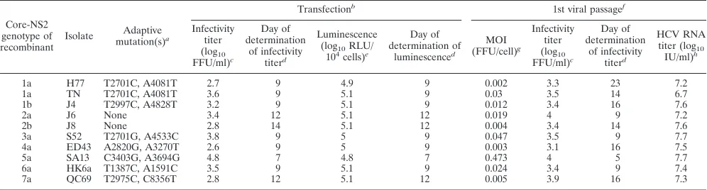
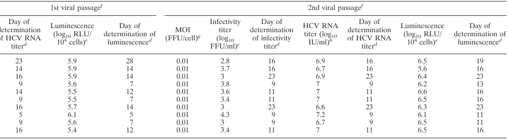
Development of a panel of JFH1-based RLuc marker viruses with core-NS2 of HCV genotypes 1 to 7. We constructed a panel of RLuc-NS5A-tagged JFH1-based recombinants with core-NS2 of HCV genotypes 1 to 7 and⌬40 in NS5A domain II. Following the transfection of Huh7.5 cells, an almost com-plete spread through the culture occurred in 7 to 14 days for viruses of all genotypes. Peak infectivity titers were 2.7 to 4.8 log10 FFU/ml [lowest for 1a(H77)-RLuc⌬40 and highest for
5a(SA13)-RLuc⌬40] (Table 3), thus being up to 10-fold lower than the infectivity titers of the respective nontagged viruses (10, 11, 16, 38, 39). At the peak of infection we measured a luminescence of ⬃5 log10RLU in lysates of 104 cells
trans-fected with RLuc-expressing recombinants, confirming the ex-pression and activity of the RLuc reporter (Table 3). In a subsequent first-viral-passage experiment, at the peak of infec-tion supernatant infectivity titers were 3.1 to 4.0 log10FFU/ml
[lowest for 4a(ED43)-RLuc⌬40 and highest for 2a(J6)-RLuc⌬40 and 5a(SA13)-RLuc⌬40] (Table 3). Luminescence
signals at the peak of infection were 5.4 to 6.1 log10 RLU
(Table 3). Direct ORF sequencing of first-passage viruses re-vealed one 50/50 coding nucleotide change for 1a(H77)- and 7a(QC69)-RLuc⌬40 in E1 and NS3, respectively. 2b(J8)-RLuc⌬40 showed one major and two 50/50 changes in E2, p7, and NS2, respectively. 3a(S52)-RLuc⌬40 acquired one 50/50 change and one major change in NS5B. No changes were observed for the other recombinants (see Table S5 in the supplemental material). In addition, for all first-passage vi-ruses, the presence of the complete RLuc gene was confirmed by the generation and sequencing of a diagnostic RT-PCR amplicon spanning the RLuc insert. For better comparisons of spread kinetics, we carried out a second-passage kinetic experi-ment, with inoculation at an MOI of 0.01. RLuc viruses spread almost to the complete culture in 9 to 23 days [fastest for 2a(J6)-RLuc⌬40, 5a(SA13)-RLuc⌬40, and 6a(HK6a)-RLuc⌬40 and slowest for 1b(J4)-RLuc⌬40 and 4a(ED43)-RLuc⌬40] (Table 3). Peak infectivity titers were 2.8 to 4.3 log10 FFU/ml [lowest for 1a(H77)-RLuc⌬40 and highest for 5a(SA13)-RLuc⌬40] (Table 3). Luminescence signals at the peak of infection were 5.6 to 6.5 log10RLU (Table 3). In addition, for all second-passage viruses,
[image:8.585.41.541.81.231.2]the presence of the complete RLuc gene was confirmed by the generation and sequencing of a diagnostic RT-PCR amplicon spanning the RLuc insert. However, apparently, for several recombinants, virus subpopulations were emerging that had partially deleted the RLuc insert, as determined by gel elec-trophoresis of diagnostic RT-PCR amplicons. The occur-rence of such in-frame deletions within the RLuc gene was confirmed by sequence analysis for 1a(H77)-RLuc⌬40, 1a(TN)-RLuc⌬40, 3a(S52)-RLuc⌬40, 5a(SA13)-RLuc⌬40, and 6a(HK6a)-RLuc⌬40 reduced-sized amplicons. In conclu-sion, we developed a panel of JFH1-based RLuc-expressing viruses with core-NS2 of HCV genotype 1 to 7 isolates.
TABLE 2. Characteristics of JFH1-based EGFP⌬40-NS5A recombinants expressing core-NS2 of genotype 1 to 7 prototype isolatesh
Core-NS2 genotype
of recombinant Isolate
Adaptive
mutation(s)a
Transfectionb
1st viral passagee
Infectivity titer
(log10FFU/ml)
c
Day of determination
of infectivity
titerd
MOI
(FFU/cell)f Infectivity titer
(log10FFU/ml)
c
Day of determination
of infectivity
titerd
HCV RNA
titer (log10
IU/ml)g
Day of determination of HCV RNA
titerd
1a H77 T2701C, A4081T 3.2 8 0.01 3.7 9 7.3 9
1a TN T2701C, A4081T 4 8 0.01 4.3 11 7.2 11
1b J4 T2997C, A4828T 3.4 10 0.01 3.7 11 7.7 11
2a J6 None 4.3 8 0.01 4.7 9 7.4 9
2b J8 None 3.5 10 0.01 4 11 7.5 11
3a S52 T2701G, A4533C 4.1 8 0.01 4.4 9 7.8 9
4a ED43 A2820G, A3270T 3.6 8 0.01 3.8 9 7.6 9
5a SA13 C3403G, A3694G 4.8 8 0.01 5.3 7 7.9 7
6a HK6a T1387C, A1591C 4.1 8 0.01 4.4 9 7.6 9
7a QC69 T2975C, C8356T 4.1 10 0.01 4.1 9 7.8 9
aCell culture-adaptive mutations required for efficient growth of the respective JFH1-based recombinant with the genotype/isolate-specific core-NS2 indicated by the
H77 absolute reference numbers (see Materials and Methods) (11).
bRNA transcripts of the indicated reporter recombinants were transfected in Huh7.5 cells.
cPeak HCV infectivity titers in culture supernatants were determined as FFU/ml as described in Materials and Methods; values are means of 3.
dDay posttransfection or postinfection of viral passage culture, at which time the respective values were obtained.
eViral passage was carried out by the inoculation of naı¨ve Huh7.5 cells at an MOI of 0.01 with supernatants derived from transfection experiments of the respective
reporter recombinants, whenⱖ80% of transfection culture cells were infected (see Materials and Methods).
fMultiplicity of infection used for the inoculation of viral passage cultures.
gHCV RNA titers in culture supernatants were determined as IU/ml as described in Materials and Methods; values are single measurements of the supernatants
yielding peak infectivity titers.
hEGFP was inserted into domain III of NS5A (immediately downstream of nucleotide 7511关amino acid 2390 by H77 absolute reference numbers兴) (31).⌬40 was
located in domain II of NS5A (nucleotides 7005 to 7136关amino acids 2222 to 2265 by H77 absolute reference numbers兴). The complete ORF of all EGFP-tagged viruses
was determined by using transfection or viral passage supernatants (see Table S3 in the supplemental material).
on November 7, 2019 by guest
http://jvi.asm.org/
Application of RLuc marker viruses in luminescence-based neutralization assays.Previously, we reported the differential sensitivities of genotype 1 to 7 HCV particles to neutralizing antibodies in sera of patients chronically infected with HCV. The assays relied on immunostaining and counting of infected
cells (11, 16, 34, 39). To develop a high-throughput lumines-cence-based assay, we first established the input dose of RLuc-tagged virus required to yield a luminescence signal above the background level. We used different doses of 5a(SA13)-RLuc⌬40, preincubated in culture medium without
neutraliz-FIG. 4. Spread kinetics of JFH1-based EGFP⌬40 marker viruses with core-NS2 of HCV genotypes 1 to 7. Huh7.5 cells were infected at an MOI of 0.01 with supernatants from transfection cultures, obtained whenⱖ80% of the respective cultures were infected. (A) Percentages of HCV NS5A antigen-positive cells were determined by immunostaining and fluorescence microscopy. (B) Percentages of EGFP-positive cells were determined by flow cytometry. (C) HCV RNA titers were determined by real-time RT-PCR; the lower cutoff was 3 log10IU/ml. (D) HCV infectivity titers in culture supernatants were determined as FFU/ml and are means of 3 replicates with SEM; the lower cutoff was 1.8 log10FFU/ml, indicated by the axis break.䉬, culture was stopped because of cell death due to viral infection;F, not done; #, ORF sequences of viruses obtained from ⱖ80%-infected cultures are shown in Table S3 in the supplemental material.
FIG. 5. Infection of Huh7.5 cells with EGFP-tagged genotype 2a and 5a core-NS2 recombinant viruses was inhibited by treatment with anti-CD81 antibodies in a fluorescence-based assay. A total of 6⫻103Huh7.5 cells per well of a 96-well plate, plated the previous day, were pretreated for 1 h with anti-CD81 (JS-81) at the indicated concentrations. Cultures were infected at MOIs of 0.6 and 1 with 2a(J6)-EGFP⌬40 and 5a(SA13)-EGFP⌬40, respectively, followed by incubation for 48 h. (A) Cells were prepared for and analyzed by flow cytometry as described in Materials and Methods. (B) Percent inhibition was calculated by relating the percentage of EGFP-expressing cells in treated cultures to the mean percentage of EGFP-expressing cells in 3 untreated cultures. Means of duplicates and SEM are shown. A control antibody specified in Materials and Methods did not show any inhibitory effect at the respective concentrations. The asterisk indicates values of⬍0.
on November 7, 2019 by guest
[image:9.585.135.449.72.345.2]ing antibodies, to infect replicate 96-well cultures. At 2 days postinfection a dose-dependent luminescence signal was de-tected (Fig. 6A), comparable to the relationship between FFU counts and input dose determined in a control experiment (Fig. 6B). We then used chronic-phase serum of a genotype 1a-infected patient (H06) to neutralize 500 FFU of 5a(SA13)-RLuc⌬40 or 5a(SA13). H06 serum was previously found to neutralize 5a(SA13) with an intermediate efficacy (11, 34, 39). For 5a(SA13)-RLuc⌬40 we found a dose-dependent titration of the luminescence signal with a reciprocal 50% neutraliza-tion titer of 100 (Fig. 6C and D). In a control experiment based on FFU counts, we obtained comparable results for 5a(SA13)-RLuc⌬40 and 5a(SA13) (Fig. 6E and F).
Previously, we found that nontagged genotype 1a(H77), 2a(J6), and 6a(HK6a) recombinants showed intermediate, low, and high sensitivities to neutralization with H06, respectively (11, 34, 39). To investigate if such differential sensitivities could be detected with the newly developed luminescence-based neutralization assay, we used H06 for the neutralization of 1a(TN)-RLuc⌬40, a genotype 1a recombinant that was not examined previously, as well as of RLuc⌬40 recombinants of genotypes 2a(J6) and 6a(HK6a) (Fig. 7). We found an inter-mediate sensitivity of 1a(TN)-RLuc⌬40, a low sensitivity of 2a(J6)-RLuc⌬40, and a high sensitivity of 6a(HK6a)-RLuc⌬40 recombinants with reciprocal 50% neutralization titers of 500 for 1a(TN)-RLuc⌬40 and 62,500 for 6a(HK6a)-RLuc⌬40; 50% neutralization of 2a(J6)-RLuc⌬40 was not achieved. Thus, we showed that RLuc-tagged viruses could be applied to lumines-cence-based neutralization assays.
RLuc marker viruses with core-NS2 of genotype 1 to 7 iso-lates showed similar sensitivities to treatment with IFN-alfa-2b.Previously, we reported similar sensitivities of JFH1-based recombinants with core-NS2 specific for genotypes 1 to 6 to treatment with a high dose of IFN-alfa-2b (500 IU/ml) (11). This assay relied on an estimation of the percentage of HCV NS5A-expressing cells by fluorescence microscopy following immunostaining and/or determinations of HCV RNA titers in cell culture supernatants. We now used the developed RLuc marker viruses to establish a high-throughput luminescence-based treatment assay, allowing the testing of a broader dose-range (see Materials and Methods). When treating genotype 1 to 7 RLuc marker viruses with IFN-alfa-2b using an 8-fold dilution series starting at 500 IU/ml, we found a depen-dent effect and no major differences between the fitted dose-response curves for viruses of various genotypes (Fig. 8). The EC50ranged from 0.44 to 0.89 IU/ml [lowest for
6a(HK6a)-RLuc⌬40 and highest for 4a(ED43)-RLuc⌬40]. The EC50 ob-tained for 2a(J6)-RLuc⌬40 was comparable to the EC50
pre-viously obtained for nontagged 2a(J6) in similar treatment assays relying on HCV antigen immunostaining and the auto-mated counting of single infected cells (40). In these assays, in 8 independent experiments with 3 replicates each, the EC50for
2a(J6) ranged from 0.18 to 1.2 IU/ml (40) (J. M. Gottwein et al., unpublished data).
RLuc marker viruses with core-NS2 of genotype 1 to 7 iso-lates were susceptible to treatment with the p7 inhibitors rimantadine and NN-DNJ. It was previously reported that putative p7 inhibitors were effective against HCV
recombi-TABLE 3. Characteristics of JFH1-based RLuc⌬40-NS5A recombinants expressing core-NS2 of genotype 1 to 7 prototype isolatesi
Core-NS2 genotype of recombinant Isolate Adaptive mutation(s)a Transfectionb
1st viral passagef
Infectivity titer (log10 FFU/ml)c Day of determination of infectivity titerd Luminescence
(log10RLU/
104cells)e
Day of determination of luminescenced MOI (FFU/cell)g Infectivity titer (log10 FFU/ml)c Day of determination of infectivity titerd HCV RNA
titer (log10
IU/ml)h
1a H77 T2701C, A4081T 2.7 9 4.9 9 0.002 3.3 23 7.2
1a TN T2701C, A4081T 3.6 9 5.1 9 0.03 3.5 14 6.7
1b J4 T2997C, A4828T 3.2 9 5.1 9 0.012 3.4 16 7.6
2a J6 None 3.4 12 5.1 12 0.019 4 9 7.2
2b J8 None 2.8 14 5.1 12 0.004 3.4 14 7.6
3a S52 T2701G, A4533C 3.8 9 5 9 0.047 3.5 9 7.7
4a ED43 A2820G, A3270T 2.6 9 5 9 0.003 3.1 16 7.5
5a SA13 C3403G, A3694G 4.8 7 4.8 7 0.473 4 5 7.7
6a HK6a T1387C, A1591C 3.5 9 5.1 9 0.024 3.4 9 7.4
7a QC69 T2975C, C8356T 2.8 12 5.1 12 0.005 3.9 16 7.3
a
Cell culture-adaptive mutations required for efficient growth of the respective JFH1-based recombinant with genotype/isolate-specific core-NS2 indicated by H77 absolute reference numbers (see Materials and Methods) (11).
b
RNA transcripts of the indicated reporter recombinants were transfected in Huh7.5 cells.
c
Peak HCV infectivity titers in culture supernatants were determined as FFU/ml as described in Materials and Methods; values are means of 3.
d
Day posttransfection or postinfection of the viral passage culture, at which time the respective value was obtained.
e
For determinations of luminescence as RLU/104
cells, cells were pelleted by centrifugation for 5 min at 13,000⫻g. For transfection and the first viral passage, cell
pellets were stored at⫺80°C until cell lysis and measurements of luminescence as described in Materials and Methods. For the second viral passage, measurements
were done immediately. Compared to RLU values in Fig. 3 and compared to second-viral-passage RLU values, overall lower RLU values were observed for transfection and the first viral passage, which were most probably due to the different handling procedures (also see the Fig. 3 legend). Values are single measurements for transfection and the first viral passage, and second-viral-passage values are means of 3 replicates. For transfection and first-viral-passage experiments, luminescence was
determined when the virus had spread toⱖ80% of cultured cells. For the second viral passage, peak luminescence measurements are given.
f
Viral passage was carried out by the inoculation of naı¨ve Huh7.5 cells at the indicated MOIs with supernatant virus stocks. These supernatants were derived from
transfection cultures (for the first viral passage) or first-viral-passage cultures (for the second viral passage) of the respective reporter viruses, whenⱖ80% of cultured
cells were infected (see Materials and Methods).
g
Multiplicity of infection used for the inoculation of viral passage cultures.
h
HCV RNA titers in culture supernatants were determined as IU/ml as described in Materials and Methods; values are single measurements of the supernatants yielding peak infectivity titers.
i
RLuc was inserted into domain III of NS5A (immediately downstream of nucleotide 7511关amino acid 2390 by H77 absolute reference numbers兴) (31).⌬40 was
located in domain II of NS5A (nucleotides 7005 to 7136关amino acids 2222 to 2265 by H77 absolute reference numbers兴). The complete ORF of all RLuc-tagged viruses
was determined by using viral passage supernatants (see Table S5 in the supplemental material).
on November 7, 2019 by guest
http://jvi.asm.org/
[image:10.585.43.541.82.216.2]nants with p7 of genotype 1 to 3 isolatesin vitro(13, 43). To evaluate the efficacy of p7 inhibitors against RLuc viruses with core-NS2 of genotypes 1 to 7, we applied two compounds exhibiting different mechanisms of action in the established treatment assay. Rimantadine is an allosteric p7 ion channel inhibitor. NN-DNJ is a p7 ion channel oligomerization inhib-itor, which in addition inhibits alfa-glucosidases, leading to an altered glycosylation of the HCV envelope proteins and the impairment of entry (43). When carrying out treatment assays with rimantadine using a 3-fold dilution series starting at 125 M, we observed a dose-dependent effect on all genotype 1 to 7 RLuc marker viruses (Fig. 9A). Also, NN-DNJ used in a 3-fold dilution series starting at 125M had a dose-dependent effect on viruses of all genotypes (Fig. 9B). We did not fit dose-response curves, because the applied maximum doses did not lead to a complete suppression of RLuc activity in the treated cultures, making curve fitting unreliable. Rimantadine and NN-DNJ at concentrations of⬎150M could not be used due to cytotoxicity. The reliability of the results obtained with the luminescence-based treatment assay was confirmed by a similar treatment assay relying on HCV-NS5A immunostain-ing and the automated countimmunostain-ing of simmunostain-ingle infected cells (40). Such control experiments were carried out with rimantadine and NN-DNJ on nontagged 2a(J6)- and 3a(S52)-infected cul-tures and yielded similar results, as shown in Fig. 9.
DISCUSSION
In this study, we identified two in-frame deletions coding for 40 and 25 amino acids (⌬40 and⌬25, respectively) in domain II of HCV NS5A that rescued the impairment of the assembly of intracellular viral particles caused by the insertion of marker genes in domain III of NS5A, thus leading to efficient HCV J6/JFH1 [2a(J6)] reporter viruses. This finding paved the way to the development of panels of JFH1-based reporter recom-binants with core-NS2 of reference isolates of genotypes 1 to 7, expressing either an EGFP⌬40- or an RLuc⌬40-NS5A fusion protein. We demonstrated that these viruses could be applied
in vitro to high-throughput fluorescence- or luminescence-based assays for studies of HCV-receptor interactions and HCV-neutralizing antibodies. In luminescence-based treat-ment assays, we found that RLuc viruses with core-NS2 of genotypes 1 to 7 responded similarly to IFN-alfa-2b. We also found that genotype 1 to 7 RLuc viruses all responded to
treatment with two p7 ion channel inhibitors with different mechanisms of action. These novel culture systems will permit further extensive genotype-specific studies of the HCV life cycle and agents interfering with the function of core-NS2.
We elected to develop a panel of monocistronic reporter viruses using an insertion site in NS5A domain III (down-stream of sequences encoding aa 2390), previously identified to allow the insertion of GFP into the Con-1 replicon (31). Var-ious modifications, such as deletions and insertions in NS5A domain III, did not have a major impact on the RNA replica-tion of the Con-1 replicon and of genotype 2a recombinant JFH1 as well as JFH1-based intragenotypic recombinants J6/ JFH1 and Jc1 (4, 5, 25, 28, 29, 45, 46). However, certain sequences of NS5A domain III and especially its C-terminal portion were shown previously to be important for the produc-tion of infectious JFH1, J6/JFH1, and Jc1 viruses (5, 28, 45). In accordance with these findings, viral viability was impaired, with a significant reduction of infectivity titers, when GFP or RLuc was inserted into NS5A domain III of the JFH1 virus at the same position as that used in our study (18, 28). Further-more, the insertion of green and red fluorescent proteins in NS5A domain III of the Jc1 virus at a site upstream of the site chosen in this study led to a⬃50-fold reduction of supernatant infectivity titers and an impairment of viral spread without a major impairment of the replication capacity (5, 37). Also, the insertion of EGFP into JFH1 at a site downstream of the site chosen in this study led to an impairment of infectivity and viral spread (50). The genetic stability of such previously de-veloped reporter viruses was not examined. In this study, fol-lowing the transfection of RNA transcripts, 2a(J6)-EGFP showed similar numbers of replicating cells but impaired viral spread compared to that of 2a(J6), indicating that the EGFP insert in NS5A domain III did not impair replication but the production of infectious virus. Experiments with CD81-defi-cient S29 cells confirmed that recombinants with the EGFP insert with or without a deletion in NS5A domain II showed only a minor reduction of the replication efficiency. In contrast, the insertion of EGFP resulted in a significant impairment of the assembly of intracellular infectious viral particles. This impairment was apparently overcome by a deletion (⌬40 or ⌬25) in domain II of NS5A (Fig. 2), which highlighted the adaptability of the HCV genome. Thus, the developed EGFP or RLuc reporter viruses with⌬40 or⌬25 were characterized overall by relatively high peak infectivity titers and efficient
on November 7, 2019 by guest
[image:11.585.46.542.81.217.2]viral spread. How deletions in NS5A domain II rescued defects in the intracellular assembly of viral particles and, thus, the viability of recombinants with insertions in domain IIIin vitro
remains to be studied. Possibly, the identified deletions could restore the NS5A conformation required for RNA binding or interactions with other viral and/or host proteins, which was perturbed by the insertion. Alternatively, the maximal length of the HCV genome might be limited due to the nature of the packaging process. We demonstrated that 2a(J6) recombinants with the identified deletions (⌬40 and⌬25), which permitted the development of efficient marker virus culture systems, were fully viablein vitro(Fig. 1 and 2). It was previously reported that even larger deletions N terminally in NS5A domain II of Jc1 were permissiblein vitroand did not lead to a major
impairment of the replication or production of infectious virus (5).
It would be of major interest to use reporter viruses forin vivostudies in order to determine the pattern of hepatic HCV infection, to localize HCV in single hepatocytes, and to identify extrahepatic sites of HCV infection. Previously developed re-porter viruses were not testedin vivo. In this study, after ob-taining ample evidence for the genetic stability of 2a(J6)-EGFP⌬40in vitro(see Table S3 in the supplemental material), we inoculated human liver chimeric mice with cell culture-derived virus. However, 2a(J6)-EGFP⌬40 turned out to be genetically unstable in thisin vivomodel (Table 1 and Fig. S1 and S2). Further studies are needed to elucidate if intrahepatic 2a(J6)-EGFP⌬40 could be detected in human liver chimeric
FIG. 6. Establishment of a luminescence-based neutralization assay using RLuc-tagged genotype 5a core-NS2 recombinant virus. (A and B) A total of 6⫻ 103 Huh7.5 cells per well of a 96-well plate, plated the previous day, were infected for 3 h with indicated doses (FFU) of 5a(SA13)-RLuc⌬40, preincubated for 1 h at 37°C. After 48 h, cell cultures were lysed on 96-well plates, and luminescence was determined as RLU/well (A), or cell cultures were immunostained for NS5A antigen, followed by determinations of the numbers of FFU/well (B). (C to F) Five hundred FFU 5a(SA13)-RLuc⌬40 or 5a(SA13) virus were preincubated with chronic-phase heat-inactivated serum of a genotype 1a-infected patient (H06) at the indicated dilutions for 1 h at 37°C. Virus-serum mixes were used to infect cell cultures on 96-well plates for 3 h. After 48 h, cell cultures were lysed, and luminescence was determined as RLU/well (C and D), or cell cultures were immunostained for NS5A antigen, followed by determinations of FFU/well (E and F). Percent neutralization was calculated by a comparison of values obtained for serum-pretreated cultures with the means of values obtained for 6 nontreated cultures (D and F). In A, B, C, and E, the origin of theyaxis reflects the mean background RLU and FFU measurements. Means of triplicates and standard errors of the means are shown.*, no neutralization was observed.
on November 7, 2019 by guest
http://jvi.asm.org/
mice shortly after inoculation, possibly allowing an assessment of early infection kinetics. Such studies can eventually be con-ducted with the chimpanzee model. We showed that ⌬40 in domain II did not impair the viability of 2a(J6) in human liver chimeric mice. However, circulating viruses acquired several coding nucleotide changes, predominantly in NS2, which could be compensating for⌬40. However, data on the genetic sta-bility of cell culture-derived viruses following the inoculation of human liver chimeric mice are limited.
We found that different sequences could be inserted into JFH1 NS5A domain III of HCV recombinants with ⌬40 in NS5A domain II. In 2a(J6) marker viruses we detected red fluorescent proteins, mCherry and DsRed-Express2, by fluo-rescence microscopy. As previously described for DSRed-Ex-press (44), an indication of the formation of intracellular ag-gregates was observed. Using⌬40, we constructed a panel of EGFP marker viruses with a genotype-specific core-NS2 se-quence showing spread kinetics and infectivity titers similar to those of the respective nontagged viruses (10, 11, 16, 38, 39). The presence of EGFP allowed us to monitor viral spread directly by fluorescence microscopy and flow cytometry (Fig. 1D and 4B). Viral spread determined by flow cytometry ap-peared to be delayed compared to estimates obtained by eval-uations of HCV-NS5A immunostained cells by fluorescence microscopy (Fig. 1D and 4A and B). This was most likely due to the fact that even though cells were trypsinized and seeded
FIG. 7. RLuc-tagged genotype 1a, 2a, and 6a core-NS2 recombinant viruses showed differential sensitivities to neutralization. A total of 250, 500, and 375 FFU of 1a(TN)-RLuc⌬40, 2a(J6)-RLuc⌬40, and 6a(HK6a)-RLuc⌬40, respectively, were preincubated with heat-inactivated H06 serum at the indicated dilutions for 1 h at 37°C. (A to C) Virus-serum mixes were used to infect 6⫻103Huh7.5 cells, plated the previous day onto 96-well plates, for 3 h. After 48 h, cell cultures were lysed, and luminescence was determined as RLU/well. (D) Percent neutralization was calculated by comparisons of values obtained with serum-pretreated cultures with the means of values obtained for 6 nontreated cultures. In A, B, and C, the origin of theyaxis reflects the mean background RLU measurements. Means of triplicates and standard errors of the means are shown.
FIG. 8. RLuc marker viruses with core-NS2 of genotype 1 to 7 isolates showed similar sensitivities to IFN-alfa-2b. A total of 5⫻103 Huh7.5 cells per well of a 96-well plate, plated the previous day, were infected for 24 h with second-passage virus stocks of the indicated RLuc marker viruses at MOIs of 0.02 to 0.2. IFN-alfa-2b, used in an 8-fold dilution series starting at 500 IU/ml, was administered at 24 and 48 h postinfection; each dose was tested in triplicates. At 72 h postin-fection, cell cultures were lysed on 96-well plates, and luminescence was determined as RLU/well. The “percent RLU related to non-treated” value was calculated by a comparison of RLU values to the mean RLU values of six nontreated cultures. After a logarithmic trans-formation of dose values, a sigmoidal dose-response curve was fitted as described in Materials and Methods. Error bars indicate SEM. Median EC50values are shown for each genotype (isolate).
on November 7, 2019 by guest
onto chamber slides on the days indicated in Fig. 4A, fixation and evaluation were carried out on the consecutive day. In contrast, cells were trypsinized, fixed, and evaluated by flow cytometry on the same day (Fig. 4B). When flow cytometry was used to detect the expression of EGFP directly as well as the expression of NS5A following intracellular staining, during the phase of exponential virus spread, similar percentages of in-fected cells were obtained (Fig. 1D). In addition, EGFP-ex-pressing cells were also stained positive by intracellular stain-ing with an anti-EGFP antibody, as analyzed by flow cytometry. Thus, it is unlikely that the EGFP maturation time or the low sensitivity of direct detection of EGFP by flow cytometry ac-counted for the differences in viral spread kinetics apparent in Fig. 4A and B. For a panel of RLuc viruses with genotype 1 to
7 core-NS2, peak infectivity titers were 0.5 to 1 log10 lower (Table 3), and spread kinetics were delayed compared to those of the respective nontagged viruses (10, 11, 16, 38, 39). Also, while genetically stable in first-passage experiments, in second-passage experiments, virus subpopulations that had deleted the RLuc insert were emerging for several RLuc recombinants. The lower efficacy of RLuc viruses than fluorescent marker viruses could be caused by a greater length of the RLuc protein (311 aa) than those of EGPF (239 aa), mCherry (236 aa), and DsRed-Express2 (225 aa) or its specific RNA or protein struc-ture. The occurrence of the RLuc deletion in several second-passage RLuc viruses could be due to a lower fitness of RLuc viruses. However, it cannot be excluded that deletions might also occur for EGFP marker viruses upon serial viral passage. The fact that the developed reporter viruses are readily detectable by fluorescence- or luminescence-based techniques allows their application in high-throughputin vitroassays. Here we showed that the developed reporter viruses could be used in such assays to study the effects of antibodies blocking HCV coreceptor CD81 (Fig. 5) and of neutralizing antibodies pres-ent in a chronic-phase serum sample from genotype 1a-in-fected patient H (H06) (Fig. 6 and 7). While results of CD81 blocking were comparable overall to results achieved previ-ously with nontagged recombinants using assays that relied on immunostaining and counting of FFU (11, 16), we observed a lower efficacy of neutralization than those previously reported (11, 16, 34, 39). This may be due to the relatively high infec-tious dose used here to achieve signals several orders of mag-nitude above background measurements. Thus, in cases of a limited availability of material such as serum, it might be ben-eficial to further optimize the described assays, e.g., by pro-longing the infection/incubation periods for neutralization as-says, allowing a smaller amount of input virus. With the established luminescence-based neutralization assay, we con-firmed that viruses of different genotypes showed differential sensitivities to neutralization. Genotype 1a isolate TN showed intermediate sensitivity, as previously found for genotype 1a isolate H77 (11, 34, 39). This observation strengthens the hy-pothesis that different HCV serotypes might exist. The devel-oped reporter virus systems could be used to further investi-gate this hypothesis, which would require high-throughput studies employing various culture-derived isolates of each ge-notype and a large panel of sera from patients infected with different HCV genotypes.
[image:14.585.58.266.68.429.2]Even though certain sequence variations in the core-NS2 region were suggested previously to have an influence on the efficacy of interferon treatment (1, 2, 8), RLuc recombinants with core-NS2 sequences specific to genotypes 1 to 7 showed similar responses to treatment with IFN-alfa-2b in the estab-lished high-throughput assay. Thus, other HCV genome re-gions might mediate differences seen in patients, or factors not present in the applied short-term assay might influence treat-ment outcomes. Previously, rimantadine and NN-DNJ were described to be effective against JFH1-based recombinants ex-pressing core-NS2 sequences of 1a(H77), 1b (Con1), 2a(JFH1), 2a(J6), and 3a(452) (13, 43). We now report an antiviral effect of these compounds on recombinants expressing core-NS2 proteins of all major HCV genotypes and the important sub-types 1b and 2b. In this study, slightly higher doses, especially of NN-DNJ, were required to achieve treatment effects similar
FIG. 9. RLuc marker viruses with core-NS2 of genotype 1 to 7 isolates responded to p7 inhibitors in a dose-dependent manner. A total of 5⫻103Huh7.5 cells per well of a 96-well plate, plated the previous day, were infected for 24 h with second-passage virus stocks of the indicated RLuc marker viruses at MOIs of 0.02 to 0.2. Riman-tadine (A) or NN-DNJ (B) used at 14, 42, and 125M was adminis-tered at 24 and 48 h postinfection; each dose was tested in triplicates. At 72 h postinfection, cell cultures were lysed on 96-well plates, and luminescence was determined as RLU/well. The “percent RLU related to nontreated” value was calculated by a comparison of RLU values to the mean RLU values of six nontreated cultures. Means of triplicates and SEM are shown.