A dissertation on
EVALUATION OF CAUSES OF ACUTE PANCREATITIS – A RETROSPECTIVE STUDY
Dissertation submitted to
THE TAMIL NADU DrM.G.R. MEDICAL UNIVERSITY CHENNAI, TAMIL NADU
With partial fulfilment of the regulations required for the award of degree of
M.S. GENERAL SURGERY BRANCH- I
COIMBATORE MEDICAL COLLEGE, COIMBATORE
DECLARATION
I solemnly declare that the dissertation titled “ EVALUATION
OF CAUSES OF ACUTE PANCREATITIS – A RETROSPECTIVE STUDY” was done by me from 2016 onwards under the guidance and supervision of PROF. DR. D.N. RENGANATHAN , M.S
This dissertation is submitted to the Tamilnadu Dr. M.G.R Medical
University towards the partial fulfillment of the requirement for the award
of M.S Degree in General Surgery (Branch I).
PLACE: DR. BALAJID.J.
CERTIFICATE
Certified that this is the bonafide dissertation done by
DR.BALAJID.J. and submitted in partial fulfillment of the requirement for the Degree of M.S. General Surgery, Branch I of the Tamilnadu Dr.
M.G.R. Medical University , Chennai.
DATE: UNIT CHIEF
DATE: PROFESSOR &HOD
Department Of General Surgery
DATE:
DEAN
Coimbatore Medical College
ACKNOWLEDGEMENT
I owe my reverential gratitude and humble thanks to Lord God
Almighty for all his mercy, for being with me and showering abundant
blessing upon me throughout the course of the study.
I am obliged to record my immense gratitude to
Dr.B.ASHOKANMch, The Dean , Coimbatore Medical College Hospital for providing all the facilities to conduct the studies.
I express my deep sense of gratitude and heartfelt thanks to
Professor Dr. V. ELANGO, M.S, Head of Department of General
Surgery for his dynamic guidance , constant help and encouragement
throughout the study.
I express my respectful gratitude and indebtedness to my guide
Professor Dr. D.N. RENGANATHAN, M.S, for his valuable guidance
and support.
I would like to express my sincere thanks to Professor
professors of surgery, for all the needful help they have provided for the
study.
I acknowledge my gratitude to our Registrar
Dr.NarayanamoorthyM.S and all my assistant professors of Department
of surgery for their encouragement and support.
I am thankful to The ETHICAL COMMITTEE of Coimbatore
Medical College for permitting me to proceed with this dissertation.
Lastly I am grateful to all the patients whose cooperation made this
work possible.
DATE: Signature of the Candidate
TABLE OF CONTENTS
S.No CONTENTS PAGE NO
1. INTRODUCTION 1
2. AIMS AND OBJECTIVES 3
3. REVIEW OF LITERATURE 4
4. METHODOLOGY 54
5. OBSERVATION AND ANALYSIS 57
6. DISCUSSION 77
7. CONCLUSION 81
8. BIBLIOGRAPHY 83
9. ANNEXURES
PROFORMA 87
LIST OF FIGURES
1. ANATOMY OF PANCREAS
2. DEVELOPMENT OF PANCREAS
3. PATHOPHYSIOLOGY OF PANCREATITIS
4. ACUTE PANCREATITIS SPECIMEN
5. X RAY SHOWING COLON CUT OFF SIGN
6. SEVERE NECROTIC PANCREATITIS
LIST OF ABBREVIATIONS USED
1. BISAP - Beside index of severity in acute pancreatitis
2. CTSI - CT severity index
3. SAP - Severe acute pancreatitis
4. PAN NEC - Pancreatic necrosis
5. CEA - carcinoembryonic antigen
6. SD - Stadard deviation
1
INTRODUCTION
Acute pancreatitis has become a common disease among the new
world population due to the increase in the socio-economic factors
affecting the general population which has led to an increase in risk
factors associated with the onset of acute pancreatitis. Acute pancreatitis
has become a formidable cause of mortality and morbidity in recent times
due to its sheer clinical features and debilitating symptoms. The increased
incidence of acute pancreatitis can be attributed to many factors and
hence it is usually caused by multifactorial risk factors which combine
from the initial damage to full blown symptoms of acute pancreatitis in a
span of few years.
The various causes attributed to the onset of acute pancreatitis has
been studied extensively and has led to an intensive research in
prevention of progression of acute pancreatitis to full blown chronic
pancreatitis leading to increased mortality and debilitating illness caused
due to complete destruction of the pancreas anatomy.
Acute pancreatitis has become a treatable entity nowadays due to
the advancement of research and study of various treatment modalities
which has been found effective in curing the symptoms and halting the
progression of the destruction of pancreas parenchyma. The treatment
2
offer a permanent solution for the treatment of this condition but they
prevent progression of the destruction of the pancreas and allows the
body to rejuvenate from the systemic illness caused by the inflammation
of the pancreas.
The only effective treatment modality which has been discovered
till date is the removal of the risk factors which initiated the event of
acute inflammation in the pancreas. Most commonly the incidence of gall
stones has increased which has led to an increased incidence of acute
pancreatitis and hence gall stones have been named as the most common
cause of acute pancreatitis. Hence the treatment has been very simplified
in terms of Endoscopic Retrograde Cholangio-Pancreaticography (
ERCP) and retrieval of the CBD stones which leads to the removal of
obstruction of pancreatic duct thereby relieving the inflammation of the
pancreas.
The risk factors of acute pancreatitis should be eliminated from the
initial stages of the disease to prevent disease progression and it has been
proven to be the most effective treatment modality for acute pancreatitis.
Evaluation of risk factors of pancreatitis has become a pivotal point in the
research of the field of pancreatic pathology and hence this study has
been undergone to evaluate various risk factors of acute pancreatitis and
3
AIMS AND OBJECTIVES OF THE STUDY
1. TO EVALUATE THE CAUSES OF ACUTE PANCREATITIS AND TO IDENTIFY THE COMMON CAUSES LEADING TO ACUTE PANCREATITIS IN OUR STUDY
POPULATION.
2. TO IDENTIFY THE SEVERITY OF ACUTE
4
REVIEW OF LITERATURE
HISTORICAL REVIEW:
In Greek Pan means “all” and Kreas means “Flesh” and it was
described first by Herophilus Chalcedon.
Rufees and Ephesees – named the pancreas.
Wirsung – described the main pancreatic duct.
Santorini – illustrated the accessory duct bearing his name.
Ringor de Graaf – gave the first reference to pancreatic lithiasis in
1664.
Reiddle – described chronic pancreatitis.
Kini – reported the first case of pancreatic calculi in India.
Elizabeth and Stephen – reported 9 cases of pancreatic calculi from
vellore.
Chuttani and Anand – reported 32 cases of pancreatitis from North
India.
Brocks and Gifferd – performed the first human homotransplant in
1959 , using fragmented pancreatic tissue.
Lillehei and collegues – performed the first human pancreatic
whole organ transplant in 1967.
Ballinger and Lacy – popularized the concept of islet cell
5
ANATOMY OF THE PANCREAS
Pancreas is a retroperitoneal organ and it lies behind the stomach ,
transverse colon and the mesocolon. The whole organ measures over 15
cm long weighs about 90 to 120g in adult, soft in consistency with
lobulated surface. It occupies the supracolic and partly the infracolic
compartment. It comprises of the head , neck , body and tail.
Head :
It is the broadest part , occupies the concavity of the duodenum ,
and lies over the inferior venacava , right and left renal veins. Its posterior
surface is indented by last part of the common bile duct. The uncinate
process is the wedge shaped lower part of the gland , lies posterior to the
superior mesenteric artery and vein and lies anterior to the aorta , at the
level of L2.
Neck :
It is the continuation of the upper part of the head , lies anterior to
the superior mesenteric vein and portal vein formation. It lies at the level
6
FIG 1 ANATOMY OF PANCREAS
Body :
The body starts from the neck and it runs across the left renal vein ,
left crus of the diaphragm, aorta, left psoas muscle, hilum or the left
kidney and the lower pole of the left supra renal gland. The Splenic artery
passes along the upper body of the tail and it is tortuous in its course.
Splenic vein gets its tributary from the inferior mesenteric vein behind the
body of the pancreas. The transverse mesocolon is attached in the lower
7
Tail :
It passes forward from the anterior surface of the left kidney at the
level of the hilum , accompanied by the splenic artery , splenic vein and
lymphatics in the two layer of lieno renal ligament and then touches the
hilum of the spleen.
Ductal system of the pancreas :
The duct of Wirsung is the major duct comes from the tail to the
head , arises from the confluence of numerous small ducts of the lobules
crossing the gland forming a “Herring bone” pattern , gradually
increasing in diameter upto 10mm joins with the common bile duct in a
dilatation , the ampulla of Vater , which opens into the duodenal papilla.
The accessory pancreatic duct drains the uncinated process and lower part
of the head of pancreas lies more on ventral plane , opens into the
duodenum 2 cm proximal to the major papilla and 7 cm distal to the
pylorus. Injury to the duct of Santorini in the pancreatic divisum during
gastrectomy results in severe hemorrhagic or recurrent pancreatitis.
Blood Supply :
Blood supply is chiefly derived from the splenic artery which
8
pancreatica magna “. The head is supplied by superior pancreatico
duodenal artery ( a branch of superior mesenteric artery ) . The right
hepatic artery is a branch of superior mesenteric artery , passes behind the
head of the pancreas or within the substance.
Venous drainage is by small veins into the splenic vein and the
head of the pancreas drains into the superior pancreatico duodenal vein
into the superior mesenteric vein which forms a landmark during
pancreatic dissection.
Lymphatic drainage :
Lymphatic drainage generally follows venous drainage in all
directions.
They drain into the following group of lymph nodes :
A. Superior nodes drain the anterior and superior half of the gland.
B. Inferior nodes drain the anterior and posterior lower half.
C. Anterior nodes drain the anterior surface of the head of the
pancreas.
D. Posterior nodes drain the posterior surface of the head.
9
Every group of lymph nodes finally drains into the coeliac and superior
mesenteric group of lymph nodes.
Nerve supply :
The afferent pain sensation from the pancreas is conducted through
the sympathetic fibres from the greater , lesser and lowest splanchnic
nerves via the central ganglia. The celiac branch of the right vagus nerve
provides the para sympathetic supply.
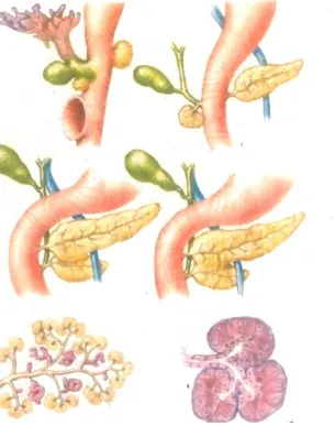
Development of the pancreas :
Pancreas develops as two separate buds each an outgrowth of the
endoderm at the junction of foregut and midgut. The ventral bud grows
into the ventral mesogastrium in common with the outgrowth of bile duct
and the dorsal bud grows into the dorsal mesogastrium. The duodenal
portion of the duct rotates and becomes adherent to the posterior
abdominal wall , the ventral bud rotates and fuses with the dorsal bud at 7
to 8 weeks of gestation. The dorsal pancreatic duct by connecting with
the ventral pancreatic duct becomes the major duct of Wirsung draining
the body and tail , the proximal end is retained as accessory pancreatic
duct of santorini. This duct opens into the duodenum separately in 70% of
the cases and in 5% of cases it becomes the major duct – pancreatic
10
The pancreatic alveoli developed by the growth of cells from the
terminal part of the branching ducts. The islet cells appear to have as
identical origin but become separated from their parent ducts and undergo
change of secretory function.
Microscopic anatomy :
This lobulated gland composed of alveoli of serous cells with ,
very few ducts without islet cells by characteristic staining reaction. In
each alveolus the basal part of the cell is deeply stained and basophilic ,
while the central part is acidophilic. The nucleus is situated towards the
basal part. The ducts are lined with simple columnar epithelium.
The islets , in section appear as pale areas , more prevalent in the
tail with Zankes – formal fixation. It varies in size from one to four times
that of pancreatic alveolus. Alpha cells produce glucagon which is
situated more in the periphery of the islets , constitute 18 to 25% of the
cells. Beta cells producing insulin has secretory granules , density of
which varies in patient to patient. The Delta cells producing somatostatin
constitutes 3 to 8 % located near the alpha cells contains granules
11
12
PHYSIOLOGY
The pancreas has both the endocrine and exocrine functions.
Exocrine pancreas– the acinar cells of the pancreas secrete enzymes and
small amount of electrolyes. The centro acinar and ductular cells secrete
water and electrolytes.
Composition – Total volume – 1500 to 2000 ml/day
Protein – 5 to 8 gm pH – alkaline ( 8.3 )
It is iso-osmotic and alkalinity is due to the bicarbonate
concentration which depends on the secretory rate ( 100 to 150 mmol/L ).
The Na+ and K+ concentration is similar to the plasma but other anions
and chlorides are inversely related to the bicarbonate concentration are
flow dependent.
Proteins in pancreatic juice :
1. Amylolytic enzymes – alpha amylase
2. Proteolytic enzymes
a. Endopeptidases – Chymotripsinogen , Trypsinogen ,
Proelastases
13
3. Lipolytic enzymes – lipase
4. Other enzymes – Phospholipase A , carbonyl ester hydrolase ,
Ribonuclease Deoxyribonuclease
5. Other proteins – Immuno globulins , Lactoferrin , CEA
REGULATION OF SECRETION
Both nervous and hormonal control.
I . Cephalic Phase
Stimuli similar to gastric secretion.
Efferent fibres – Vagus nerve
Volume of secretion – small
Enzyme – High
Hormone – Gastrin from antrum.
II. Gastric Phase
Secretion further stimulated.
14
Distension of body of stomach excite tension in the wall of the
Vasovagal reflex and causes increased enzyme output , Gastrin release
due to chemical or mechanical stimuli which produce enzyme rich small
volume secretion.
III. Intestinal phase
Acid chime enters the duodenum and causes the release of
hormone secretin from the endocrine cells of the mucosa. Secretin
stimulates watery secretion and an iso osmotic solution of bicarbonates.
Pancreozymin hormone from the I cells in crypts and villi of
15
ACUTE PANCREATITIS
Defined as pancreatic inflammation followed by clinical and
biological restitution of gland if the primary cause is eliminated. Different
stages are distinguished in the development of acute pancreatitis. There
are a number of known and unknown etiological factors capable of
initiating pancreatic inflammation in a variety of ways that finally results
in pancreatic necrosis.
Pancreatic involvement may be confined to the initial damage and
may cease spontaneously or give rise to an activation of digestive
enzymes within the pancreas thereby self perpetuating pancreatic auto
digestion with fat necrosis and hemorrhage.
ETIOLOGICAL FACTORS
A number of factors either acting alone or a combination of them
may be responsible for the pancreatic onslaught, they can be :
I. METABOLIC :
a. Alcohol
b. Hyperlipoprotenemia
16 d. Drugs
e. Scorpion venom
II. MECHANICAL :
a. Cholelithiasis
b. Post operative ( gastric , biliary )
c. Post traumatic
d. Obstruction of the duct
e. Pancreatic tumor
f. Duodenal obstruction
III. VASCULAR :
a. Post operative ( cardio pulmonary bypass )
b. Polyarteritis nodosa
c. Atheroembolism
IV. INFECTIONS :
a. Mumps
17
DEVELOPMENT OF ACUTE PANCREATITIS :
Etiological factors described above initiate the process of bile
reflux and causes the pancreatic injury. The pancreatic injury is
manifested as edema , vascular injury , and pancreatic acinar damage.
This injury causes the activation of the pancreatic enzymes such as
trypsin , phospholipase A etc. This leads to autodigestiion and pancreatic
necrosis.
MECHANISM BY WHICH COMMON ETIOLOGICAL FACTORS
CAUSE ACUTE PANCREATITIS :
A)ALCOHOL – MECHANISM OF INJURY :
a. Pancreatic exocrine hypersecretion in the presence of partial
ampullary obstruction.
b. Alcohol is a stimulant of gastric acid secretion and the
resultant duodenal acidification releases secretin which
increases the exocrine pancreatic secretion of water and
bicarbonate.
c. Alcohol also increases the resistance of sphincter of Oddi
causing partial obstruction to the flow of pancreatic
18
d. Alcohol increases the intraductal pressure in pancreatic ducts
and also increases permeability of ducts to macro molecules.
e. Alcohol initiates enzyme extravasation and cause pancreatic
injury as a result of protein obstruction of the pancreatic
duct.
f. Intermediate state of hyper triglyceredemia following
alcohol ingestion. Toxic levels of free fatty acids , produced
from the lipolysis of triglycerides may cause acinar cell or
capillary endothelial cell injury in the pancreas.
B) GALL STONES
Mechanism :
Gall stone migration through the ampulla of Vater , causes
diversion of bile into the pancreatic duct and subsequent bile
induced pancreatic parenchymal injury.
The evidences are:
Presence of gall stones in stools of 90% of patients with
19
Cholangiographic studies show a common channel between
CBD and pancreatic duct in 90% of patients with gall stone
pancreatitis.
Intraoperative cholangiogram after cholecystectomy shows
that pancreatic duct reflux in 60% of the patients with history of
pancreatitis.
Endoscopic recovery of stones impacted at the Ampulla of
Vater within 48 hours of onset of symptoms.
C) HYPERLIPOPROTEINEMIA
Pancreatitis associated with various primary hyperlipoprotenemic
conditions are as follows :
1. Fredrickson – Type I – 30 % of occurrence of pancreatitis.
2. Fredrickson – Type IV – 15% of possibility of pancreatitis.
3. Fredrickson – Type V – 27 – 41 % of possibility of pancreatitis.
Since type IV is the commonest form of Hyperlipoprotenemia , this
accounts for most examples of lipid associated pancreatitis. Free
fatty acids released by pancreatic lipase may exert a toxic influence
20
D)HYPERPARATHYROIDISM AND HYPERCALCEMIA
Incidence – 7-19%
Mechanism :
Calcium induced trypsinogen activation and subsequent auto
destruction.
Calcium associated stone precipitation in the duct causing
obstruction.
Calcium stimulated pancreatic exocrine hypersecretion.
Direct toxic effect on parenchyma of pancreas by
21
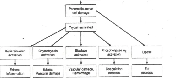
MECHANISM OF ACUTE PANCREATITIS
1. INTRA PANCREATIC ACTIVATION OF PANCREATIC
ZYMOGENS
The cardinal mechanism is the activation of the trypsinogen to
trypsin and this enzyme activates the other enzymes and the pathology
continues. Whatever may be the etiology finally it lands upon the above
given mechanism.
A concept known as the intrapancreatic activation of the enzymes
is postulated. The release of the pancreatic enzyme is hindered and they
join the intracellular lysosomes and this results in activating all known
pancreatic zymogens like chymotripsinogen to active chymotrypsin ,
proelastase to elastase and prophospholipase to lipase A. Only lipase
already synthesized in active form is independent of trypsin. Every
activated enzyme has its own function and it is summarized in the flow
chart.
Of all the etiological factors alcohol is the most common cause of
acute pancreatitis so its mechanism is discussed. The mechanism is as
22
Hyper secretion of the exocrine pancreatic secretion in the presence
of partial ampullary obstruction.
Enzyme extravasations initiating the pancreatic injury.
Alcoholics usually have hypertriglyceridemia this also initiates
pancreatitis.
The next common cause is the gallstone pancreatitis. Gall stone
migrates into the ampulla of Vater which causes the diversion of the bile
into the pancreatic duct which results in the bile induced pancreatic
injury.
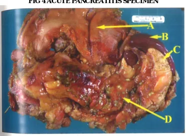
PATHOLOGICAL CHANGES IN ACUTE PANREATITIS :
Mildest pathological change - Edema of the gland. May be
accompanied by infiltration of the intra lobular septa by inflammatory
cells.
Microscopy - fat necrosis in the pancreas and surrounding tissues.
If extensive necrosis - Whitish yellow plaques occur due to
necrosis and calcium deposition.
Vascular thrombosis or disruption results in pancreatic necrosis or
23
Increased levels of active pancreatic enzymes occur :
1. Within the pancreas
2. In the peritoneal exudate
24
25
FIG 4 ACUTE PANCREATITIS SPECIMEN
26
CHANGES IN VARIOUS SYSTEMS OF THE BODY IN ACUTE
PANCREATITIS:
The pathophysiology alters many systems in the body. The changes
affects the following systems:
1. Fluid and electrolyte changes.
2. Cardiovascular changes.
3. Respiratory changes.
4. Renal changes.
5. Local changes.
FLUID AND ELECTROLYTE CHANGES :
Circulating blood volume is decreased due to loss from
intravascular space of plasma into the retroperitoneum and
systematically. Additional loss occurs following vomiting or naso gastric
aspiration. Hypocalcemia and Hypomagnesemia are frequent. Decreased
ionised calcium levels also occur due to trapping of calcium in areas of
27 CARDIOVASCULAR CHANGES :
Hypotension , Tachycardia , increased total peripheral vascular
resistance and decreased cardiac output - sequelae of hypovolemia also
observed in acute pancreatitis similar to septic shock or hepatic cirrhosis
are due to circulatory vasoactive substances. Hypotension persists despite
restoration of intravascular volume.
RESPIRATORY COMPLICATIONS :
Early features of acute pancreatitis - arterial hypoxemia
Pulmonary function studies - Decreased respiratory lung
volume with decreased pulmonary compliance and
decreased diffusion capacity.
Early respiratory failure resolves with subsidence of acute
pancreatitis.
Severe or unresolving pancreatitis may develop progressive
28
FACTORS IMPLICATED FOR PULMONARY
COMPLICATIONS:
1. Abdominal distension and elevation of diaphragm.
2. Alteration in the lecithin of pulmonary surfactant by circulating
pancreatic lecithinase.
3. Pulmonary thromboembolism.
4. Circulating free fatty acids.
5. Circulating products of the proteolyitc cleavage complement.
RENAL FAILURE :
Major factor for the cause of death in patients with acute
pancreatitis. Due to principally hypovolemia. Therefore , many patients
land up in acute renal failure. Pathologically it is due to deposition of the
fibrin complexes in the glomeruli.
OTHER SYSTEMIC FEATURES :
Abnormal liver function tests - elevation of serum bilirubin and
liver enzymes such as Alkaline phosphatase are raised and it is mainly
29
Early intravascular thrombosis with decreased platelet count and
fibrinogen level occur due to the effects of pancreatic proteolytic
enzymes. May be followed by marked thrombocytosis and
hyperfibrinogenemia.
LOCAL SEQUELAE :
Intra abdominal complications include :
1. Paralytic ileus
2. Duodenal / biliary obstruction
3. Release of pancreatic enzymes with peripancreatic fluid
collection and fluid in general peritoneal cavity.
4. Destruction of tissues adjacent to pancreas.
5. Rarely cause gross disruption of the pancreatic ductal system
which is usually self limited.
6. Persistent chronic pseudocyst in 1% of the patients.
7. Infected pancreatic abscess due to secondary infection occur in
30
8. Extension of local necrosis to involve colonic wall causing
colonic perforation occurs in 1% of the patients and occurs in the left
transverse colon or splenic flexure.
CLINICAL MANIFESTATIONS AND DIAGNOSIS
The classical feature of acute pancreatitis is its severity of
symptoms and paucity of physical signs.
1. Abdominal pain - 85 - 100%
Upper abdominal pain may radiate to the back and may be severe.
Pain is aggravated by intake of food or intake of alcohol. Pain is resistant
to analgesics. Patient assumes various postures in an effort to obtain relief
from the pain.
2. Nausea and vomiting - 92%
Vomiting is usually non projectile and it of low volume and it
contains gastric and duodenal content and it is non feculent.
3. Physical examination :
Restless patient.
31
Arterial hypotension
Abdomen - moderately distended with epigastric dullness.
Tenderness is marked in the upper abdomen.
Moderate muscle spasm is present.
GREY TURNERS SIGN - Grey green discoloration of the
flank present in patients with peripancreatic hemorrhage.
CULLEN'S SIGN - bluish discoloration of periumbilical
region.
4. Extra abdominal manifestations :
Left sided pleural effusion
Acute pulmonary failure marked by Tachypnoea and dyspnoea.
Central or peripheral cyanosis due to :
a. Circulating Phospholipase A
b. Circulating free fatty acids from triglycerides from lipolysis
c. Pulmonary surfactant loss
32
5. Central Nervous System manifestations -
Nonlateralizing nature , including billigerence , confusion , psychosis and
coma. This is due to hyperosmolarity , hypoperfusion , hypoxia , cerebral
fat embolism or disseminated intravascular coagulation.
LABORATORY INVESTIGATIONS :
DIAGNOSIS OF ACUTE PANCREATITIS :
LABORATORY TEST
RADIOGRAPHIC PROCEDURES
Serum Amylase Chest X-ray
Serum Amylase isoenzymes Plain Abdominal X-ray
Urine Amylase Ultrasonography
Amylase-Creatinine
clearance Ratio
33 1. BLOOD COUNT :
Leucocytosis - 10,000 to 20,000 occurs early in all cases
Hematocrit - is high in most patients at the onset
Hemoglobin decreased value of more than 2.5gm% without
detectable blood loss is found in patients with pancreatic necrosis.
2. SERUM AMYLASE :
Elevated in 95% of patients with Acute pancreatitis. But this is not
an ideal marker because it is elevated in other conditions such as :
Perforated peptic ulcer
Biliary Lithiasis
Intestinal obstruction
Mesenteric Infarction.
Also in patients with acute pancreatitis , serum amylase in normal
levels can occur due to :
Hyper triglyceredemia - Latescent serum
34
Previous attach has destroyed most glandular tissues.
Present attack is associated with massive destruction of gland.
Serum amylase in Acute pancreatitis is elevated within 24 hours of
onset of symptoms and returns to normal in 7 days.
3.SERUM ISOAMYLASE - P :
As it is produced only from pancreas it has a higher specificity in
detection and confirmation of acute pancreatitis.
4.SERUM LIPASE :
Serum lipase is solely of pancreatic origin hence serum lipase level
is more specific than amylase. Recent development of an enzyme
immunoassay of lipase is reliable and is of great value in Acute
pancreatitis. Duration of Hyper Lipasemia exceeds hyper amylossemia.
5. PLUERAL AND PERITONEAL FLUID AMYLASE :
Pleural effusion shows raised levels of amylolytic activity in
pancreatitis. High activities of amylase may also be found in fluids
35 6. OTHER BIOCHEMICAL INDICES :
Hyperglycemia and Glycosuria - Non specific , transient cause -
relative hypoinsulinemia and Hyperglucognemia
Hypocalcemia - Well recognised entity in acute pancreatitis but can
also occur in perforated peptic ulcer.
Cause - Deposition of calcium in areas of fat necrosis. Release of
glucagon , Inadequate parathyroid response and Dilutional hypo
albuminemia.
Methemalbumin :
Appearance in serum indicates necrotic rather than
edematous pancreatitis.
Liver function tests :
Slight increases in alkaline phosphatase and amino transfer
are with raise in serum bilirubin - transitions. Markedly elevated
serum aspartate and alanine amino transferase are within 48 hours
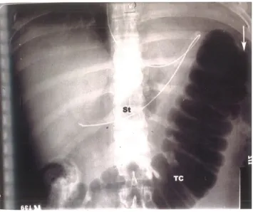
36 RADIOGRAPHIC FINDINGS :
I. PAIN X-RAYS :
i) Plain X-ray Abdomen :
a) Segmental small bowel ileus or a "SENTINEL LOOP" in the left
upper quadrant.
b) Dilatation of the transverse colon - "COLON CUT OFF SIGN"
A. Increase epigastric soft tissue density.
B. Obscured psoas muscle margins.
C. Presence of gall stones.
D. Pancreatic calcification - may not be an acute pancreatitis.
ii) Plain X-Ray Chest :
a) Pleural effusion
b) Atelectasis
c) Pneumonia
37
FIG 5 X RAY SHOWING COLON CUT OFF SIGN
CONTRAST STUDIES WITH WATER SOLUBLE CONTRAST :
Upper Gastrointestinal study :
a) Widening of "C" loop
b) Anterior displacement of stomach
38 ABDOMINAL ULTRASONOGRAM :
a) Enlargement and edema of pancreas
b) Pseudocysts of pancreas
c) Delineates pancreatic abscess
d) Dilatation of Bile duct and presence of stone in gall bladder and
common bile duct.
COMPUTED TOMOGRAPHY :
Except in early or mild eases it is useful in assessing CT scan
reveals many findings in patients with Acute pancreatitis. Pancreas is
usually enlarged and there is pancreatic edema. Pancreatic necrosis is
characterized CT scan. In the peri pancreatic area there is collection ,
obliteration of the fat plane and thickening of the fat plane. Other findings
are paralyitc ileus and Pleural effusion.
ENDOSCOPY :
To detect biliary pancreatitis.
Therapeutically used for papillotomy and removal of stones
40
TREATMENT
MEDICAL MANAGEMENT :
A. NUTRITION :
1. Enteral nutrition :
Previously it was thought that enteral nutrition stimulates the
pancreas and results in pain in pancreatitis , but now it is found that
pancreas is actually in a state of rest in acute pancreatitis so it better to
stimulate the pancreas. So enteral feeding does no harm to the patient.
2. Total parenteral nutrition :
TPN is associated with many complications such as arterial injury ,
pneumothorax , thrombosis , and catheter embolism. Many studies
confirm that enteral nutrition is better than TPN.
B. GASTRITIS PREVENTION :
Patients suffering from severe AP have risk to develop peptic
ulcers or erosive gastritis Histamine 2 antagonists is indicated in patients
on mechanical ventilation and patients with adult respiratory distress
41 C. FLUID MANAGEMENT :
Adequate fluid management is the main stay in the management of
acute pancreatitis. If missed it can lead to serious complications. Plenty of
fluids are sequestered in third spaces. So crystalloids and colloids are
used in the ratio of 3:1 . The fluid loss may be 6-10 litres. It is said that
when hematocrit is less than 30% if dextran is used it improves the
microcirculation. Good fluid resuscitation is indicated by adequate urine
output , CVP around 8-12 cm of water , hematocrit of 35-40 %.
D. PAIN MANAGEMENT :
As a result of activation of pancreatic enzymes there are increase in
release of the inflammatory mediators. These mediators irritate the
sensory fibers of the celiac plexus ( T5 TO T9 ) and cause severe pain
radiating to the back. The following drugs are used in management of
pain :
Non steroidal analgesia
Meperidine
Tramadol
42 E. THE ROLE OF ANTIBIOTICS :
There is a great controversy regarding used of antibiotics in acute
pancreatitis. Since there is great risk in developing necrosis in acute
pancreatitis , there is also a problem in developing abscess formation ,
antibiotics are used. The most common antibiotics used are imipenum ,
meropenum , metronidazole , fluoroquinolones and cephalosporins. These
antibiotics penetrate the pancreas well , but aminoglycosides do not
penetrate the pancreas. Over use of antibiotics result in fungal infection.
F. SUPPRESSION OF PANCREATIC EXOCRINE SECRETION :
These are done by nasogastric suction , histamine H-2 receptor
antagnoists , antacids , atropine , glucagon , calcitonin and stomatostatin.
SURGICAL MANAGEMENT : INDICATIONS AND TIMING OF
INTERVENTION :
1. Uncertainty of diagnosis
2. Treatment of pancreatic sepsis
3. Correction of associated biliary tract disease
4. Progressive clinical deterioration despite optimal supportive care.
43
6. Severe sterile necrosis
7. Symptomatic organized pancreatic necrosis
A) BILIARY OPERATIONS IN PATIENTS WITH
CHOLELITHIASIS :
a. Cholecystostomy
b. Common duct drainage
c. Cholecystectomy
d. Early endoscopic papillotomy.
In patients with severe gall stone pancreatitis early intraabdominal
surgery has been associated with higher mortality than early non
operative treatment. Surgical correction of cholelithiasis to prevent
recurrent pancreatitis undertaken once evidence of pancreatitis has
subsided usually during the same hospital admission.
B) SURGICAL MANAGEMENT : PROCEDURES
1. RESECTION
2. PANCREATIC DEBRIDEMENT
3. MINIMALLY INVASIVE APPROACHES
44
Percutaneous necrosectomy and sinus tract endoscopy
RESECTION :
Pancreatic resection is primarily of historical interest only and is
not recommended currently.
PANCREATIC DEBRIDEMENT :
All pancreatic debridement and post debridement care based on :
1. Wide removal of devitalized and necrotic tissue.
2. The assurance of post operative removal of the products of
ongoing local inflammation and infection.
TECHNIQUE OF DEBRIDEMENT :
1. Debridement and closed drainage.
2. Open packing for pancreatic necrosis.
3. Debridement and continuous Closed post operative Lavage of the
Lesser Sac.
DIFFERENTIAL DIAGNOSIS :
In addition to the diagnosis of acute pancreatitis , the general
surgeon should also have a clinical suspicion of the various other disease
45
mimic acute pancreatitis and can cause a clinical dilemma in the
diagnosis of Acute pancreatitis.
Various differential diagnosis of Acute pancreatitis are :
Acute Mesenteric Ischemia
Acute Respiratory Distress Syndrome
Bacterial Pneumonia
Cholangitis
Cholecystitis
Chronic Pancreatitis
Colon Cancer
Colonic Obstruction
Community-Acquired Pneumonia (CAP)
Emergent Treatment of Gastroenteritis
Gallstones (Cholelithiasis)
Gastric Cancer
Irritable Bowel Syndrome
Myocardial Infarction
46
Pancreatic Pseudocysts
Peptic Ulcer Disease
Viral Hepatitis
THE COMPLICATIONS OF ACUTE PANCREATITIS : The complications of Acute pancreatitis are :
1. Local complications :
Fluid collection
Pancreatic ascites / pleural effusion
Pancreatic Pseudocyst
Pancreatic necrosis
Infected pancreatic abscess
Hemorrhagic / Pseudo aneurysm.
2. Regional complications :
Venous thrombosis
Paralytic ileus
Intestinal obstruction
Intestinal ischemia / necrosis
47
3. Systemic complications :
Systemic inflammatory response syndrome
Multiple organ dysfunction syndrome
ARDS / Pulmonary failure
Renal failure
Cardiovascular complications
Hypocalcemia
Hyperglycemia
Disseminated intravascular coagulopathy
Protein calorie malnutrition.
A) PANCREATIC ABSCESS :
Pancreatic abscess - incidence 9%. Most common in patients with
post-operative pancreatitis.
Clinical features :
Persistent or recurrent fever
Abdominal distension
Abdominal mass
48
Pneumonia / Effusion
Renal failure
Coma
Elevated serum Amylase
Leucocytosis ( more than 11,000 / mm3 )
Radiographic diagnosis :
1. Upper GI contrast studies showing displacement of stomach or
duodenum. Gas outside of GIT.
2. Ultrasound abdomen can delineate pancreatic abscess
3. Computed Tomography sensitive and specific.
4. Percutaneous aspiration under CT guidance.
Treatment :
Adjuvant :
1. Vigorous supportive management.
2. Meticulous respiratory care and Nutritional support.
49 Specific :
1. Percutaneous drainage by catheter.
2. Laprotomy - Debridement and packing of pancreatic bed.
3. Surgical correction of other complications like involvement of
colon by colostomy.
4. Feeding jejunostomy to correct nutritional imbalance.
B) PSEUDOCYSTS :
Pseudocysts following acute pancreatitis spontaneous
disappearance of pseudocysts is a common occurrence in acute
pancreatitis. These are carefully monitored by serial ultrasonogram or CT
and operative intervention is needed only when they go in for further
complications. It is dealt in detail along with treatment of pseudocysts
following chronic pancreatitis.
C) PANCREATIC ASCITES :
Pancreatic ascites - more common following chronic pancreatitis.
But may also follow acute pancreatitis secondary to trauma, pseudocysts
and rarely pancreatic neoplasm. Treatment is by drainage procedures as
50 PROGNOSTIC ASSESSMENT
PROGNOSTIC INDICATORS :
Because of the variability and unpredictability of acute pancreatitis,
clinical scoring systems have been made to predict the severity of acute
pancreatitis.
1. RANSON'S CRITERIA
2.BISAP SCORE
3. CT SEVERITY INDEX
4. OTHER PROGNOSTIC SYSTEMS :
The Acute Physiology and Chronic Health Evaluation II
(APACHE II ) Score.
C-reactive protein (CRP ) assays.
Trypsinogen-activating peptide ( TAP ) assays.
ATLANDA CLASSIFICATION :
1. ACUTE EDEMATOUS PANCREATITS - MILDER
51
2. ACUTE NECROTISING PANCREATITIS - Incidence 20%
and characterized by pancreatic necrosis. MORTALITY - 15
- 20%
GRADING OF ACUTE PANCREATITIS :
This uses Ct scan primarily to grade the severity. CT scan is used
to find pancreatic changes and necrosis. Either contrast or plain CT is
used. This is developed by BALTHAZAR and Acute pancreatitis is
graded from A to E. It gives points according to the following criteria :
1. Nature of necrosis
2. Peri pancreatic changes.
Mild pancreatitis , Interstitial pancreatitis :
Patients with pancreatitis having no collection or necrosis. They
have a mild pancreatitis. Balthazar grade A-C comes under this group.
CTSI is 2
Severe pancreatitis or necrotizing pancreatitis :
They occur in 20 of patients. They have uneven clinical course and
high mortality rate. They are more than fluid collections. The grade is D
52
due to fat necrosis. This group of patients with necrosis has most
complication and they have to be identified. There is a separate type
called extra pancreatic necrosis which has no pancreatic necrosis the
CTSI is 4.
Balthazar Scoring for the Grading of Acute Pancreatitis
Grade A – normal CT
Grade B – focal or diffuse enlargement of the pancreas
Grade C – Pancreatic gland abnormalities and peri pancreatic
inflammation.
Grade D – Fluid collection in a single location.
Grade E – two or more collections and/or gas bubbles in or
adjacent to pancreas.
CT severity index = CT grade point + points for necrosis
First grade points are calculated. The grade A,B,C,D, E have
0,1,2,3 and 4 respectively. The points for necrosis are based on the
percentage of necrosis. When the necrosis is <30% the point is 2 ,
53
CT grade points are added to points assigned for percentage of
necrosis to determine the CT severity index. So the patients with score
54
METHODOLOGY
MATERIALS AND METHODS
STUDY AREA :
GOVERNMENT COIMBATORE MEDICAL COLLEGE AND
HOSPITAL.
STUDY POPULATION :
Patients admitted in CMCH with symptoms suggestive of Acute
pancreatitis.
INCLUSION CRITERIA :
1. Clinical findings suggestive of Abdominal pain characteristic of
acute pancreatitis.
2. Elevated serum amylase
3. CT Scan with findings suggestive of acute pancreatitis.
The following criteria are studied :
1. Severity of Acute pancreatitis.
2. Pancreatic Necrosis.
55 DEFINITION :
1. MILD OR SEVERE AP :
Depending on the organ failure the patients are classified into mild
or severe Acute pancreatitis. The presence of organ failure is again
dictated by the presence of following factor :
Pulmonary failure ( Pco2 - 60 mmHg )
Renal failure ( Serum Creatinine levels more than 2 mg/dL )
Severe Shock.
2. PANCREATIC NECROSIS :
This finding is easily found by CECT scan. This is detected by the
absence of enhancement in the CECT Scan in the pancreatic parenchyma.
EXCLUSION CRITERIA :
1. Pediatric patients were excluded from the study.
2. Pregnant and postpartum patients were excluded.
STUDY PERIOD : June 2016 to July 2017
56
All patients eligible by inclusion and exclusion criteria are to be included
in this study.
STUDY DESIGN :
57
RESULTS
STATISTICAL ANALYSIS :
The data are reported as the mean +/- SD or the median ,
depending on their distribution.
[image:68.612.146.525.300.363.2]A) SEX DISTRIBUTION :
Table -1 – Sex distribution :
Male Female
95 5
The above tabular column 1 gives the sex distribution of the disease in
both the gender groups.
The total number of patients studied in the study were 100, it comprises
58
SEX DISTRIBUTION:
The total number of patients studied in the study were 100, it comprises
of 95 males and 5 females.
In our population males are commonly affected than the female
population , this factor has link with the etiology. In our population
alcohol consumption is the common cause of acute pancreatitis. Alcohol
consumption is not prevalent in female population so female are not
commonly affected by acute pancreatitis 95
5 0
10 20 30 40 50 60 70 80 90 100
59
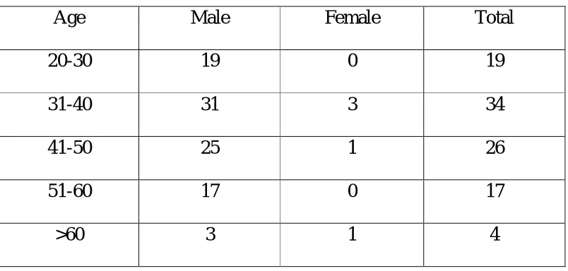
[image:70.612.123.525.139.331.2]AGE DISTRIBUTION
Table 2 – Age distribution
Age Male Female Total
20-30 19 0 19
31-40 31 3 34
41-50 25 1 26
51-60 17 0 17
>60 3 1 4
The above tabular column 1 gives the age sex distribution of the disease
in various age groups.
Maximum number of patients affected between the age group of 31-40
years and it is 34 patients.
Minimum number of patients affected in the age group of 0f > 60 years
60
Maximum number of patients affected between the age group of 31-40
years and it is 34 patients.
Minimum number of patients affected in the age group of 0f > 60 years
and its
4 patients
The pie diagram shows the same
19
34 26
17 4
AGE DISTRIBUTION
20-30YEARS
31-40 YEARS
41-50 YEARS
51-60 YEARS
61
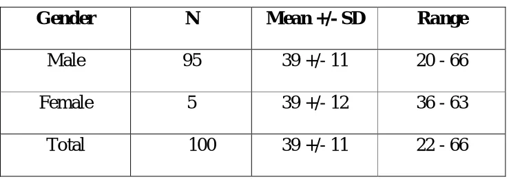
Table -3 – Mean SD for Age
Gender N Mean +/- SD Range
Male 95 39 +/- 11 20 - 66
Female 5 39 +/- 12 36 - 63
Total 100 39 +/- 11 22 - 66
The above tabular column gives the mean and the SD for the age.
The explanation as follows ,
The total number of male patients in the study population are 95
with a mean of 39 +/- 11 and an age group of range 20 – 66.
The total number of female patients in the study population are 5
with a mean of 39 +/- 11 and an age group of range 36 – 63.
The overall mean of age in the study population is 39 +/- 11 and
62 AGE DISTRIBUTION – FEMALE :
This pie diagram shows the number of patients affected with acute
pancreatitis in different age group.
Maximum number of female patients affected between the age
group of 31-40 years and it is 3 patients.
Minimum number of female patients affected in the age group of 0f
51-60 years and 21-30 years of age group of study population which is
nil.
The mean age of female patients affected with acute pancreatitis in
this study is 39 +/- 11.
3 1
1
AGE DISTRIBUTION - FEMALE
20-30
31-40
41-50
51-60
63
AGE DISTRIBUTION – MALE :
This graph shows the number of patients affected with acute
pancreatitis in different age group.
Maximum number of male patients affected between the age group
of 31-40 years and it is 31 patients.
Minimum number of male patients affected in the age group of 0f
> 60 years and its 3 patients
The bar diagram shows the same. 19
31
25
17
3 0
5 10 15 20 25 30 35
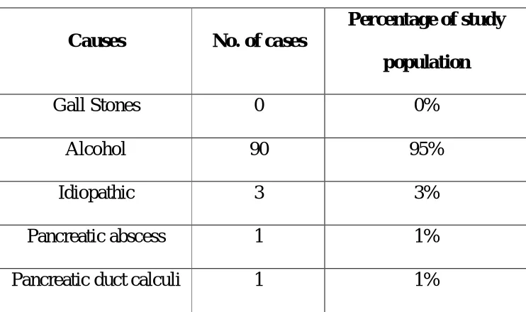
64 ETIOLOGY :
Table 4 – Etiology – Male
Causes No. of cases
Percentage of study population
Gall Stones 0 0%
Alcohol 90 95%
Idiopathic 3 3%
Pancreatic abscess 1 1%
Pancreatic duct calculi 1 1%
The above tabular column gives the various etiological factors
implicated in the onset of acute pancreatitis in male patients in the study
population.
In our area the most prevalent cause is chronic alcohol intake.
In our study 90 male patients have history of chronic alcohol
intake. 3 patients the cause of acute pancreatitis was due to idiopathic
causes . The unknown causes may be due to drugs, increase in cholesterol
65
CAUSES OF ACUTE PANCREATITIS - MALE :
This bar diagram shows the different causes of acute pancreatitis in
male patients prevalent in our study population.
In our area the most prevalent cause is chronic alcohol intake.
In our study 90 male patients have history of chronic alcohol
intake. 3 patients the cause of acute pancreatitis was due to idiopathic
causes . The unknown causes may be due to drugs, increase in cholesterol
and hypercalcemia . The unknown causes needs further evaluation, even
in unknown causes 2 patients had history of alcohol intake during their
earlier days and stopped recently for various reasons. 90
0 3 1 1
0 10 20 30 40 50 60 70 80 90 100
ALCOHOL INTAKE GALL STONES OTHER CAUSES PANCREATIC
ABSCESS
66
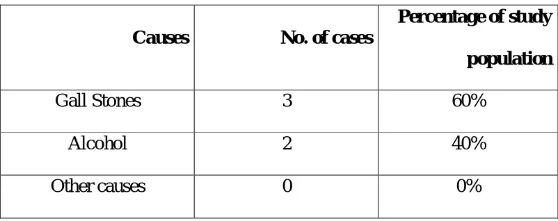
Table 5 – Etiology – Female
Causes No. of cases
Percentage of study population
Gall Stones 3 60%
Alcohol 2 40%
Other causes 0 0%
The above tabular column gives the various etiological factors
implicated in the onset of acute pancreatitis in female patients of the
study population.
In our study 3 female patients have been diagnosed to have gall
stones which caused the onset of acute pancreatitis.
In our study , 2 female patients have been diagnosed to have acute
67
CAUSES OF ACUTE PANCREATITIS IN FEMALES :
This pie diagram shows the different causes of acute pancreatitis in
female patients prevalent in our study population.
In our area the most prevalent cause of acute pancreatitis in female
patients is gall stones.
In our study 3 female patients have been diagnosed to have gall
stones which caused the onset of acute pancreatitis.
In our study , 2 female patients have been diagnosed to have acute
pancreatitis due to the intake of alcohol.
In our study , the incidence of idiopathic causes of acute
pancreatitis in female patients was nil.
2
3
CAUSES OF ACUTE PANCREATITIS
ALCOHOL
GALL STONES
68
Table 6 – History of Alcohol Intake
Alcohol Intake Percentage of Study population
Present 96%
Absent 4%
From the above tabular column it is shown that 96% of the study
population has a history of intake of chronic alcohol and 4% does not
have a history of intake of alcohol. 96% of the study population has
shown that alcohol is implicated as the causative factor of Acute
pancreatitis.
96% of the study population has shown that alcohol is implicated
as the causative factor of Acute pancreatitis.
96% 4%
prevalance of alcohol intake
chronic alcohol intake present
69
COMPARISON OF MALE AND FEMALE DISTRIBUTION OF CAUSES:
The most common cause for acute pancreatitis in male is chronic
alcohol intake. Out of 95 male patients with acute pancreatitis in our
study 94 patients had history of chronic alcohol intake.
Out of the 5 female patients in our study 3 patients had gall stones
and 2 had history of alcohol intake. On linking the etiology with the sex
Alcoholic pancreatitis is common in males
Gall stone pancreatitis is common in females.
70
Table 7 – Severity of Acute pancreatitis - Male:
Severity Number of patients
Percentage of Study population
Mild 69 73%
Moderate 26 27%
Severe 0 0%
From the above tabular column it is shown that 73% of the study
population has severity graded as Mild according to CT severity Index.
27% of the study population has severity graded as Moderate.
71
SEVERITY OF ACUTE PANCREATITIS - MALE :
From the above bar graph it is shown that 69 male patients of the
study population has severity graded as Mild according to CT severity
Index.
26 male patients of the study population has severity graded as
Moderate.
0 male patients of the study population has severity graded as
Severe. 69
26
0 0
10 20 30 40 50 60 70 80
72
Table 8 – Severity of Acute Pancreatitis – Female:
Severity Number of patients
Percentage of Study population
Mild 2 40%
Moderate 1 20%
Severe 2 40%
From the above tabular column it is shown that 40% of the females
in the study population have a severity graded as Mild according to CT
severity Index.
20% of the females are graded as Moderate.
73
SEVERITY IN FEMALE PATIENTS
From the above pie diagram it is shown that 2 of the females in the
study population have a severity graded as Mild according to CT severity
Index.
1 of the females are graded as Moderate.
2 of the female patients were graded into Severe category.
2
1
2 MILD
MODERATE
74
SEVERITY OF ACUTE PANCREATITIS :
72% of the study population were divided into mild grade of Acute
pancreatitis by the use of CT severity index.
26% of the study population were classified into moderate grade
and 2% of the population were classified into severe grade.
72% 26%
2%
MILD
MODERATE
75
Table 9 – Management of Acute Pancreatitis :
Management Number of patients Percentage of Study
population
Surgery 0 0%
Conservative 100 100%
Conservative management was the treatment option for all grades of
Acute pancreatitis patients and surgical intervention was not performed in
the study population.
All cases can be managed conservatively and does not require emergency
76
COMPARISION OF SEVERITY AND ITS MANAGEMENT FOR ACUTE PANCREATITIS :
According to this graph ,
72% of the study population were divided into mild grade of Acute
pancreatitis by the use of CT severity index.
26% of the study population were classified into moderate grade and
2% of the population were classified into severe grade.
Conservative management was the treatment option for all grades of
Acute pancreatitis patients and surgical intervention was not performed in
the study population. 72
26
2
0 0 0
72 26 2 0 10 20 30 40 50 60 70 80
MILD MODERATE SEVERE
NUMBER OF PATIENTS
EMERGENCY SURGERY
77
DISCUSSION
In this study , Out of 100 patients of the study population 95
patients were found to be male patients and 5 patients were female
patients. This shows the acute pancreatitis is common in Male patients in
comparison to female patients.
The age group in which the onset of acute pancreatitis is more
common is between 31-40 which is 31% of the study population in both
male and female patients of the study population. The second most
common age group affected by acute pancreatitis is 41-50 age group in
males and in female population we have found to have similar incidence
in both 41-50 age group and more than 60 years of age group population.
The mean age of population affected by Acute pancreatitis is 39 for
both males and female patients in the study population. The range of the
age group affected in male patients is 20 – 66 and for female patients is
36 – 63. This indicates that the patients commonly affected by acute
pancreatitis usually fall into the age group of more than 20 years and an
78
The most common cause of acute pancreatitis in male patients of
the study population in this study is found to be chronic intake of alcohol
which is 94% of the study population and 1 % of male patients were
found to have acute pancreatitis due to other causes. This shows that , the
most common cause of acute pancreatitis in male patients was found to
excessive intake of alcohol from this study. Gall stones were not found to
be the cause of acute pancreatitis in male patients of the study population
and hence in this study it is shown that gall stones are the least common
cause of acute pancreatitis in male patients.
From this study , it is found that the most common cause of acute
pancreatitis in the female patients of the study population is gall stones
which is 3 % of the study population and the second most common cause
of acute pancreatitis in female patients was alcohol which is 2% of the
study population and pancreatitis due to other causes was not reported in
this study in female patients of the study population.
The severity graded by CT severity index of acute pancreatitis in
male patients was mild , moderate and severe. 69 male patients were
graded as mild grade which was 73% of the study population. 26 male
patients were graded as moderate grade which was 27% of the study
79
This shows that in the study population , the most prevalent grade of
acute pancreatitis was mild grade which has minimal complications and
can be managed conservatively. It has a good prognosis and better
outcome with medical management alone and does not need any surgical
intervention.
The severity in female patients was found out to be as , Mild grade
in 2 female patients which was 40% of the study population. Moderate
grade was found in 1 female patients which was 20% of the study
population. Severe grade was found in 2 patients which was 40% of the
study population. This shows that the most prevalent grade in females of
the study population is mild and severe acute pancreatitis. Severe acute
pancreatitis is most commonly caused by gall stones and is treated with
surgical management and has good prognosis.
The management of acute pancreatitis is divided into two
categories : medical and surgical management. In this study , mild grade
of acute pancreatitis was managed conservatively with medical
management such as intravenous fluids , intravenous antibiotics ,
parenteral analgesics and intravenous octreotide. Moderate grade and
severe of acute pancreatitis patients were also managed conservatively
80
improvement with resolution of symptoms after the completion of the
conservative management. Hence , this study shows that the management
of both moderate and severe grades of acute pancreatitis can be done with
medical management and does not require surgical management for
81
CONCLUSION
To conclude, Acute pancreatitis has become a common entity in
the day to day practice of general surgeons and it has become vital to
evaluate the underlying causes leading to the onset of the disease and to
properly categorize the patients according to the severity of the disease
and accordingly manage the patients to prevent morbidity and mortality
caused by the disease.
By this study , we have concluded the following :
1. Acute pancreatitis is more prevalent in the male population.
2. Acute pancreatitis most commonly involves the age group of
31- 40 years of age.
3. The most common cause leading to the onset of acute
pancreatitis is chronic intake of alcohol in the male population
and gall stones as a cause of acute pancreatitis has become rare
due to its early detection and treatment.
4. According to the CT severity index grading system , the most
common grade of acute pancreatitis is found to be mild grade
and segmental involvement which has an overall good
82
5. Conservative management is the treatment of choice of patients
presenting with Acute pancreatitis in this study which causes an
83
BIBLIOGRAPHY
1. Susan Stranding PhD DSc. The 39th edition of Gray’s Anatomy ;
88 pancreas : spleen and suprarenal and spleen
2. Michael J. Zinner , MD, FACS. Maingot’s Abdominal Operations
11th edition. Chapter 36; Management of Acute pancreatitis.
3. Banks PA – acute pancreatitis ; medical & surgical management.
4. Beger H G et al – natural course of acute pancreatitis; world j surg;
21: 130-135.
5. Sakorafas GH – ETIOLOGY and pathogenesis of acute
pancreatitis; j clin gastro 2000;30: 343-356.
6. RANSON J : diagnostic standards of acute pancreatitis; world j
surg 1997 ; 21 : 136.
7. Balthazar EJ , RANSON JH. Acute pancreatitis : value of CT in
establishing prognosis. Radiology 1990;174(2):331-6.
8. Balthazar EJ, Freeny PC, van Sonnenberg E. Imaging and
intervention in acute pancreatitis. Radiology 1994; 193:297-306.
9. Golub R, et al. Role of antibiotics in acute pancreatitis: a meta