0022-538X/10/$12.00 doi:10.1128/JVI.01350-09
Copyright © 2010, American Society for Microbiology. All Rights Reserved.
Inability of Plasmacytoid Dendritic Cells To Directly Lyse HIV-Infected
Autologous CD4
⫹
T Cells despite Induction of Tumor Necrosis
Factor-Related Apoptosis-Inducing Ligand
䌤
Jihed Chehimi,
1Emmanouil Papasavvas,
1Costin Tomescu,
1Bethsebah Gekonge,
1Shaheed Abdulhaqq,
1Andrea Raymond,
1Aidan Hancock,
1Kavita Vinekar,
1Craig Carty,
2Griffin Reynolds,
1Maxwell Pistilli,
1Karam Mounzer,
3Jay Kostman,
2and Luis J. Montaner
1*
The Wistar Institute, Philadelphia, Pennsylvania 191041; University of Pennsylvania, Philadelphia, Pennsylvania 191042; and
The Philadelphia Field Initiating Group for HIV-1 Trials (Philadelphia FIGHT), Philadelphia, Pennsylvania 191073
Received 1 July 2009/Accepted 30 November 2009
The function of plasmacytoid dendritic cells (PDC) in chronic human immunodeficiency virus type 1 (HIV-1) infection remains controversial with regard to its potential for sustained alpha interferon (IFN-␣) production and induction of PDC-dependent tumor necrosis factor (TNF)-related apoptosis-inducing ligand (TRAIL)-mediated cytotoxicity of HIV-infected cells. We address these areas by a study of chronically HIV-1-infected subjects followed through antiretroviral therapy (ART) interruption and by testing PDC cytolytic function against autologous HIV-infected CD4ⴙT cells. Rebound in viremia induced by therapy interruption showed a positive association between TRAIL and viral load or T-cell activation, but comparable levels of plasma IFN-␣/ were found in viremic ART-treated and control subjects. While PDC from HIV-infected subjects expressed less interferon regulator factor 7 (IRF-7) and produced significantly less IFN-␣ upon Toll-like receptor 7/9 (TLR7/9) engagement than controls, membrane TRAIL expression in PDC from HIVⴙsubjects was increased. Moreover, no significant increase in death receptor 5 (DR5) expression was seen in CD4ⴙT cells from viremic HIVⴙsubjects compared to controls or followingin vitro infection/exposure to infectious and noninfectious virus or exogenous IFN-␣, respectively. Although activated PDC killed the DR5-expressing HIV-infected Sup-T1 cell line, PDC did not lyse primary autologous HIVⴙCD4ⴙT cells yet could provide accessory help for NK cells in killing HIV-infected autologous CD4ⴙT cells. Taken together, our data show a lack of sustained high levels of soluble IFN-␣in chronic HIV-1 infectionin vivoand document a lack of direct PDC cytolytic activity against autologous infected or uninfected CD4ⴙT cells.
Human immunodeficiency virus (HIV) infection is associ-ated with chronic immune activation, progressive immune sup-pression, and deletion of memory adaptive responses, resulting in increased susceptibility to opportunistic infections (23, 51, 52). Loss of CD4⫹T cells is the hallmark of HIV infection, with multiple mechanisms proposed as contributing to this loss (activation-induced cell death, direct cytopathic effect, immune cells, and death receptor-mediated apoptosis induction) (re-viewed in references 33 and 34). One of the most puzzling observations in AIDS pathogenesis has been the progressive depletion of bystander T cells, especially in lymphoid tissues (25, 33, 34, 55). While antiretroviral therapy (ART) initiated in the early stages of HIV infection, when CD4⫹T-cell counts are high (⬎500 cells/l), may prevent the destruction of lymph node (LN) tissue and the massive depletion of CD4⫹T lym-phocytes by decreasing the rate of virally induced apoptosis (20), a persistent, albeit decreased, level of apoptosis of pe-ripheral blood CD4⫹and CD8⫹T cells is seen in ART-treated HIV⫹subjects despite long-term viral suppression (36).
A member of the tumor necrosis factor (TNF) family, TNF-related apoptosis-inducing ligand (TRAIL), has been shown to
be involved in HIV-1-associated T-cell apoptosis (33, 34). TRAIL (soluble or membrane bound) induces apoptosis upon binding to death receptor 4 (DR4; also named TRAIL-R1) or DR5 (also named TRAIL-R2, TRICK2, or Killer/DR5).
On the basis of thein vitroobservation that alpha interferon (IFN-␣) and interferon regulator factor 7 (IRF-7) are in-creased in plasmacytoid dendritic cells (PDC) exposed to HIV-1 (40), the hypothesis that PDC activation by HIV-1 is responsible for an increased level of IFN-␣throughout chronic disease has been proposed. It has also been proposed that the activation of the PDC compartment by HIV-1 participates in the initial immune activation following acute infection and contributes to CD4⫹ T-cell depletion by inducing, through IFN-␣, the production of TRAIL, which mediates apoptosis of DR5-expressing CD4⫹T cells following HIV-1 infection (37, 38, 40). However, several lines of evidence question the direct involvement of PDC in the loss of T cells during HIV infection, as PDC numbers are depleted during chronic HIV infection and PDC remaining in circulation are functionally impaired (10). Recent data show that circulating PDC in HIV-infected subjects, although unable to secrete IFN-␣after Toll-like re-ceptor (TLR)-mediated activation, constitutively express an increased level of IFN-␣mRNA, indicating that during HIV infection PDC are activated yet impaired (71). Rodriguez et al. demonstrated the prevention of spontaneous apoptosis of CD4⫹and CD8⫹T cells by IFN-␣ (63), a major product of
* Corresponding author. Mailing address: The Wistar Institute, 3601 Spruce Street, Philadelphia, PA 19104. Phone: (215) 898-9143. Fax: (215) 573-7008. E-mail: montaner@wistar.org.
䌤Published ahead of print on 30 December 2009.
2762
on November 8, 2019 by guest
http://jvi.asm.org/
PDC following HIV-1 stimulation (3, 28). In addition, Audige et al. (2) showed that blockade of IFN-␣and IFN-␣receptor duringin vitroHIV infection of CD4⫹T cells isolated from human tonsils did not prevent apoptosis or TRAIL production, suggesting a lack of a central link between IFN-␣production and apoptosis of tonsillar CD4⫹ T cells in HIV-1 infection. These data are also consistent with the observation that, in the human peripheral blood lymphocyte-transplanted SCID mouse (hu-PBL-SCID) model, IFN-␣efficiently increases the survival of CD4⫹T cells (49). Thus, controversy remains on the role of IFN-␣as an indirect or direct inducer of apoptosis of CD4⫹T cells through PDC/TRAIL induction, whereas the possibility that IFN-␣acts as an antiviral agent by controlling HIV-1 replication and thus reducing virally mediated T-cell loss appears to be supported by several studies (reviewed in references 26, 47, and 58). In this regard, endogenous IFN-␣ produced by PDC has been shown to play an important role in controlling HIV infection in the human thymus (35), upregu-lating host antiviral factors such as APOBEC (1, 32, 44, 70) and stimulating NK cell-mediated cytotoxic activity against au-tologous HIV-infected targets (72).
In this report, we investigated thein vivocorrelates of vire-mia in chronically infected subjects by studying the relationship between therapy interruption-associated viremia and plasma IFN-␣and TRAIL levels. We also testedin vitrothe functional outcome of HIV-1-activated PDC in terms of their ability to mediate lysis of primary autologous CD4 T cells (infected or not with HIV-1), compared to indirect PDC-mediated lysis effects on the NK-dependent antiviral cytotoxic response.
MATERIALS AND METHODS
Study subjects.Two cohorts recruited at the Jonathan Lax Immune Disorders Clinic (Philadelphia FIGHT) were used in this study. The first cohort was composed of 21 suppressed HIV-1 infected subjects on ART (asymptomatic without opportunistic infections), who had CD4 counts of⬎400 cells/l and plasma HIV-1 RNA levels of⬍500 copies/ml for⬎6 months and⬍50 copies/ml at recruitment; these subjects were participating in a parent study investigating the effect of treatment interruption (TI) on the viral set point (set point⫽ average plasma HIV-1 RNA of the first three consecutive measurements within a⬍0.5 log difference) and immune reconstitution. The details and main findings of this cohort were published elsewhere (60). The second cohort (without op-portunistic infection) was composed of 69 subjects with chronic HIV-1 infection (24 females and 45 males), whose CD4 numbers and viral loads ranged from 111 to 1,408 cells/l and 50 to 268,933 HIV RNA copies/ml, respectively (Table 1). Healthy HIV-1-seronegative donors from the Wistar Institute Blood Donor Program were included as controls. Informed consent was obtained from all study participants at enrollment. The study was approved and monitored by Institutional Review Boards at Philadelphia FIGHT and The Wistar Institute.
PBMC preparation and phenotypic analysis.Blood was processed within 6 h of being drawn. Peripheral blood mononuclear cells (PBMC) were separated on a Ficoll-Paque (Amersham Pharmacia Biotech, Uppsala, Sweden) density gra-dient and resuspended in complete medium (RPMI 1640 medium supplemented with 10% heat-inactivated fetal bovine serum, 2 mML-glutamine, 100 U/ml penicillin, and 100g/ml streptomycin sulfate).
Multicolor fluorescence-activated cell sorter (FACS) analysis was performed on isolated PBMC as described previously (10, 11). All monoclonal antibodies were purchased from BD Biosciences, except where indicated. PDC, NK cells, and activated T cells were identified using the following monoclonal antibody combinations: PDC, BDCA2-allophycocyanin (APC)/BDCA4-APC (Miltenyi Biotech, Auburn, CA); NK cells, CD3-PacB/CD14-APC/CD56-phycoerythrin (PE)/CD16-fluorescein isothiocyanate (FITC)/CD161-APC-Cy5; T-cell activa-tion, CD3-PE/CD8-APC/CD4-APC/CD38-PerCP/CD95-FITC/HLA-DR/PerCp. Irrelevant, isotype-matched monoclonal antibodies were used in each staining experiment. Fluorescence data on 150,000 to 200,000 events were acquired using a CyAn flow cytometer (Dako North America, Inc., Carpinteria, CA). Detection thresholds were set according to isotype-matched negative control antibodies.
Results were expressed as percentages of positive events and as mean fluores-cence intensity (MFI). Data analysis was performed using FloJo software (Tree Star, San Carlos, CA).
Cell culture and cell purification.Sup-T1 (CD4⫹T-cell line), K562 (erythro-leukemia cell line), and WM793 (TRAIL-sensitive melanoma cell line, kindly provided by M. Herlyn, The Wistar Institute) cells were grown in complete medium. CD4⫹primary T cells were isolated from PBMC by positive selection using anti-CD4 magnetic beads (Miltenyi Biotech). CD4⫹T-cell purity was tested by flow cytometry using monoclonal antibodies to CD3 and CD4 and was routinely found to be greater than 96%. Purified NK cells were isolated from whole blood by negative selection using the NK Rosette cocktail (Stem Cell Technologies, Vancouver, British Columbia, Canada) according to the manufac-turer’s instructions. NK cell purity was tested by flow cytometry using monoclo-nal antibodies to the cell surface markers (CD3, CD14, CD16, and CD56) and routinely found to be greater than 95%. PDC were purified from PBMC using a PDC negative selection cocktail (Diamond isolation kit; Miltenyi Biotech), fol-lowed by a secondary depletion of NK cells and monocytes using anti-CD56⫹ and anti-CD14⫹magnetic beads in conjunction with an LD depletion column (Miltenyi Biotech). PDC cell purity was tested by flow cytometry using BDCA-2/BDCA-4/CD56/CD14/CD3 cell surface markers and routinely found to be ⱖ97%. Contaminating NK cells were present at less than 0.5% of the total purified PDC culture.
Viral strains.All HIV-1 strains (IIIB, NL4-3, Bal, DOGE, and SHIP), used at 150 to 300 ng/ml in this study, were isolated, expanded, and titered at the University of Pennsylvania Center for AIDS Research Viral Core Facility (UPENN CFAR). In some experiments, aldrithiol-2 (AT2)-treated HIV strains (Bal and NL4-3, provided by UPENN CFAR) were used at 300 ng/ml p24 antigen (Ag) equivalent.
HIV-1 infection.PBMC were stimulated for 2 days with 10g/ml phytohem-agglutinin (PHA-P; Sigma Aldrich, St. Louis, MO) and 100 U/ml human inter-leukin-2 (hIL-2) (BD Pharmingen, San Jose, CA) prior to CD4⫹primary T-cell isolation and infected as previously described (72). HIV-1-infected and unin-fected CD4⫹primary T cells were then grown in complete medium with 100 U/ml IL-2 for 4 days, and nonviable cells were removed by Ficoll density gradient separation. At day 4 postinfection, the percentages of infected CD4⫹primary T cells varied between 25 and 70% according to intracellular p24 Ag content as measured by flow cytometry.
Detection of mTRAIL, DR5 (CD262), and IRF-7.Freshly isolated PBMC were used to detect constitutive membrane TRAIL (mTRAIL) expression on PDC and DR5 expression on CD4⫹T cells. Cells (106
[image:2.585.300.542.80.147.2]cells/100l) were incubated for 20 min at room temperature in FACS buffer (BD Bioscience) with PE-conju-gated mouse IgG1 anti-human TRAIL monoclonal antibody (clone RIK-2, 0.5 mg/100 ml; eBioscience, San Diego, CA) and APC-conjugated anti-human BDCA2/BDCA4 monoclonal antibodies (Miltenyi Biotech) for PDC, CD3-PacB/CD4-APC (BD Bioscience), and DR5-PE (clone DJR2-4, 0.5g/100l; eBioscience). Irrelevant PE-conjugated isotype-matched monoclonal antibodies for TRAIL- or DR5-IgG1 (clone MOPC-21/P3; eBioscience) were used. Sup-T1, K562, and WM793 were always used as positive controls for DR5 detection. Intracellular staining for IRF-7 detection was performed on freshly isolated PBMC as previously described by Dai et al. (15) using intracellular staining (IRF-7) combined with surface staining (PDC). Briefly, PDC were first stained with APC-conjugated BDCA2/BDCA4 for 20 min at 4°C and then washed, fixed with paraformaldehyde, and permeabilized for 15 min at room temperature. Cells were then incubated with rabbit anti-human IRF-7 (500 ng/ml; Santa Cruz Biotechnology, Santa Cruz, CA) for 30 min at room temperature. Cells were washed and incubated with goat anti-rabbit IgG–FITC for 30 min. Normal rabbit IgG was used as a control in all IRF-7 stainings. Cells were washed twice, and fluorescence data on 150,000 to 200,000 events were acquired. Results were expressed as percentages of positive events or MFI. Data analysis was performed using FloJo software (Tree Star, San Carlos, CA).
TABLE 1. Study population
Cohort (n)
Median level (25th–75th percentile) at study entry of:
CD4 (cells/ml) CD8 (cells/ml) Viral RNA (copies/ml)
Aviremic (32) 527 (477–756) 822 (617–1,001) ⬍50
Viremic (37) 356 (240–481) 842 (592–1,072) 32,365 (16,169–268,933) All patients (69) 474 (305–615) 843 (597–1,027) 8,380 (50–36,432)
on November 8, 2019 by guest
http://jvi.asm.org/
Killing assays.PBMC, NK cells, or purified PDC were activated for 18 h with CpG oligodeoxynucleotide (ODN) class A (CpG-2216, 10g/ml; Invivogen, San Diego, CA), resiquimod (3M Pharmaceuticals, St. Paul, MN), or autologous HIV-infected targets and used as effector cells. These cells were used in a chromium release assay of K562 cells, Sup-T1 cells (infected or not with HIV-1), and autologous primary CD4⫹T cells infected or not with HIV-1 as previously described (10, 72). Target cells (2 ⫻ 106
viable cells) were labeled with Na251CrO4 (⬃100Ci) for 2 to 3 h at 37°C, washed, and resuspended in
complete medium (5⫻104
cells/ml). Effectors and51
Cr-labeled targets were incubated in duplicate at an effector-to-target cell (E:T) ratio of 10:1 in a 0.2-ml volume in round-bottom 96-well plates and incubated for 6 h. Cell-free super-natant was collected, and51Cr release was measured. The percentage of specific
lysis was determined as described previously (11, 12). In some experiments, concanamycin A (CMA) was used at 1g/ml to selectively block the perforin-based lytic pathway.
sTRAIL and IFN-␣/assays.All reagents used were selected for their low levels of endotoxin contamination. PBMC (2.5⫻106
/well) were cultured in 24-well plates for 18 h with (i) CpG-2216 (10g/ml), (ii) resiquimod (10g/ml), (iii) primary autologous CD4⫹T cells infected or not with HIV-1, or (iv) AT2-treated virus (300 ng/ml p24 Ag equivalent). Cell-free supernatants and cryo-preserved plasma samples from controls and HIV⫹subjects were evaluated for soluble TRAIL (sTRAIL) (Diaclone, Stamford, CT), IFN-␣(multisubtypes with no cross-reactivity with human IFN-␥, IFN-, and IFN-), or IFN-(both IFN-␣ and IFN-were from PBL Biomedical Lab, Piscataway, NJ) by enzyme-linked immunosorbent assays (ELISAs) according to the manufacturer’s specifications. All measurements were based on the average of duplicate samples. Limits of detection for IFN-␣/IFN-and TRAIL were 12 to 27 pg/ml and 120 pg/ml, respectively.
Statistical analysis.Differences between groups were tested using the Mann-Whitney U test. Correlations between variables at specific time points were tested using Spearman’s rank correlation tests. Between-time-point comparisons were performed using parametric or nonparametric analysis of variance (ANOVA) or pairedttests depending on the data distribution. All tests were two tailed, andPvalues of⬍0.05 were considered statistically significant (␣ ⫽0.05). Analysis was performed with JMP (SAS, Carley, NJ) or Prism (Graphpad Soft-ware Inc., San Diego, CA).
RESULTS
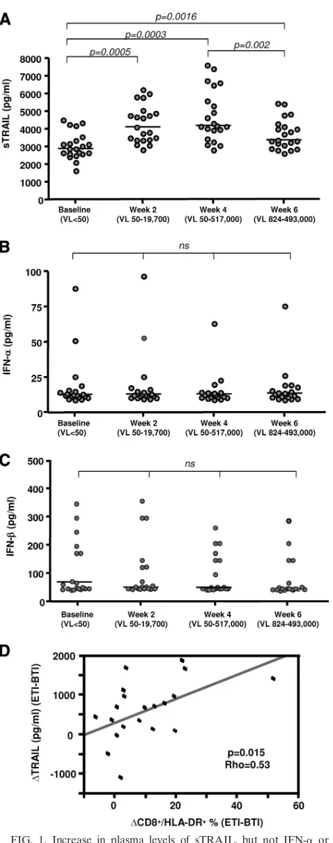
Immune activation after therapy interruption or upon chronic viremia is associated with an increase in sTRAIL but not in plasma IFN-␣/IFN-.IFN-␣has been shown to induce robust TRAIL expression, and PDC have been proposed to contribute to increased levels of sTRAIL and IFN-␣in chronic HIV-1 infection (40). Similar to TNF, biologically active TRAIL can be present as both soluble and membrane-bound forms. To study the relationship between plasma TRAIL and IFN-␣/IFN- in the presence of viral replication during chronic HIV-1 infection, we evaluated plasma levels of TRAIL and IFN-␣/IFN-in a longitudinal cohort (n⫽21) before and after ART interruption and viral rebound. As previously ported (60), a 6-week ART interruption resulted in viral re-bound (from⬍50 copies/ml to a range of 824 to 493,000 cop-ies/ml) and activation-induced changes in both CD4 and CD8 T-cell compartments. As shown in Fig. 1A, ART interruption induced an acute increase in TRAIL plasma levels. This in-crease was noticeable as early as 2 weeks after TI, when viral rebound was minimum, and peaked at week 4, before decreas-ing at week 6 at maximum viral load. However, no changes in the observed circulating IFN-␣ or IFN- plasma levels were detected at any time point tested (Fig. 1B and C). Correlation analysis showed a positive association between TRAIL and viral load (P⫽0.023; Spearman’s ⫽0.493). The change in the frequency of CD8⫹HLA-DR⫹T cells (used as a marker of viremia-induced immune activation) and the change in soluble TRAIL levels during a 6-week TI were also correlated (as well
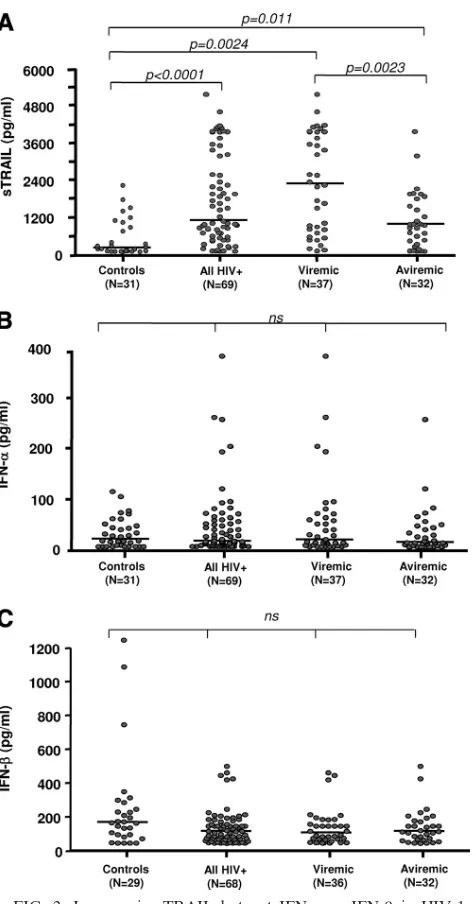
as 2- and 4-week TI [not shown]; CD8⫹HLA-DR⫹,P⫽0.015, Spearman’s ⫽0.5353) (Fig. 1D), yet no association was noted between T-cell activation and plasma IFN-␣/IFN-levels (data not shown). The differential regulation between plasma levels of TRAIL and IFN-␣/IFN-was also reconfirmed in a cross-sectional comparison of 69 HIV⫹ subjects with uninfected controls (Fig. 2A). While plasma levels of TRAIL were signif-icantly higher in HIV⫹viremic subjects than in aviremic and uninfected control subjects, no difference in levels of IFN-␣ (Fig. 2B) or IFN-was observed (median, 170 pg/ml in con-trols, n ⫽ 24, versus 118 pg/ml in HIV⫹ subjects, n ⫽ 68; difference not significant) (Fig. 2C). Our results clearly dem-onstrated that, in our cohorts, increased viral replication is associated with increased plasma sTRAIL but not IFN-␣ or IFN-.
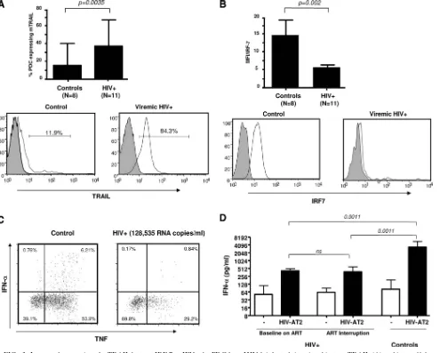
HIV-1 replication increases mTRAIL expression on circu-lating PDC in association with lower IRF-7 levels and no increase in TLR-mediated IFN-␣production.Having shown a lack of association between soluble TRAIL and soluble
IFN-␣/IFN-〉plasma levels, we evaluated mTRAIL expression on PDC from HIV⫹subjects and uninfected controls. A signifi-cant increase in the percentage of PDC expressing mTRAIL (as well as MFI) was observedex vivoin HIV-infected subjects compared to uninfected controls (median, 37.20% in HIV⫹ subjects versus 16.80% in controls; P ⫽ 0.0035) (composite data and representative diagrams are shown in Fig. 3A). Based on several reports, including ours, describing a profound im-pairment in IFN-␣production by PDC during chronic viremia following TLR engagement (13, 14, 49) and because IRF-7 has been identified as the key player in induced type I IFN pro-duction (45, 46), we measured levels of intracellular IRF-7 in PDC. Using a polyclonal antibody that recognizes total IRF-7 (phosphorylated and unphosphorylated forms [16, 31, 56]), we observed that the constitutive intracellular levels of IRF-7 were lower in circulating PDC from chronically HIV-1-infected sub-jects than in those from uninfected control subsub-jects (P⬍0.002) (Fig. 3B shows composite data and representative diagrams). In agreement with our previous work (10), we observed a low intracellular expression of IFN-␣ and TNF-␣ following PDC stimulation with resiquimod in HIV-infected PBMC (Fig. 3C). We also evaluated PDC of HIV⫹subjects, before and after short-term ART interruption, to determine if PDC be-came immediately refractory to stimulation with HIV upon viremia. We stimulated PBMC for 18 h with HIV AT2 (300 ng/ml) and tested for the presence of IFN-␣ by ELISA. As depicted in Fig. 3D and consistent with a partial recovery of PDC function under ART (10), we observed a “net IFN-␣ response” to HIV AT2 stimulation in samples before ART interruption (median, 52 pg/ml without stimulation versus 409 pg/ml after stimulation) and after 6 weeks of ART interruption (62 pg/ml without stimulation versus 378 pg/ml after stimula-tion). Levels measured were significantly higher than those during long-term viremia in chronic infection (data not shown) (10) but lower than levels seen in controls (median, 2,100 pg/ml), indicating that, in spite of increases in TRAIL and a partial retention of PDC function upon acute viremia, levels of plasma IFN-␣remained unchanged, as shown above.
DR5 expression is not increased in circulating primary CD4ⴙT cells from chronically infected HIVⴙsubjects or after
in vitroHIV-1 exposure/infection of primary CD4ⴙT cells.We
on November 8, 2019 by guest
http://jvi.asm.org/
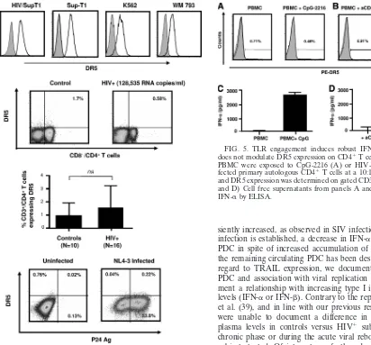
then sought to determine whether an increase in CD4⫹T cells expressing TRAIL receptor (DR5) was present in HIV-1 in-fection, allowing for direct effects by TRAIL-expressing PDC as a potential effector mechanism of CD4 T-cell death. To determine the specificity of the anti-DR5 antibody used by others (38) and in this study, we observed that Sup-T1 (in-fected or not), K562, and WM793 cells expressed high DR5 levels (Fig. 4A). Thus, we used four independent experimental approaches to determine whether HIV infection was associ-ated with DR5 induction in primary CD4⫹T cells. First, we measured DR5 expression in circulating CD4⫹T cells from a cross-sectional HIV⫹cohort including subjects with long-term viremia and subjects undergoing an acute viremic episode (i.e., as a result of a 6-week ART interruption) utilized for the studies described above. As shown in Fig. 4B and C, and consistent with results described by others (36, 68), circulating CD4⫹T cells from all HIV⫹subjects tested showed similar low levels of DR5 expression compared to those from uninfected controls. No increase in DR5 levels (percent positive cells and MFI) was observed during rebound viremia following ART interruption in 3 HIV⫹ subjects tested at week 6 of ART interruption (data not shown). Second, analysis of in vitro -isolated primary CD4⫹ T cells infected for 3 to 4 days with HIV-1NL4-3did not show upregulation of DR5 expression on
CD3⫹ CD4⫹ gated p24 antigen-positive cells (also showing downregulation of CD4 expression), indicating that in vitro
infection was not able to induce measurable membrane DR5 expression (Fig. 4D). Similar results were obtained using X4 and R5 HIV-1 isolates (not shown). Third, stimulation of PBMC from control uninfected subjects with a TLR9 agonist
orin vitroHIV-infected primary autologous CD4⫹T cells for
[image:4.585.43.282.65.669.2]24 h did not result in an increase in DR5 expression in CD4⫹ T cells gated from PBMC (Fig. 5A and B) despite the fact that in the same cultures high levels of IFN-␣, indicative of an intact PDC response to activation in uninfected controls, were observed (Fig. 5C and D). Finally, we exposed control unin-fected PBMC to conditions that have been reported to induce an increase in DR5 mRNA expressionin vitro(40), i.e., infec-tious HIV-1NL4-3, noninfectious HIV-1NL4-3(AT2 treated and heat killed), exogenous IFN-␣ (10,000 U/ml), and various other stimuli (resiquimod at 10 g/ml, heat-killed influenza virus PR8 at 10 hemagglutinin (HA)/ml, and lipopolysaccha-ride [LPS] at 100 ng/ml) for 24 h. As shown in Table 2, DR5 expression within CD4⫹T cells did not increase after 24 h of exposure (as well as 48 and 72 h [data not shown]) regardless of stimuli used, although robust secretion of IFN-␣ and sTRAIL was observed as described above. Of interest, we have tested several currently available TRAIL-R2/DR5 anti-bodies (eBioscience [used throughout this study], Biolegend, and R&D Systems), and none of them detected an upregula-tion in DR5 expression on primary CD4⫹T cells, while all of
FIG. 1. Increase in plasma levels of sTRAIL, but not IFN-␣ or IFN-, in HIV-1-infected subjects after viral rebound during ART interruption. Soluble TRAIL (A), IFN-␣ (B), and IFN- (C) levels were determined by ELISA in cryopreserved plasma samples from HIV-1-infected subjects at baseline (n⫽21) and after 2, 4, and 6 weeks of ART interruption (n⫽21). Horizontal bars represent the
median values for each group, and brackets indicate statistical signif-icance by Wilcoxon signed rank tests. ns, not significant; VL, viremia level (copies of HIV-1 RNA/ml). (D) Association between increased levels of TRAIL and T-cell activation was determined by the Spear-man rank correlation. ETI, end of 6-week treatment interruption; BTI, beginning of 6-week treatment interruption.
on November 8, 2019 by guest
http://jvi.asm.org/
them detected high levels of DR5 in various cell lines (K562, Sup-T1 infected or not with HIV, and WM793). Taken to-gether, these results failed to document an increased DR5 expression in primary CD4⫹ T cells in vivo during viral re-bound, during chronic HIV-1 viremia, or afterin vitroHIV-1 exposure/PDC activation, indicating that PDC activation alone is unlikely to account for major induction of CD4 T-cell apop-tosis.
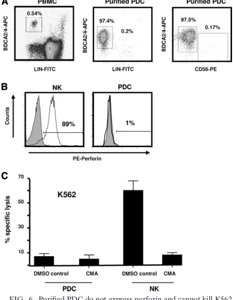
Activated PDC provide accessory help for NK cell-mediated lysis of primary autologous CD4ⴙ T cells. Previous studies have documented the ability of PDC to lyse tumor targets (9), and a recent report by Hardy et al. (37) predicted that PDC could kill autologous HIV-infected targets based on the ability of PDC to lyse transformed cells expressing DR5, such as Sup-T1 cells. In order to understand the lytic ability of PDC, we first evaluated the presence of perforin in highly purified PDC depleted of residual NK cells (Fig. 6A). While PDC expressed variable amounts of TRAIL (Fig. 3A), expression of intracellular perforin was very low or absent compared to that by purified NK cells, used as a positive control (Fig. 6B). While these results make it unlikely that PDC could mediate per-forin-dependent lysis, we directly evaluated the cytotoxic po-tential of purified activated PDC depleted of residual NK cells compared to that of purified NK cells for lytic activity against K562 cells, which express high levels of DR5 but are highly resistant to TNF-␣, Fas-L, and TRAIL-induced apoptosis (43). As shown in Fig. 6C, activated PDC (containing⬃0.1 to 0.5% contaminating NK cells) exhibited very low to no cytotoxic activity against K562 at an E:T ratio of 10:1 compared to purified NK cells. Of interest, when an E:T ratio of 20:1 was used in preliminary experiments, the percentage of specific killing did not increase and remained similar to the level seen at an E:T ratio of 10:1 (results not shown). Addition of con-canamycin A (CMA), a potent inhibitor of perforin-mediated killing, produced a dramatic reduction in NK cell-mediated lysis but had no effect on the activity of PDC (Fig. 6C).
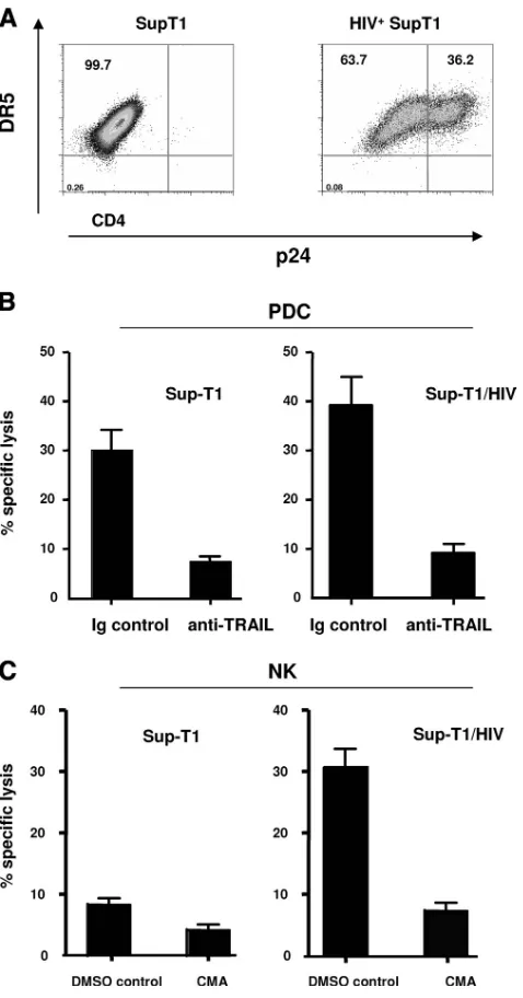
We then analyzed PDC-mediated cytotoxic killing of DR5-expressing Sup-T1 (infected or uninfected) cells. Sup-T1 cells were acutely infected for 3 days and used as targets. Activated NK-depleted purified PDC were incubated with chromium-labeled Sup-T1 cells at an E:T ratio of 10:1 for 6 h. Figure 7A depicts the levels of DR5 and p24 Ag in the infected cells. As shown in Fig. 7B, and consistent with the observations of Hardy et al. (37) describing the killing of Sup-T1 cells by PDC, activated PDC efficiently induced cytolysis of DR5-expressing HIV-infected Sup-T1 cells. In addition, we also show that PDC killing of HIV-infected Sup-T1 cells was dependent on TRAIL, as addition of neutralizing antibodies against TRAIL abolished the killing (Fig. 7B). In parallel experiments and as a control, we show that granule release plays a dominant role in the NK-mediated killing of Sup-T1 cells.
Having shown a different lytic mechanism between PDC and NK cells, we next sought to determine whether PDC activated
byin vitro HIV-1-infected autologous primary CD4⫹T cells
[image:5.585.44.282.63.515.2](HIV/aCD4) alone could provide accessory help to activate NK cells and enhance NK cell-mediated cytotoxic killing. As depicted in Fig. 8A, total PBMC and isolated PDC produced high levels of IFN-␣ when stimulated with HIV/aCD4. HIV/ aCD4-stimulated PBMC but not HIV/aCD4-stimulated PDC-depleted PBMC triggered activation of NK cells, as indicated by CD69 upregulation (Fig. 8B) and enhanced lysis of HIV/ aCD4 cells (Fig. 8C). The latter results extended our previ-ously reported data using TLR-9 agonist-activated PDC (72). Because the lysis of physiologically relevant targets such as autologous CD4 primary T cells by PDCs has not been fully investigated, we tested whether the interaction between acti-vated PDC that expressed TRAIL andin vitroHIV-infected autologous CD4 T-cell targets could result in CD4⫹ T-cell
FIG. 2. Increase in sTRAIL but not IFN-␣ or IFN- in HIV-1-infected subjects during chronic viremia or after ART-induced viral suppression. Soluble TRAIL (A), IFN-␣ (B), and⌱F⌵-(C) levels were determined by ELISA in cryopreserved plasma samples from controls and HIV-1-infected subjects. Data from HIV-1⫹ subjects were segregated into viremic (n⫽37) and aviremic (n⫽32) groups. Horizontal bars represent the median values for each group, and brackets indicate statistical significance by Mann-Whitney U tests. ns, not significant.
on November 8, 2019 by guest
http://jvi.asm.org/
lysis. Acutely infected autologous CD4⫹T cells containing 30 to 60% p24 Ag⫹ cells were used as targets as described by Tomescu et al. (72). As a positive control and in agreement with our previous report (72), PDC-activated NK cells were cytotoxic for autologous infected targets (Fig. 9) through a perforin-mediated killing mechanism (not shown) (76). Unlike what was found for Sup-T1 cells and as depicted in Fig. 9, PDC failed to show any cytotoxic activity against in vitro HIV-1-infected primary autologous CD4⫹ T cells (Fig. 9). In two different experiments, the use of a higher E:T ratio (20:1) did not increase the level of cell-mediated killing by PDC (not shown).
Taken together, our data are consistent with the lack of activity by PDC to mediate TRAIL/DR5-dependent lysis of HIV/aCD4 cells, while reconfirming in the same autologous
system the potential for IFN-␣-producing PDC to activate functional NK cells to induce lysis of primary HIV/aCD4⫹T cells.
DISCUSSION
[image:6.585.46.541.69.466.2]In this report, we provide the first evidence that plasma levels of sTRAIL and its expression on the PDC membrane are associated with HIV-1 replication in vivo, in the absence of detectable plasma IFN-␣or IFN-. We also provide new data on the lack of activity by PDC to mediate CD4 T-cell apoptosis by showing that PDC activated following interaction with HIV-infected primary CD4 T cells do not directly lyse HIV-HIV-infected autologous CD4⫹T cells via TRAIL/DR5 but rather enhance NK cell-mediated cytotoxicity.
FIG. 3. Increased expression of mTRAIL but not IRF-7 or IFN-␣by PDC from HIV-1-infected viremic subjects. mTRAIL (A) and intracellular IRF-7 (B) levels were determined by flow cytometry on freshly isolated PBMC-gated BDCA2/4⫹PDC. Bar graphs represent composite data from 19 different donors. Representative diagrams for controls and HIV⫹subjects are shown below the graphs. Medians and ranges for percentages of PDC expressing mTRAIL and fluorescence intensity (MFI) for IRF-7 are shown. (C) Determination of the intracellular expression of IFN-␣and TNF-␣was performed by intracellular flow cytometry on freshly isolated PBMC after resiquimod stimulation. (D) IFN-␣production by PDC from 10 HIV⫹subjects before and after ART interruption and from 16 controls in response to 18 h of HIV-AT2 stimulation was determined by ELISA. Brackets indicate statistical significance by Mann-Whitney U tests or Wilcoxon signed rank tests.
on November 8, 2019 by guest
http://jvi.asm.org/
The activation of innate and adaptive responses following acute HIV-1 infection would predict that TRAIL and IFN-␣ could be positively correlated if responses by PDC remained functional in spite of continued viral replication. Indeed, re-cent data from acute infection by simian immunodeficiency virus in sooty mangabeys (SIVsm) show a relationship between increasing IFN-␣ plasma levels and acute viremia, yet IFN-␣ levels subsequently decrease in spite of sustained viremia (19). Studies of acute infection in humans have also shown a severe loss of circulating PDC, with conflicting data regarding sus-tained plasma IFN-␣ levels, which are expected to be
tran-siently increased, as observed in SIV infection. Once chronic infection is established, a decrease in IFN-␣production from PDC in spite of increased accumulation of IFN-␣mRNA in the remaining circulating PDC has been described (50). With regard to TRAIL expression, we document its induction in PDC and association with viral replication yet do not docu-ment a relationship with increasing type I interferon plasma levels (IFN-␣or IFN-). Contrary to the report by Herbeuval et al. (39), and in line with our previous results (10, 11), we were unable to document a difference in IFN-␣ or IFN- plasma levels in controls versus HIV⫹ subjects during the chronic phase or during the acute viral rebound in all HIV⫹ subjects tested. Of interest, we further document that, even with retained partial PDC function during a short-term period of viremia, no change in plasma IFN-␣/IFN-or increase in IFN-␣ expression was noted in PDC upon viral replication. With chronic viremia, an increasingly impaired PDC response is evidenced by a decrease in IRF-7 as a central transcriptional mediator of IFN production. Decreased IRF-7 levels further indicate an absence of a positive feedback of IFN receptor activation, leading to increases of IRF-7, as otherwise expected if PDC were continually responding to type I receptor activa-tion and in return had the potential for higher IFN-␣ produc-tion upon activaproduc-tion.
As suggested by Kim et al. (48), in human and nonhuman primates the presence of significantly high levels of sTRAIL but only very low levels of IFN-␣ in HIV⫹ subjects tested suggests that IFN-␣alone is likely not a major factor sustaining elevated TRAIL expression upon chronic viremia. Besides apop-tosis induction, TRAIL has also been shown to have antiapop-totic (prosurvival), regulatory, and proliferative functions (7, 21, 50, 64, 73), and its role in HIV infections is likely to be more complex than proposed. Interestingly, a recent report by Shepard et al. clearly showed the beneficial effect of TRAIL by demonstrating a reduction in viral load without significant death of bystander cells (65).
[image:7.585.55.467.66.449.2]Because binding of TRAIL to its cognate receptors induces
FIG. 4. DR5 surface expression in cell lines and primary cells. (A) DR5 surface expression was determined on HIV-1-infected Sup-T1, uninfected Sup-Sup-T1, and K562 cells and the melanoma cell line WM793, used as the control. Gray histograms represent the isotype control, and open histograms represent DR5. (B) Representative staining for DR5 expression on freshly isolated CD8⫺CD4⫹gated T cells is shown for one control subject and one viremic subject. (C) Composite data showing medians and ranges for DR5 expression from 10 controls and 16 HIV-1 infected subjects. Brackets indicate statistical differences by Mann-Whitney U tests. ns, not significant. (D) Purified primary CD4ⴙT cells from a control donor were infected
in vitrowith HIV-1NL4-3for 4 days and stained for DR5 expression
along with intracellular p24 Ag. One representative experiment of five is shown.
FIG. 5. TLR engagement induces robust IFN-␣ production but does not modulate DR5 expression on CD4⫹T cells. Freshly isolated PBMC were exposed to CpG-2216 (A) or HIV-1-infected or unin-fected primary autologous CD4⫹T cells at a 10:1 ratio (B) for 18 h, and DR5 expression was determined on gated CD3⫹CD4⫹T cells. (C and D) Cell free supernatants from panels A and B were tested for IFN-␣by ELISA.
on November 8, 2019 by guest
http://jvi.asm.org/
[image:7.585.43.258.68.449.2]apoptosis, we analyzed in depth the expression of DR5 during HIV infection using several experimental approaches. Al-though Herbeuval et al. reported an increase of DR5 mRNA expression (38, 40, 41), we were unable to document an
in-crease in DR5 surface expression in primary cell cultures ex-posed to or followingin vitroinfection with HIV-1 to establish TRAIL/DR5 as a major virally induced mechanism of lympho-cyte depletion. Most importantly, we failed to demonstrate an ability of activated PDC to lyse primary autologous CD4 T-cell targets whether infected or not with HIV-1, although we ob-served TRAIL-dependent lysis of a DR5-expressing Sup-T1 cell line (infected or not with HIV). Our inability to document direct lysis stands in contrast to a recent report by Stary and colleagues showing an increase in DR4 expression in CD4⫹T cells from HIV⫹subjects compared to controls (5% in viremic subjects, 2% in ART-treated subjects, and 1% in controls), where approximately 30% killing of autologous CD4⫹T cells by PDC was shown (68). Data in the above-mentioned report that remain unexplained include the high level of killing along with a low level of DR4 expressed on autologous CD4⫹T cells, which we have also confirmed is similar to the level of DR5, as DR4 is not upregulated in infected T-cell targets (data not shown).
While our study clearly showed that circulating CD4 cells do not express higher levels of DR5 as a result of exposure to increasing levels of viremia following therapy interruption, it remains to be determined whether surface DR5 or another death receptor is expressed in tissue lymphocytes from HIV-1-infected patients (68). With regard to tissue distribution, PDC in tissues have not been consistently reported as highly prevalent, as reported by Biancotto et al., who used LN of HIV⫹ subjects (4). Furthermore, Brown et al., using rhesus macaques infected with pathogenic SIV (6), also showed that tissue PDC are lost rather than recruited to lymphoid tissue in advanced HIV or SIV infection. Although these data do not identify the mechanisms responsible for the decrease in num-ber and function of circulating PDC during chronic HIV in-fection (i.e., homing, direct inin-fection, apoptosis [10, 11, 18, 24, 37, 40, 54, 57]), they support a loss of PDC both in blood and tissues. Because HIV can directly infect and kill PDC (57), data to date have not conclusively addressed whether the sys-temic loss of PDC during chronic HIV-1 infection is due to virally mediated depletion or redistribution of PDC to tissues. The role of IFN-␣in the pathogenesis of HIV-1 is, at this time, unclear and paradoxical (26, 27, 42, 47, 58). Besides its well-documented potent antiviral propertiesin vivoandin vitro
[image:8.585.46.543.80.189.2]and the stimulation of the Th-1 response described to be im-portant in HIV-1 resistance (13), early work, pioneered by
TABLE 2. TRAIL, IFN-␣, and DR5 levels followingin vitrostimulationa
Treatment
Level (pg/ml) of induced:
% DR5⫹CD4⫹T cells
IFN-␣ sTRAIL
None 157 (110–192) 199 (110–299) 3.8 (2.5–5)
NL4-3 5,706 (4,210–7,260) 8,475 (6,541–8,674) 2.3 (1.8–2.8)
AT2–NL4-3 6,647 (5,297–8,210) 8,111 (7,023–9,201) 2.0 (1.1–2.9)
HI–NL4-3 137 (99–202) 131 (98–176) 3.1 (1.5–4.7)
IFN-␣ NAb 11,095 (8,941–14,231) 1.55 (1.2–2.1)
PR8 5,648 (4,987–6,945) 7,633 (5,120–8,457) 1.63 (0.9–2.1)
Resiquimod 10,806 (9,847–12,324) 6,781 (5,634–8,457) 1.15 (0.56–1.9)
LPS 212 (125–299) 9,290 (8,457–10,123) 1.02 (0.75–1.3)
a
Freshly isolated PBMC were incubated in the presence or absence of 300 ng/ml p24 Ag equivalent NL4-3, AT2-treated NL4-3, heat-inactivated (HI) NL4-3, human IFN-␣(10,000 U/ml), influenza virus strain PR8 (10 HA/ml), resiquimod (10g/ml), or LPS (100 ng/ml) for 18 h. Following incubation, DR5 expression was tested on CD3⫹CD4⫹T cells and cell-free supernatants were assayed for sTRAIL and IFN-␣by ELISA. Values are means (ranges) of 3 different experiments.
b
NA, not applicable.
FIG. 6. Purified PDC do not express perforin and cannot kill K562 target cells. (A) PDC were enriched to high purity from PBMC by negative selection with magnetic beads and stained with monoclonal antibodies to BDCA-2/4, lineage (LIN) cocktail mix, and CD56. The frequencies of PDC before and after purification and the frequency of contaminating CD56⫹NK cells are shown. (B) Perforin expression in CD56⫹ CD3⫺ gated NK cells and BDCA-2⫹/4⫹ LIN⫺ gated PDC. (C) Purified PDC and NK cells were used as effector cells in a chro-mium release assay with DR5-expressing K562 targets in the presence or absence of 1,000 nM concanamycin A (CMA) at a 10:1 E:T ratio for 6 h (PDC) or 4 h (NK). Means ⫾ standard errors of 4 different experiments are shown. DMSO, dimethyl sulfoxide.
on November 8, 2019 by guest
http://jvi.asm.org/
[image:8.585.41.280.306.614.2]Gendelman’s group and others (8, 29, 30, 69), demonstrated biological differences in IFN-␣proteins produced during ad-vanced HIV infection compared to those of the controls. A limitation of our study is that the HIV⫹cohort analyzed does not include patients with opportunistic infections or with end stage disease (i.e., CD4 counts ⬍ 50 copies/ml), which may represent an immunopathogenic situation different from those described in the present study, with steady-state viremia and higher CD4 counts.
Our data strongly support a predominant antiviral role for PDC and IFN-␣in the pathogenesis of HIV-1, although their possible contribution to the induction of factors associated with immunodeficiency such as indoleamine 2,3-dioxygenase (IDO), as shownin vitro(5), remains to be establishedin vivo. Furthermore, recent work by the Bhardwaj laboratory (53) elegantly described a dichotomy in the response of PDC to HIV: enhancing innate and adaptive responses to HIV and at the same time limiting chronic immune activation through generation of regulatory T cells (Tregs). While the in vivo
activation of IFN-inducible genes in circulating PBMC during chronic HIV infection suggests the activation of IFN-mediated signaling, described in several reports (reviewed in reference 47), it remains to be determined to what extent this IFN sig-nature reflects the effect of IFN-␣ alone rather than IFN-␥, known to be produced by activated CD8 T cells in the lym-phoid organs of HIV-1-infected patients (17, 22). Our findings do not support a sustained role for PDC through TRAIL expression and IFN-␣/IFN-secretion as being responsible for a progressive T-cell loss over the extended period of chronic infection but instead indicate that PDC are decreased in num-ber and impaired in function without a potential to mediate CD4 T-cell loss through TRAIL. Our data instead support the hypothesis that, if PDC are functional and activated in the presence of functional NK cells, as observed in long-term non-progressors (67), a predominant antiviral response, including activation of NK cell-mediated responses to limit viral replica-tion, would be expected.
[image:9.585.43.282.66.522.2]HIV-induced dysregulation of dendritic cell (DC)/NK cell cross talk may represent a major mechanism through which HIV
FIG. 7. PDC efficiently kill DR5-expressing Sup-T1 cells (infected or not with HIV) but lack lytic activity against HIV-infected primary autologous CD4⫹T cells. (A) DR5 and p24 Ag expression in Sup-T1 cells. (B) Purified PDC and NK cells were incubated with51Cr-labeled
DR5-expressing Sup-T1 cells or HIV-infected Sup-T1 cells in the pres-ence of 10g/ml control antibody or 10g/ml anti-TRAIL (B) or 1,000 nM CMA (C) for 6 h at a 10:1 E:T ratio. Results represent means⫾standard errors for 4 different donors.
FIG. 8. PDC accessory cell help is required to activate NK cells to lyse HIV-1-infected primary aCD4⫹T cells. (A) Total PBMC, PDC-depleted PBMC, or purified PDC were incubated with HIV-1-infected or uninfected aCD4ⴙT cells for 18 h at a 10:1 E:T ratio, and super-natants were tested for IFN-␣secretion by ELISA. (B) Staining with CD69 antibody is shown on CD3ⴚCD56ⴚgated NK cells. Gray grams represent isotype-matched control antibody, and open histo-grams represent CD69.
on November 8, 2019 by guest
http://jvi.asm.org/
escapes immune surveillance (14, 61, 62), and several observa-tions support a role for PDC/NK cell responses in HIV-1 control (10, 27, 66, 72). Our data predict that, in contrast to blocking systemic production of IFN-␣, as recently suggested (42, 71, 74), IFN-␣would mediate viral control in the presence of ART-me-diated immune reconstitution, as already suggested by the de-creased HIV titer observed in HIV/hepatitis C virus (HCV)-coinfected subjects treated with IFN-␣/ribavirin (59). Current clinical studies sponsored by our laboratory in which patients on ART are treated with pegylated IFN-␣2a upon ART interruption are directly testing this hypothesis (http://www.clinicaltrials.org/ identifier: NCT00594880). Taken together, our findings challenge the hypothesis of a detrimental role for PDC in AIDS pathogen-esis and suggest an IFN-␣-independent pathway for the progres-sive loss of circulating CD4 T cells during HIV infection.
ACKNOWLEDGMENTS
This work was supported by National Institutes of Health grants AI 51225, UO1 AI065279, and AI 073219 (to L.J.M.); AI 0587780 (to J.C.); and AI 068405 (to C.T.). Support was also provided by the Philadelphia Foundation and the Fund from the Commonwealth Uni-versal Research Enhancement Program, Pennsylvania Department of Health.
J.C., E.P., and C.T. contributed equally to this work.
We thank all the study participants; C. Gallo and the nursing staff at Philadelphia FIGHT/Jonathan Lax Clinic; D. Davis (phlebotomist at the Wistar Blood Donor Program); J. S. Faust, D. E. Ambrose, and D. Hussey at The Wistar Institute Flow Cytometry Core facility; and F. Shaheen (supervisor, Center for AIDS Research Viral Core Facility, University of Pennsylvania) for all HIV-1 strains used in this study.
REFERENCES
1.Argyris, E. G., E. Acheampong, F. Wang, J. Huang, K. Chen, M. Mukhtar, and H. Zhang.2007. The interferon-induced expression of APOBEC3G in human blood-brain barrier exerts a potent intrinsic immunity to block HIV-1 entry to central nervous system. Virology367:440–451.
2.Audige, A., M. Urosevic, E. Schlaepfer, R. Walker, D. Powell, S. Hallen-berger, H. Joller, H. U. Simon, R. Dummer, and R. F. Speck.2006. Anti-HIV state but not apoptosis depends on IFN signature in CD4⫹T cells. J. Im-munol.177:6227–6237.
3.Beignon, A. S., K. McKenna, M. Skoberne, O. Manches, I. DaSilva, D. G. Kavanagh, M. Larsson, R. J. Gorelick, J. D. Lifson, and N. Bhardwaj.2005. Endocytosis of HIV-1 activates plasmacytoid dendritic cells via Toll-like receptor-viral RNA interactions. J. Clin. Investig.115:3265–3275. 4.Biancotto, A., J. C. Grivel, S. J. Iglehart, C. Vanpouille, A. Lisco, S. F. Sieg,
R. Debernardo, K. Garate, B. Rodriguez, L. B. Margolis, and M. M. Led-erman.2007. Abnormal activation and cytokine spectra in lymph nodes of people chronically infected with HIV-1. Blood109:4272–4279.
5.Boasso, A., J. P. Herbeuval, A. W. Hardy, S. A. Anderson, M. J. Dolan, D. Fuchs, and G. M. Shearer.2007. HIV inhibits CD4⫹T-cell proliferation by inducing indoleamine 2,3-dioxygenase in plasmacytoid dendritic cells. Blood
109:3351–3359.
6.Brown, K. N., A. Trichel, and S. M. Barratt-Boyes.2007. Parallel loss of myeloid and plasmacytoid dendritic cells from blood and lymphoid tissue in simian AIDS. J. Immunol.178:6958–6967.
7.Budd, R. C.2002. Death receptors couple to both cell proliferation and apoptosis. J. Clin. Investig.109:437–441.
8.Capobianchi, M. R., F. De Marco, P. Di Marco, and F. Dianzani.1988. Acid-labile human interferon alpha production by peripheral blood mono-nuclear cells stimulated by HIV-infected cells. Arch. Virol.99:9–19. 9.Chaperot, L., A. Blum, O. Manches, G. Lui, J. Angel, J. P. Molens, and J.
Plumas.2006. Virus or TLR agonists induce TRAIL-mediated cytotoxic activity of plasmacytoid dendritic cells. J. Immunol.176:248–255. 10.Chehimi, J., L. Azzoni, M. Farabaugh, S. A. Creer, C. Tomescu, A. Hancock,
A. Mackiewicz, L. D’Alessandro, S. Ghanekar, A. S. Foulkes, K. Mounzer, J. Kostman, and L. J. Montaner.2007. Baseline viral load and immune acti-vation determine the extent of reconstitution of innate immune effectors in HIV-1-infected subjects undergoing antiretroviral treatment. J. Immunol.
179:2642–2650.
11.Chehimi, J., D. E. Campbell, L. Azzoni, D. Bacheller, E. Papasavvas, G. Jerandi, K. Mounzer, J. Kostman, G. Trinchieri, and L. J. Montaner.2002. Persistent decreases in blood plasmacytoid dendritic cell number and func-tion despite effective highly active antiretroviral therapy and increased blood myeloid dendritic cells in HIV-infected individuals. J. Immunol.168:4796– 4801.
12.Chehimi, J., S. E. Starr, I. Frank, M. Rengaraju, S. J. Jackson, C. Llanes, M. Kobayashi, B. Perussia, D. Young, E. Nickbarg, et al.1992. Natural killer (NK) cell stimulatory factor increases the cytotoxic activity of NK cells from both healthy donors and human immunodeficiency virus-infected patients. J. Exp. Med.175:789–796.
13.Clerici, M., and G. M. Shearer.1993. A TH13TH2 switch is a critical step in the etiology of HIV infection. Immunol. Today14:107–111.
14.Conry, S. J., K. A. Milkovich, N. L. Yonkers, B. Rodriguez, H. B. Bernstein, R. Asaad, F. P. Heinzel, M. Tary-Lehmann, M. M. Lederman, and D. D. Anthony.2009. Impaired PDC-NK activity in viremic HIV infection attrib-utable to impairments in both PDC and NK function. J. Virol.83:11175– 11187.
15.Dai, J., N. J. Megjugorac, S. B. Amrute, and P. Fitzgerald-Bocarsly.
2004. Regulation of IFN regulatory factor-7 and IFN-alpha production by enveloped virus and lipopolysaccharide in human plasmacytoid dendritic cells. J. Immunol.173:1535–1548.
16.Danis, B., T. C. George, S. Goriely, B. Dutta, J. Renneson, L. Gatto, P. Fitzgerald-Bocarsly, A. Marchant, M. Goldman, F. Willems, and D. De Wit.
2008. Interferon regulatory factor 7-mediated responses are defective in cord blood plasmacytoid dendritic cells. Eur. J. Immunol.38:507–517. 17.Di Domizio, J., A. Blum, M. Gallagher-Gambarelli, J. P. Molens, L.
Chap-erot, and J. Plumas.2009. TLR7 stimulation in human plasmacytoid den-dritic cells leads to the induction of early IFN-inducible genes in the absence of type I IFN. Blood114:1794–1802.
18.Dillon, S. M., K. B. Robertson, S. C. Pan, S. Mawhinney, A. L. Meditz, J. M. Folkvord, E. Connick, M. D. McCarter, and C. C. Wilson.2008. Plasmacy-toid and myeloid dendritic cells with a partial activation phenotype accumu-late in lymphoid tissue during asymptomatic chronic HIV-1 infection. J. Acquir Immune Defic. Syndr.48:1–12.
19.Diop, O. M., M. J. Ploquin, L. Mortara, A. Faye, B. Jacquelin, D. Kunkel, P. Lebon, C. Butor, A. Hosmalin, F. Barre-Sinoussi, and M. C. Muller-Trutwin.
2008. Plasmacytoid dendritic cell dynamics and alpha interferon production during simian immunodeficiency virus infection with a nonpathogenic out-come. J. Virol.82:5145–5152.
20.Ehrhard, S., M. Wernli, G. Kaufmann, G. Pantaleo, G. P. Rizzardi, F. Gudat, P. Erb, and M. Battegay.2008. Effect of antiretroviral therapy on FIG. 9. PDC lack lytic activity against HIV-infected primary
autol-ogous CD4⫹ T cells. (A) Purified NK cells were incubated in the presence or absence of autologous PDC and the TLR9 agonist CpG-2216 for 18 h at a 10:1 NK cell/PDC ratio. Cells were then incubated with chromium-labeled primary HIV-1-infected aCD4⫹ T cells at a 10:1 E/T ratio for 6 h in a standard chromium release assay. (B) Rest-ing or activated PDC or activated NK cells were incubated with chro-mium-labeled primary HIV-1-infected aCD4⫹ T cells at a 10:1 E/T ratio for 6 h. Means⫾standard errors of 4 to 6 different experiments
are shown.
on November 8, 2019 by guest
http://jvi.asm.org/
apoptosis markers and morphology in peripheral lymph nodes of HIV-infected individuals. Infection36:120–129.
21.Ehrhardt, H., S. Fulda, I. Schmid, J. Hiscott, K. M. Debatin, and I. Jer-emias.2003. TRAIL induced survival and proliferation in cancer cells resis-tant towards TRAIL-induced apoptosis mediated by NF-kappaB. Oncogene
22:3842–3852.
22.Emilie, D., M. Peuchmaur, M. C. Maillot, M. C. Crevon, N. Brousse, J. F. Delfraissy, J. Dormont, and P. Galanaud.1990. Production of interleukins in human immunodeficiency virus-1-replicating lymph nodes. J. Clin. Investig.
86:148–159.
23.Fauci, A. S.1993. Immunopathogenesis of HIV infection. J. Acquir Immune Defic. Syndr.6:655–662.
24.Feldman, S., D. Stein, S. Amrute, T. Denny, Z. Garcia, P. Kloser, Y. Sun, N. Megjugorac, and P. Fitzgerald-Bocarsly.2001. Decreased interferon-alpha production in HIV-infected patients correlates with numerical and func-tional deficiencies in circulating type 2 dendritic cell precursors. Clin. Im-munol.101:201–210.
25.Finkel, T. H., G. Tudor-Williams, N. K. Banda, M. F. Cotton, T. Curiel, C. Monks, T. W. Baba, R. M. Ruprecht, and A. Kupfer.1995. Apoptosis occurs predominantly in bystander cells and not in productively infected cells of HIV- and SIV-infected lymph nodes. Nat. Med.1:129–134.
26.Fitzgerald-Bocarsly, P., J. Dai, and S. Singh.2008. Plasmacytoid dendritic cells and type I IFN: 50 years of convergent history. Cytokine Growth Factor Rev.19:3–19.
27.Fitzgerald-Bocarsly, P., and D. Feng.2007. The role of type I interferon production by dendritic cells in host defense. Biochimie89:843–855. 28.Fonteneau, J. F., M. Larsson, A. S. Beignon, K. McKenna, I. Dasilva, A.
Amara, Y. J. Liu, J. D. Lifson, D. R. Littman, and N. Bhardwaj.2004. Human immunodeficiency virus type 1 activates plasmacytoid dendritic cells and concomitantly induces the bystander maturation of myeloid dendritic cells. J. Virol.78:5223–5232.
29.Gendelman, H. E., R. M. Friedman, S. Joe, L. M. Baca, J. A. Turpin, G. Dveksler, M. S. Meltzer, and C. Dieffenbach.1990. A selective defect of interferon alpha production in human immunodeficiency virus-infected monocytes. J. Exp. Med.172:1433–1442.
30.Gendelman, H. E., R. M. Friedman, S. Joe, L. M. Baca, J. A. Turpin, G. Dveksler, M. S. Meltzer, and C. Dieffenbach.1991. A selective defect of interferon alpha production in human immunodeficiency virus-infected monocytes. J. Exp. Med.173:277.
31.George, T. C., P. J. Morrissey, C. Cui, S. Singh, and P. F. Bocarsly.2009. Measurement of cytoplasmic to nuclear translocation. Curr. Protoc. Cytom., chapter 9, unit 9.28.
32.Goila-Gaur, R., and K. Strebel.2008. HIV-1 Vif, APOBEC, and intrinsic immunity. Retrovirology5:51.
33.Gougeon, M. L.2003. Apoptosis as an HIV strategy to escape immune attack. Nat. Rev. Immunol.3:392–404.
34.Gougeon, M. L.2004. Apoptotic pathways triggered by HIV and conse-quences on T cell homeostasis and HIV-specific immunity. Prog. Mol. Sub-cell. Biol.36:95–115.
35.Gurney, K. B., A. D. Colantonio, B. Blom, H. Spits, and C. H. Uittenbogaart.
2004. Endogenous IFN-alpha production by plasmacytoid dendritic cells exerts an antiviral effect on thymic HIV-1 infection. J. Immunol.173:7269– 7276.
36.Hansjee, N., G. R. Kaufmann, C. Strub, R. Weber, M. Battegay, and P. Erb.
2004. Persistent apoptosis in HIV-1-infected individuals receiving potent antiretroviral therapy is associated with poor recovery of CD4 T lympho-cytes. J. Acquir Immune Defic. Syndr.36:671–677.
37.Hardy, A. W., D. R. Graham, G. M. Shearer, and J. P. Herbeuval.2007. HIV turns plasmacytoid dendritic cells (pDC) into TRAIL-expressing killer pDC and down-regulates HIV coreceptors by Toll-like receptor 7-induced IFN-alpha. Proc. Natl. Acad. Sci. U. S. A.104:17453–17458.
38.Herbeuval, J. P., J. C. Grivel, A. Boasso, A. W. Hardy, C. Chougnet, M. J. Dolan, H. Yagita, J. D. Lifson, and G. M. Shearer.2005. CD4⫹T-cell death induced by infectious and noninfectious HIV-1: role of type 1 interferon-dependent, TRAIL/DR5-mediated apoptosis. Blood106:3524–3531. 39.Herbeuval, J. P., A. W. Hardy, A. Boasso, S. A. Anderson, M. J. Dolan, M.
Dy, and G. M. Shearer.2005. Regulation of TNF-related apoptosis-inducing ligand on primary CD4⫹T cells by HIV-1: role of type I IFN-producing plasmacytoid dendritic cells. Proc. Natl. Acad. Sci. U. S. A.102:13974–13979. 40.Herbeuval, J. P., J. Nilsson, A. Boasso, A. W. Hardy, M. J. Kruhlak, S. A. Anderson, M. J. Dolan, M. Dy, J. Andersson, and G. M. Shearer.2006. Differential expression of IFN-alpha and TRAIL/DR5 in lymphoid tissue of progressor versus nonprogressor HIV-1-infected patients. Proc. Natl. Acad. Sci. U. S. A.103:7000–7005.
41.Herbeuval, J. P., J. Nilsson, A. Boasso, A. W. Hardy, M. Vaccari, V. Cecchi-nato, V. Valeri, G. Franchini, J. Andersson, and G. M. Shearer.2009. HAART reduces death ligand but not death receptors in lymphoid tissue of HIV-infected patients and simian immunodeficiency virus-infected ma-caques. AIDS23:35–40.
42.Herbeuval, J. P., and G. M. Shearer.2007. HIV-1 immunopathogenesis: how good interferon turns bad. Clin. Immunol.123:121–128.
43.Hietakangas, V., M. Poukkula, K. M. Heiskanen, J. T. Karvinen, L.
Sis-tonen, and J. E. Eriksson.2003. Erythroid differentiation sensitizes K562 leukemia cells to TRAIL-induced apoptosis by downregulation of c-FLIP. Mol. Cell. Biol.23:1278–1291.
44.Holmes, R. K., F. A. Koning, K. N. Bishop, and M. H. Malim. 2007. APOBEC3F can inhibit the accumulation of HIV-1 reverse transcription products in the absence of hypermutation. Comparisons with APOBEC3G. J. Biol. Chem.282:2587–2595.
45.Honda, K., Y. Ohba, H. Yanai, H. Negishi, T. Mizutani, A. Takaoka, C. Taya, and T. Taniguchi.2005. Spatiotemporal regulation of MyD88-IRF-7 signal-ling for robust type-I interferon induction. Nature434:1035–1040. 46.Honda, K., H. Yanai, H. Negishi, M. Asagiri, M. Sato, T. Mizutani, N.
Shimada, Y. Ohba, A. Takaoka, N. Yoshida, and T. Taniguchi.2005. IRF-7 is the master regulator of type-I interferon-dependent immune responses. Nature434:772–777.
47.Hosmalin, A., and P. Lebon.2006. Type I interferon production in HIV-infected patients. J. Leukoc. Biol.80:984–993.
48.Kim, N., A. Dabrowska, R. G. Jenner, and A. Aldovini.2007. Human and simian immunodeficiency virus-mediated upregulation of the apoptotic fac-tor TRAIL occurs in antigen-presenting cells from AIDS-susceptible but not from AIDS-resistant species. J. Virol.81:7584–7597.
49.Lapenta, C., S. M. Santini, E. Proietti, P. Rizza, M. Logozzi, M. Spada, S. Parlato, S. Fais, P. M. Pitha, and F. Belardelli.1999. Type I interferon is a powerful inhibitor of in vivo HIV-1 infection and preserves human CD4(⫹) T cells from virus-induced depletion in SCID mice transplanted with human cells. Virology263:78–88.
50.Levina, V., A. M. Marrangoni, R. DeMarco, E. Gorelik, and A. E. Lokshin.
2008. Multiple effects of TRAIL in human carcinoma cells: induction of apoptosis, senescence, proliferation, and cytokine production. Exp. Cell Res.
314:1605–1616.
51.Levy, J. A.1993. HIV and host immune responses in AIDS pathogenesis. J. Clin. Apher.8:19–28.
52.Levy, J. A.2006. HIV pathogenesis: knowledge gained after two decades of research. Adv. Dent. Res.19:10–16.
53.Manches, O., D. Munn, A. Fallahi, J. Lifson, L. Chaperot, J. Plumas, and N. Bhardwaj. 2008. HIV-activated human plasmacytoid DCs induce Tregs through an indoleamine 2,3-dioxygenase-dependent mechanism. J. Clin. In-vest.118:3431–3439.
54.Martinelli, E., C. Cicala, D. Van Ryk, D. J. Goode, K. Macleod, J. Arthos, and A. S. Fauci.2007. HIV-1 gp120 inhibits TLR9-mediated activation and IFN-{alpha} secretion in plasmacytoid dendritic cells. Proc. Natl. Acad. Sci. U. S. A.104:3396–3401.
55.McCune, J. M. 2001. The dynamics of CD4⫹ T-cell depletion in HIV disease. Nature410:974–979.
56.Megjugorac, N. J., E. S. Jacobs, A. G. Izaguirre, T. C. George, G. Gupta, and P. Fitzgerald-Bocarsly.2007. Image-based study of interferongenic interac-tions between plasmacytoid dendritic cells and HSV-infected monocyte-derived dendritic cells. Immunol. Invest.36:739–761.
57.Meyers, J. H., J. S. Justement, C. W. Hallahan, E. T. Blair, Y. A. Sun, M. A. O’Shea, G. Roby, S. Kottilil, S. Moir, C. M. Kovacs, T. W. Chun, and A. S. Fauci.2007. Impact of HIV on cell survival and antiviral activity of plasma-cytoid dendritic cells. PLoS One2:e458.
58.Muller-Trutwin, M., and A. Hosmalin.2005. Role for plasmacytoid dendritic cells in anti-HIV innate immunity. Immunol. Cell Biol.83:578–583. 59.Neumann, A., M. Polis, L. Rozenberg, J. Jackson, K. Reitano, M.
McLaugh-lin, C. Koratich, R. Dewar, H. Masur, B. Haagmans, and S. Kottilil.2007. Differential antiviral effect of PEG-interferon-alpha-2b on HIV and HCV in the treatment of HIV/HCV co-infected patients. AIDS21:1855–1865. 60.Papasavvas, E., J. R. Kostman, K. Mounzer, R. M. Grant, R. Gross, C.
Gallo, L. Azzoni, A. Foulkes, B. Thiel, M. Pistilli, A. Mackiewicz, J. Shull, and L. J. Montaner.2004. Randomized, controlled trial of therapy interrup-tion in chronic HIV-1 infecinterrup-tion. PLoS Med.1:e64.
61.Quaranta, M. G., A. Napolitano, M. Sanchez, L. Giordani, B. Mattioli, and M. Viora.2007. HIV-1 Nef impairs the dynamic of DC/NK crosstalk: differ-ent outcome of CD56(dim) and CD56(bright) NK cell subsets. FASEB J.
21:2323–2334.
62.Reitano, K. N., S. Kottilil, C. M. Gille, X. Zhang, M. Yan, M. A. O’Shea, G. Roby, C. W. Hallahan, J. Yang, R. A. Lempicki, J. Arthos, and A. S. Fauci.
2009. Defective plasmacytoid dendritic cell-NK cell cross-talk in HIV infec-tion. AIDS Res. Hum. Retroviruses25:1029–1037.
63.Rodriguez, B., M. M. Lederman, W. Jiang, D. A. Bazdar, K. Garate, C. V. Harding, and S. F. Sieg.2006. Interferon-alpha differentially rescues CD4 and CD8 T cells from apoptosis in HIV infection. AIDS20:1379–1389. 64.Secchiero, P., A. Gonelli, E. Carnevale, D. Milani, A. Pandolfi, D. Zella, and
G. Zauli.2003. TRAIL promotes the survival and proliferation of primary human vascular endothelial cells by activating the Akt and ERK pathways. Circulation107:2250–2256.
65.Shepard, B. D., D. De Forni, D. R. McNamara, A. Foli, S. A. Rizza, R. S. Abraham, K. Knutson, P. J. Wettstein, F. Lori, and A. D. Badley.2008. Beneficial effect of TRAIL on HIV burden, without detectable immune consequences. PLoS One3:e3096.
66.Siegal, F.2003. Interferon-producing plasmacytoid dendritic cells and the pathogenesis of AIDS. Res. Initiat. Treat. Action8:10–13.
on November 8, 2019 by guest
http://jvi.asm.org/
67.Soumelis, V., I. Scott, F. Gheyas, D. Bouhour, G. Cozon, L. Cotte, L. Huang, J. A. Levy, and Y. J. Liu.2001. Depletion of circulating natural type 1 interferon-producing cells in HIV-infected AIDS patients. Blood98:906– 912.
68.Stary, G., I. Klein, S. Kohlhofer, F. Koszik, T. Scherzer, L. Mullauer, H. Quendler, N. Kohrgruber, and G. Stingl.2009. Plasmacytoid dendritic cells express TRAIL and induce CD4⫹T cell apoptosis in HIV-1 viremic pa-tients. Blood114:3854–3863.
69.Swindells, S., T. Baldwin, C. Kelly, L. Baca-Regen, L. Loomis, D. Post, B. Brichacek, M. Stevenson, E. A. Dominguez, R. Reddy, R. Klein, M. J. Liao, D. Testa, T. McDonald, J. Bellanti, S. Skurkovich, and H. E. Gendelman.
1996. Regulation and characterization of the interferon-alpha present in patients with advanced human immunodeficiency virus type 1 disease. J. In-terferon Cytokine Res.16:127–137.
70.Takeuchi, H., and T. Matano.2008. Host factors involved in resistance to retroviral infection. Microbiol. Immunol.52:318–325.
71.Tilton, J. C., M. M. Manion, M. R. Luskin, A. J. Johnson, A. A. Patamawenu,
C. W. Hallahan, N. A. Cogliano-Shutta, J. M. Mican, R. T. Davey, Jr., S. Kottilil, J. D. Lifson, J. A. Metcalf, R. A. Lempicki, and M. Connors.2008. Human immunodeficiency virus viremia induces plasmacytoid dendritic cell activation in vivo and diminished alpha interferon production in vitro. J. Vi-rol.82:3997–4006.
72.Tomescu, C., J. Chehimi, V. C. Maino, and L. J. Montaner.2007. NK cell lysis of HIV-1-infected autologous CD4 primary T cells: requirement for IFN-mediated NK activation by plasmacytoid dendritic cells. J. Immunol.
179:2097–2104.
73.Vilimanovich, U., and V. Bumbasirevic.2008. TRAIL induces proliferation of human glioma cells by c-FLIPL-mediated activation of ERK1/2. Cell. Mol. Life Sci.65:814–826.
74.Zagury, D., H. Lecoq, I. Gervi, H. Le Buanec, J. F. Zagury, B. Bizzini, A. Burny, P. Hermans, M. Perja, E. Santagostino, and A. Gringeri.1999. Anti-IFN alpha immunization raises the IFN alpha-neutralizing capacity of serum—an adjuvant to antiretroviral tritherapy. Biomed. Pharmacother.53:
90–92.