1
PREVALENCE AND CLINICAL ASSOCIATIONS OF
ANTI-C1Q ANTIBODIES IN PEDIATRIC SYSTEMIC LUPUS
ERYTHEMATOSUS
2
CERTIFICATE
This is to certify that the dissertation titled
“Prevalence and clinical
association of anti C1q antibodies in pediatric SLE”
is the bonafide,
original work done by
Dr. George Ipe Vettiyil,
under my guidance,
during his academic term from
April 2016
to
March 2018
, at the
Christian Medical College, Vellore, in partial fulfillment of the rules and
regulations of The Tamil Nadu Dr. MGR Medical University, Chennai, for
the award of the degree of MD Paediatrics.
Dr. Sathish Kumar (Guide)
Professor and Head
Department of Child Health Unit 2
Christian Medical College
3
CERTIFICATE
This is to certify that the dissertation titled
“Prevalence and clinical
association of anti C1q antibodies in pediatric SLE”
is the bonafide,
original work done by
Dr. George Ipe Vettiyil,
under the guidance of Dr.
Sathish Kumar, during his academic term from
April 2016
to
March
2018
, at the Christian Medical College, Vellore, in partial fulfillment of
the rules and regulations of The Tamil Nadu Dr. MGR Medical
University, Chennai, for the award of the degree of MD Paediatrics.
Dr. Indira Agarwal
Professor and Head
4
CERTIFICATE
This is to certify that the dissertation titled
“Prevalence and clinical
association of anti C1q antibodies in pediatric SLE”
is the bonafide,
original work done by
Dr. George Ipe Vettiyil,
during his academic term
from
April 2016
to
March 2018
, at the Christian Medical College,
Vellore, in partial fulfillment of the rules and regulations of The Tamil
Nadu Dr. MGR Medical University, Chennai, for the award of the degree
of MD Paediatrics.
Dr. Anna Pulimood
Principal
5
CERTIFICATE
This is to certify that the dissertation titled
“Prevalence and clinical
association of anti C1q antibodies in pediatric SLE”
is the bonafide,
original work done by meunder the guidance of Dr. Sathish Kumar,
during my academic term from
April 2016
to
March 2018
, at the
Christian Medical College, Vellore, in partial fulfillment of the rules and
regulations of The Tamil Nadu Dr. MGR Medical University, Chennai, for
the award of the degree of MD Paediatrics.
Dr. George Ipe Vettiyil
PG Registrar
6
7
CERTIFICATE - II
This is to certify that this dissertation work titled “
Prevalence and
clinical association of anti C1q antibodies in pediatric SLE”
the
candidate
Dr. George Ipe Vettiyil,
with registration Number
201617451 for the award of Degree of MD Paediatrics branch VII. I
personally verified the urkund.com website for the purpose of
plagiarism Check. I found that the uploaded thesis file contains from
introduction to conclusion pages and result shows
TWO
percentage of
plagiarism in the dissertation.
8
ACKNOWLEDGEMENTS
I thank God for His abundant blessings and Grace, which has sustained me in the
past year
I am extremely grateful to Dr. Sathish Kumar T., who has guided me in this
endeavor and constantly supported me in every phase of this study.
I would also like to thank Dr. Jeyakanthan from the rheumatology laboratory for
running the antibody tests and providing data, timely assistance and support.
I am grateful to Mrs.Visali, my statistician for guiding me with data analysis.
I thank my parents and siblings who have stood by me and encouraged me
throughout this endeavor.
9
Sl. No
CONTENTS
PAGE No
1.
INTRODUCTION
12
2.
AIMS AND OBJECTIVES
17
3.
REVIEW OF LITERATURE
19
4.
MATERIAL AND METHODS
52
5.
RESULTS
56
6.
DISCUSSION
68
7.
SUMMARY
77
8.
CONCLUSIONS
80
9.
LIMITATIONS
82
10.
BIBLIOGRAPHY
84
10 ABSTRACT
Background:
Anti-C1q has been associated with systemic lupus erythematosus (SLE) as well as in
other connective tissue diseases. They have been considered as a marker for disease
activity and presence of nephritis in previous studies
Objectives:
Aim of this study was to determine the prevalence of anti- C1q antibodies in the pediatric
SLE population and to determine clinical associations of elevated anti- C1q antibody
levels especially with lupus nephritis.
Methods:
Sera of 150 pediatric SLE patients who fulfilled ACR criteria for SLE were recruited.
After obtaining informed consent, blood samples were tested for anti- C1q antibody by
commercially available ELISA kit. Prevalence of anti-C1q and its association with lupus
nephritis were determined. .
Results:
Out of total 150 children with SLE, anti- C1q positivity was present in 95 children (64%),
at a cut off value of 20U/ml. Children with proteinuria, low C3, low C4 and anti dsDNA
positivity had were significantly more likely to have anti- C1q antibody positivity.
11
positive than children without renal involvement (74% vs. 51% , p= 0.02). Among the
children with lupus nephritis, children with active renal disease were more likely to have
anti- C1q positivity than in children with quiescent disease (88% vs. 53% , p= 0.002).
Anti-C1q antibodies had a sensitivity of 74% and specificity of 54% at a cut off value of
22U/L , for renal disease in pSLE
Conclusion:
Out study confirms previous findings of the association of anti-C1q antibodies with
nephritis and disease activity in pSLE. Anti-C1q antibody titers were found to have
positive correlation with renal disease in children with pediatric SLE, and could be used
as an adjunctive biomarker in monitoring disease activity in children with lupus nephritis
.
12
13 INTRODUCTION
Systemic lupus erythematosus (SLE) is a chronic, autoimmune disease characterized by
multi-system inflammation and the production of autoantibodies directed against
self-antigens. SLE is described as one of the great mimics in medicine, as symptoms
attributed to the disease process are myriad and can involve any organ system in the
body. Diagnosis of the disease requires a high degree of suspicion. Compared to adults,
children with SLE have more severe disease process and greater organ involvement.
Renal disease has been found to be commoner in pediatric SLE as compared to adults.
Complements play a very important role in our body and are one of the first lines of
defense against pathogenic micro-organisms. There are three complement pathways
which act via activation of numerous complement components. The final common
pathway leads to formation of a membrane attack complex, which helps in opsonization
of bacteria.
Deficiency of early complement components are genetically associated with development
of SLE.(1) Individuals with deficiency of C1q, the first component of the classical
complement pathway, have the highest prevalence of SLE and the most severe disease
manifestations. The strongest association is seen in patients with homozygous C1q
deficiency, of whom 88% developed SLE and 30% developed glomerulonephritis.(2) In
vitro, physiological concentrations of C1q inhibit interferon alpha production, by
plasmacytoid dendritic cells stimulated with nucleic acid containing immune complexes,
14
shown to help in the clearance of immune complexes. In patients with SLE, C1q levels
were reduced in glomerulonephritis flares.(3) In patients with renal lupus, presence of
C1q at the time of renal biopsy was associated with worse renal outcomes.(4) Acquired
antibodies against the collagen like region of C1q were present in the glomerular
basement membrane of patients with proliferative lupus nephritis, at a much higher
concentration than in the serum, suggesting a role for this antibody in the pathogenesis of
lupus nephritis.(5) C1q were aggregated within immunoglobulin G in renal
sub-endothelial deposits in active proliferative lupus nephritis, as seen on electron
microscopy, further supporting a pathogenic role of anti-C1q.(5) In another study, an
increase in anti-C1q level preceded renal flare by 2.3 months and was more specific for
renal flare than increase in anti- dsDNA levels.(6) The presence of anti c1q at the time off
renal biopsy was associated with worse renal outcome by the ACR renal response
criteria. Patients with active lupus nephritis had a higher prevalence of anti c1q than those
without lupus nephritis, 74% vs. 32% (p<0.0001).(7) Anti-C1q increased almost 6
months prior to renal involvement in 50% of patients with SLE (8) and were associated
with proliferative forms of lupus nephritis.(9) Anti-C1q concentration correlated with
activity on modified SELENA SLEDAI and the SLICC renal activity score.(10) With
immunosuppressive therapy for membranoproliferative lupus nephritis with either
cyclophosphamide or azathioprine, anti-C1q disappeared by week 12 and remained
undetectable throughout one year of follow up.(11) Several studies have proved that
anti-C1q antibody is superior to other serological markers in identifying a flare of lupus
15
FQ Wu and colleagues investigated 90 pediatric SLE patients, 43 in active stage and 47
in remission. They demonstrated significantly higher levels of anti-C1q antibodies in
children with active disease which had a positive correlation with SLE disease activity
index (SLEDAI) score. The sensitivity of C1q antibody in pediatric SLE was sound to be
95.6% and specificity was 97.5%. (12)
Renal disease is the most dreaded complication of SLE and is the main cause of mortality
in children. Renal biopsy is the current gold standard to diagnosed lupus nephritis. This
procedure is invasive, painful and requires in-hospital care. A biomarker with high
sensitivity and specificity for lupus nephritis, which can predict nephritis or flare much
before the onset or renal function abnormalities or histopathological changes can help
avoid unnecessary invasive procedures in children.
In one of the few studies on pediatric lupus nephritis, Mohammed Kader and colleagues
(13), evaluated levels of anti-C1q antibodies in children and adolescents, after classifying
them into three groups- group 1 with SLE and active lupus nephritis, group 2 with SLE
and without active lupus nephritis, group 3 with healthy controls. They found
significantly higher levels of these antibodies in children with active lupus nephritis and
those without and healthy controls. They concluded that anti-C1q antibodies can be used
as a considerable marker for lupus nephritis in the pediatric population with 97.5%
sensitivity and 65% specificity with a cut off value of 12U/L. (13)
As detailed above, evidence suggests that anti-C1q is associated, not only with lupus
16
India on the efficacy of anti-C1q are few and far in between. In fact there is no published
trial evaluating the role of anti-C1q in Indian children with SLE. Hence this study is
being undertaken with an aim to evaluate and understand the role of this antibody as a
marker for renal injury in pediatric SLE and also to elucidate its association with other
17
18
AIMS AND OBJECTIVES
AIMS
To evaluate the prevalence of anti-C1q antibody in pediatric SLE
To determine its clinical correlation with disease manifestations in pediatric SLE
OBJECTIVES
1. To determine the prevalence of anti- C1q antibodies in the pediatric SLE
population presenting to pediatric rheumatology OPD
2. To determine clinical associations of elevated anti- C1q antibody levels
3. To define the association of anti- C1q with renal involvement in pediatric SLE and
to determine the sensitivity and specificity of anti-C1q antibodies in pediatric
lupus nephritis.
19
20
LITERATURE REVIEW
INTRODUCTION
Systemic lupus erythematosus (SLE) is a heterogeneous, chronic autoimmune disease
characterized by multi-system inflammation and the production of antibodies directed against
self- antigens. It can cause significant morbidity and mortality and can occur at any age,
though it is most frequent in women of child–bearing age. Childhood onset SLE constitutes
about 15 – 20% of all cases of SLE. Although, its etiopathogenesis, clinical features and
laboratory abnormalities are similar to adult onset SLE, children have more severe disease
process. They also have problems with physical and emotional growth, drug toxicities and
life-long burden of chronic disease. Renal disease is commoner in children and is the most
common cause of morbidity and mortality in pediatric SLE. SLE as a disease process was
described in the 13th century; however diagnosis of SLE is a field of active research even today.
Complements play a very important role in the human body. They are components of both
innate and adaptive immunity and help in the opsonization of pathogens. They are one of the
first lines of defense against infection in humans. There are three complement pathways of
equal importance and C1q is the initiation molecule of the classical complement pathway.
Genetic deficiency of early complement components has been classically associated with
21
homozygous C1q deficiency, 88% developed SLE and 30% developed lupus nephritis. SLE
is an autoimmune process characterized by formation of autoantibodies against self-antigens
and studies in the last decade have demonstrated the presence of antibodies against C1q in
patients with SLE. These antibodies are not specific for SLE and were initially described in
hypocomplementemic urticarial vasculitis. Other diseases where anti-C1q antibodies are
described are mixed connective tissue disorders, Felty syndrome, hypocomplementemic
urticarial vasculitis syndrome and SLE. These antibodies are also described in healthy people
with population prevalence described between 2 – 8%.(14,15) However the role of these
antibodies and association with clinical disease have been best described in SLE, especially
lupus nephritis. Several studies have proven that anti-C1q is superior to other serological
markers in identifying a flare of lupus nephritis.(16)
There are various studies which have demonstrated that higher titers of anti-C1q correlated
with active SLE disease. Anti-C1q also showed a positive correlation with SLEDAI score,
which is one of the disease activity scores used for both children and adults. The sensitivity
of C1Q antibody in pediatric SLE was found to be 95.6% and specificity was 97.5%.(12)
Renal disease is the most dreaded complication of SLE and is the main cause of mortality in
children. Renal biopsy is the current gold standard to diagnose lupus nephritis. This
procedure is invasive, painful, and expensive and requires in-hospital care. A biomarker with
a high sensitivity and specificity for lupus nephritis, which can predict nephritis or flare much
before the onset of proteinuria, renal function abnormalities or histopathological changes can
22
Mohammed Kader and colleagues evaluated levels of anti-C1q antibodies in Egyptian
children and adolescents, after classifying them into three groups- Group 1 with SLE and
active lupus nephritis, Group 2 with SLE and without active lupus nephritis, Group 3 with
healthy controls. They found significantly higher levels of these antibodies in children with
active lupus nephritis than those without and healthy controls, with a median (range) of
[27.5(14-83), 9(2.5-30), 7 (2-13)], respectively.(13) They concluded that anti-C1q antibodies
can be used as a considerable marker for lupus nephritis in the pediatric population with
97.5% sensitivity and 65% specificity with a cut-off value of 12U/L.(13) Anti-C1q antibodies
were found to rise almost 6 months prior to the onset of proteinuria and renal function
abnormalities and were higher titers was found in patients with proliferative lupus nephritis.
Hence, anti-C1q antibodies seem to be a promising new biomarker for diagnosing active
lupus nephritis and for predicting disease flare. However, most studies on this novel
biomarker are done in the adult population. Studies from India evaluating the efficacy of
anti-C1q are few and far in between. In fact there is no published trial evaluating the role of anti-
C1Q in Indian children. Hence this study is being undertaken with an aim to evaluate and
23
HISTORY OF LUPUS
Lupus was first named in the 13th century by Rogerius, with the Latin word for wolf, as the cutaneous manifestations of the disease described looked similar to a wolf bite. Osler first
described systemic manifestations of the disease without skin involvement. The history of
lupus is divided into classical, neoclassical and modern periods. In the classical
period(1230-1856), various physicians described the cutaneous manifestations of lupus, including malar
rash and discoid lupus.(17) During the neoclassical period (1872 – 1948), Kaposi first
described systemic manifestations of lupus. Kaposi proposed that there were two forms of
lupus: Disseminated form and discoid form. In 1904, Osler in Baltimore and Jadassohn in
Vienna established the existence of the systemic form of the disease. Autopsy studies over
the next 30 years lead to the establishment of Libman Sacks endocarditis and wire loop
lesions in lupus nephritis. This led Kemperer and colleagues to propose that SLE was a
‘collagen vascular disease’, a term which is still widely used more than 80 years later.(17)
1948 was an important year in the history of lupus with Hargraves and colleagues
discovering LE cells, which marked the beginning of the modern era of Lupus. LE cells were
found in the bone marrow of individuals with acute disseminated lupus and the investigators
postulated that the cell is the result of phagocytosis of free nuclear material. This discovery
ushered in the era of immunology in lupus research. This, along with discovery of the
pharmacological uses of cortisone, changed the way lupus was viewed up until that point in
time. Friou, applied the technique of indirect immunofluorescence to demonstrate
24
described. The familial occurrence of SLE was first described by Leonhardt in 1954 at the
Johns Hopkins institute.
In the last decade, significant advances have been made in the diagnosis and treatment
options for lupus. Prognosis is better now than it was in the last century with earlier
recognition of disease, better diagnostic tests with higher sensitivity and specificity, drugs
which control the disease better and the application of biologics to treat the disease.
Although, SLE is a chronic multisystem autoimmune disease affecting children and adults,
manifestations of the disease and severity of organ involvement differs between children and
adults. Children with SLE have more widespread organ involvement and more severe
disease. They also have problems with physical and emotional growth and development,
25 EPIDEMIOLOGY:
Global burden of disease
Childhood onset SLE constitutes about 15-20% of all SLE cases(19). In absolute numbers,
pediatric SLE is a rare disease, with an incidence reported of 0.3 – 0.9 per 100,000 children,
and prevalence of 3.3 – 8.8 per 100,000 children, which is lower than the prevalence
described in adults (20-70/100,000). A higher frequency of SLE is reported in African, Asian
and Afro-American children.(20) These ethnic groups are also found to have more severe
disease process. In most studies the median age at onset of SLE is 11 -12 years and the
disease is quite rare in children younger than 5 years of age. There is an impressive female
preponderance for the disease with more than 80% of children with SLE reported to be
girls.(21) There is a 4:1 female : male ratio pre-puberty, with the ratio changing to 8:1 post
puberty.(22)
Indian burden of disease
Screening of a north Indian population by Malaviya et al. in 1993 gave an SLE point
prevalence of 3.2 per 100,000 population. This was a much lower figure than reported from
the west, which varied from 12.5 per 100,000 adults in England to 124 per 100,000 adults in
the USA. Although pediatric SLE is a rare disease, Habibi et al in 2011, presented data which
26
ratio.(14) There are no published studies from India evaluating population prevalence of
pediatric SLE.
According to a study published from the National Institute of Immunohematology, in 2013,
84% of the study population with SLE was girls and the mean age at onset was 9.2 years
(range 5-14 years). However in a retrospective chart review from a tertiary care center in
Kerala, female : male ratio was found to be 2.3:1, which was much less than previously
described.(23) Pediatric patients had more CNS involvement and lupus nephritis was present
in 52% of children at the time of presentation(24). In a study conducted by Samanta et al,
27 ETIOPATHOGENESIS:
The etiology of SLE is complex, multifactorial and polygenic. In a genetically susceptible
individual, loss of the normal control over immunologic reactivity to antigens combined with
environmental triggers leads to clinical manifestations of the disease.
SLE is caused by loss of self-tolerance, with development of antibodies against self-antigens,
most commonly nucleic acids. These antibodies develop many years prior to the development
of clinical features. Nucleic acid antigens are usually inaccessible to antibody producing cells
and are cloistered within cells. During apoptosis these antigens are released. In patients with
SLE there is either an excess of apoptosis or decrease in the debris clearing capacity.(25)Skin
cells in patients with SLE are particularly susceptible to UV light and undergo early
apoptosis in the presence of UV light. These cells also release their nucleic acid materials,
which act as targets for antibody production. Genetic predisposition to SLE is found in
individuals with congenital deficiencies of complement factors C1q, C2 and C4. In addition,
certain HLA types such as HLA B8, DR2 and DR3 are found with greater frequency in
patients with SLE than in the general population. Although, clearly a genetically mediated
disease SLE occurrence in families is sporadic and its concordance is incomplete (2-5%
among dizygotic twins and 25-60% among monozygotic twins).
Increased incidence of SLE in females especially in the reproductive years implies the role of
hormones in the pathogenesis of the disease. Estrogen directly affects cytokine network and
28
There are environmental influences, which produce epigenetic modifications (DNA
methylation, modification of histone tails and non-coding RNAs) and these contribute to
expression of SLE phenotype.(26) According to a report published by the National Institute
of Environmental Health Sciences in 2010, occupational exposure to silica is the only definite
environmental exposure, which predisposes to SLE.(27) Cigarette smoking is classified as
‘likely’ to cause autoimmune diseases and it is known that smoke exposure worsens flares in
people with SLE.
Many organisms have been proposed as SLE triggers and these include bacteria, viruses and
parasites. Notable among these is Ebstein Bar virus and it is only against this organism that
some conclusive proof exists to prove its association with autoimmune diseases. Certain
antigens in SLE, specifically Ro and La have been found to have similarity with EBV nuclear
antigen 1. This is thought to initiate molecular mimicry, which is one of the explanations for
the etiopathogenesis of SLE. In the matched case control study conducted by Parks et al,
EBV IgA seroprevalence was strongly associated with development of SLE (OR 5.6, [95%
CI] 3.0-10.6).(28) They also found that the strongest association was found in those above 50
years of age, and hypothesized that repeated EBV infection had a causal relationship with
SLE. James et al conducted similar studies in children and adolescents and found that EBV
exposure was associated with SLE in a significant proportion of children (odds ratio [OR]
49.9, 95% confidence interval (95% CI 9.3-1,025). (29)
Drugs can induce a lupus like syndrome and common drugs implicated include hydralazine,
29
resolve with the discontinuation of the offending drug. This clinical entity is associated with
anti-histone antibodies.
Autoantibodies form circulating immune complexes and deposit on various tissues, leading to
local complement activation, initiation of a pro-inflammatory cascade and tissue damage.
Both innate and adaptive immunity have been implicated in the immune system
dysregulation seen in SLE. There is an increase in the production of interferon α by
plasmacytoid dendritic cells. This increases production of pro-inflammatory cytokines such
as IL-1, IL-2, IL-6, IL-10 etc., T and B cell auto-reactivity and loss of self-tolerance which is
seen in SLE. This type of cytokine profile is called as the Type 1 interferon signature and is
seen in many patients with SLE. (25)
Both B and T cells have functional impairment in SLE. B cells have increased
auto-reactivity, enhancing their ability to form auto-antibodies following exposure to self-antigens.
Also cytokines such as B lymphocyte stimulator promotes abnormal B cell number and
function. There is decrease in T regulatory cells, with increase in T memory cells. SLE T
cells also demonstrate abnormal signaling and increased auto-reactivity and are resistant to
30
CLINICAL MANIFESTATIONS OF DISEASE
SLE is a multisystem disease which can affect any organ system, however skin
manifestations; arthritis, neuropsychiatric problems and renal disease are the commonest
presenting features. SLE is one of the great mimics, in that it can impersonate numerous
diseases. In the absence of the typical malar rash, diagnosis of SLE can be delayed and
requires a high degree of suspicion.
Non-specific constitutional symptoms
Fever, fatigue, weight loss, anorexia, arthralgia and alopecia can be the initial presenting
symptoms of SLE in children. They can also present with generalized lymphadenopathy and
hepatosplenomegaly.(21) Thus, SLE can be a differential diagnosis for numerous pediatric
illnesses such as fever of unknown origin, lymphoreticular malignancies, other autoimmune
conditions, infiltrative disorders such as Langerhans cell histiocytosis etc.
Cutaneous manifestations:
The classical rash in SLE is called the malar rash or butterfly rash. It is an erythematous,
non-pruritic, non-scarring rash which extends over the nasal bridge, sparing the nasolabial folds.
It can affect the chin and ears as well. The rash in SLE is frequently photosensitive, with
flares of the rash heralding a flare of the systemic manifestations. Discoid rash is seen less
frequently in pediatrics as compared to adults and it is a scarring erythematous scaly lesion
31
There are various other cutaneous manifestations of lupus including photosensitivity,
Raynaud’s phenomenon, alopecia, vasculitic rash, palmo-plantar/periungual erythema, and
bullous lesions.(21)
In an inception cohort study of 256 SLE patients at the Hospital for Sick kids, Toronto, malar
rash was seen in 66% of the patients, making it one of the most common clinical
manifestations.(30) In their study arthritis was seen in 67%, nephritis in 55%, and CNS
disease in 27% of the children.
Children can also present with oral or nasal hyperemia, shallow painless oral ulcers on the
hard palate, or even nasal septal perforation.
Musculo-skeletal manifestations
Musculo-skeletal involvement in SLE can be due to active disease process or due to side
effects of drugs used to treat the disease, including steroids. Children can present with
arthralgia or arthritis, bone fragility, fractures and secondary pain amplification. Arthritis in
children with SLE is one of the commonest presenting features. It is usually symmetrical
polyarthritis, affecting both large and small joints. SLE can develop in children earlier
diagnosed to have JIA.(25) Varying figures have been quoted for the incidence of arthritis in
pediatric SLE. Levy et al found arthritis in 80% of children with SLE, while Hiraki quoted an
32
sciences,Kerala, arthralgia was found in 65% of children with SLE and it was one of the
commonest presenting feature along with fever.(23)
Osteoporosis and avascular necrosis are frequently seen, and are attributed to corticosteroid
use. However these manifestations are more commonly seen in children with SLE than with
other similar illnesses which are treated with long term corticosteroids.
Hematological and immunological manifestations
Cytopenias are common in SLE and at least 50% of children present with decrease in atleast
one cell line.(30) Leukopenia, along with lymphopenia and/or neutropenia can be a
presenting feature and can be a marker of active disease or disease flares. Neutropenia can
also be due to drugs used to treat the disease such as cyclophosphamide. Anemia is usually
present and it can be – normocytic normochromic anemia of chronic disease, iron deficiency
anemia or coombs positive hemolytic anemia due to autoantibodies. GLADEL study in Latin
America showed hemolytic anemia in 15% patients.(31) Mild to severe thrombocytopenia is
frequently seen, though it rarely requires transfusion. The risk of severe bleeding in SLE
related thrombocytopenia is similar to the risk in immune thrombocytopenic purpura and
hence transfusion is limited to patients with platelet count less than 20,000/cumm. Children
with isolated thrombocytopenia, with a diagnosis of immune thrombocytopenic purpura
should have screening tests done for SLE, as the initial manifestation may be limited to
33
Antiphospholipid antibodies are seen in almost 40% of children with SLE and are usually
associated with hypercoagulability. However thrombotic or thromboembolic phenomenon are
usually seen in less than half of these patients. Commonest manifestations are deep vein
thrombosis, cerebral venous thrombosis and pulmonary thromboembolism.(32) Arterial
thromboembolism is less frequent.
Neuropsychiatric manifestations
SLE can involve both central and peripheral nervous systems and there are various SLE
related neuropsychiatric syndromes which are described. Up to 25% of children with SLE
have neuropsychiatric manifestations and 70% of them present within the first year after
diagnosis(33). Headache (66%), psychosis (36%), cerebrovascular disease (24%) and
cognitive dysfunction (27%) are the commonest neurological manifestations. Children can
also present with seizures, chorea or cerebrovascular accidents. Overall prognosis is
generally good, with 95-97% survival, however about one fourth of children may have
34
Lupus Nephritis
Renal disease can be seen in up to two thirds of children with SLE and presents mostly
within the first two years after diagnosis(34). It is the main cause of morbidity and mortality
in childhood SLE. Lupus nephritis in children is mostly asymptomatic initially and this
underscores the need for careful systematic monitoring of urinalysis and blood pressure.
SLE mainly affects the glomerulus and presents as glomerulonephritis, however it can affect
the renal interstitium as well. Manifestations of renal disease can range from minimal
proteinuria and microscopic hematuria to frank renal failure and nephrotic syndrome. It can
also present with features of thrombotic microangiopathy such as atypical hemolytic uremic
syndrome or thrombotic thrombocytopenic purpura.(34)
As per the ACR criteria, lupus nephritis is defined as
- persistent proteinuria (> 0.5g/day , or spot UP/UC > 0.5 or >3+ by dipstick) and /or
- casts (cellular, RBC, granular or mixed casts)
Renal biopsy is the current gold standard for diagnosis of lupus nephritis. It is recommended
when a child presents with any of the following:
- elevated creatinine without other explanation
- confirmed proteinuria
- Combinations of the following – proteinuria with hematuria or proteinuria with
35
In the study conducted by Hiraki et al in Toronto, lupus nephritis was seen in 55% of the
study population. Children with renal and CNS disease had the highest SLE disease activity
index (SLEDAI) scores (30). Kadar Ismail et al, reviewed the data for 39 children admitted
with acute renal failure in four tertiary care hospitals in Somalia and found SLE to be the
cause for ARF in 5% of cases
Histology of Lupus nephritis:
The most recent classification for lupus nephritis is the one proposed by the International Society
of Nephrology/Renal Pathology Society (ISN/RPS) in 2003.They classified lupus nephritis into 6
histological types, based on light microscopy, electron microscopy and immunofluorescence. Of
36
Class Name Clinical features Pathological features
Class I Minimal mesangial
lupus nephritis
No renal findings Normal light microscopy, minimal
mesangial deposits in immunofluorescence
Class II Mesangial proliferative LN Mild to moderate proteinuria Hypercellularity and mild mesangial expansion on LM,
mesangial deposits evident on immunofluorescence, subepithelial or subendothelial deposits on EM
Class III Focal proliferative LN Urinary sediments, greater
proteinuria, hypertension
Lesions present in <50% glomeruli,
classified as
A – active lesions
C – Chronic scarring lesions
A/C – both active and scarred lesions
Class IV Diffuse proliferative LN Most severe renal involvement, active sediments,
heavy proteinuria,
often with reduced GFR.
Serology mostly positive
Lesions in > 50% glomeruli,
may be segmental or global,
again classified as acute,
chronic or acute/chronic
Class V Membranous LN Nephrotic range proteinuria
with less active serology
Thickening of glomerular
basement membrane,
global or segmental
immune deposits, may have
mesangial expansion
37
Renal biopsy is the current gold standard for diagnosing lupus nephritis. However as this is an
invasive procedure, it cannot be used for routine monitoring of renal status or progress of
the disease. Several clinical indices have been developed for adults with lupus nephritis, and
these have been validated in children. Of these the SLEDAI – renal domain remains the best
standard to monitor kidney function in children.
The SLEDAI-R score is the sum of 4 renal related items in the SLEDAI 2K score. Each item is
given a score of 4, with a maximum total score of 16. 0 is considered as inactive lupus nephritis.
Items in the SLEDAI-R score include – Cellular casts, proteinuria >0.5g/day, hematuria >5
38 DIAGNOSIS OF SLE
The diagnosis of SLE requires detailed clinical and laboratory evaluation to establish the
disease as well as to rule out other differentials such as malignancy. The most commonly
used classification criteria for SLE was the American college of Rheumatology criteria (ACR
criteria), which was initially published in 1982 and revised in 1997. According to this
criteria, four of the following eleven criteria, simultaneously or cumulatively over time
39
The ACR criteria for diagnosis of SLE are as follows:
1) Malar rash
2) Discoid rash
3) Oral or nasal ulcers
4) Photosensitivity
5) Arthritis - Non erosive, ≥2 joints
6) Serositis
7) 7. Renal manifestations- Consistent renal biopsy
- Persistent proteinuria or renal casts
8) Seizures or psychosis
9) 9. Hematological manifestations
Hemolytic anemia
Leukopenia (Leukocytes < 4000/mm3)
Lymphopenia ( Lymphocytes< 1500/mm3)
Thrombocytopenia (Platelets < 100,000/mm3)
10.Immunological criteria
Anti ds-DNA or anti Smith positive
False positive rapid plasma regain test, positive lupus
anticoagulant or elevated anti-cardiolipin
40
SLICC:Systemic lupus international collaborating clinics (SLICC) is an international group dedicated to SLE research, and they undertook a revision of the ACR SLE classification
criteria. Data was reviewed from 716 patients from 25 sites and data for each patient was
reviewed by at least 25 clinicians. A consensus diagnosis was achieved for 98% of the 716
patient data submitted for review and from this data, 17 criteria, which were associated with
SLE diagnosis, were identified. These criteria were classified as clinical and immunological,
and they are as follows:
Clinical criteria
1. Acute cutaneous lupus
a. Including lupus malar rash
b. Bullous lupus
c. Toxic epidermal necrolysis variant of SLE
d. Maculopapular lupus rash
e. Photosensitive lupus rash – in the absence of dermatomyositis
2. Chronic cutaneous lupus
a. Discoid rash – localized or generalized
b. Hypertrophic (verrucous lupus)
c. Lupus panniculitis
d. Mucosal lupus
e. Chilblains lupus
41
3. Oral ulcers or nasal ulcers – in the absence of other causes such as
vasculitis/Behcet/inflammatory bowel disease/reactive arthritis and acidic foods.
4. Non scarring alopecia – in the absence of other causes such as alopecia areata, iron
deficiency and androgenic alopecia.
5. Synovitis involving two or more joints, or tenderness in two or more joints and thirty
minutes of more of morning stiffness
6. Serositis for more than one day , in the absence of other causes such as infection, uremia
and Dressler’s pericarditis
7. Renal
a. 24 hour urine protein more than 500 mg
b. Red blood cell casts
8. Neurological
a. Seizures
b. Psychosis
c. Mononeuritis multiplex in the absence of other known causes such as primary
vasculitis
d. Myelitis
e. Peripheral or cranial neuropathy
f. Acute confusional state in the absence of other causes including toxins, uremia or
drugs
9. Hemolytic anemia
42
11.Thrombocytopenia (<100,000/mm3) atleast once
Immunological criteria
1. ANA above laboratory reference range
2. Anti- dsDNA above laboratory reference range
3. Anti-Smith
4. Antiphospholipid antibody: any of the following
a. Lupus anticoagulant
b. False positive RPR
c. Medium or high titreAnticardiolipin (IgA, IgG or IgM)
d. Anti β2 glycoprotein (IgA, IgG, IgM)
5. Low complement
6. Direct Coomb’s test in the absence of hemolytic anemia
SLICC criteria for SLE require:
a) Four criteria, simultaneously or cumulatively, of which one is clinical and one
immunological. Or
b) Lupus nephritis, with either ANA or anti-dsDNA positivity.
The new scoring system incorporates additional features such as low complements,
Direct coombs test positivity and gives greater clarity for neurological and skin
manifestations of SLE(37).
Experts found that use of SLICC classification resulted in fewer misclassifications
43
p<0.0001) and equal specificity (92% vs. 93%, p = 0.39) as compared to ACR
criteria.(37)
MONITORING DISEASE ACTIVITY IN SLE – SLEDAI AND BILAG SCORES
Due to the protean manifestations of SLE, assessing patients at each visit with regard to
disease remission or flare is complicated. There are various scoring systems that are used to
measure the reversible inflammation in SLE, and these are internationally validated. Some of
these scoring systems are British Isles Lupus Assessment group (BILAG), Systemic lupus
activity measure (SLAM), European consensus lupus activity measure (ECLAM) and SLE
disease activity index (SLEDAI). Of these the BILAG score is the most comprehensive and
aims to show disease activity in individual organs. It is a transitional index, with each item
measured as new, same, improving or worse.(38) However with 86 items, it is the longest of
the SLE activity indices and requires time and training to perform.
SLEDAI score consists of 24 items, of which 16 are clinical questions related to various
organs and systems. 8 of the 24 are laboratory results such as urinalysis, complement levels,
ANA activity etc. SLEDAI is the shortest of the SLE disease scores and it does not contain
subjective measures such as fatigue, joint pain etc. Scores are based on whether symptoms
were present or absent in the past 10 days. Organ involvement is weighted, like neurological
symptoms are multiplied by 8, while joint pain and kidney disease are multiplied by four.
The weighted scores are summed into a final score, which can range from 0 – 105. A
SLEDAI score more than 6 has been shown to be consistent with active disease requiring
44
a worsening of 8 points.(39) There are three versions of SLEDAI – original version, SLEDAI
2k and MEX-SLEDAI. The MEX SLEDAI has the added advantage of avoiding costly
investigations as it does not include complements and ds-DNA.
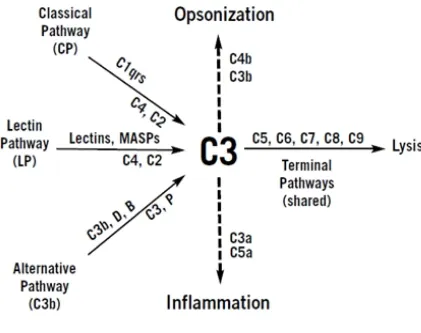
COMPLEMENT SYSTEM
The complement system consists of numerous plasma proteins , which interact with each
other to opsonize pathogens and induce inflammatory responses which help the body to fight
infections. It is the first line of defense against microorganisms and plays a very important
role in both innate and acquired immunity. There are three pathways for initiation of the
complement system, with a final common pathway. These three pathways are
a. Classical pathway activated by immune complexes
b. Alternative pathway activated by bacterial surfaces
c. Mannose binding lectin pathway
The common final pathway results in formation of a membrane attack complex. The first
complement component C1, is composed of three subunits- C1q, C1r and C1s. C1qis a
polypeptide multimer with 18 chains and a molecular weight of 460 kDa. C1q has to bind
to atleast two heavy chains in order to change its conformation and activate the classical
pathway.(6) Hence C1q is activated only by immune complexes containing
immunoglobulins and multivalent antigens. Genes coding for the three arms of human
45
1p34.1 – 1p36.3. Mutations leading to homozygous C1q deficiency are inherited in an
46
47
[image:47.612.73.361.75.472.2](40)
48
C1q specifically binds to early apoptotic cells and clears dying cells by activating the
classical complement pathway.
ANTI-C1q IN LUPUS NEPHRITIS
Complement pathway plays an important role in the pathogenesis of SLE, as
proved by genetic predisposition to SLE in children with early complement pathway
deficiencies. This also leads to the proposition that complement antibodies are associated
with SLE. Most work in this field has been done on anti C1Q and its association with
lupus nephritis.
Pathogenic association of anti C1q with SLE was demonstrated by the work of
Mart Mannik and Mark Wener, which was first published in 1997. They obtained kidney
biopsies at autopsy, of 12 patients who were diagnosed to have SLE. Antibodies to C1q
collagen like region were found in 4 of the 12 patients. All four patients were found to
have proliferative lupus nephritis. There was one patient with lupus nephritis, who did
not have significant anti-C1q titers.(5) Anti-C1q antibodies are not specific for SLE and
are found in numerous conditions. Other conditions with high anti-C1q titers include
hypocomplementemic urticarial vasculitis (100%), scleroderma, rheumatoid
arthritis(32%), undifferentiated connective tissue disease (94%) and Felty
syndrome(76%) Patients with hepatitis C are also found to have high antibody titers.
Anti-C1q antibodies are present in nearly one third of patients with SLE,
49
high antibody titers, the levels of C1q, C3 and C4 are usually low. In patients with
homozygous C1Q deficiency, 88% were found to have SLE and 30% developed
glomerulonephritis.(2) Levels of C1q were found to be low in patients with SLE with
glomerulonephritis flare. Nived et al assessed the outcomes in 52 Swedish adults with
biopsy proven lupus nephritis and they found that treatment response at 6 months and
levels of anti-C1q positively correlated with outcomes of disease.(41)
FQ Wu and colleagues investigated 90 pediatric SLE patients, 43 in active stage and
47 in remission. They demonstrated significantly higher levels of anti-C1q
antibodies in children with active disease which had a positive correlation with SLE
disease activity index (SLEDAI) score.(12)
Marto and Bertolaccini studied 151 patients with SLE, of which 77 patients had
lupus nephritis, and their disease activity was categorized according to the BILAG
(British Isles Lupus Assessment Group) renal score. They measured the sera of these
patients for anti C1Q by enzyme immunoassay and found that patients with active lupus
nephritis had a higher prevalence of anti-c1Q as compared to patients without lupus
nephritis (74% vs. 32%, RR= 2.3, 95% CI 1.6 to 3.3, p<0.0001). They did not find
significant difference between patients without lupus nephritis and those who had
inactive nephritis. Levels of anti C1Q were also found to be higher in patients with
nephritis as compared to patients without nephritis (36 U/ml vs. 7.3 U/ml, p<0.001).
They further conducted a retrospective cohort study in 83 patients without renal disease,
50
went on to develop lupus nephritis. None of the patients who had a negative anti C1Q
developed renal disease and hence the negative predictive value of the test was 100%.
The authors found positive correlation of anti C1Q with anti dsDNA and a negative
correlation with levels of C3 and C4, which are established markers of disease activity
and flare. The authors hence concluded that anti-C1Q was positively associated with
lupus nephritis and that monitoring anti C1Q could be useful in predicting renal flares of
the disease.(7)
In an international, multicenter collaborative study, a sample of patients who were
assembled to derive the Systemic Lupus collaborating criteria(SLICC), samples for
anti- C1q testing were collected from 308 patients with SLE and 389 patients with
other rheumatological illnesses. Anti- C1q was found in 28% of patients with SLE,
and in 13% of controls with other rheumatological illnesses.(42)
Genetic and ethnic factors play a role in the levels of anti-C1q antibody titers.
Asians are found to have higher titers of antibody to C1q as compared to Caucasians
and Afro-Americans. Also younger people less than 35 years of age have higher
antibody levels as compared to older individuals.
Kabeerdoss et al conducted a retrospective chart review of all SLE patients attending
the rheumatology OPD of Christian Medical College, Vellore from March 2013 to
January 2015. Clinical and laboratory data was retrieved from the electronic records
of patients. Disease activity was scored according to SLEDAI scores and patients
51
and >18 respectively). Renal SLEDAI score was used to assess the renal disease
activity. Of the 126 patients included in the study, 54.76% of patients had lupus
nephritis. 42.8% of SLE patients had positive anti-C1q titers, defined as anti c1q
level >10U/ml, while 50.7% of the lupus nephritis subgroup had positive titers. The
group found significantly higher levels of antibody in patients with lupus nephritis as
compared to patients who had SLE without lupus nephritis. There was also a
statistically significant difference between patients who had active lupus nephritis
and those with quiescent disease. In multivariate analysis, mucocutaneous disease
(OR 4.72), low C4 (OR 3.11) and higher renal SLEDAI scores (OR 1.35) were
found to have significant correlation with positive anti-C1q antibody. If renal
SLEDAI score was removed from the analysis, UP/UC ratio was found to attain
significance (OR 1.77, p<0.05). This study further confirms the utility of C1q
antibody as a novel biomarker for renal involvement in patients with SLE.(43)
Most studies evaluating C1q antibodies in SLE, and its correlation with lupus
nephritis have been done in adults. Pediatric studies are few and far in between and there
are no studies from India. Even though the etiopathogenesis of SLE in children and adults
is similar, children differ from adults in their clinical manifestations and disease severity.
Hence studies evaluating the role of this novel biomarker in Indian children with SLE are
52
53
MATERIALS AND METHODS
Study population recruitment
This study was conducted in the Pediatric Rheumatology OPD and Pediatric wards from
November 2016 to August 2017.
Inclusion criteria
All patients aged 6 – 18 years of age diagnosed with SLE according to the ACR
criteria, attending the above clinic /ward, after obtaining parental consent
Exclusion criteria
a. Known case of congenital complement deficiencies
b. Other glomerulonephritis
c. Ambiguity in diagnosis
Design of data collection:
This was a prospective cohort study. Data was collected from children fulfilling the
inclusion criteria and entered in a proforma, which has been approved by the ethics
committee of CMC, Vellore. Data was collected from hospital records and laboratory
54
After obtaining parental consent, a portion of blood samples which were sent for various
immunological tests was frozen and batches of blood samples were sent to the
rheumatology laboratory.
Anti-C1q antibody assay was done by commercially available ELISA kit [IMTEC-
anti-C1q-antibodies 9ITC590330, Germany]. Reference level of 20 IU/ml was taken as cut
off as recommended by the manufacturers. A level of assay more than 20IU/ml was taken
as positive.
Sample size calculation:
The required sample size to show that the prevalence of anti c1q with 95% confidence
limits was found to be 267 SLE children when the anticipated proportion of anti-C1q was taken as 30% based on previous studies with precision of about 5%.
The required sample size to show that anti-C1q was associated with renal involvement
was found to be 223 SLE children with 80% power and 5% level of significance and
55 Statistical methods:
The categorical variables were analysed using frequencies and percentages. The
prevalence of anti-C1q antibody was presented as percentage and 95% confidence
interval. Continuous variables are presented as mean with standard deviation or median
with interquartile range based on the distribution of data. The association of C1q with
various clinical manifestations was obtained by calculating odd’s ratio, adjusted for
confounders by performing logistic regression analysis. The sensitivity and specificity
with 95% confidence interval for anti-C1q antibodies in renal lupus was obtained. All
statistical analysis were done using SPSS software version 17.0 or later. This study was
56
57
Table 1: Baseline characteristic features of children with SLE
Parameters Number (%)
n=150
Age 6 – 10 years 22 (15%)
10 – 15 years 85 (57%)
Sex (M:F) 23 :127
Clinical features Cutaneous 10 (6.7)
Arthritis 13 (8.6)
Serositis 1 (0.6)
Seizures 6 (4)
Psychosis 1 (0.6)
Lab parameters Proteinuria 55(37)
Leukopenia 19(13)
Thrombocytopenia 6(4)
Low C3 73 (49)
Low C4 61 (41)
DS DNA 79 (53)
Drugs used HCQ 149 (97)
NSAIDS 3 (2)
Steroids 77 (51)
MMF 53 (35)
Cyclophosphamide 1 (0.6)
Azathioprine 23 (15)
Rituximab 7 (5)
Anti-C1Q positive 95 (64)
58
Table 2.Biopsies were done in a total of 72 children.Anti C1Q was positive in 59 (82%) of children who had undergone renal biopsy.
CLASS OF NEPHRITIS NUMBER (%)
(n=72)
CLASS II 7 (10)
CLASS III 21 (28)
CLASS IV 40 (54)
[image:58.612.121.489.162.659.2]CLASS V 4 (6)
FIGURE 1
7
21
40
4
RENAL BIOPSY RESULTS
CLASS II
CLASS III
CLASS IV
59
Table 3.Clinical and lab parameters compared with Anti-C1q status
Parameter Anti–C1q
positive N= 95 Anti-C1q Negative N=55 P value
Malar rash N(%)
(n=10)
7 (70) 3 (30) 0.73
Arthritis N (%)
(n=13)
11 (85) 2 (15.4) 0.13
Seizures N(%)
(n=6)
2 (33) 4 (67) 0.2
PsychosisN(%)
(n=1)
1 (100) 0 (0) 1
ProteinuriaN(%)
(n=55)
42 (76) 13 (24) 0.009
LeukopeniaN(%)
(n=19)
11 (58) 8 (42) 0.8
Thrombocytopenia N(%)
(n=6)
3 (50) 3 (50) 0.67
Low C3N(%)
(n=73)
58 (80) 15 (20) <0.0001
Low C4N(%)
(n=61)
49 (80) 12 (20) <0.0001
Anti-dsDNAN(%)
(n=79)
60
Table 4. Anti-C1q status in pSLE with or without renal involvement
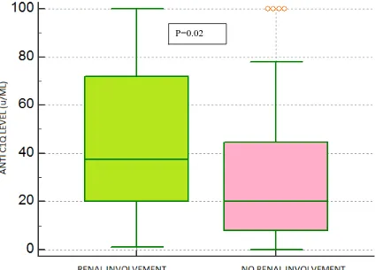
Figure 2: Anti-C1q status in pSLE with or without renal involvement
Anti- C1q antibody was positive in 58 (74%) in pSLE with renal involvement and was positive in 37(51%) in pSLE without renal involvement (p=0.02)
Renal involvement
N (%)
(n = 78)
No renal involvement
N (%)
(n = 72)
Anti-C1q 58 (74%) 37(51%) P=0.02
[image:60.612.79.512.342.647.2]61
Table 5. Anti-C1q status in pSLE with active or inactive renal disease
Active Renal disease
(n =48)
Inactive renal disease
(n = 30)
Anti-C1q positive 42 (88%) 16(53%) P=0.0015
Of the 78 children with lupus nephritis, 48 of them had active renal disease, defined as the presence of proteinuria, urinary cast or pyuria. Among the 48 children with active renal involvement, 42 had a positive value for anti-C1q (88 %), which was statistically significant.
Figure 3.Anti-C1q status in pSLE with active or inactive renal disease
[image:61.612.80.520.333.678.2]62
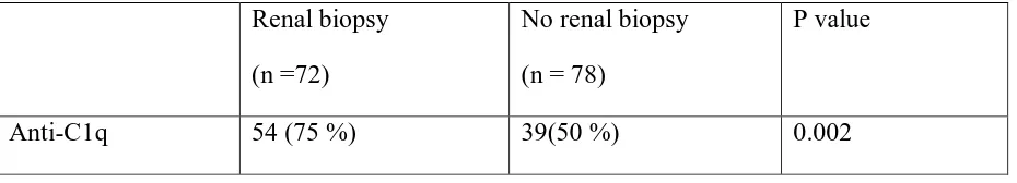
Table 6. Comparison of Anti-C1q in children who underwent renal biopsy
Renal biopsy
(n =72)
No renal biopsy
(n = 78)
P value
Anti-C1q 54 (75 %) 39(50 %) 0.002
63 0 10 20 30 40 50 60
II III IV V
NU
M
B
ER
S
RENAL BIOPSY CLASS
RENAL BIOPSY RESULT AND AVERAGE
C1Q
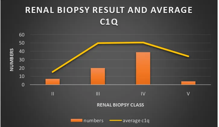
[image:63.612.111.576.126.283.2]numbers average c1q
Table 7: Comparison of renal biopsy report with mean Anti-C1q level
Renal Biopsy Number
(n=72)
Mean Ani-C1q (U/ml)
Class II 7 15.29
Class III 21 49.85
Class IV 40 50.53
Class V 4 34
Class III and IV nephritis were commonest, and the mean Anti-C1q values were highest in these two groups.
[image:63.612.126.489.435.646.2]64
Figure 5.Correlation between positive SLEDAI and Anti-C1q antibody titers
There was no significant correlation between average c1q antibody titers and SLEDAI positivity
65
Table 8.Correlation between positive SLEDAI and Anti-C1q antibody titers
SLEDAI (>8)
(n =43)
SLEDAI (<8)
(n = 105)
P value
Mean Anti-C1q level (U/ml) 59 30 0.086
66
Figure 6. ROC curve for sensitivity and specificity for Anti-C1q levels
Area under curve :0.659 Significance level : 0.0004
At anti C1q value > 22, sensitivity was 74.4% and specificity was 54.3%
0
20
40
60
80
100
rslt
0
20
40
60
80
100
100-Specificity
Se
n
s
it
iv
it
y
Sensitivity: 74.4 Specificity: 54.367
Figure 7.ROC curve for sensitivity and specificity, taking SLEDAI as disease indicator
Area under curve = 0.742 Significance level = P<0.0001
At Anti-C1q value >44, sensitivity 63.64 and specificity 78.85
0
20
40
60
80
100
rslt
0
20
40
60
80
100
100-Specificity
Se
n
s
it
iv
it
68
69 DISCUSSION
This observational study was performed to evaluate the diagnostic value of anti-C1q in
the clinical follow-up of SLE patients with regard to disease activity in general and to
renal involvement in particular. Between November 2016 to August 2017, 912 children
attended the pediatric rheumatology clinic at Christian Medical College, Vellore. Of
these, 271 Children had SLE and they constituted 30% of the total number of children.
150 children were recruited in the study and 121 children were excluded due to either
lack of parental consent, age <6 years, or lack of adequate sample.
In our study, children in the age group of 11 to 15 years constituted more than half of the
study population (57%), followed by children more than 16 years of age (29%). The
mean age at onset for SLE in our study population was 11 years, which was similar to
that seen in other studies, which reported a mean age at onset of 11 years (21,34). There
was a significant female preponderance noted in the study with more than 80% of the
study population being females. All the children with SLE less than 10 years of age were
females. In the age group of 11-15 years, about 77% of the study population was females
whereas in the age group more than 16, about 90% of the total number of patients were
females.
Renal involvement was the most common clinical feature with 55 children having active
proteinuria at the time of recruitment to the study and 78 children (52%) with established
70
described a lupus nephritis incidence of 55% in their SLE population.(30) Sinha and
Raut proposed that 50 – 75% of pediatric SLE patients have lupus nephritis.(34) They
also found that lupus nephritis was more common in males than in females. However in
our study, of the 78 patients with lupus nephritis, 68 were females and there were only 10
boys with lupus nephritis. The difference was not statistically significant.
Malar rash was seen in 10 children (6.67%) and no child had discoid rash. In the study
from Toronto,(30) cutaneous manifestations were the most common, with malar rash
being seen in 66% of the study population. In the study from West India, the incidence of
malar rash was reported at 44%.(24)
Arthritis in our study was seen in 13 children (9%), which is again less than that reported
previously from literature. Levy et al reported an arthritis incidence of 80% in pediatric
SLE while Hiraki reported arthritis in about 67% of children.(21,30)Patwardhan et al also
reported an arthritis incidence of 60% from West India, as did studies from the Amrita
Institute in Kerala.(23,24) This low incidence of arthritis in our study may be attributed
to the fact that majority of the children recruited were on follow up for many years and
the disease was quiescent.
CNS manifestations are one of the most dreaded complications of pediatric SLE. In our
study, there were 6 children with seizures and one child with psychosis. Hence CNS
manifestations were present in 4.8% of the study population. In a study from South India
on adult SLE, Robert and colleagues reported a seizure incidence of 20% and psychosis
71
Low complement levels are commonly seen in SLE and are used as a marker of disease
activity. Low complement levels are seen during disease flares. In our study low
complements were seen in 77 children (51%). 79 children had anti dsDNA positive. 25
children had cytopenias(17%), which included mild to moderate thrombocytopenia and
leukopenia. There were no children with severe anemia.
There were 78 (52%) children with lupus nephritis in our study population, of whom 48
children had active nephritis- defined as presence of a) urinary casts (heme-granular or
RBC), b) proteinuria (>0.5g/24 hours, new onset proteinuria or increase >0.5g/24 hours)
c) pyuria (>5 WBC/ hpf, excluding infection). Each criteria gets a score of 4 points.(45)
72 of the children with lupus nephritis underwent renal biopsy. Class IV lupus nephritis
(Diffuse proliferative lupus nephritis) was the most common, followed by Class III (focal
proliferative lupus nephritis). In the study from the Hospital for Sick Kids, Toronto, Class
III and IV nephritis constituted about 80% of the renal biopsy samples.(30) The average
anti-C1q values were found to be higher in Class III and IV lupus nephritis, reflecting a
more severe disease course. There was however, no statistically significant difference
between Class III and IV nephritis with regard to anti-C1q values.
Anti-C1q was found in the renal biopsy in 59 children with lupus nephritis. There was no
significant difference between anti-C1q positivity among those with C1q in the renal
biopsy and those without. There was also no statistically significant difference in the
average C1q level between those with C1q in the renal biopsy and those without.
72
compared with children without renal involvement (74.4% vs. 48.6%, p=0.0001, OR
1.77, 95% CI 1.2 – 2.6). The mean anti-C1q value was also higher in the lupus nephritis
group as compared to children without lupus nephritis (46.29U/L vs. 29.77U/L) and the
difference was statistically significant (p=0.011). This is consistent with the study
published by Marto and Bercolonni, where children with lupus nephritis were found to
73 ANTI-C1q ANTIBODIES:
Antibodies to initial complement factors have been found to be increased in patients with
SLE. Most of these studies have been undertaken in adults or mix