0022-538X/10/$12.00 doi:10.1128/JVI.00918-10
Copyright © 2010, American Society for Microbiology. All Rights Reserved.
Interferons Accelerate Decay of Replication-Competent
Nucleocapsids of Hepatitis B Virus
䌤
Chunxiao Xu,
1Haitao Guo,
1Xiao-Ben Pan,
1Richeng Mao,
1Wenquan Yu,
2Xiaodong Xu,
2Lai Wei,
3Jinhong Chang,
1Timothy M. Block,
1,2and Ju-Tao Guo
1*
Drexel Institute for Biotechnology and Virology Research, Department of Microbiology and Immunology, Drexel University College of
Medicine,1and Institute for Hepatitis and Virus Research, Hepatitis B Foundation,23805 Old Easton Road,
Doylestown, Pennsylvania 18902, and Peking University People’s Hospital and
Peking University Hepatology Institute, Beijing 100044, China3
Received 28 April 2010/Accepted 29 June 2010
Alpha interferon (IFN-␣) is an approved medication for chronic hepatitis B. Gamma interferon (IFN-␥) is a key mediator of host antiviral immunity against hepatitis B virus (HBV) infectionin vivo. However, the molecular mechanism by which these antiviral cytokines suppress HBV replication remains elusive. Using an immortalized murine hepatocyte (AML12)-derived cell line supporting tetracycline-inducible HBV replication, we show in this report that both IFN-␣ and IFN-␥ efficiently reduce the amount of intracellular HBV nucleocapsids. Furthermore, we provide evidence suggesting that the IFN-induced cellular antiviral response is able to distinguish and selectively accelerate the decay of HBV replication-competent nucleocapsids but not empty capsids in a proteasome-dependent manner. Our findings thus reveal a novel antiviral mechanism of IFNs and provide a basis for a better understanding of HBV pathobiology.
Hepatitis B virus (HBV) is a noncytopathic hepatotropic
DNA virus which belongs to the familyHepadnaviridae (11, 44). Despite the fact that most adulthood HBV infections are transient, approximately 5 to 10% of infected adults and more than 90% of infected neonates fail to clear the virus and de-velop a lifelong persistent infection, which may progress to chronic hepatitis, cirrhosis, and primary hepatocellular carci-noma (4, 33, 34). It has been shown by several research groups that resolution of HBV and other animal hepadnavirus
infec-tionin vivodepends on both killing of infected hepatocytes by
viral antigen-specific cytotoxic T lymphocytes and noncytolytic suppression of viral replication, which is most likely mediated by inflammatory cytokines, such as gamma interferon (IFN-␥) and tumor necrosis factor␣(TNF-␣) (10, 12, 15, 20, 26, 27, 48). Moreover, together with five nucleoside or nucleotide analogs that inhibit HBV DNA polymerase, alpha IFN (IFN-␣) and pegylated IFN-␣are currently available antiviral medications for the management of chronic hepatitis B. Com-pared to the viral DNA polymerase inhibitors, the advantages of IFN-␣therapy include a lack of drug resistance, a finite and defined treatment course, and an increased likelihood for hep-atitis B virus surface antigen (HBsAg) clearance (8, 39). How-ever, only approximately 30% of treated patients achieve a sustained virological response to a standard 48-month pegy-lated IFN-␣therapy (6, 32). Thus far, the antiviral mechanism of IFN-␣and IFN-␥and the parameters determining the suc-cess or failure of IFN-␣therapy in chronic hepatitis B remain elusive. Elucidation of the mechanism by which the cytokines
suppress HBV replication represents an important step toward understanding the pathobiology of HBV infection and the mo-lecular basis of IFN-␣therapy of chronic hepatitis B.
Considering the mechanism by which IFNs noncytolytically control HBV infectionin vivo, it is possible that the cytokines either induce an antiviral response in hepatocytes to directly limit HBV replication or modulate the host antiviral immune response to indirectly inhibit the virus infection. However, due to the fact that IFN-␣and -␥do not inhibit or only modestly inhibit HBV replication in human hepatoma-derived cell lines (5, 22, 23, 30), the direct antiviral effects of the cytokines and their antiviral mechanism against HBV have been studied with either an immortalized hepatocyte cell line derived from HBV transgenic mice or duck hepatitis B virus (DHBV) infection of primary duck hepatocytes (37, 53). While these studies re-vealed that IFN treatment significantly reduced the amount of encapsidated viral pregenomic RNA (pgRNA) in both mouse and duck hepatocytes, further mechanistic analyses suggested that IFN-␣inhibited the formation of pgRNA-containing nu-cleocapsids in murine hepatocytes (52) but shortened the half-life of encapsidated pgRNA in DHBV-replicating chicken hep-atoma cells (21). Moreover, the fate of viral DNA replication intermediates or nucleocapsids in the IFN-treated hepatocytes was not investigated in the previous studies.
To further define the target(s) of IFN-␣and -␥in the HBV life cycle and to create a robust cell culture system for the identification of IFN-stimulated genes (ISGs) that mediate the antiviral response of the cytokines (25), we established an immortalized murine hepatocyte (AML-12)-derived stable cell line that supported a high level of HBV replication in a tetra-cycline-inducible manner. Consistent with previous reports, we show that both IFN-␣and IFN-␥potently inhibited HBV rep-lication in murine hepatocytes (37, 40). With the help of small molecules that inhibit HBV capsid assembly (Bay-4109) (7, 47) and prevent the incorporation of pgRNA into nucleocapsids
* Corresponding author. Mailing address: Drexel Institute for Bio-technology and Virology Research, Department of Microbiology and Immunology, Drexel University College of Medicine, 3805 Old Easton Road, Doylestown, PA 18902. Phone: (215) 4929. Fax: (215) 489-4920. E-mail: [email protected].
䌤Published ahead of print on 7 July 2010.
9332
on November 8, 2019 by guest
http://jvi.asm.org/
(AT-61) (9, 29), we obtained evidence suggesting that the IFN-induced cellular antiviral response is able to distinguish and selectively accelerate the decay of HBV replication-com-petent nucleocapsids but not empty capsids in a proteasome-dependent manner. Our findings provide a basis for further studies toward better understanding of IFN⬘s antiviral mech-anism, which might ultimately lead to the development of strategies to improve the efficacy of IFN therapy of chronic hepatitis B.
MATERIALS AND METHODS
Establishment of murine hepatocyte-derived cell line that supports high-level HBV replication.AML12 cells (a gift of Chen Liu at Florida State University, Jacksonville, FL) were maintained in Dulbecco’s modified Eagle medium, nutrient mixture F12 (DMEM/F12) (Invitrogen), supplemented with 10% fetal bovine serum, 100 U/ml penicillin, and 100g/ml streptomycin. To obtain cell lines that support tetracycline-inducible HBV replication, the AML12 cells were cotransfected with the plasmid pTet-off (Clontech), which expresses a tetracy-cline (Tet)-responsive transcriptional activator (tTA), and the plasmid pTREHBVDES, in which HBV pgRNA expression is controlled by a cytomega-lovirus early promoter with a tetracycline-responsive element (17). Transfected cells were selected with 500g/ml G418 in the presence of 1g/ml tetracycline. G418-resistant colonies were picked and expanded into cell lines. HBV replica-tion was induced by culturing cells in tetracycline-free medium, and the levels of viral DNA replicative intermediates were determined by Southern blot hybrid-ization. One of the cell lines with the highest level of HBV replication, desig-nated AML12HBV10, was chosen for this study.
Viral DNA and RNA analysis.Intracellular viral core DNA was extracted as described previously (19). One-half of the DNA sample from each well of 12-well plates was resolved by electrophoresis into a 1.5% agarose gel and transferred onto a Hybond-XL membrane. For viral RNA analysis, total cellular RNA was extracted with TRIzol reagents (Invitrogen). Five micrograms of total RNA was resolved in a 1.5% agarose gel containing 2.2 M formaldehyde and transferred onto a Hybond-XL membrane in 20⫻SSC buffer (1⫻SSC is 0.15 M NaCl plus 0.015 M sodium citrate). Encapsidated HBV RNA was extracted as previously described (21). One-half of the RNA sample from each well of 12-well plates was resolved in 1.5% agarose gel containing 2.2 M formaldehyde and transferred onto Hybond-XL membrane in 20⫻SSC buffer. For the detection of HBV DNA and RNA, membranes were probed with either an [␣-32P]UTP (800 Ci/mmol;
Perkin Elmer)-labeled minus-strand- or plus-strand-specific full-length HBV ri-boprobe as described previously. The membrane was exposed to a phospho-rimager screen, and hybridization signals were quantified with the QuantityOne software program (Bio-Rad).
Viral nucleocapsid analysis.HBV capsids (cores) and associated viral DNA were analyzed essentially as previously described, with minor modifications (16, 17). Briefly, AML12HBV10 cells were lysed by addition of 300l buffer con-taining 10 mM Tris-HCl (pH 7.6), 100 mM NaCl, 1 mM EDTA, and 0.1% NP-40 to each well of a 12-well plate. Cell debris was removed by centrifugation at 5,000⫻gfor 10 min. Ten microliters of the clarified cell lysates were fractionated by electrophoresis through nondenaturing 1% agarose gels and transferred to a nitrocellulose filter by blotting with TNE buffer (10 mM Tris-HCl, pH 7.6, 150 mM NaCl, and 1 mM EDTA). HBV capsids were detected by probing the membrane with an antibody against the HBV core protein (Dako). Bound antibody was revealed by IRDye secondary antibodies and visualized by the Li-COR Odyssey system. To detect capsid-associated HBV DNA, the mem-branes were treated with buffer containing 0.5 N NaOH and 1.5 M NaCl for 5 min, followed by neutralization with buffer containing 1 M TRIS-HCl and 1.5 M NaCl for 5 min. The viral DNA was detected by hybridization with a [␣-32
P]UTP (800 Ci/mmol; Perkin Elmer)-labeled minus-strand-specific full-length HBV ri-boprobe.
Viral core protein analysis.AML12HBV10 cells were lysed with 1⫻Laemmli buffer. A fraction of cell lysate was separated on sodium dodecyl sulfate–12% polyacrylamide gels and electrophoretically transferred to a polyvinylidene di-fluoride (PVDF) membrane (Bio-Rad). Membranes were blocked with phos-phate-buffered saline (PBS) containing 5% nonfat dry milk and probed with antibodies against HBcAg (18) or-actin (Chemicon International). Bound antibody was revealed by IRDye secondary antibodies and visualized and quan-tified by the Li-COR Odyssey system.
IFN and chemicals.Recombinant murine IFN-␣, IFN-␥, TNF-␣, interleukin-1 (IL-1), and IL-6 were purchased from PBL Interferon Source and PeproTech.
AT-61 and Bay-4109 were synthesized in-house by following the published pro-cedures (7, 29). Lamivudine is a gift from William S. Mason, Fox Chase Cancer Center, Philadelphia, PA.
RESULTS
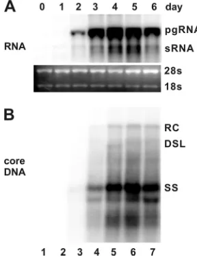
IFN-␣and IFN-␥potently inhibit HBV replication in mu-rine hepatocytes.To investigate the antiviral effects and illus-trate the antiviral mechanism of IFNs and other inflammatory cytokines, we first established an immortalized mouse hepato-cyte (AML12)-derived cell line, designated AML12HBV10, that allows tetracycline (Tet)-inducible transcription of HBV pgRNA and viral DNA replication. In the presence of Tet, the transcription of viral pgRNA is suppressed. However, upon removal of Tet from cultural media, HBV mRNAs are tran-scribed and accumulated to a detectable level at 24 h, reaching the peak level at 96 h (Fig. 1, upper panel). As expected, following pgRNA transcription, HBV DNA replication inter-mediates were detected at 48 h and reached the steady-state level at 96 h after Tet removal (Fig. 1, lower panel).
[image:2.585.346.490.70.257.2]To determine the antiviral effects of IFN-␣, IFN-␥, and three other inflammatory cytokines that are induced in the liver during HBV infection and potentially play a role in noncytopathic suppression of HBV replication (20, 24), AML12HBV10 cells were cultured in the absence of Tet and presence of the indicated concentrations of murine IFN-␣, IFN-␥, TNF-␣, IL-1, or IL-6 for 4 days. The results show that while IL-1 and IL-6 do not affect the levels of intracellular HBV DNA, a modest reduction of HBV DNA replication intermediates can be observed in cells treated with higher
FIG. 1. Kinetics of HBV RNA transcription and DNA replication in a murine hepatocyte-derived cell line. AML12HBV10 cells were maintained in the medium containing 1g/ml of tetracycline to inhibit viral pgRNA transcription and then cultured in the medium without the antibiotic for the indicated number of days. Total cellular RNA and cytoplasmic core DNA were extracted and analyzed by Northern and Southern blot hybridization, respectively. (A) For viral RNA anal-ysis, each lane was loaded with 5 g of total RNA. pgRNA, pre-genomic RNA; sRNA, mRNAs specifying the three envelope proteins. rRNA (28S and 18S) served as loading controls. (B) For core DNA analysis, each lane represents the amount of viral DNA extracted from one-half of cells in a well of a 12-well plate. RC, relaxed circular DNA; DSL, double-stranded linear DNA; SS, single-stranded DNA.
VOL. 84, 2010 IFNs PROMOTE DECAY OF HBV NUCLEOCAPSIDS 9333
on November 8, 2019 by guest
http://jvi.asm.org/
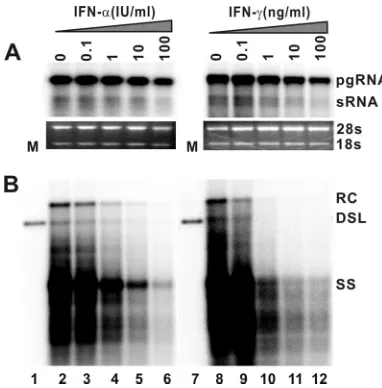
concentrations of TNF-␣ (data not shown). However, in marked contrast, treatment of the cells with either IFN-␣or IFN-␥ dramatically reduces the levels of intracellular HBV DNA replication intermediates in a dose-dependent manner (Fig. 2B). Moreover, under the concentrations at which HBV replication is inhibited, the levels of viral pgRNA are not affected or are only slightly reduced (Fig. 2A). Hence, the results imply that the type I and type II IFNs inhibit one or several posttranscriptional steps of HBV replication in the murine hepatocytes.
IFN-␣ and IFN-␥ reduce the level of intracellular HBV nucleocapsids.To map the replication steps that are inhibited by IFNs, the levels of viral gene products (pgRNA and core protein) and replication intermediates (capsids, encapsidated pgRNA, capsid-associated DNA, and DNA replication inter-mediates) were determined in cells that were either left un-treated or un-treated with the indicated concentrations of IFN-␣, IFN-␥, Bay-4109, AT-61, or lamivudine (3TC) for 4 days. The three antiviral compounds inhibited HBV replication in a dose-dependent manner in the murine hepatocytes (data not shown). Because each of these antiviral compounds inhibits a well-defined step of HBV replication, they are used in this study as references to help dissect the antiviral mechanism of IFNs.
[image:3.585.65.256.67.261.2]Our results from this experiment revealed the following. First, none of the treatments affected the levels of pgRNA (Fig. 3A). Second, none of the three antiviral compounds af-fected the levels of the HBV core protein, but the amount of the core protein is modestly reduced in cells treated with either IFN-␣or IFN-␥(Fig. 3B). Third, in accordance with the inhi-bition of HBV DNA polymerase, lamivudine treatment did not affect pgRNA encapsidation (Fig. 3C) and capsid formation
FIG. 3. IFN-␣and IFN-␥treatment reduce the level of intracel-lular HBV nucleocapsids. AML12HBV10 cells were cultured in the absence of tetracycline and left untreated (control) or treated with 10 IU/ml IFN-␣, 5 ng/ml IFN-␥, 25M AT-61, 2M Bay-4109, or 50M lamivudine (3TC) for 4 days, and cells were then harvested. (A) Intracellular viral RNA was determined by Northern blot hy-bridization. (B) HBV core protein was determined by a Western blot assay with an antibody recognizing the carboxyl-terminal 14 amino acid residues of HBcAg. (D) The amounts of nucleocapsids were determined by a particle gel assay (see Materials and Methods for details) that measures intact nucleocapsids. (E) Nucleocapsid-associated HBV DNA was quantified by alkaline treatment of nu-cleocapsids on the membrane following the particle gel assay and hybridized with a full-length HBV riboprobe that anneals to the minus strand of HBV DNA. Encapsidated pgRNA (C) and HBV DNA replication intermediates (F) were extracted and determined by Northern blot and Southern blot hybridizations, respectively. FIG. 2. IFN-␣and IFN-␥treatment potently reduce the level of
HBV DNA. AML12HBV10 cells were cultured in the absence of tetracycline and in the presence of the indicated concentrations of mu-rine IFN-␣or IFN-␥for 4 days. Total cellular RNA and cytoplasmic core DNA were extracted and analyzed by Northern and Southern blot hybridization, respectively. (A) For viral RNA analysis, each lane was loaded with 5g of total RNA. rRNAs (28S and 18S) serve as loading controls. (B) For core DNA analysis, each lane represents the amount of viral DNA extracted from one-half of cells in a well of 12-well plate.
on November 8, 2019 by guest
http://jvi.asm.org/
[image:3.585.315.525.89.549.2](Fig. 3D) but reduced the levels of capsid-associated viral DNA replication intermediates (Fig. 3E and F). The observed poor antiviral activity of lamivudine in this cell line is most likely due to its inefficient phosphorylation in rodent hepato-cytes (57). Fourth, treatment of cells with Bay-4109, a capsid assembly inhibitor (7, 47), completely inhibited capsid forma-tion, as revealed by the particle gel assay which detects intact nucleocapsids (17) (Fig. 3D). As expected, pgRNA encapsida-tion (Fig. 3C) and HBV DNA replicaencapsida-tion (Fig. 3E and F) did not occur in Bay-4109-treated cells. Fifth, consistent with the previous reports (9, 29), treatment of cells with an HBV pgRNA encapsidation inhibitor, AT-61, did not affect capsid formation (Fig. 3D) but eliminated encapsidated pgRNA (Fig. 3C). As a consequence, viral DNA replication was also inhib-ited (Fig. 3E and F). Finally, unlike all the three antiviral compounds, treatment of cells with either IFN-␣ or IFN-␥ significantly reduced the levels of HBV capsids (Fig. 3D). Con-sistently, the levels of encapsidated pgRNA (Fig. 3C) and capsid-associated viral DNA replication intermediates (Fig. 3E and F) were also proportionally reduced. Taken together, the distinct profile of HBV core protein and capsid accumulation in IFN-␣- or IFN-␥-treated cells suggests that the cytokines may inhibit HBV capsid assembly, which results in the degra-dation of the unassembled core protein and reduced pgRNA encapsidation and DNA replication. Alternatively, IFNs may promote the degradation of nucleocapsids.
[image:4.585.90.496.72.357.2]IFN promotes the decay of intracellular HBV nucleocapsids in cells in which pgRNA transcription is inhibited.To distin-guish the two possibilities described above, we took advantage of our Tet-inducible replication system to determine whether IFN treatment shortens the half-life of HBV nucleocapsids. To this end, AML12HBV10 cells were cultured in the absence of Tet for 4 days to allow the accumulation of nucleocapsids and replication intermediates. At this time (0 h), Tet was added back to culture medium to stop pgRNA transcription and thus the assembly of nucleocapsids. As shown in Fig. 4A, upon inhibition ofde novopgRNA transcription, the level of preex-isting pgRNA quickly declined and IFN-␣ treatment appar-ently did not accelerate the rate of pgRNA decrease. Interest-ingly, treatment of the cells with IFN-␣reduced the half-lives of nucleocapsids and capsid-associated viral DNA from ap-proximately 24 h to less than 12 h (Fig. 4B and C). To further determine whether IFN treatment has a differential effect on the decay of nucleocapsids containing different DNA replica-tion intermediates, we performed a Southern blot analysis on the intracellular core-associated HBV DNA and found that IFN treatment drastically accelerated the rate of decay of sin-gle-stranded DNA containing nucleocapsids but only modestly promoted the decay of nucleocapsids containing mature re-laxed circular (rc) DNA (Fig. 4D). Similar results were ob-tained with IFN-␥treatment (data not shown).
FIG. 4. IFN-␣promotes the decay of intracellular HBV nucleocapsids in cells in which pgRNA transcription is blocked. AML12HBV10 cells were cultured in the absence of tetracycline for 4 days to allow HBV replication, and tetracycline was then added back to stop pgRNA transcription from viral transgenes integrated in the cellular chromosome. At the same time, cells were left untreated (control) or treated with 10 IU/ml IFN-␣. Cells were harvested at the indicated period of time post-IFN treatment. Intracellular viral RNA (A), nucleocapsid (B), core-associated HBV DNA (C), and HBV DNA replication intermediates (D) were determined as described in Materials and Methods.
VOL. 84, 2010 IFNs PROMOTE DECAY OF HBV NUCLEOCAPSIDS 9335
on November 8, 2019 by guest
http://jvi.asm.org/
IFN accelerates the decay of viral DNA-containing nucleo-capsids in cells in which pgRNA encapsidation is inhibited.
Although the amount of preexisting pgRNA declined quickly upon addition of Tet to culture medium (Fig. 4A), pgRNA-containing capsid formation might not stop promptly. There-fore, the apparently accelerated decay of nucleocapsids ob-served in the above experiment could be alternatively explained as a result of IFN-induced inhibition of pgRNA-containing capsid formation. To rule out this possibility, the effect of IFN-␣ on nucleocapsid decay kinetics was further examined in the presence of AT-61, which inhibits the forma-tion of pgRNA-containing nucleocapsids but not empty cap-sids (9). Briefly, AML12HBV10 cells were cultured in the absence of Tet for 4 days to allow the accumulation of nucleo-capsids and replication intermediates. At this time (0 h), Tet was added to culture medium to stop pgRNA transcription. At the same time, the cells were either left untreated or treated with IFN-␣in the absence or presence of AT-61. The results showed that compared with untreated controls, AT-61 treat-ment only modestly increased the rates of capsid and capsid-associated HBV DNA decline (Fig. 5). However, consistent with the results presented above (Fig. 4B and C), treatment of the cells with IFN-␣ in the absence of AT-61 resulted in a profound reduction of both capsids and capsid-associated viral DNA (Fig. 5). Similar results were obtained with IFN-␥ treat-ment (data not shown). These results suggest that although assembly of pgRNA-containing nucleocapsids may occur after cessation of pgRNA transcription, it is obvious that inhibition of pgRNA-containing capsid formation under this experimen-tal condition could not account for the profound reduction in
HBV nucleocapsids and viral DNA in IFN-treated cells. Hence, our results favor a hypothesis that an IFN-induced antiviral response promotes the decay of HBV nucleocapsids. However, it was rather unexpected that compared with IFN-␣monotreatment, treatment of the cells with IFN-␣in the presence of AT-61 resulted in a slightly slower decay of capsids but a higher rate of capsid-associated HBV DNA decay (Fig. 5). One possible explanation for this intriguing observation is that whereas HBV nucleic acid-containing capsids or the rep-lication-competent nucleocapsids are sensitive to IFN-induced decay, the empty capsids formed in the presence of AT-61 might be resistant to the cytokine-induced antiviral response.
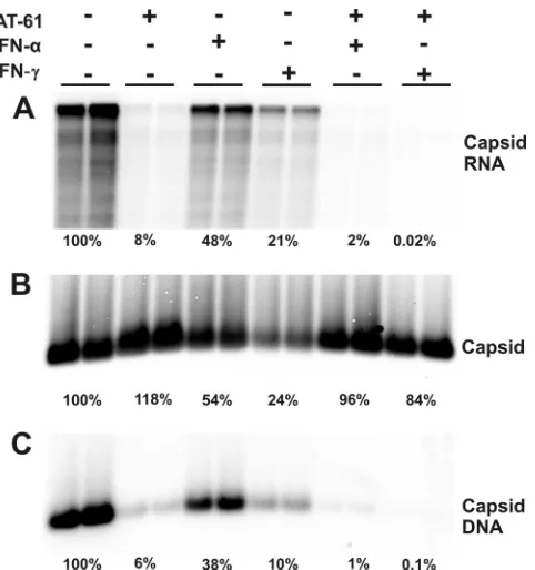
IFN selectively promotes the decay of HBV nucleocapsids but not empty capsids formed in the presence of AT-61.To test this hypothesis, we first investigated the effect of IFN-␣and IFN-␥on the accumulation of empty capsids in AT-61-treated cells. Briefly, AML12HBV10 cells were cultured in the absence of Tet and left untreated or treated with appropriate concen-trations of AT-61, IFN-␣, or IFN-␥, alone or in combinations, for 4 days. Cells were then harvested, and levels of intracellular capsids, encapsidated pgRNA, and capsid-associated HBV DNA were determined. As shown in Fig. 6, consistent with the results presented in Fig. 3, AT-61 treatment did not affect capsid formation but efficiently inhibited pgRNA encapsida-tion and thus DNA synthesis. Moreover, treatment of the cells with either IFN-␣ or IFN-␥ reduced the levels of capsids, encapsidated pgRNA, and HBV DNA. However, as predicted by the hypothesis, neither IFN-␣ nor IFN-␥ significantly re-duced the levels of capsids in AT-61-treated cells. In contrast, the residual amounts of encapsidated pgRNA (8%) and
cap-FIG. 5. IFN-␣ accelerates the decay of DNA-containing capsid in cells in which pgRNA-containing capsid formation is inhibited. AML12HBV10 cells were cultured in the absence of tetracycline for 4 days, and tetracycline was then added back to stop pgRNA transcription. At the same time, cells were left untreated (control) or treated with 25M AT-61, 10 IU/ml IFN-␣, or a combination of 25M AT-61 and 10 IU/ml mouse IFN-␣(mIFN-␣). Cells were harvested at the indicated periods of time posttreatment. Intracellular viral capsids (left panel) and core-associated HBV DNA (right panel) were determined as described in Materials and Methods.
on November 8, 2019 by guest
http://jvi.asm.org/
[image:5.585.112.469.64.326.2]sid-associated DNA (6%) in AT-61-treated cells were further reduced by the cytokine treatment.
To further determine the differential sensitivities of replica-tion-competent nucleocapsids and empty capsids to the IFN-induced antiviral response, AML12HBV10 cells were cultured in the absence of Tet and left untreated or treated with serial concentrations of IFN-␣in the absence or presence of 10M AT-61. Under these concentrations, AT-61 only partially in-hibits pgRNA-containing capsid formation and reduces core-associated HBV DNA by approximately 40% (Fig. 7). In
sup-port of our hypothesis, the results (Fig. 7) showed that compared with IFN-␣ treatment alone, in the presence of AT-61, IFN-␣induced a much less profound reduction of total capsids but a more profound reduction of capsid-associated HBV DNA in a dose-dependent manner. Furthermore, as observed in AT-61-treated AMLHBV10 cells, treatment of a stable AML12-derived cell line expressing the HBV core pro-tein with either IFN-␣or IFN-␥did not apparently alter the level of intracellular empty capsids, as revealed by a particle gel assay (data not shown). The results presented above thus imply that the IFN-induced antiviral response is able to distinguish HBV nucleic acid-containing capsids (nucleocapsids) from empty capsids and selectively promotes the decay of the former but not the latter.
IFN-induced decay of HBV nucleocapsids, at least in part, depends on proteasome activity.Expression of IFN-inducible proteasome catalytic and regulatory subunits correlates with the IFN-␣- and IFN-␥-mediated noncytopathic inhibition of HBV in transgenic mice and murine hepatocytes and with clearance of the virus in acutely infected chimpanzees (50, 54). It was also demonstrated previously that proteasome inhib-itors were able to abrogate the antiviral activity of IFN- and IFN-␥ against HBV in immortalized HBV transgenic mouse hepatocytes (41). It is therefore possible that the accelerated decay of HBV DNA-containing nucleocapsids by IFN-␣and IFN-␥in AML12HBV10 cells is mediated by cellular proteasome activity.
[image:6.585.46.287.66.323.2]To test this hypothesis, we examined the effect of epoxomi-cin, an irreversible proteasome inhibitor, on the IFN-induced reduction of nucleocapsids. Because treatment of AML12 cells with epoxomicin for more than 8 h induces apoptosis (data not shown), the cells were treated for only 6 h with the inhibitor. Briefly, AML12HBV10 cells were cultured in the absence of Tet for 4 days to allow the accumulation of HBV DNA-con-taining nucleocapsids. The cells were then left untreated or treated with 50 IU/ml of IFN-␣or 10 ng/ml of IFN-␥. Six hours later, epoxomicin was added to the culture medium and incu-bated for an additional 6 h. As shown in Fig. 8, treatment of the cells with epoxomicin for only 6 h after IFN treatment atten-uated the cytokine-induced reduction of HBV capsids and core-associated viral DNA, suggesting that IFN-induced decay
FIG. 6. Effects of combinational treatment of AT-61 and IFN on HBV nucleocapsids. AML12HBV10 cells were left untreated or treated with 10 IU/ml IFN-␣, 5 ng/ml IFN-␥, or a combination of 25
M AT-61 and 10 IU/ml IFN-␣or 5 ng/ml IFN-␥for 4 days. Cells were then harvested, and encapsidated viral RNA (A), capsids (B), and core-associated HBV DNA (C) were determined as described in Ma-terials and Methods.
FIG. 7. Effects of combinational treatment of AT-61 and IFN-␣on HBV nucleocapsids. AML12HBV10 cells were left untreated or treated with 10 IU/ml IFN-␣in the absence or presence of 10M AT-61 for 4 days. Cells were then harvested, and encapsidated viral RNA, capsids, and core-associated HBV DNA were determined as described in Materials and Methods.
VOL. 84, 2010 IFNs PROMOTE DECAY OF HBV NUCLEOCAPSIDS 9337
on November 8, 2019 by guest
http://jvi.asm.org/
[image:6.585.104.490.546.696.2]of HBV nucleocapsids, at least in part, depends on cellular proteasome activity.
DISCUSSION
IFN-␣ and IFN-␥ are key mediators of host innate and adaptive immunity against viral infection (45). In the case of HBV infection, it has been demonstrated that induction of IFN-␥and the cytokine-induced cellular gene expression in the livers of HBV-infected chimpanzees is correlated with suppres-sion of HBV replication and ultimately clearance of the virus infection (15, 50). In contrast, unlike the case with hepatitis C virus (HCV) infection, induction of type I IFN-stimulated gene (ISG) expression was not detectable in the livers of chimpan-zees during the initial phase of HBV infection, suggesting that HBV may have evolved a strategy to either evade the detection or inhibit the activation of the type I IFN-mediated innate defense system (51). However, IFN-␣is an approved antiviral medication for chronic hepatitis B and is able to significantly reduce the viral load in 30% of hepatitis B virus e antigen (HBeAg)-positive patients and 40% of HBeAg-negative pa-tients after a standard 48-month pegylated IFN-␣therapy (8, 39). Although it was shown that IFN-␣and IFN-␥were able to modestly reduce the levels of HBV DNA at concentrations higher than 100 IU/ml in HBV genome-stably transfected hu-man hepatoma cell lines, the relatively poor antiviral activity of the cytokines in these cell lines prevented the further dissec-tion of their antiviral mechanism (5, 22).
The AML12 cell line consists of well-differentiated and non-tumorigenic murine hepatocytes derived from the liver of a transgenic mouse overexpressing human transforming growth factor alpha (55). Because this immortalized murine hepato-cyte cell line has typical features of normal hepatohepato-cytes and expresses high levels of hepatocyte-specific proteins, such as albumin,␣1-antitrypsin, and transferrin (55), it has been used as a cell culture model to study hepatocellular metabolism and effects of the HBV X protein and other exogenous or host cellular proteins on the growth and differentiation of hepato-cytes (46, 49). Taking advantages of this well-differentiated murine hepatocyte cell line, we created a stable cell line
[image:7.585.59.268.66.175.2]ex-pressing HBV pgRNA in a Tet-inducible manner to study HBV replication and its inhibition by IFN and other inflam-matory cytokines. Our results show that in spite of robust HBV DNA replication, the murine hepatocytes fail to support co-valently closed circular DNA (cccDNA) formation (data not shown), which is consistent with that observed in HBV trans-genic micein vivo(13). Moreover, in agreement with a previ-ous report suggesting that both type I and type II IFNs, but not TNF-␣, inhibit HBV replication in an HBV transgenic mouse hepatocyte-derived cell line (37), we found that IFN-␣ and IFN-␥ potently inhibit HBV replication via a posttranscrip-tional mechanism. Furthermore, although the possibility that the cytokines inhibit the pgRNA-containing nucleocapsid for-mation, as suggested by others (52), could not be directly investigated in our cell culture system, we obtained compelling evidence supporting the notion that the IFN-induced antiviral response accelerates the decay of HBV nucleocapsids (as de-picted in Fig. 9), which is consistent with our previous obser-vation that IFN-␣treatment shortens the half-life of encapsi-dated DHBV pgRNA in a chicken hepatoma-derived cell line (dstet5) (21). Furthermore, we provide evidence suggesting that the accelerated decay of HBV nucleocapsids by IFNs requires cellular proteasome activity.
FIG. 8. Epoxomicin attenuates IFN-induced decay of HBV nucleo-capsids. AML12HBV10 cells were cultured in the absence of tetracy-cline for 4 days, and tetracytetracy-cline was then added back to stop pgRNA transcription. At the same time, cells were left untreated (control [NT]) or treated with 50 IU/ml IFN-␣. Six hours later, epoxomicin (Epox) was added to culture medium to a final concentration of 0.75
M. Cells were harvested 6 h after addition of epoxomicin. Capsids and core-associated HBV DNA were determined as described in Ma-terials and Methods.
FIG. 9. Graphic representation of the antiviral mechanisms of IFNs and two key antiviral compounds used in this study. Bay-4109 inhibits HBV capsid assembly. AT-61 does not affect capsid assembly but abrogates pgRNA encapsidation. As revealed in this study, IFNs promote the decay of nucleocapsids (especially unmatured nucleocap-sids) but not empty capsids.
on November 8, 2019 by guest
http://jvi.asm.org/
[image:7.585.303.541.67.389.2]Interestingly, we observed in this study that the IFN-induced antiviral response is able to distinguish the replication-compe-tent (DNA-containing) nucleocapsids from empty capsids and selectively promote the decay of viral nucleic acid-containing but not empty capsids. Furthermore, we also observed that single (minus)-stranded DNA-containing capsids are more prone to IFN-induced degradation than mature double-stranded (rc) DNA-containing capsids (Fig. 4D and 9). These intriguing observations imply that the distinct structural fea-tures displayed by the different capsids might be differentially recognized by the innate immune response and targeted for degradation. This hypothesis is supported by recent structural and genetic analyses showing that nucleic acid-containing and empty HBV capsids display distinct structural features on their surfaces (36, 42). Alternatively, the phosphorylation status of the HBV core protein, the structural component of capsids, has been shown to affect capsid stability, pgRNA encapsida-tion, and HBV DNA synthesis (2, 31, 35, 56) and is dynamically regulated during viral capsid maturation (3, 38). It is therefore possible that the differential phosphorylation or dephosphor-ylation of the core protein on replication-competent nucleo-capsids could serve as a molecular tag for recognition by IFN-induced antiviral proteins.
Moreover, our finding that the IFN-induced antiviral re-sponse selectively purges replication-competent but not empty capsids may actually bear important biological significance. For example, early electron microscopic studies suggested that the majority of capsids in HBV-infected livers do not contain viral DNA (43) and DNA-negative “empty” Dane particles are predominant in sera (1, 28). Importantly, it was reported by Chisari’s group in a chimpanzee study that during the recovery phase of an acute HBV infection, while the levels of HBV DNA in the livers were reduced by more than 10-fold, the HBV core antigen (HBcAg)-positive hepatocytes remains at the same levels during this period of time and started to de-cline only at later time points (15). Accordingly, these authors concluded that viral DNA was more sensitive to cytokine-induced noncytolytic clearance mechanisms than HBcAg, sug-gesting that the turnover of HBcAg was slower than that of viral DNA in infected cells (15). Because HBV DNA is syn-thesized exclusively in nucleocapsids, the results imply that in HBV-infected livers, DNA-containing capsids are differentially eliminated early during the noncytolytic clearance phase, which is believed to be mediated by inflammatory cytokines, including IFNs (14, 15, 53).
Nevertheless, elucidation of the molecular mechanism by which IFN accelerates the decay of HBV nucleocapsids and identification of IFN-induced cellular proteins that mediate the robust antiviral response in murine hepatocytes will ulti-mately lead to a better understanding of the IFN antiviral mechanism against HBV in human hepatocytes. Such knowl-edge should be helpful for development of novel therapeutic strategies to improve the antiviral efficacy of IFN-␣and cure chronic HBV infections.
ACKNOWLEDGMENTS
This work was supported by a grant from the National Institutes of Health (AI061441) and by the Hepatitis B Foundation through an appropriation of the Commonwealth of Pennsylvania. Ju-Tao Guo is the Bruce Witte Scholar of the Hepatitis B Foundation.
REFERENCES
1.Alberti, A., S. Diana, G. H. Scullard, W. F. Eddleston, and R. Williams.1978. Full and empty Dane particles in chronic hepatitis B virus infection: relation to hepatitis B e antigen and presence of liver damage. Gastroenterology 75:869–874.
2.Barrasa, M. I., J. T. Guo, J. Saputelli, W. S. Mason, and C. Seeger.2001. Does a cdc2 kinase-like recognition motif on the core protein of hepadna-viruses regulate assembly and disintegration of capsids? J. Virol.75:2024– 2028.
3.Basagoudanavar, S. H., D. H. Perlman, and J. Hu.2007. Regulation of hepadnavirus reverse transcription by dynamic nucleocapsid phosphoryla-tion. J. Virol.81:1641–1649.
4.Block, T. M., H. Guo, and J. T. Guo.2007. Molecular virology of hepatitis B virus for clinicians. Clin. Liver Dis.11:685–706, vii.
5.Caselmann, W. H., M. Meyer, S. Scholz, P. H. Hofschneider, and R. Koshy. 1992. Type I interferons inhibit hepatitis B virus replication and induce hepatocellular gene expression in cultured liver cells. J. Infect. Dis.166:966– 971.
6.Cooksley, W. G., T. Piratvisuth, S. D. Lee, V. Mahachai, Y. C. Chao, T. Tanwandee, A. Chutaputti, W. Y. Chang, F. E. Zahm, and N. Pluck.2003. Peginterferon alpha-2a (40 kDa): an advance in the treatment of hepatitis B e antigen-positive chronic hepatitis B. J. Viral Hepat.10:298–305. 7.Deres, K., C. H. Schroder, A. Paessens, S. Goldmann, H. J. Hacker, O.
Weber, T. Kramer, U. Niewohner, U. Pleiss, J. Stoltefuss, E. Graef, D. Koletzki, R. N. Masantschek, A. Reimann, R. Jaeger, R. Gross, B. Becker-mann, K. H. Schlemmer, D. Haebich, and H. Rubsamen-Waigmann.2003. Inhibition of hepatitis B virus replication by drug-induced depletion of nu-cleocapsids. Science299:893–896.
8.Dienstag, J. L.2009. Benefits and risks of nucleoside analog therapy for hepatitis B. Hepatology49:S112–S121.
9.Feld, J. J., D. Colledge, V. Sozzi, R. Edwards, M. Littlejohn, and S. A. Locarnini.2007. The phenylpropenamide derivative AT-130 blocks HBV replication at the level of viral RNA packaging. Antiviral Res.76:168–177. 10.Guidotti, L. G., K. Ando, M. V. Hobbs, T. Ishikawa, L. Runkel, R. D. Schreiber, and F. V. Chisari.1994. Cytotoxic T lymphocytes inhibit hepatitis B virus gene expression by a noncytolytic mechanism in transgenic mice. Proc. Natl. Acad. Sci. U. S. A.91:3764–3768.
11.Guidotti, L. G., and F. V. Chisari.2006. Immunobiology and pathogenesis of viral hepatitis. Annu. Rev. Pathol.1:23–61.
12.Guidotti, L. G., T. Ishikawa, M. V. Hobbs, B. Matzke, R. Schreiber, and F. V. Chisari.1996. Intracellular inactivation of the hepatitis B virus by cytotoxic T lymphocytes. Immunity4:25–36.
13.Guidotti, L. G., B. Matzke, H. Schaller, and F. V. Chisari.1995. High-level hepatitis B virus replication in transgenic mice. J. Virol.69:6158–6169. 14.Guidotti, L. G., A. Morris, H. Mendez, R. Koch, R. H. Silverman, B. R.
Williams, and F. V. Chisari.2002. Interferon-regulated pathways that con-trol hepatitis B virus replication in transgenic mice. J. Virol.76:2617–2621. 15.Guidotti, L. G., R. Rochford, J. Chung, M. Shapiro, R. Purcell, and F. V. Chisari.1999. Viral clearance without destruction of infected cells during acute HBV infection. Science284:825–829.
16.Guo, H., C. E. Aldrich, J. Saputelli, C. Xu, and W. S. Mason.2006. The insertion domain of the duck hepatitis B virus core protein plays a role in nucleocapsid assembly. Virology353:443–450.
17.Guo, H., D. Jiang, T. Zhou, A. Cuconati, T. M. Block, and J. T. Guo.2007. Characterization of the intracellular deproteinized relaxed circular DNA of hepatitis B virus: an intermediate of covalently closed circular DNA forma-tion. J. Virol.81:12472–12484.
18.Guo, H., R. Mao, T. M. Block, and J. T. Guo.2010. Production and function of the cytoplasmic deproteinized relaxed circular DNA of hepadnaviruses. J. Virol.84:387–396.
19.Guo, H., W. S. Mason, C. E. Aldrich, J. R. Saputelli, D. S. Miller, A. R. Jilbert, and J. E. Newbold.2005. Identification and characterization of avi-hepadnaviruses isolated from exotic anseriformes maintained in captivity. J. Virol.79:2729–2742.
20.Guo, J.-T., H. Zhou, C. Liu, C. Aldrich, J. Saputelli, T. Whitaker, M. I. Barrasa, W. S. Mason, and C. Seeger.2000. Apoptosis and regeneration of hepatocytes during recovery from transient hepadnavirus infection. J. Virol. 74:1495–1505.
21.Guo, J. T., M. Pryce, X. Wang, M. I. Barrasa, J. Hu, and C. Seeger.2003. Conditional replication of duck hepatitis B virus in hepatoma cells. J. Virol. 77:1885–1893.
22.Hayashi, Y., and K. Koike.1989. Interferon inhibits hepatitis B virus repli-cation in a stable expression system of transfected viral DNA. J. Virol. 63:2936–2940.
23.Hiraga, N., M. Imamura, T. Hatakeyama, S. Kitamura, F. Mitsui, S. Tanaka, M. Tsuge, S. Takahashi, H. Abe, T. Maekawa, H. Ochi, C. Tateno, K. Yoshizato, T. Wakita, and K. Chayama.2009. Absence of viral interfer-ence and different susceptibility to interferon between hepatitis B virus and hepatitis C virus in human hepatocyte chimeric mice. J. Hepatol.51:1046– 1054.
24.Hosel, M., M. Quasdorff, K. Wiegmann, D. Webb, U. Zedler, M.
Broxter-VOL. 84, 2010 IFNs PROMOTE DECAY OF HBV NUCLEOCAPSIDS 9339
on November 8, 2019 by guest
http://jvi.asm.org/
mann, R. Tedjokusumo, K. Esser, S. Arzberger, C. J. Kirschning, A. Lan-genkamp, C. Falk, H. Buning, S. Rose-John, and U. Protzer.2009. Not interferon, but interleukin-6 controls early gene expression in hepatitis B virus infection. Hepatology50:1773–1782.
25.Jiang, D., H. Guo, C. Xu, J. Chang, B. Gu, L. Wang, T. M. Block, and J. T. Guo.2008. Identification of three interferon-inducible cellular enzymes that inhibit the replication of hepatitis C virus. J. Virol.82:1665–1678. 26.Jilbert, A. R., T. T. Wu, J. M. England, P. M. Hall, N. Z. Carp, A. P.
O’Connell, and W. S. Mason.1992. Rapid resolution of duck hepatitis B virus infections occurs after massive hepatocellular involvement. J. Virol. 66:1377–1388.
27.Kajino, K., A. R. Jilbert, J. Saputelli, C. E. Aldrich, J. Cullen, and W. S. Mason.1994. Woodchuck hepatitis virus infections: very rapid recovery after a prolonged viremia and infection of virtually every hepatocyte. J. Virol. 68:5792–5803.
28.Kimura, T., N. Ohno, N. Terada, A. Rokuhara, A. Matsumoto, S. Yagi, E. Tanaka, K. Kiyosawa, S. Ohno, and N. Maki.2005. Hepatitis B virus DNA-negative Dane particles lack core protein but contain a 22-kDa precore protein without C-terminal arginine-rich domain. J. Biol. Chem.280:21713– 21719.
29.King, R. W., S. K. Ladner, T. J. Miller, K. Zaifert, R. B. Perni, S. C. Conway, and M. J. Otto.1998. Inhibition of human hepatitis B virus replication by AT-61, a phenylpropenamide derivative, alone and in combination with (-)beta-L-2⬘,3⬘-dideoxy-3⬘-thiacytidine. Antimicrob. Agents Chemother.42: 3179–3186. (Erratum,43:726.)
30.Ladner, S. K., M. J. Otto, C. S. Barker, K. Zaifert, G. H. Wang, J. T. Guo, C. Seeger, and R. W. King.1997. Inducible expression of human hepatitis B virus (HBV) in stably transfected hepatoblastoma cells: a novel system for screening potential inhibitors of HBV replication. Antimicrob. Agents Che-mother.41:1715–1720.
31.Lan, Y. T., J. Li, W. Liao, and J. Ou.1999. Roles of the three major phosphorylation sites of hepatitis B virus core protein in viral replication. Virology259:342–348.
32.Lau, G. K., T. Piratvisuth, K. X. Luo, P. Marcellin, S. Thongsawat, G. Cooksley, E. Gane, M. W. Fried, W. C. Chow, S. W. Paik, W. Y. Chang, T. Berg, R. Flisiak, P. McCloud, and N. Pluck.2005. Peginterferon alfa-2a, lamivudine, and the combination for HBeAg-positive chronic hepatitis B. N. Engl. J. Med.352:2682–2695.
33.Liang, T. J.2009. Hepatitis B: the virus and disease. Hepatology49:S13–S21. 34.McMahon, B. J. 2005. Epidemiology and natural history of hepatitis B.
Semin. Liver Dis.25(Suppl. 1):3–8.
35.Melegari, M., S. K. Wolf, and R. J. Schneider.2005. Hepatitis B virus DNA replication is coordinated by core protein serine phosphorylation and HBx expression. J. Virol.79:9810–9820.
36.Pairan, A., and V. Bruss.2009. Functional surfaces of the hepatitis B virus capsid. J. Virol.83:11616–11623.
37.Pasquetto, V., S. F. Wieland, S. L. Uprichard, M. Tripodi, and F. V. Chisari. 2002. Cytokine-sensitive replication of hepatitis B virus in immortalized mouse hepatocyte cultures. J. Virol.76:5646–5653.
38.Perlman, D. H., E. A. Berg, P. B. O’Connor, C. E. Costello, and J. Hu.2005. Reverse transcription-associated dephosphorylation of hepadnavirus nucleo-capsids. Proc. Natl. Acad. Sci. U. S. A.102:9020–9025.
39.Perrillo, R.2009. Benefits and risks of interferon therapy for hepatitis B. Hepatology49:S103–S111.
40.Robek, M. D., B. S. Boyd, S. F. Wieland, and F. V. Chisari.2004. Signal
transduction pathways that inhibit hepatitis B virus replication. Proc. Natl. Acad. Sci. U. S. A.101:1743–1747.
41.Robek, M. D., S. F. Wieland, and F. V. Chisari.2002. Inhibition of hepatitis B virus replication by interferon requires proteasome activity. J. Virol.76: 3570–3574.
42.Roseman, A. M., J. A. Berriman, S. A. Wynne, P. J. Butler, and R. A. Crowther.2005. A structural model for maturation of the hepatitis B virus core. Proc. Natl. Acad. Sci. U. S. A.102:15821–15826.
43.Sakamoto, Y., G. Yamada, M. Mizuno, T. Nishihara, S. Kinoyama, T. Koba-yashi, T. Takahashi, and H. Nagashima.1983. Full and empty particles of hepatitis B virus in hepatocytes from patients with HBsAg-positive chronic active hepatitis. Lab. Invest.48:678–682.
44.Seeger, C., and W. S. Mason.2000. Hepatitis B virus biology. Microbiol. Mol. Biol. Rev.64:51–68.
45.Sen, G. C.2001. Viruses and interferons. Annu. Rev. Microbiol.55:255–281. 46.Shang, X. Z., H. Zhu, K. Lin, Z. Tu, J. Chen, D. R. Nelson, and C. Liu.2004. Stabilized beta-catenin promotes hepatocyte proliferation and inhibits TNFalpha-induced apoptosis. Lab. Invest.84:332–341.
47.Stray, S. J., and A. Zlotnick.2006. BAY 41–4109 has multiple effects on hepatitis B virus capsid assembly. J. Mol. Recognit.19:542–548.
48.Summers, J., A. R. Jilbert, W. Yang, C. E. Aldrich, J. Saputelli, S. Litwin, E. Toll, and W. S. Mason.2003. Hepatocyte turnover during resolution of a transient hepadnaviral infection. Proc. Natl. Acad. Sci. U. S. A.100:11652– 11659.
49.Tarn, C., L. Zou, R. L. Hullinger, and O. M. Andrisani.2002. Hepatitis B virus X protein activates the p38 mitogen-activated protein kinase pathway in dedifferentiated hepatocytes. J. Virol.76:9763–9772.
50.Wieland, S., R. Thimme, R. H. Purcell, and F. V. Chisari.2004. Genomic analysis of the host response to hepatitis B virus infection. Proc. Natl. Acad. Sci. U. S. A.101:6669–6674.
51.Wieland, S. F., and F. V. Chisari.2005. Stealth and cunning: hepatitis B and hepatitis C viruses. J. Virol.79:9369–9380.
52.Wieland, S. F., A. Eustaquio, C. Whitten-Bauer, B. Boyd, and F. V. Chisari. 2005. Interferon prevents formation of replication-competent hepatitis B virus RNA-containing nucleocapsids. Proc. Natl. Acad. Sci. U. S. A.102: 9913–9917.
53.Wieland, S. F., L. G. Guidotti, and F. V. Chisari.2000. Intrahepatic induc-tion of alpha/beta interferon eliminates viral RNA-containing capsids in hepatitis B virus transgenic mice. J. Virol.74:4165–4173.
54.Wieland, S. F., R. G. Vega, R. Muller, C. F. Evans, B. Hilbush, L. G. Guidotti, J. G. Sutcliffe, P. G. Schultz, and F. V. Chisari.2003. Searching for interferon-induced genes that inhibit hepatitis B virus replication in trans-genic mouse hepatocytes. J. Virol.77:1227–1236.
55.Wu, J. C., G. Merlino, and N. Fausto.1994. Establishment and character-ization of differentiated, nontransformed hepatocyte cell lines derived from mice transgenic for transforming growth factor alpha. Proc. Natl. Acad. Sci. U. S. A.91:674–678.
56.Zheng, Y., X. D. Fu, and J. H. Ou.2005. Suppression of hepatitis B virus replication by SRPK1 and SRPK2 via a pathway independent of the phos-phorylation of the viral core protein. Virology342:150–158.
57.Zhou, T., J. T. Guo, F. A. Nunes, K. L. Molnar-Kimber, J. M. Wilson, C. E. Aldrich, J. Saputelli, S. Litwin, L. D. Condreay, C. Seeger, and W. S. Mason. 2000. Combination therapy with lamivudine and adenovirus causes transient suppression of chronic woodchuck hepatitis virus infections. J. Virol.74: 11754–11763.