This is a repository copy of Peptidergic control of the crop of the cabbage root fly, Delia radicum (L.) Diptera: Anthomyiidae): a role for myosuppressin..
White Rose Research Online URL for this paper: http://eprints.whiterose.ac.uk/135329/
Version: Accepted Version
Article:
Bell, P, Down, RE, Matthews, HJ et al. (2 more authors) (2019) Peptidergic control of the crop of the cabbage root fly, Delia radicum (L.) Diptera: Anthomyiidae): a role for
myosuppressin. General and Comparative Endocrinology, 278. pp. 50-57. ISSN 0016-6480
https://doi.org/10.1016/j.ygcen.2018.08.001
© 2018 Elsevier Inc. Licensed under the Creative Commons
Attribution-NonCommercial-NoDerivatives 4.0 International License (http://creativecommons.org/licenses/by-nc-nd/4.0/).
eprints@whiterose.ac.uk https://eprints.whiterose.ac.uk/ Reuse
This article is distributed under the terms of the Creative Commons Attribution-NonCommercial-NoDerivs (CC BY-NC-ND) licence. This licence only allows you to download this work and share it with others as long as you credit the authors, but you can’t change the article in any way or use it commercially. More
information and the full terms of the licence here: https://creativecommons.org/licenses/ Takedown
If you consider content in White Rose Research Online to be in breach of UK law, please notify us by
1
Peptidergic control of the crop of the cabbage root fly, Delia radicum (L.) Diptera: Anthomyiidae): a role for myosuppressin.
Petra Bella,b , Rachel E. Downb , H. June Matthewsb, R. Elwyn Isaaca+ and Neil
Audsleyb
aSchool of Biology, University of Leeds, Leeds LS2 9JT, UK
bFERA Science, Sand Hutton, York YO41 1LZ, UK
+Corresponding author:
Email r.e.isaac@leeds.ac.uk
Phone +44 113 3432903
Abstract
There is much interest in targeting neuropeptide signaling for the development of new
and environmentally friendly insect control chemicals. In this study we have focused
attention on the peptidergic control of the adult crop of Delia radicum (cabbage root
fly), an important pest of brassicas in European agriculture. The dipteran crop is a
muscular organ formed from the foregut of the digestive tract and plays a vital role in
the processing of food in adult flies. We have shown using direct tissue profiling by
MALDI-TOF mass spectrometry that the decapeptide myosuppressin
(TDVDHVFLRFamide ) is present in the crop nerve bundle and that application of
this peptide to the crop potently inhibits the spontaneous contractions of the muscular
lobes with an IC50 of 4.4 x 10⁻⁸ M. The delivery of myosuppressin either by oral
administration or by injection had no significant detrimental effect on the adult fly.
This failure to elicit a response is possibly due to the susceptibility of the peptide to
degradative peptidases that cleave the parent peptide to inactive fragments. Indeed,
we show that the crop of D. radicum is a source of neuropeptide-degrading endo- and
amino-peptidases. In contrast, feeding benzethonium chloride, a non-peptide agonist
of myosuppressin, reduced feeding rate and increased the rate of mortality of adult D.
radicum. Current results are indicative of a key role for myosuppressin in the
2
basis for subsequent studies aimed at developing insecticidal molecules targeting the
peptidergic control of feeding and food digestion in this pest species.
Keywords: Delia radicum, myosuppressin, neuropeptide, crop, mass spectrometry,
myoinhibition.
Abbreviations:
FMRFamide-related peptides, FLPs; crop nerve bundle, CNB; matrix assisted laser
desorption ionization time of flight mass spectrometry, MALDI-TOF MS;
benzethonium chloride, Bztc; 7-amino-4-methylcoumarin , AMC; phosphate buffered
saline, PBS; Triton X-100 in phosphate buffer saline, PBST;
-cyano-4-hydroxycinnammic acid, HCCA
1. Introduction
The cabbage root fly, Delia radicum (L.) (Diptera: Anthomyiidae), is a pest of brassicas
in Europe and North America and poses a major and chronic threat to the commercial
production of brassica crops (Blackshaw et al., 2012). The flies overwinter as pupae
and in the spring the emerging females lay their eggs on the soil close to the base of
cruciferous plants. Following egg hatch, the larvae feed on the host plant’s root system
and it is this life stage that is the most damaging (Biron et al., 1998). Insecticide options
for controlling D. radicum in brassica crops are now limited, and there is a need to
develop alternatives that may replace or at least extend the useful life of conventional
insecticides (Myrand et al., 2015). Insect neuropeptides and their receptors are
considered important targets for the development of novel pesticides because of their
role in the regulation of diverse physiological and behavioural processes (Audsley and
Down, 2015; Gäde and Goldsworthy, 2003; Scherkenbeck and Zdobinsky, 2009). One
group of peptides that potentially could be utilized is the FMRFamide-like peptides
(FLPs), which all share the common RFamide (Arg-Phe-NH2) C-terminal moiety. In
particular, myosuppressins are FLPs with important roles in visceral muscle motility
(e.g. heart, gut) in a wide range of insects. The first myosuppressin identified was
leucomyosuppressin, isolated from the cockroach Leucophaea maderae showing a
3
Nachman et al., 1997). This peptide inhibits contractions of the foregut and hindgut in
several cockroach species (Periplaneta americana, Leucopheae maderaea and Blatella
germanica) and inhibits midgut contractions in Diploptera punctata (reviewed by
Orchard et al. 2001). Leucomyosuppressin has also been shown to inhibit foregut
peristalsis in vitro in Lepidoptera larvae. When injected into 5th instar Lacanobia
oleracea and Spodoptera littoralis it suppresses feeding and reduces survival, which
was most likely due to the inhibitory actions on the gut (Matthews et al., 2009).
The dipteran myosuppressin peptides identified to date are structurally very similar to
leucomyosuppressin (pEDVDHVFLRFamide) except for one amino acid substitution
(T or S) at the terminus (S/TDVDHVFLRFamide). An antibody specific for the
N-terminal region of myosuppressin has been used to identify and localize the peptide to
neuronal cell bodies and processes in adult Drosophila melanogaster , while
myosuppressin-like material was reported in the house fly, Musca domestica, blow flies
Phormia regina and Protophormia terraenovae, and the horse fly Tabanus
nigrovittatus, (Angioy et al., 2007; Haselton et al., 2008, 2004; McCormick and
Nichols, 1993). In addition, myosuppressin has been identified by matrix assisted laser
desorption ionization time of flight mass spectrometry (MALDI-TOF MS) in the brain
and retrocerebral complex, comprising the endocrine glands corpus allatum and corpus
cardiacum, from a range of dipteran species (Caers et al., 2015; Hauser et al., 2010;
Predel et al., 2010; Rahman et al., 2013; Wegener et al., 2006), including both larval
and adult stages of Delia radicum (Audsley et al., 2011; Zoephel et al., 2012). Mass
spectrometry has also identified myosuppressin in the crop nerve bundle (CNB) of adult
D. radicum and Drosophila suzukii (Audsley et al., 2015).
The crop is present in almost all adult dipterans and has a critical role in the
transfer of food to the midgut. Dysfunctionality of the crop can result in profound
reductions in survival of the adult fly (Peller et al., 2009; Ren et al., 2014) and
therefore targeting the neuronal control of the crop is an attractive strategy in the
search for new insect control chemicals.
The crop is an anterior section of the alimentary canal formed by an impermeable
cuticle that is shaped into the form of expandable bi-lobed sac connected to the foregut
by the crop duct (Imms, 1957). Muscles in the wall of the crop allow it to expand and
collapse, and peristaltic waves of contractions of the crop and crop duct allows
4
sphincters (Stoffolano et al., 2013; Thomson, 1975) . The crop nerve bundle extends
from the retrocerebral complex and branches out over the muscle of the crop lobes.
Myosuppressin immunoreactivity has been localised to the adult crop nerve and
processes that cover the external surface of the crop of M. domestica, P. regina, D.
melanogaster and D. suzukii and the peptide has been shown to reduce the spontaneous
contractions of the crop in these insects (Duttlinger et al., 2002; Gough et al., 2017;
Haselton et al., 2004; Richer et al., 2000). Interfering with myosuppressin signaling that
regulates crop contractions is expected to disrupt the movement and digestion of food,
which could potentially lead to the development of more targeted and environmentally
safe control measures.
In this study we have investigated the role of FLPs in regulating crop motility
of D. radicum. We confirm that myosuppressin is the dominant peptide in the crop
nerve bundle and that this FLP is a potent inhibitor of crop muscle contractions. We
have also undertaken experiments to assess the potential of myosuppressin and the
myosuppresin receptor agonist benzethonium chloride (Bztc) to disrupt gut function in
this important pest of brassica crops in our efforts to identify targets for the
development of new insect control chemicals.
2. Materials and methods
2.1. Insect maintenance
Delia radicum were reared at 20 °C, a photoperiod of 16L: 8D and 65 % R.H and adults
were maintained on a diet consisting of dry yeast powder, sugar, dried skimmed milk
powder as previously described (Finch and Coaker, 1969).
2.2. Peptides and chemicals
Myosuppressin (TDVDHVFLRFamide) and truncated short neuropeptide F (sNPF4-11,
SPSLRLRFamide) were custom synthesized by Biomatik, Cambridge, Ontario,
Canada). 7-Amino-4-methylcoumarin (AMC), Dulbecco’s Phosphate Buffered Saline
(PBS) and Benzethonium chloride (Bztc) were all purchased from Sigma-Aldrich
Company Ltd., Gillingham, U.K. AMC-RPPGFSAFK(DNP) and L-Threonine
7-amido-4-methylcoumarin (Thr-AMC) were purchased from Enzo Life Sciences (UK)
Ltd, Exeter, U.K. and Insight Biotechnology Ltd., Wembley, U.K., respectively.
5
Adult flies were anaesthetized with CO2 and chilled on ice before being dissected in
Phormia fly saline (Chen and Friedman, 1975). Samples were fixed in 4% (wt/v)
paraformaldehyde at 4°C overnight. Tissues were then permeabilized in 0.3% (v/v)
Triton X-100 in phosphate buffer saline (PBST), and blocked by 10% (v/v) goat serum
in PBST for 1h at room temperature to reduce non-specific binding. Samples were then
incubated with a primary rabbit cross-reactive anti-FMRFamide antibody (1:1000,
Peninsula, California) made up in 5% PBST for 48 hours at 4°C. Following incubation,
tissues were rinsed five times in PBST, and further incubated in secondary antibody
solution containing goat anti-rabbit IgGAlexa Fluor 594 (1:500, Invitrogen) in
corresponding blocking buffer overnight at 4 °C. Excess reagent was washed away with
PBST and samples were mounted in Dapi-Fluorount-G mounting media (2BScientific,
UK) on microscope slides and sealed with nail varnish. Slides were stored in the dark
at 4 °C. Immunolabeled samples were analyzed with Zen 2011 viewing software (Zeiss)
and pictures taken by Zeiss confocal laser inverted microscope LSM700 (Carl Zeiss,
Germany). One set of control samples omitted primary antibody whereas secondary
controls were incubated with blocking peptide consisting of primary antibody
pre-absorbed with myosuppressin peptide (100 g/ml).
2.4. Mass analysis of crop nerve bundle (CNB)
The CNB from D. radicum were directly transferred onto MALDI-TOF MS plate into
1 l of HPLC-grade water. Blotting with filter paper removed excess water and 0.5 l
matrix solution ( -Cyano-4-hydroxycinnammic acid (HCCA), Sigma-Aldrich; 10mg/ml in 70% acetonitrile 0.1% trifluoroacetic acid (TFA)) was added and allowed
to dry at room temperature. Samples were analyzed using a Voyager DE STR MALDI
TOF MS (Applied Biosystems, Warrington, UK). Settings for laser intensity and the
number of sub-spectra were adjusted to individual sample. The measured monoisotopic
masses ([M+H]+) were compared to the monoisotopic masses of reference peptides
calculated using the Applied Biosystems Data Explorer software. A calibration of the
Voyager was performed with an external calibration mixture containing
des-Arg-bradykinin, angiotensin 1, Glu-fibrinopeptide B and neurotensin (Applied Biosystems)
or angiotensin I, angiotensin II, substance P, bombesin, ACTH clip 1-17, ACTH clip
18-39, and somatostatin 28 (Bruker Daltronic) (Audsley et al 2015).
6
Twenty crops were dissected under Phormia saline and disrupted using a glass
homogenizer (0.1 ml glass Wheaton Micro Tissue Grinder, Fisher Scientific) in 100 l
of 0.1 M HEPES buffer (pH 7.5, 10 M Zn) with 20 upward down strokes. The
homogenate was stored in aliquots at 4 °C until required. To study the degradation of
myosuppressin, 10 µl of the crop homogenate diluted 5-fold was added to 200 µl of
100 M myosuppressin in 0.1 M HEPES buffer, pH 7.5 and incubated at 24 OC. At
different time points 20 l aliquots were removed and added to 5 l 8% (v/v) TFA to
terminate enzyme activity. Each reaction was performed in quadruplicate. Acidified
samples were centrifuged (4°C, 12,000 x g for 20 min) and the supernatant was diluted
10-fold with 0.1% TFA prior to reversed-phase high-performance liquid
chromatography (Beckman gold chromatography system, Beckman Coulter, U.K. Ltd)
using a 150 x 4.6 mm Kinetex reverse phase column (Phenomenex, Macclesfield, U.K.),
eluted with a 10-60% acetonitrile 0.1% TFA gradient at a flow rate at 1mL/min over
25 min. HPLC fractions (1 ml) of were collected and concentrated using a Savant Speed
Vac concentrator (Thermo Electron, U.K.) to less than 10 l. The mass of
HPLC-purified metabolic breakdown products was determined by MALDI-TOF mass
spectrometry using a Voyager DE STR MALDI TOF mass spectrometer. A single 0.5
l droplet of sample from HPLC fractions was mixed with 0.5 l of matrix (HCCA)
and spotted onto a MALDI-TOF plate. The collected mass spectra fragmentation
patterns were compared with those generated by Protein Prospector software
(University of California, U.S.A.). The UV (214 nm) peak area (uVmin) of
myosuppressin in samples were measured and the reduction in peak area after
incubation with crop homogenate at different time periods was used to determine the
half-life (t1/2) of myosuppressin. Under the separation conditions described above,
myosuppressin eluted at 9.6 min.
2.6. Detection of membrane and soluble crop peptidases
To prepare a high-speed membrane and supernatant preparation homogenizing, 15
crops were homogenised in 0.5 ml of PBS,) using a glass homogeniser (Jencons, East
Grinstead, U.K.) and 20 up and down strokes of the pestle. The resulting homogenate
was centrifuged at 55,000 g for 1 h at 4oC using a Beckman Optima™ MAX bench-top
ultracentrifuge and TLA110 rotor (Beckman Instruments Inc, Palo Alto, Ca, U.S.A.).
The pellet was re-suspended in 0.5 ml of PBS and both the pellet and supernatant were
7
initial rate of increase in fluorescence from cleavage of the quenched substrate 1.6 µM
AMC-RPPGFSAFK(DNP) by 10µl of enzyme in 100µl of MES buffer, pH 6.5 in a
96-well black plastic plate (Corning Life Sciences, High Wycombe, U.K.) using a
FLUOstar Omega (BMG LABTECH GmbH, Offenburg, Germany) with the excitation
set at 330nm and emission set at 410nm. The same assay conditions were used for detecting aminopeptidase activity using 1.6 µM Thr-AMC, except that the initial rate
of increase in fluorescence was determined with the excitation set at 355nm and emission set at 460nm. The amount released was calculated from a standard curve of AMC. Activities for both enzymes are expressed as pmol of substrate cleaved/h/crop
equivalent.
2.7. Crop Bioassay
Three-day old adult females were deprived of food and water for a 24 h period prior to
use to ensure that their crop was devoid of food. Flies were then fed with a 4 ul droplet
of blue-colored 1M sucrose mixture (Natural Blue Food Colouring, Ocado). As soon
as each fly has stopped feeding, it was anesthetized with CO2 and the crop was exposed
under a drop of Phormia physiological saline. The crop duct was cut from the
proventriculus (cardia) and transferred immediately into 40 l of saline in a cavity slide
for viewing using a stereo dissecting microscope (GXM-XTL, GT Vision Ltd,
Stansfield, Suffolk, U.K.). For routine assays, contractions were counted by direct
observation using the following protocol. After allowing 1 min for acclimatization, to
determine the basal rate, the saline was replaced by test solutions using the ‘two-pipette
transfer system’ which limits disturbance to the crop tissue (Stoffolano et al., 2013). After a 1 min adjustment period, contractions were counted for the following minute
and compared to the basal contraction rate for each tissue. Test solutions were washed
out with physiological saline to observe the recovery of muscle activity. A further
procedural control was performed where saline was substituted with carrier saline only.
Each crop was used only once. Dose-response plots were generated and analyzed using
Prism version 7 (Prism Software Corporation, U.S.A.). The graphic presented in Fig.
4b was generated by video recording the experiment using a GXCAM camera attached
to the microscope (see supplementary Fig. 3 and videos in the supplementary section).
8
and using AviLine software (http://biolpc22.york.ac.uk/avianal/avi_line/) to record
changes in pixel brightness in successive frames as described by Norville et al., (2010).
2.8. Survival and food intake assays
Adult females (3-5 days post eclosion) were transferred to 25 x 95 mm vials containing
either 6 ml of 5% sucrose (wt /vol) and 2% agar (wt/vol) (control diet) or the same
sucrose/agar mixture containing 5 mM Bztc (treatment diet). For both control and
treatment groups, the diet was replenished weekly until all flies were dead. For
measuring food intake, 0.5% bromophenol blue (wt/vol, Sigma-Aldrich) was included
in both the control and the treatment (5 mM Bztc) sucrose/agar diet. Flies were allowed
to feed for 24 h before pairs of females were transferred to 6 mm diameter glass tubes
and fed from a drop (10 l) of 5% (wt/vol) sucrose solution without added dye. After
24h, the flies were removed and the empty tubes were washed with 300 l of distilled
water. Pooled washes from three tubes, containing excreta from six flies, were
measured at the absorbance of 595 nm wavelength using a SpectraMax 340PC
Microplate Reader Spectrophotometer (n=6)
All flies were 3-5 days post-eclosion and were maintained at 26°C, 12:12 light regime
and 65% relative humidity.
2.9 Injection of adult D. radicum with myosuppressin
Females (2 days post-eclosion) were anaesthetized under CO2 and injected with either
1 µl of PBS (controls) or 1ul PBS containing 6.4 g of myosuppressin. Flies were
monitored twice daily until all flies had died.
2.10. Statistical analysis
All graphs and statistical analyses were performed using GraphPad Prism 7 for
Windows. Survival curves were compared by the Kaplan-Meier log-rank survival
analysis for each treatment group.
3. Results
3.1. Peptidergic innervation of the D. radicum crop
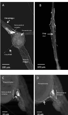
Immuno-staining of a whole mount preparation of the crop of adult D. radicum using
9
innervation by a network of immunoreactive fibres extending over the central region of
the crop sac with individual projections reaching towards the lobes (Fig. 1). These
immuno-reactive fibres originate from two axons emanating from the retrocerebral
complex (Fig. 2A) that travel along the lateral sides of the crop duct (Fig. 2B) towards
the crop sac. Reaching the base of the crop, they undergo prominent division (Fig. 1A).
Figure 2C shows stained cells of the retrocerebral complex as well as processes that
project over the proventriculus (Fig. 2D) and terminate on the surface of the anterior
midgut. No differences were noted between male and female crop preparations. The
antibody specificity was confirmed when tissues were incubated either with secondary
antibody alone or with antibody pre-absorbed with peptide, which abolished the
immuno-reactivity (supplementary Figure 1). The staining in the midgut is the result
of cross-reactivity to FLPs present in the enteroendocrine cells.
3.2. Mass analysis of crop nerve bundle (CNB) peptides
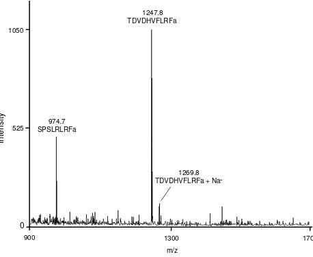
In the mass range of 500-2500 Da two prominent monoisotopic mass ion peaks m/z,
974.7 and 1247.8 were present in the mass spectra obtained from single tissue extracts
of the CNB (Fig. 3). These signals correspond to the monoisotopic masses of
myosuppressin (TDVDHVFLRFamide) and sNPF4-11 (SPSLRFamide), respectively.
The sodium adduct of the myosuppressin ion (m/z, 1269.8) was also present (Fig. 3).
3.3. Inhibition of crop muscle contractions by myosuppressin and Bztc
Myosuppressin inhibited spontaneous contractions of semi-isolated preparations of
adult D. radicum crop in a dose-dependent manner (Fig. 4) with an apparent EC50 of
4.4 x10⁻⁸ M. Spontaneous contractions were recovered when the peptide solution was
removed and washed from crop preparations with physiological saline (Fig. 4B). The
application of the non-peptide agonist Bztc to the isolated crop tissue also reduced the
frequency of spontaneous contractions, but was less potent (EC50 7.2 x10⁻⁶ M) than
myosuppressin (Fig. 4A). Importantly, the crop tissues recovered from inhibitory effect
of 1 and 10 µM Bztc, but not 100 µM Bztc, when washed with fresh saline. sNPF4-11
had no significant effect on spontaneous contractions of the crop even at high
concentrations (10-4 M) (Fig. 4A).
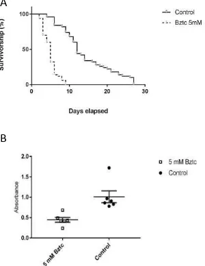
3.4. In vivo effect of peptides and Bztc
Injection of myosuppressin into adult female D. radicum had no effect on survival
10
P=0.667, 0.416) (supplementary Fig. 2). In contrast, there was a significant difference
in the survivorship between the controls and flies maintained on 5 mM Bztc/agar diet
(Log-rank test, P<0.0001) (Fig. 5A). The survival median was 5 days for Bztc-fed flies,
whereas control flies lived for up to 12 days. All the flies fed with Bztc died by day 10,
whereas it took 27 days for all the control flies to die. When Bztc was included in the
diet containing a food dye, the amount of colored food passing into the faeces was much
less than that occurring in the absence of the agonist, suggesting reduced consumption
of food (Fig. 5B).
3.5. Degradation of myosuppressin by crop peptidases
HPLC with uv detection was used to monitor the reduction in 2 nmoles myosuppressin
when incubated with crop homogenate. Myosuppressin degradation by the crop
peptidases was rapid with an estimated half-life of c. 2 min (R2= 0.9398). MALDI-TOF
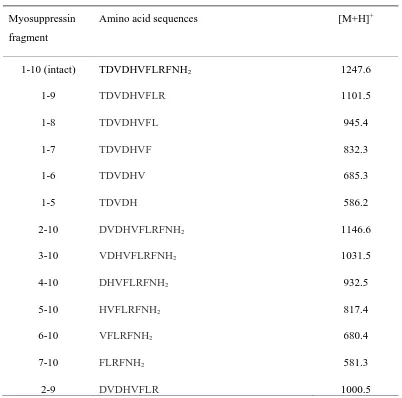
mass analysis of HPLC fractions identified a number of degradation products (Table 1,
Fig. 6), five of which retained the amino terminus (N-terminus) and six had Phe-amide
at the carboxy terminus (C-terminus). One peptide fragment (DVDHVFLR) was
truncated at both termini. The mass spectrometric data indicated the involvement of
crop aminopeptidases as well as endopeptidase activity capable of cleaving the
Arg-Phe peptide bond. In support of this hypothesis, we used fluorogenic aminopeptidase
and endopeptidase substrates to show that the crop possessed both peptidase activities
(Fig. 6). These enzyme activities were measured in both a soluble and a membrane
fraction separated from each other by high-speed centrifugation. Around 85% of the
endopeptidase (85 ± 2 pmoles/h) and 68% of the aminopeptidase activity (3.11 ± 0.08
pmole/h) were located in the soluble fraction.
4. Discussion
The release of regulatory peptides in response to external and internal cues is well
known to have direct impact on feeding activity from the control of levels of digestive
enzymes in response to food stimuli to effectively maneuvering a food bolus through
the gut via coordinated muscle contractions (reviewed by Audsley and Weaver, 2009,
Spit et al., 2012). Previous studies have demonstrated that myosuppressins have a role
11
food intake in the cockroach Blattella germanica (Aguilar et al., 2004), while injection
of myosuppressin into Spodoptera littoralis resulted in anti-feeding behavior
(Vilaplana et al., 2008). In the pea aphid, Acyrthosiphon pisum, myosuppressin
suppressed feeding resulting in mortality, most likely due to the inhibition of gut
motility preventing the movement of food (Down et al., 2011). Feeding the non-peptide
myosuppressin agonist Bztc to adult M. domestica and D. suzukii resulted in early
mortality suggesting that myosuppressin signaling has potential as an insecticide target
(Gough et al., 2017; Haselton et al., 2004).
In the present study we established the presence of myosuppressin and sNPF4-11
within the CNB of D. radicum and provide several pieces of evidence in support of
myosuppressin, but not sNPF4-11, as an important regulator of crop function in this
pest species. Myosuppressin was demonstrated in the CNB of D. radicum by direct
peptide profiling using the same approach used by Audsley and colleagues (2015) to
show the presence of myosuppressin in the CNB of D. suzukii. The current findings
however differ from the previous study by the co-occurrence of myosuppressin and
sNPF4-11. Our identification of these two peptides were based on monoisotopic peaks
(M+H]+) that are in accordance with the peptide sequences and masses reported by
Zoephel et al. (2012) and Audsley et al. (2011) in their peptidomics studies of the
larval and adult central nervous system of D. radicum, respectively. Commercially
available antiserum recognizing the C-terminus of FMRFamide was used to support
the claim that myosuppressin/ sNPF4-11 neurons extend to the crop muscle of flies
(Gough et al. 2017). Myosuppressin and sNPF4-11, as well as other insect FLPs, share
the Arg-Phe-amide sequence with FMRFamide and are expected to cross-react with
FMRFamide antibodies. Consistent with this expectation, pre-incubation of the
antiserum with synthetic myosuppressin blocked the staining of the D. radicum
nervous system. The immuno-staining of the axons in the crop nerve that project from
the retrocerebral complex to the crop muscle and spread over the surface of the crop is
consistent with previous reports on the spatial distribution of FLPs in other dipteran
species including the housefly M. domestica (Haselton et al., 2004), the fruitfly D.
melanogaster (Dickerson et al., 2012), blowfly P. regina (Richer et al., 2000), horn
fly H. irritans and stable fly Stomoxys calcitrans (Meola et al., 1996). The widespread
occurrence of FMRFamide-like immunoreactive material in the central and
12
general regulatory role for FLPs in regulating feeding and digestion in this group of
insects.
Consistent with an important role for myosuppressin in regulating crop
function, the synthetic peptide powerfully inhibited the spontaneous contractions of
the D. radicum crop musculature. Myoinhibition was observed with nM doses of
peptide (EC50, 44 nM) and the effect was immediate and long lasting, but reversible.
Such potency is typical of insect peptide receptors and compares well with the
potency (EC50, 40 nM) of myosuppressin at activating two cloned G protein-coupled
receptor genes (DmsR-1 and DmsR-2 ) from D. melanogaster expressed in
mammalian cell lines (Egerod et al., 2003a; Johnson et al., 2003). Both DmsR1 and
DmsR-2 are expressed in the crop of D. melanogaster, but DmsR-1 appears to be
more important for myosuppressin signal transduction in the crop of this fruit fly (P.
Bell, unpublished data, Chintapalli et al., 2007). In contrast, sNPF4-11failed to elicit a
myoinhibitory response when applied to the crop at concentrations even as high as 0.1
mM leading us to conclude that myosuppressin probably works alone to inhibit D.
radicum crop contractions. At present we have no functional information for the
CNB sNPF4-11.
When fed or injected into adult D. radicum, myosuppressin had no measurable
effect on feeding behaviour or mortality. This lack of a response could have resulted
from a failure to reach target gut tissues and/or rapid inactivation. Myosuppressin was
rapidly degraded (t1/2< 2min) when incubated with a homogenate of the D. radicum
crop. MALDI-MS revealed a complex mixture of myosuppressin fragments that
suggested multiple initial attacks by aminopeptidase and endopeptidase enzymes.
Indeed, we confirmed the presence of both aminopeptidase and endopeptidase activities
predominantly in a soluble fraction of the crop homogenate. A very similar pattern of
rapid degradation was reported for leucomyosuppressin (pEDVDHVFLRF-NH2) by
gut juices of two moths, Lacanobia oleracea and Spodoptera littoralis (Matthew et al.,
2009; Down et al., 2011). A structure-activity study of the inhibitory activity of
N-terminally truncated myosuppressin peptides on adult D. melanogaster crop
contractions showed that removal of the N-terminal tripeptide resulted in loss of activity
(Dickerson et al., 2012). When tested the same peptides were tested on larval gut, only
the parent 10-mer peptide gave a full inhibitory response. A similar study of
13
identified VFLRFamide as the core fragment, although this activity was at least two
orders of magnitude below that of the intact peptide (Nachman et al., 1993). In
conclusion, myosuppressin is susceptible to rapid breakdown by gut peptidases present
in the crop. Many of the fragments generated are expected to have weak or no agonist
activity on the D. radicum crop. These studies emphasise the need for myosuppressin
analogues that are resistant to degradation by gut peptidases when testing as oral
activity.
Benzethonium chloride (Bztc), a quaternary ammonium salt, was the first
non-peptide compound to be described as a myosuppressin analogue capable of
mimicking the myoinhibitory actions of myosuppressin on heart, visceral and skeletal
muscle from different a variety of insect species, including the crop of dipterans such
as M. domestica, P. regina, or D. melanogaster and D.suzukii (Duttlinger et al., 2002;
Gough et al., 2017; Haselton et al., 2004; Lange et al., 1995; Richer et al., 2000). The
evidence for Bztc being a myosuppressin agonist included shared structural features
and the competitive displacement of radioactively labelled myosuppressin from both
high- and low-affinity myosuppressin receptors in locust oviduct membranes (Lange
et al., 1995) . In the present study, the inhibitory effect of Bztc was 100-fold less
potent compared to myosuppressin and the recovery of spontaneous contractions after
the Bztc was replaced with saline was noticeably slower compared to the peptide. Our
results are in accordance with findings reported by Stoffolano et al., (2013) and Lange
et al., (1995), where in both instances Bztc reversibly inhibited muscle contractions.
Furthermore, Richer (et al., 2000) described Bztc action in mM range to be equivalent
to myosuppressin peptide, terminating spontaneous crop contractions in the blowfly
P. regina. However, it remains unclear how Bztc mimics the effect of myosuppressin
on muscle contractions. Egerod et al., (2003) could not demonstrate that
heterologously expressed Dms-R1 and Dms-R2 of D. melanogaster were activated by
Bztc in a specific manner and it is possible that some of the physiological effects of
this quaternary ammonium salt results from its weak surfactant properties. Using
dye-labelled food to follow food ingestion and excretion in adult D. radicum, we showed
that Bztc had a significant effect on food intake which probably contributed to the
toxicity of the chemical as revealed by a strong reduction in life-span. A fuller
understanding of the mechanisms leading to this toxicity is required before we can
14 Acknowledgements:
PB, REI and NA received generous support from the Department for Environment,
Food and Rural Affairs (Defra), project code PH0453. REI was also supported by the
European Union’s Horizon 2020 under grant agreement 634361.
References:
Aguilar, R., Maestro, J.L., Vilaplana, L., Chiva, C., Andreu, D., Bellés, X., 2004.
Identification of leucomyosuppressin in the German cockroach, Blattella
germanica, as an inhibitor of food intake. Regul. Pept. 119, 105 12.
https://doi.org/10.1016/j.regpep.2004.01.005
Angioy, A.M., Muroni, P., Barbarossa, I.T., McCormick, J., Nichols, R., 2007.
Evidence dromyosuppressin acts at posterior and anterior pacemakers to
decrease the fast and the slow cardiac activity in the blowfly Protophormia
terraenovae. Peptides 28, 585 593.
https://doi.org/10.1016/j.peptides.2006.10.015
Audsley, N., Down, R.E., 2015. G protein coupled receptors as targets for next
generation pesticides. Insect Biochem. Mol. Biol. 67.
https://doi.org/10.1016/j.ibmb.2015.07.014
Audsley, N., Down, R.E., Isaac, R.E., 2015. Genomic and peptidomic analyses of the
neuropeptides from the emerging pest, Drosophila suzukii. Peptides 68, 33
42. https://doi.org/10.1016/j.peptides.2014.08.006
Audsley, N., Matthews, H.J., Down, R.E., Weaver, R.J., 2011. Neuropeptides
associated with the central nervous system of the cabbage root fly, Delia
radicum (L). Peptides 32, 434 440.
https://doi.org/10.1016/j.peptides.2010.08.028
Biron, D., Langlet, X., Boivin, G., Brunel, E., 1998. Expression of early and
late-emerging phenotypes in both diapausing and non-diapausing Delia radicum
15
https://doi.org/10.1023/A:1003227812242
Blackshaw, R.P., Vernon, R.S., Prasad, R., 2012. Reduction of Delia radicum attack
in field brassicas using a vertical barrier. Entomol. Exp. Appl. 144, 145 156.
https://doi.org/10.1111/j.1570-7458.2012.01271.x
Caers, J., Boonen, K., Van Den Abbeele, J., Van Rompay, L., Schoofs, L., Van Hiel,
M.B., 2015. Peptidomics of Neuropeptidergic Tissues of the Tsetse Fly
Glossina morsitans morsitans. J. Am. Soc. Mass Spectrom. 26, 2024 2038.
https://doi.org/10.1007/s13361-015-1248-1
Chen, A.C., Friedman, S., 1975. An isotonic saline for the adult blowfly, Phormia
regina, and its application to perfusion experiments. J. Insect Physiol. 21,
529 536. https://doi.org/10.1016/0022-1910(75)90158-4
Chintapalli, V.R., Wang, J., Dow, J. a T., 2007. Using FlyAtlas to identify better
Drosophila melanogaster models of human disease. Nat. Genet. 39, 715 20.
https://doi.org/10.1038/ng2049
Dickerson, M., McCormick, J., Mispelon, M., Paisley, K., Nichols, R., 2012.
Structure-activity and immunochemical data provide evidence of
developmental- and tissue-specific myosuppressin signaling. Peptides 36,
272 279. https://doi.org/10.1016/j.peptides.2012.05.002
Down, R.E., Matthews, H.J., Audsley, N., 2011. Oral activity of FMRFamide-related
peptides on the pea aphid Acyrthosiphon pisum (Hemiptera: Aphididae)
and degradation by enzymes from the aphid gut. Regul. Pept. 171, 11 18.
https://doi.org/10.1016/j.regpep.2011.05.013
Duttlinger, A., Berry, K., Nichols, R., 2002. The different effects of three
Drosophila melanogaster dFMRFamide-containing peptides on crop
contractions suggest these structurally related peptides do not play
redundant functions in gut. Peptides 23, 1953 1957.
https://doi.org/10.1016/S0196-9781(02)00179-1
Egerod, K., Reynisson, E., Hauser, F., Cazzamali, G., Williamson, M.,
Grimmelikhuijzen, C.J.P., 2003a. Molecular cloning and functional expression
of the first two specific insect myosuppressin receptors. Proc. Natl. Acad. Sci.
U. S. A. 100, 9808 13. https://doi.org/10.1073/pnas.1632197100
Egerod, K., Reynisson, E., Hauser, F., Cazzamali, G., Williamson, M.,
16
expression of the first two specific insect myosuppressin receptors. Proc.
Natl. Acad. Sci. U. S. A. 100, 9808 9813.
https://doi.org/10.1073/pnas.1632197100
Finch, S., Coaker, T.H., 1969. A method for the continuous rearing of the cabbage
root fly Erioischia brassicae (Bch.) and some observations on its biology.
Bull. Entomol. Res. 58, 619. https://doi.org/10.1017/S0007485300057345
Gäde, G., Goldsworthy, G.J., 2003. Insect peptide hormones: A selective review of
their physiology and potential application for pest control. Pest Manag. Sci.
59, 1063 1075. https://doi.org/10.1002/ps.755
Gough, C.S., Fairlamb, G.M., Bell, P., Nachman, R.J., Audsley, N., Isaac, R.E., 2017.
Peptidergic control in a fruit crop pest: The spotted-wing drosophila,
Drosophila suzukii. PLoS One 12, 1 12.
https://doi.org/10.1371/journal.pone.0188021
Haselton, A.T., Stoffolano, J.G., Nichols, R., Yin, C.-M., 2004. Peptidergic
innervation of the crop and the effects of an ingested nonpeptidal agonist on
longevity in female Musca domestica (Diptera: Muscidae). J. Med. Entomol.
41, 684 90.
Haselton, A.T., Yin, C.-M., Stoffolano, J.G., 2008. FMRFamide-like
immunoreactivity in the central nervous system and alimentary tract of the
non-hematophagous blow fly, Phormia regina, and the hematophagous
horse fly, Tabanus nigrovittatus. J. Insect Sci. 8, 1 17.
https://doi.org/10.1673/031.008.6501
Hauser, F., Neupert, S., Williamson, M., Predel, R., Tanaka, Y., Grimmelikhuijzen,
C.J.P., 2010. Genomics and peptidomics of neuropeptides and protein
hormones present in the parasitic wasp nasonia vitripennis. J. Proteome
Res. 9, 5296 5310. https://doi.org/10.1021/pr100570j
Holman, G.M., Nachman, R.J., Wright, M.S., Schoofs, L., Hayes, T.K., Deloof, A.,
1991. Insect Myotropic Peptides - Isolation, Structural Characterization, and
Biological-Activities. Acs Symp. Ser. 453, 40 50.
Imms, A.D., 1957. A General Textbook of Entomology, in: Richards, O. W., Davies,
R.G. (Ed.), A General Textbook of Entomology. Methuen & Co. LTD., London,
pp. 597 596.
17
P.H., 2003. Identification of Drosophila Neuropeptide Receptors by G
Protein-coupled Receptors-??-Arrestin2 Interactions. J. Biol. Chem. 278,
52172 52178. https://doi.org/10.1074/jbc.M306756200
Lange, a B., Orchard, I., Wang, Z., Nachman, R.J., 1995. A nonpeptide agonist of
the invertebrate receptor for SchistoFLRFamide (PDVDHVFLRFamide), a
member of a subfamily of insect FMRFamide-related peptides. Proc. Natl.
Acad. Sci. U. S. A. 92, 9250 3. https://doi.org/10.1073/pnas.92.20.9250
Matthews, H.J., Audsley, N., Weaver, R.J., 2009. Degradation of
leucomyosuppressin by enzymes in the hemolymph and midgut of
Lacanobia oleracea and Spodoptera littoralis (Lepidoptera: Noctuidae)
larvae. Peptides 30, 565 570.
https://doi.org/10.1016/j.peptides.2008.12.020
McCormick, J., Nichols, R., 1993. Spatial and temporal expression identify
dromyosuppressin as a brain-gut peptide in Drosophila melanogaster. J.
Comp. Neurol. 338, 279 288. https://doi.org/10.1002/cne.903380210
Myrand, V., Buffet, J.P., Guertin, C., 2015. Susceptibility of cabbage maggot larvae
(Diptera: Anthomyiidae) to hypocreales entomopathogenic fungi. J. Econ.
Entomol. 108, 34 44. https://doi.org/10.1093/jee/tou019
Nachman, R.J., Giard, W., Favrel, P., Suresh, T., Sreekumar, S., Holman, G.M., 1997.
Insect Myosuppressins and Sulfakinins Stimulate Release of the Digestive
Enzyme -Amylase in Two Invertebrates: The Scallop Pecten maximus and
Insect Rhynchophorus ferrugineus. Ann. N. Y. Acad. Sci. 814, 335 338.
https://doi.org/10.1111/j.1749-6632.1997.tb46178.x
Nachman, R.J., Holman, G.M., Hayes, T.K., Beier, R.C., 1993. Structure-activity
relationships for inhibitory insect myosuppressins: Contrast with the
stimulatory sulfakinins. Peptides 14, 665 670.
https://doi.org/10.1016/0196-9781(93)90095-X
Norville, K., Sweeney, S.T., Elliott, C.J.H., 2010. Postmating change in physiology
of male Drosophila mediated by serotonin (5-HT). J. Neurogenet. 24, 27 32.
https://doi.org/10.3109/01677060903477601
Peller, C.R., Bacon, E.M., Bucheger, J.A., Blumenthal, E.M., 2009. Defective gut
function in drop-dead mutant Drosophila. J. Insect Physiol. 55, 834 839.
18
Predel, R., Neupert, S., Garczynski, S.F., Crim, J.W., Brown, M.R., Russell, W.K.,
Kahnt, J., Russell, D.H., Nachman, R.J., 2010. Neuropeptidomics of the
Mosquito Aedes aegypti. J. Proteome Res. 9, 2006 2015.
https://doi.org/10.1021/pr901187p
Rahman, M.M., Neupert, S., Predel, R., 2013. Neuropeptidomics of the Australian
sheep blowfly Lucilia cuprina (Wiedemann) and related Diptera. Peptides
41, 31 37. https://doi.org/10.1016/j.peptides.2012.12.021
Ren, J., Zhu, H., Chi, C., Mehrmohamadi, M., Deng, K., Wu, X., Xu, T., 2014. Beadex
affects gastric emptying in Drosophila. Cell Res. 24, 636 639.
https://doi.org/10.1038/cr.2014.24
Richer, S., Stoffolano, J.G., Yin, C.M., Nichols, R., 2000. Innervation of
dromyosuppressin (DMS) immunoreactive processes and effect of DMS and
benzethonium chloride on the Phormia regina (Meigen) crop. J. Comp.
Neurol. 421, 136 42.
Scherkenbeck, J., Zdobinsky, T., 2009. Insect neuropeptides: Structures, chemical
modifications and potential for insect control. Bioorg. Med. Chem. 17, 4071
4084. https://doi.org/10.1016/j.bmc.2008.12.061
Stoffolano, J.G., Danai, L., Chambers, J., 2013. Effect of channel blockers on the
smooth muscle of the adult crop of the queen blowfly, Phormia regina. J.
Insect Sci. 13, 97. https://doi.org/10.1673/031.013.9701
Thomson, A.J., 1975. Regulation of crop contraction in the blowfly Phormia
regina Meigen. Can. J. Zool. 53, 451 455.
Vilaplana, L., Pascual, N., Perera, N., Leira, D., Bellés, X., 2008. Antifeeding
properties of myosuppressin in a generalist phytophagous leafworm,
Spodoptera littoralis (Boisduval). Regul. Pept. 148, 68 75.
https://doi.org/10.1016/j.regpep.2008.02.002
Wegener, C., Reinl, T., Jänsch, L., Predel, R., 2006. Direct mass spectrometric
peptide profiling and fragmentation of larval peptide hormone release sites
in Drosophila melanogaster reveals tagma-specific peptide expression and
differential processing. J. Neurochem. 96, 1362 74.
https://doi.org/10.1111/j.1471-4159.2005.03634.x
Zoephel, J., Reiher, W., Rexer, K.-H.H., Kahnt, J., Wegener, C., 2012. Peptidomics of
19
radicum (diptera: Anthomyiidae). PLoS One 7, e41543.
https://doi.org/10.1371/journal.pone.0041543
[image:20.595.87.489.233.631.2]
Table 1
Monoisotopic masses ([M+H]+) and sequences of myosuppressin and hydrolysis products
identified in HPLC fractions after incubation with peptidases from the crop of adult Delia radicum.
Myosuppressin
fragment
Amino acid sequences [M+H]+
20 Figure legends
Fig.1. Immunostaining of the crop of D. radicum using an antibody recognising the
RFamide epitope. A) Whole mount showing FLP material in a network of filaments
covering the central region. Enteroendocrine cells of the midgut are also visible with
this antiserum. B) Higher magnification view of the region highlighted by the square
box in (A).
Fig. 2. Immunostaining of neuronal FLP peptides in whole mounts of the foregut,
retrocerebral complex and crop duct of adult D. radicum. A) Axons on the surface of
the oesophagus enter the retrocerebral complex. Stained axons run across the
proventriculus surface where they divide passing over the anterior midgut (A) and
along crop duct surface (B) to the crop lobes. C and D) Confocal z-stack images of
the retrocerebral complex showing prominently stained. D) Immunostained axons
(arrow) originating in the retrocerebral complex cover the proventriculus.
Fig.3. Mass spectrum of direct analysis of a single tissue of the D. radicum crop. Fig. 4. Inhibition of crop contractions. A) The effect of myosuppressin, sNPF4-11 and
Bztc on the spontaneous contractions of the crop. Data are expressed as the %
inhibition of the contractions counted in a 1 min period after the addition of the
agonist as described in the methods section. Values are the mean of 5 determinations
using fresh tissues for each determination. Non-linear regression analysis (GraphPad
Prism 7.01) was performed to calculate EC50 values. B) Graphical representation of
the inhibition of crop contractions by 10 µM myosuppressin and recovery after
washing with fresh saline. Muscle contractions generated tissue movement that was
video recorded (see supplementary Fig.3 and videos) and analysed as described by
Norville et al., (2010).
Fig. 5. The effect of feeding Bztc to adult D. radicum. A) 5 mM Bztc in the diet
increases mortality rate. B) Bztc reduces ingestion and excretion of sucrose/food dye.
The amount of dye in the faeces after 24 h of feeding was determined
spectrophotometrically (595 nm) and the results are expressed as the mean ± SEM (n
= 6). Differences in the means values are statistically significant (t-test, P <0.001).
Fig.6. Predicted scissile peptide bonds of myosuppressin and the structures of
substrates used to measure endopeptidase and aminopeptidase activities of the crop.
100 m
Anterior midgut
Crop
Figure 2
A B
Retrocerebral complex Oesophagus
Anterior midgut Cropduct
Crop duct Proventriculus
100 m 100 m
Proventriculus
Proventriculus
C D
Retrocerebral complex
20 m
20 m
0
525 1050
In
te
n
si
ty
900 1300 1700
m/z 1247.8 TDVDHVFLRFa
974.7 SPSLRLRFa
[image:24.595.107.336.186.374.2]1269.8 TDVDHVFLRFa + Na+
5 s 10 µM MS wash
A
[image:25.595.122.306.94.297.2]A
[image:26.595.120.270.162.356.2]Figure 6
Myosuppressin Thr-Asp-Val-Asp-His-Val-Phe-Leu-Arg-Phe-NH2
Endopeptidase substrate Mca-Arg-Pro-Pro-Gly-Phe-Ser-Ala-Phe-Lys(Dnp)
Aminopeptidase substrate Thr-Mca
1
1
2 2