Int. J. Electrochem. Sci., 13 (2018) 12000 – 12009, doi: 10.20964/2018.12.259
International Journal of
ELECTROCHEMICAL
SCIENCE
www.electrochemsci.org
Mini Review
Magnetic Particles in Electrochemical Analyses
Miroslav Pohanka
Faculty of Military Health Sciences, University of Defence, Trebesska 1575, CZ-500 01 Hradec Kralove, Czech Republic
E-mail: [email protected]
Received: 23 August 2018 / Accepted: 8 September 2018 / Published: 5 November 2018
Magnetic particles are an actual tool which has multiple use in many subjects including industrial processes, chemical separation, analytical chemistry and medicine. The major progress in this field has been made in the last few years. In this mini review, specific field of electrochemical analyses is surveyed and description of new methods where magnetic particles play a significant role is elucidated. Discussion about the current electrochemical protocols written in the current literature and expectation for the next development of biosensors construction, portable sensor systems and electrochemical methods where magnetic particles are a vital part necessary for analyte isolation is given here.
Keywords: affinity; amperometry; antibody; biosensor; biorecognition; immunochemistry; immunoglobulin; label free assay; piezoelectric; quartz crystal microbalance; voltammetry
1. INTRODUCTION
In the field of electroanalysis, wide spectrum of advanced materials including micro and nanoparticles has been developed as well. There is a number of promising materials discovered in the recent time and lot of them exert unique electrochemical properties or other promising physicochemical parameters making the electrochemical assay more competitive to the standard analytical protocols. In this minireview, electrochemical methods that are based on magnetic particles are surveyed and their promising parameters are introduced and discussed.
2. PRINCIPLE OF MAGNETIC PARTICLES
In the current literature, the term of magnetic particles is widely used though it does not precisely indicate mechanism how the particles work. Magnetism is a phenomenon related to ability to attract or repulse mass through magnetic field created by an electric or permanent magnet. The magnetic field is a space where magnetism has its impact and it is described by vectors as the magnetic field is polarized (magnetic polarization – North and South poles). To understand the principle, basic terms should be explained. Magnetic field strength H in amperes per meters is proportional to the electric current I going through the mass and it can be defined by formula:
𝐻 = 𝐼
2 × 𝜋 × 𝑟
The material surrounding the source of magnetic field influences the real magnetic strength which is called flux density B expressed in tesla (T) units and proportional to the magnetic field strength and magnetic permeability µ expressed in henry per meter (H/m). The magnetic permeability is an ability of the matter exposed to a magnetic field to support or weaken the field. The relation between magnetic strength H and flux density B is expressed in the formula:
𝐵 = 𝜇 × 𝐻
The magnetic permeability µ can be used for characterization of materials but frequently its relative value µr considering the permeability of vacuum µ0 is used:
𝜇𝑟 = 𝜇
𝜇0
We can sort materials according the relative magnetic permeability to diamagnetic (µr <1),
paramagnetic (µr >1), and ferromagnetic (µr >5). A paramagnetic and ferromagnetic materials actually
fluctuates due to temperature and it can occur only in nanoparticles and the superparamagnetic particles can acquire single domain and the magnetization does not variate in the mass of the nanoparticle.
Magnetic force is calculated in compliance with the Lorentz Force Law that considers the electric force caused by an electric field E and magnetic force onto a particle with a charge q and velocity v. When there is a magnetic field only, the E is equal to zero. The Lorentz Force Law is expressed by equation:
𝐹 = 𝑞 × 𝐸 + 𝑞 × 𝑣 × 𝐵
The Lorentz Force Law describes typical particles like charged molecules. Calculation of force between magnetized surfaces can be done according equation considering flux density, permeability and surface of area S where the interaction is done:
𝐹 =𝐵
2× 𝑆
2𝜇
Magnetic particles produced in limited series for research purposes and the commercially available ones typically exert paramagnetic properties, but the both ferromagnetic and even ferrimagnetic nanoparticles exist are frequently prepared in small amounts for research purposes and became available. The magnetic particles can be manufactured from many materials from which iron (II,III) oxide can be taken for a gold standard. The properties of iron oxides can be further improved by adding of other materials. Nanoparticles prepared from iron (II,III) oxide and titanium (IV) oxide [18], nanocomposite of iron (II,III) oxide/titanium (IV) oxide/silver silicate [19], cerium coated iron oxide and magnetic silica – cerium [20], composite from iron oxide and calcium oxide [21], composite from cobalt oxide – calcium oxide [21], Cr alloyed Nd2 (Fe,Co)14 B permanently magnetic nanoparticles
[22], Nd2Fe12B flake shaped permanently magnetic nanoparticles [23], and superparamagnetic
nanoparticles [24] can be introduced as functional examples. The use of magnetic particles can be focused on various areas. Tomographic technique magnetic particle imaging [25], magnetic separation of a wide number of compounds and cells [26-28] and analytical flow through and microfluidic devices [29-32] are common areas of application. In the field of chemistry, the magnetic particles are well suited for isolation of various compounds as the complex magnetic particle – analyte/another target molecule can be easily separated by an external magnetic field.
3. SENSORS BASED ON MAGNETIC PARTICLES
formation can further improve physical and chemical properties of magnetic particles but the unmodified magnetic particles can also exert promising applicability.
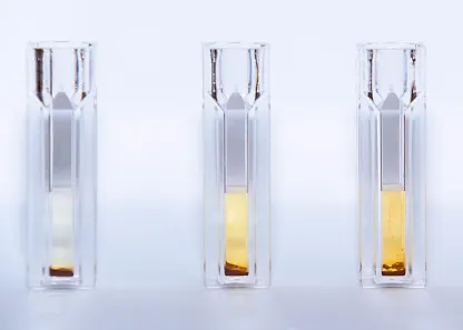
[image:4.596.91.507.375.672.2]The magnetic particles have some interesting physical and chemical properties that allow to incorporate them as a vital part of an assay. Apart of the ability to be attracted by a permanent magnet, they appear to exert some catalysing properties. In the past, pseudoperoxidase activity of iron oxides and iron containing structures like haemoglobin was described [33]. The same is also valid for magnetic particles containing the iron oxides. A colorimetric biosensor taking advantage of the magnetic particles as a pseudoperoxidase was constructed for the determination of glucose [34]. In another work, magnetic particles with Prussian blue as a modifier were interdigitated into a chitosan membrane in order to determine hydrogen peroxide in a voltammetry technique on screen printed electrodes [35]. The concept of magnetic particles performance like a catalyser was discussed in the aforementioned papers and it was stated that replace enzyme hydrogen peroxidase in an assay and would be interesting in the next development process where the assays will be introduced into praxis. Because the production of magnetic particles became cheap and parameters of the particles are standardized, use in a commercial device or analytical kit would be quite easy. Visualized pseudoperoxidase reaction of magnetic particles is exampled in figure 1.
Various materials are suitable for the surface inorganic or organic modification of magnetic particles. In an overview, magnetic nanoparticles with surface covered by polydopamine and silver were found to be able to reduce carcinogenic Cr (VI) to the less toxic Cr (III) [36]. The authors were motivated by an idea to prepare a decontamination tool but the electrochemical adaptation in a chromium assay is possible in the future. Liu and co-workers prepared a tool for biodiesel production which was based on iron oxide with modification by bamboo charcoal [37]. The particles can act catalytically in the fuel production due to the charcoal and be easily separated by an external magnetic field. In a consensus with the previous example, the application was not focused on electrochemistry but an electrochemical application can be easily adopted in the future. Analytical application of magnetic polymer was described in work by Syed et al [38]. They synthesized magnetic β-cyclodextrin with modification by toluene diisocyanate sporopollenin and it was further performed for nonsteroidal anti-inflammatory drugs like indoprofen, ketoprofen, ibuprofen and fenoprofen isolation from water samples. Desorption and identification by chromatography followed the isolation. This magnetic material appears to be promising and would be employed in the next construction of an electrochemical biosensor.
4. BIOSENSORS AND BIO-ASSAYS BASED ON MAGNETIC PARTICLES
The magnetic particles are readily to be modified on their surface by macromolecules of biological origin like enzyme, antibody or receptor and the final modified particle can work together with a sensor. The connection of both gives an analytical biosensor selectively determining analyte. The principle of magnetic particles use in a specific electrochemical assay: voltammetry on screen printed electrodes was widely reviewed in the cited paper [39]. When isolation of an analyte from solution is intended, an antibody would be the first choice how to modify surface of the magnetic particles. The antibodies can be prepared by several ways and there are typically available polyclonal, monoclonal and recombinant antibodies on the market. The availability can differ for various analytes. While the recombinant and monoclonal antibodies are sold relatively pure, the polyclonal antibody is frequently provided as a serum or plasma from hyperimmunized animals containing other plasma proteins including other antibodies [40]. The recombinant and monoclonal antibodies contain also other proteins but there should be only one type of immunoglobulin. The impurities can be further removed and immunoglobulins from medium purified prior to the biosensor construction. On the other hand, any purification is a time-consuming process which increases the costs.
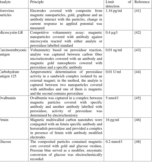
Table 1. Survey of bio-assays based on magnetic particles
Analyte Principle Limit of
detection
Reference Norovirus
particles
Electrodes covered with composite from magnetic nanoparticles, gold, graphene and an antibody interact with the particles, change in current response to applied potential was measured
1.16 pg/ml [41]
Microcystin-LR Competitive voltammetry assay: magnetic nanoparticles covered with antibody against microcystin reacted with either analyte or peroxidase labelled standard
0.4 µg/l [42]
Carcinoembryonic antigen
Voltammetry based on peroxidase reaction, analyte was captured between carbon fiber microelectrodes cowered with an antibody and magnetic gold nanospheres cowered with peroxidase and a specific antibody
0.01 ng/ml [43]
Carbohydrate antigen 125
Amperometric determination of peroxidase activity in a sandwich complex isolated by an external magnet; in the method, the analyte is captured between two nanoparticles covered with antibodies and one of them is magnetic and the second contains peroxidase
0.01 U/ml [44]
Ovalbumin Ovalbumin was captured in a complex between magnetic particles covered with specific antibody and another antibody labelled with peroxidase, activity of peroxidase was determined by electrochemistry
5 nmol/l [45]
Fetuin Magnetic multiwalled carbon nanotubes were conjugated with an fetuin specific antibody and horseradish peroxidase and provided a complex in presence of fetuin with antibody modified electrodes
16 pg/ml [46]
Glucose The composited particles contained magnetic core covered with gold and glucose oxidase, Prussian blue served as a modifier, enzymatic conversion of glucose was electrochemically recorded
0.2 mmol/l [48]
was stopped by microcystin-LR. An enzyme in connection with magnetic particles was chosen by Jung et al. for the measurement of glycemia, a glucose level in blood [48]. The authors selected gold covered magnetic particles with comprehended glucose oxidase on their surface and used also Prussian blue as a modifier. In the electrochemical assay, they were able to record signal of glucose in a range wide from at least 0.2 mmol/l to 10 mmol/l. Comparing to the immunosensor, the electrochemical bio-assays containing an enzyme interdigitated on magnetic particle are not such frequent and this area deserves and more experiments on this issue and further development of platform outcoming from it.
5. CONCLUSION
Magnetic particles appear as a promising tool in the electrochemistry. Considering the methods cited in this paper, they are readily for practical application where an antibody or enzyme is immobilized on their surface. Such electrochemical assays were proved and can be taken for a reliable platform. Economic impact of such methods can be expected in the near future. Physical properties of magnetic particles with regards to their pseudocatalytical abilities and affinity to various compounds are not fully utilized in the electrochemical analyses and primary research in this field is expected in the next years.
ACKNOWLEDGEMENTS
This work was supported by a Ministry of Defence of the Czech Republic - long-term organization development plan Medical Aspects of Weapons of Mass Destruction of the Faculty of Military Health Sciences, University of Defence.
References
1. M. A. Sani and A. Ehsani, Microb. Pathog., 6 (2018) 30332. 2. D. J. McClements, Biotechnol. Adv., 4 (2018) 30136.
3. D. J. McClements, Adv. Colloid Interface Sci., 253 (2018) 1.
4. J. Kleynhans, A. F. Grobler, T. Ebenhan, M. Sathekge and J. R. Zeevaart, J Control Release, 4 (2018) 30461.
5. J. C. Stendahl and A. J. Sinusas, J. Nucl. Med., 56 (2015) 1469. 6. M. Pohanka, Mini Rev. Med. Chem., 20 (2017) 20.
7. W. J. Mulder, G. J. Strijkers, G. A. van Tilborg, D. P. Cormode, Z. A. Fayad and K. Nicolay, Acc. Chem. Res., 42 (2009) 904.
8. M. Gautam, K. Poudel, C. S. Yong and J. O. Kim, Int. J. Pharm., 549 (2018) 31.
9. P. Singh, S. Pandit, V. Mokkapati, A. Garg, V. Ravikumar and I. Mijakovic, Int. J. Mol. Sci., 19 (2018).
10. S. Kobayashi, M. Akimoto, K. Takatoh, Y. Shiraishi, H. Sawai, N. Toshima, K. Takeuchi, K. Kotani, M. Kaneoya, K. Takeishi and H. Takatsu, Mol. Cryst. Liquid Cryst., 594 (2014) 21. 11. D. Choi, S. J. Hong and Y. Son, Materials, 7 (2014) 7662.
12. H. S. Liu and S. C. Jeng, Opt. Mater., 35 (2013) 1418.
13. W. Suthabanditpong, M. Tani, C. Takai, M. Fuji, R. Buntem and T. Shirai, Adv. Powder Technol., 27 (2016) 454.
15. R. Mandal, A. Baranwal, A. Srivastava and P. Chandra, Biosens. Bioelectron., 117 (2018) 546. 16. Z. Li and G. Y. Chen, Nanomaterials, 8 (2018).
17. N. Elahi, M. Kamali and M. H. Baghersad, Talanta, 184 (2018) 537.
18. H. Su, Z. Li, L. Lazar, Y. Alhamoud, X. Song, J. Li, Y. Wang, S. S. Fiati Kenston, M. Z. Lqbal, A. Wu, Q. Hua, M. Ding and J. Zhao, Environ. Toxicol., 11 (2018) 22631.
19. H. Chen, N. Chen, C. Feng and Y. Gao, J. Colloid. Interface Sci., 515 (2018) 119.
20. A. Dados, E. Kartsiouli, T. Chatzimitakos, C. Papastephanou and C. D. Stalikas, Talanta, 130 (2014) 142.
21. V. Chaudhary, Y. Zhong, H. Parmar, V. Sharma, X. Tan and R. V. Ramanujan, ChemistryOpen, 7 (2018) 590.
22. V. Chaudhary, Y. Zhong, H. Parmar, X. Tan and R. V. Ramanujan, Chemphyschem, 7 (2018) 201800318.
23. N. G. Akdogan, G. C. Hadjipanayis and D. J. Sellmyer, Nanotechnology, 21 (2010) 0957. 24. Z. Zhu, Z. Wang, Y. Hao, C. Zhu, Y. Jiao, H. Chen, Y. M. Wang, J. Yan, Z. Guo and X. Wang,
Chem. Sci., 7 (2016) 2864.
25. X. Y. Zhou, Z. W. Tay, P. Chandrasekharan, E. Y. Yu, D. W. Hensley, R. Orendorff, K. E. Jeffris, D. Mai, B. Zheng, P. W. Goodwill and S. M. Conolly, Curr. Opin. Chem. Biol., 45 (2018) 131.
26. I. K. Herrmann, A. A. Schlegel, R. Graf, W. J. Stark and B. Beck-Schimmer, J. Nanobiotechnology, 13 (2015) 015.
27. M. Iranmanesh and J. Hulliger, Chem. Soc. Rev., 46 (2017) 5925.
28. B. D. Plouffe, S. K. Murthy and L. H. Lewis, Rep. Prog. Phys., 78 (2015) 0034. 29. B. M. Dincau, Y. Lee, J. H. Kim and W. H. Yeo, Sensors, 17 (2017).
30. Y. Zhang and N. T. Nguyen, Lab Chip, 17 (2017) 994.
31. A. Karimi, S. Yazdi and A. M. Ardekani, Biomicrofluidics, 7 (2013) 4799787. 32. N. Pamme, Lab Chip, 6 (2006) 24.
33. J. N. Daka and T. M. Chang, Biomater. Artif. Cells Artif. Organs, 17 (1989) 553.
34. P. Martinkova, R. Opatrilova, P. Kruzliak, I. Styriak and M. Pohanka, Mol. Biotechnol., 58 (2016) 373.
35. P. Martinkova and M. Pohanka, Int. J. Electrochem. Sc., 11 (2016) 10391.
36. C. L. Chen, K. R. Zhu, K. Chen, A. Alsaedi and T. Hayat, J. Colloid Interface Sci., 526 (2018) 1.
37. K. Liu, R. Wang and M. Q. Yu, Renew. Energy, 127 (2018) 531.
38. S. F. F. Syed Yaacob, M. A. Kamboh, W. A. Wan Ibrahim and S. Mohamad, R. Soc. Open Sci., 5 (2018).
39. P. Yanez-Sedeno, S. Campuzano and J. M. Pingarron, Sensors, 16 (2016). 40. M. Pohanka, J. Appl. Biomed., 7 (2009) 115.
41. J. Lee, K. Takemura, C. N. Kato, T. Suzuki and E. Y. Park, ACS Appl. Mater. Interfaces, 9 (2017) 27298.
42. L. Reverte, D. Garibo, C. Flores, J. Diogene, J. Caixach and M. Campas, Environ. Sci. Technol., 47 (2013) 471.
43. D. Tang, R. Yuan and Y. Chai, Anal. Chem., 80 (2008) 1582.
44. D. Tang, B. Su, J. Tang, J. Ren and G. Chen, Anal. Chem., 82 (2010) 1527.
45. M. Cadkova, R. Metelka, L. Holubova, D. Horak, V. Dvorakova, Z. Bilkova and L. Korecka, Anal. Biochem., 484 (2015) 4.
46. E. Sanchez-Tirado, A. Gonzalez-Cortes, P. Yanez-Sedeno and J. M. Pingarron, Biosens. Bioelectron., 113 (2018) 88.
47. S. Delshadi, G. Blaire, P. Kauffmann, M. Fratzl, T. Devillers, D. Delabouglise, M.
48. H. Y. Jung, J. H. Park, S. W. Hwang and J. Kwak, J. Biomed. Nanotechnol., 9 (2013) 901. 49. M. Seenuvasan, G. Vinodhini, C. G. Malar, N. Balaji and K. S. Kumar, IET Nanobiotechnol.,
12 (2018) 535.
50. A. B. Muley, A. S. Thorat, R. S. Singhal and K. Harinath Babu, Int. J. Biol. Macromol., 9 (2018) 31423.
51. H. Torabizadeh and M. Mikani, Int. J. Biol. Macromol., 117 (2018) 134.