-·---
...
ASPECTS ON THE FORMATION AND TEMPERATURE
MODIFICATION OF LEAF SURFACE WAX
IN
Brassica napusL.
by
Douglas John Armstrong
B.Sc. Hon. (A. N. U. )
Thesis submitted for the
degree of Doctor of Philosophy
at the Australian National University
a
This thesis contains no material which has been accepted for the award of any other degree or diploma in any University and, to the best of my knowledge and belief, it contains no material previously published or the result of work by another person, except where due reference is made in the text.
Canberra, July 1973.
D.J. ARMSTRONG
iii
ACKNOWLEDGEMENTS
I am indebted to my supervisor, Dr. M.I. Whitecross, for suggesting the project, for advice and guidance throughout its development, and for supervision during the preparation of the Whitecross
&
Armstrong (1972) paper. I also wish to thank Professor L.D. Pryor for allowing me the opportunity to use the facilities within the Botany Department, S.G.S., A.N.U., and for his supervision during 1971.I am indebted to Dr. D.J. Goodchild, C.S.I.R.O., Canberra, for making available to me the facilities of the Electron Microscope Unit. Other C.S.I.R.O. members to whom I am indebted include Dr. C. MacDonald for making available the services of the Mass Spectrometry Unit;
Dr. B. Filshie for scanning electron microscope studies; Mr. N.A.
Pyliotis for helpful discussion and to the staff of the Ceres Phytotron for their care and maintenance of plants.
Thanks also go to Dr. R. Sands, C.S.I.R.O., Melbourne, for assistance with radioassays. The final preparative stages of this thesis were achieved with the helpful assistance of Anne Farrell and Mervyn Cormnons.
Finally, I am grateful for the financial assistance provided by the award of a Commonwealth Postgraduate Scholarship to carry out this study.
iv
SUMMARY
Electron microscopy, gas-liquid chromatography and mass spectrometry have been used to elucidate many of the phenomena concerned in the
formation and temperature modification of leaf surface waxes in Brassica napus L.
A characteristic wax pattern was observed for plants growing over a range of temperature regimes. The change observed in wax pattern was one of wax rods at low growing temperature and complex wax plates at high growing temperature (up to 36 °C).
Low and high temperature waxes were found to differ chemically by a small but consistent reduction in the major C29 and C31 hydrocarbons accompanied by an increase in the C2 9 symmetric ketone. The presence of C42 to C45 ester compounds in Brassica spp. leaf wax is reported here for the first time.
The effects of different growing temperatures, wax mutants and
dewaxing herbicides did not reveal any marked cytological differences in epidermal cell fine structure, which could be related to wax synthesis.
An autoradiographic study implicated endoplasmic reticulum and Golgi vesicles in the manufacture and transport of waxes or wax precursors
respectively. Movement of the waxes to the leaf surface appears to take place purely by diffusion across the cell wall and the adjacent
subcuticular region whereafter wax reaches the surface by means of cuticular channels and surface pores.
The cuticular pores were found to play no role in the formation of wax patterns characteristic for a given growing temperature. This
pattern indeed was largely attributed to the small differences in wax chemistry and conditions under which the wax crystallised.
The 'growth ring' appearance characteristic of wax rods was shown not to result from extrusion of the wax on the leaf surface as has been suggested previously.
Low temperature leaf wax was found to be modified significantly within hours of transferring the plant concerned to a higher temperature
CONTENTS
GENERAL INTRODUCTION
1. The Leaf Cuticle - Its Nature and Function 2. Biosynthesis of Cuticular Waxes
3. Aims of the Investigation
CHAPTER 1: MODIFICATION IN FINE STRUCTURE OF EPIDERMAL CELLS AND ASSOCIATED SURFACE WAXES
INTRODUCTION METHODS
Light Microscopy
Scanning Electron Microscopy Transmission Electron Microscopy Plant Growth
Herbicide Treatment of Plants Sampling RESULTS (i) (ii) (iii) (iv) (v) (vi) DISCUSSION
Effect of Growing Temperature on Epidermal Cells Mutant and Chemical Effects on Epidermal Cells
Fine Structure of Outer Epidermal Cell Wall
Effect of Growing Temperature on Fine Structure of Wax
Mutant and Chemical Effects on Wax Fine Structure Effect of Rapid Temperature Changes on Wax Fine
Structure V 1 1 4 5 6 6 7 7 7 8 9 10 11 11 11 13 14 16 17 18 20
CHAPTER 2: ELECTRON MICROSCOPE INVESTIGATION ON CUTICULAR PORES
AND MICROCHANNELS 24
INTRODUCTION METHODS
Surface Replicas
Freeze-Fractured Replicas RESULTS
DISCUSSION
CHAPTER 3: EFFECT OF TEMPERATURE ON THE CHEMISTRY OF LEAF SURFACE WAXES
INTRODUCTION METHODS
Isolation of Leaf Waxes Thin Layer Chromatography
Infra-red Spectroscopy Gas-liquid Chromatography Mass Spectrometry
RESULTS
Thin Layer Chromatography and Infra-red Spectroscopy G.L.C. and Mass Spectrometry
DISCUSSION
CHAPTER
4:
RECRYSTALLISATION STUDIES ON HIGH AND LOW TEMPERATURE WAXESINTRODUCTION METHODS RESULTS DISCUSSION vi 33 33 33 34 37 37 38 40 42
CHAPTER 5: INCORPORATION OF 14C-PALMITATE INTO EPIDERMAL CELLS
AND SURFACE WAXES 44
INTRODUCTION
METHODS
Experimental Standards
Preparation of Radioactive Palmitate
Incorporation of Palmitate and Wax Removal Autoradiography of Whole Leaves
Combined Electron Microscopy and Autoradiography
RESULTS
DISCUSSION
GENERAL CONCLUSIONS
APPENDIX I:
APPENDIX II:
LEAF THICKNESS MEASUREMENT
DEHYDRATION VARIANTS FOR E.M.-AUTORADIOGRAPHY OF LIPIDS
APPENDIX III: PROCEDURE FOR COATING NUCLEAR EMULSION ON ELECTRON MICROSCOPE SECTIONS
GENERAL INTRODUCTION
1. THE LEAF CUTICLE - ITS NATURE AND FUNCTION (i) The Problem of Definition
Aerial parts of the primary plant body are invested with a
protective cuticle. Consisting primarily of lipoidal substances, the cuticle overlies and merges into the outer walls of the epidermal cells. Because of this close association with the epidermis, a strict
morphological definition of the cuticle has proved difficult. Indeed, the term has been used in different senses and modified in the literature
(Anderson, 1935; Priestley, 1943; van Overbeek, 1956; Goodman, 1962; Sitte
&
Rennier, 1963; Crisp, 1965; Martin&
Juniper, 1970).Crisp (1965), following definitions of Esau (1953), defined the outer epidermal wall as comprising stratified layers of cellulosic, pectinaceous and lipoidal materials, the outermost layer being the cuticle. This definition would suffice in most cases, except that the three layers are by no means always clearly defined. The pectic layer may be virtually absent, with the cellulosic layer and the cuticle merging gradually into one another as in
Clivia
(Roelofsen, 1952). In such circumstances, an arbitrary distinction is usually made between the cuticularised (or cutinised) cell wall layer containing both cellulose and cuticular substances, and the cuticle proper which contains nocellulose. The cuticle is thus defined in a positional sense.
(ii) Morphological Description
The recent review by Martin
&
Juniper (1970) sets out in detail the diversity of cuticular morphology as well as the basic similarities2
the cuticle. For one thing there is every likelihood that the surface waxes and the embedded waxes are continuous, if only by means of surface
pores (Hall & Donaldson, 1962; Hall, 1967a). Schieferstein & Loomis (1959) reported that only about 50% of the species they studied had waxy leaf cuticles. It has been shown since (D.M. Hall, personal
communication) that the former authors, because of the replica methods used for their observations, failed to recognise the large flat sheets of non-structural wax on their 'non-waxy' species. It is still true to say, however, that the proportions of cutin to wax (embedded and superficial) vary widely between species.
(iii) Basic Chemistry
Chemically, the cutin framework is a polyester consisting of
polymerised dicarboxylic and hydroxy-substituted aliphatic acids. The hydroxy acids are commonly substituted in the omega-position allowing head to tail condensation while mid-chain substitution, frequently in the 9,10-position, permits three-dimensional polymerisation. The result is a closely-meshed elastomer with a tendency to lamination (Linskens
et al.,
1965; Eglinton & Hamilton, 1967; Martin & Juniper, 1970).
The chemistry of plant waxes has most recently been reviewed by Eglinton & Hamilton (1967), Mazliak (1968), and Martin & Juniper (1970), while rapid advances in their characterisation has been discussed by Lindeman & Annis (1960), Eglinton
et al.
(1966), and Holloway & Challen(1966). In general, plant waxes are mixtures of long chain alkanes, primary and secondary alcohols, ketones, ketols, acetals, esters and acids plus true waxes in the chemical sense. At first sight, these compounds may appear simple; however the variability and complexity of plant waxes cannot be over-emphasised.
(iv) Physiological Functioning
The cuticle and its associated waxy layer has been ascribed numerous barrier functions between the environment and the plant body.
(a) Suppression and stimulation at the leaf surface of infection by fungi has received a considerable amount of attention. The results,
however, have been rather conflicting, and reference is made to Martin & Juniper (1970), where a detailed account of the work in this field is given. What appears to be of major importance in the role of the surface
3
(b) While a high degree of glaucousness does not necessarily
indicate extreme waxiness, the presence of surface wax has an effect on the light reflection characteristics of leaves. Variations in light
reflectance due to leaf surface features have been studied by Billings &
Morris (1951), Moss
& Loomis (1952), Cameron (1964), Howard (1966), and
Armstrong (unpublished data). The light scattering character of the rough textured wax layer and the light absorbing powers of polyphenolics in the cutin may well shield the plant from excess ultraviolet radiation.(c) Of agricultural importance is the contact angle made by water drops on a leaf surface. A considerable amount of work has been done in measuring and correlating the magnitude of the contact angle with surface roughness and waxiness. The more recent workers in this field include
Thrower
et al.
(1965), Wortman (1965), Martin (1966), Sargent (1966), Troughton&
Hall (1967), and Armstrong (unpublished data). Holloway(1969) has studied the orientation of constituent molecules in surface wax in relation to water repellency but found that hydrophobic properties
generally give little indication of chemical composition. Penetration of externally applied solutions, particularly in the study of herbicides, has been investigated more recently by Franke (1964, 1967), Hallam (1964), Middleton & Sanderson (1964), Martin (1966), Sargent (1966), and Sands
(1972).
(d) The more recent work on cuticular transpiration indicates that surface waxes are important in preserving the water balance of the plant. Increased transpiration due to disruption of the wax surface by
mechanical or chemical means has been noted by Pfeiffer
et al.
(1957), Hall & Jones (1961), Daly (1964), Horrocks (1964), Bain & McBean (1967), Possinghamet al.
(1967), and Denna (1970). Grncarevic & Radler (1967) have noted that aliphatic components such as the hydrocarbon, alcohol and aldehyde fractions of the surface wax gave the greatest reduction in4
2. BIOSYNTHESIS OF CUTICULAR WAXES (i) Genetics of Wax Inheritance
The chemical genetics of wax formation have recently been examined for Pisum and Brassiaa spp. by Macey & Barber (1969, 1970a,b). In
Brassiaa mutants, the genes of at least two different loci were found to be responsible for the glossy leaf character of the mutant concerned. In both studies, the mutants contained reduced amounts of paraffin, ketone and secondary alcohol fractions, while the aldehyde primary alcohol and ester components were more prominent when compared with the normal waxy plant. The genetic control of diketone fractions, in relation to the synthesis of wheat and barley waxes respectively, has been examined by Barber
&
Netting (1968) and Wettstein-Knowles (1972). Clines inglaucousness have been correlated with changes in frost severity. Such clines appear to represent changes in allelic frequency at one or more loci controlling the development of wax (Barber, 1955; Barber
&
Jackson,1957) . Macey (1967) reported that it is likely that several genes are active in determining different surface wax patterns.
(ii) Variations in Wax Amount, Type and Chemistry
Variations in cuticular composition within species have been demonstrated, and have been shown to depend on leaf age, leaf surface
(adaxial or abaxial), and environmental growth conditions (Kurtz, 1950; Mueller et al., 1954; Skoss, 1955; Juniper, 1959b, 1960; Baker, 1963;
Hall & Donaldson, 1963; Leigh and Matthews, 1963; Whitecross, 1963; Baker
&
Martin, 1967; Eglinton&
Hamilton, 1967; Herbin&
Robins,1968a,b, 1969; Hallam
&
Chambers, 1970; Hawthorn&
Stewart, 1970; Banks & Whitecross, 1971; and Whitecross & Armstrong, 1972).The more recent work on the biosynthesis of plant surface wax components has made use of 14C-labelled fatty acid precursors in an attempt to evaluate the mechanisms involved (Kaneda, 1966, 1967, 1968; Kasprzyk & Wojciechowski, 1969; Kolattukudy, 1965, 1966, 1967a,b,c, 1968,
1970a,b,c; Kolattukudy et al., 1968; Kolattukudy
&
Tsui-Yun, 1970; Kolattukudy&
Walton, 1972; Marekov et al., 1968; and Mitchie&
Reid,1968). A detailed account of the various biosynthetic mechanisms in relation to leaf waxes has been recently reviewed by Kolattukudy
(1970b,c). Alkanes are considered to be formed from fatty acids either by elongation followed by decarboxylation or by ~head to head'
specific decarboxylation of one of them. Fatty acyl-CoA is reduced to
the aldehyde which in turn is reduced to the alcohol. The alcohol is
then esterified with acyl moieties from acyl-CoA or phospholipids.
(iii) Biosynthetic Site and Pathway for Wax Precursors
5
While it is generally agreed that the epidermal cell layer of plant
leaves is the site for the production of leaf wax precursors (Kolattukudy,
1968, 1970b,c), there is much speculation within the literature as to how
these components traverse the cell wall and cuticle to become established
on the leaf surface (Linskens et al., 1965). Whether the mechanisms
involved are achieved via some specific wax microchannel cum surface pore
arrangement, or else by purely diffusive means along a concentration
gradient, or perhaps by a combination of these factors, has not been
clearly established for any plant. Even assuming that the mechanisms
involved could be determined for a particular plant species, one could
not be certain that the same pathway operated in all waxy plants, though
differences between species would most likely be small.
3. AIMS OF THIS INVESTIGATION
In view of the significant qualitative differences observed in the
leaf surface wax pattern of Brassica napus when grown under a variety of
temperature regimes (Whitecross & Armstrong, 1972), it seemed appropriate
to make a comprehensive study of this plant in the hope of answering some
specific questions on formation of leaf surface wax and its modification
in response to temperature changes, viz.
(i) What specific organelles within the cells of a leaf are
responsible for the manufacture of wax precursors?
(ii) What pathway operates in the transport of waxes or their
precursors from the cells across the outer cell wall and cuticle regions,
and how do the waxes finally reach the leaf surface?
(iii) Once on the leaf surface, is the wax pattern characteristic
of a particular growing temperature determined by morphological and
surface characteristics of the leaf, the chemical composition of the wax,
or simply the physical conditions encountered by the wax as it deposits?
(iv) How readily adaptable is a growing plant to alter its leaf
CHAPTER 1
MODIFICATION IN FINE STRUCTURE OF EPIDERMAL CELLS
AND ASSOCIATED SURFACE WAXES
INTRODUCTION
6
Waxy substances are components of the leaf cuticle from the very
earliest stages of leaf development. During leaf expansion, wax is found
on the surface in a semi-crystalline form and appears to be produced
continuously during this phase of growth. Regeneration of structural wax
on damaged areas of the leaf surface may continue until a late stage of
leaf expansion (Juniper, 1960; Hallam, 1970c).
The presence of structural wax on plant surfaces raises the question
of how such fine structures develop and whether the modification
attributed to the changing of environmental factors can be characterised.
Whitecross (1963) has described the effects of light, temperature
and water stress on the structure and composition of leaf wax of Brassica oZeracea. More recently the effect of light and temperature on the waxes of Brassica napus has also been examined (Whitecross
&
Armstrong, 1972). In each case, structural alteration of the wax resulted from differentgrowing temperatures, while the effect of light was to alter the wax
quantitatively. Water stress apparently affected the chemical
proportions in the wax (Whitecross, 1963).
The earliest investigations on the effects of varying growth
temperatures have been restricted in several ways. For instance, the
range did not include the region of possible stress above, say, 30 °C.
Further, the observed variation in fine structure of surface wax
represented only the end result of a chain of physiological events
capable of being affected by temperature. There was thus a definite need
to extend the investigation to include a study of the effects of higher
temperatures and also to examine the effects throughout the range at the
site of wax synthesis in the cytoplasm of the epidermal cells themselves
(Kolattukudy, 1968, 197Gb).
whereby the leaves appear green as opposed to those of the typically
glaucous waxy varieties. Another greening effect has been observed as a
result of inhibiting wax synthesis with T.C.A. (Kolattukudy, 1965,
1970b,c; Hallam
&
Juniper, 1971).7
Pre-emergent soil treatment with T.C.A. almost completely suppressed
surface wax formation in peas (Juniper, 1959a; Dewey
et al.,
1961) while changes induced by related herbicides on epidermal cell membrane systemshave been observed by Hallam (1970a).
The question arose as to whether the natural mutants, in which wax
formation is affected, have anything in common at the cellular level with
plants affected by T.C.A. (trichloroacetic acid), or other related
compounds such as dalapon (2,2-dichloropropionic acid). This
investigation sought to make this comparison possible also.
Finally, the pathway linking the site of wax formation with its
ultimate repository demanded examination. The effects of temperature,
such as those observed, may have resulted neither from direct effects at
the cuticular surface nor from effects at the metabolic level in the
cytoplasm, but rather from effects on the membranes, cell walls and
cuticle which the wax or its precursors traverse.
METHODS
Light Microscopy
Light micrographs of leaf cross sections were prepared by cutting
1 µm sections of epoxy-embedded material and staining with a solution of
1% aqueous Azure Band methylene blue containing 1% borax (A.E. Ashford,
personal communication).
Low power leaf surface characteristics were prepared by the use of
replicating tape (Ladd Corporation).
Scanning Electron Microscopy
Fresh representative leaves were cut and glued onto specimen stubs.
The exposed leaf surface was initially coated with carbon ("' 60 nm)
followed with 60/40 gold/palladium alloy. The specimens were viewed
directly in a JEOL scanning electron microscope and photographed on to
Transmission Electron Microscopy
Thin Sections
Leaf material was prepared according to the following schedule:
1. Leaf pieces< 1 nnn2 were rapidly cut from a representative leaf and placed in 3% glutaraldehyde in 0.025 M sodium phosphate buffer, pH 7.2,
for two hours at room temperature, vacuum infiltration being applied
where necessary.
8
2. The tissue was washed in 0.025 M sodium phosphate buffer, pH 7.2, for
a minimum of forty-five minutes, generally one hour with four changes.
3. Post-fixation was carried out in 2% osmium tetroxide in the same
buffer for one and a half hours at room temperature.
4. The tissue was further washed in the same buffer for ten minutes (two
changes) before being dehydrated in a graded series of acetone solutions
(20% - 30 minutes; 40% - 25 minutes; 60% - 20 minutes; 80% - 15
minutes; and 100% (two changes) - 15 minutes, at room temperature).
5. After dehydration the tissue was infiltrated in 1:1 acetone-Texas
mixture for thirty minutes followed by a one hour infiltration (two
changes) in 100% Texas mixture at room temperature. Vacuum infiltration
of the embedding medium was not used. (Texas I mixture - Araldite M,
Epon 812, D.D.S.A., dibutyl phthalate, DMP-30 (Mollenhauer, 1964).)
6. Polymerisation was carried out in silicone rubber moulds at 60 °C for
six hours followed by four hours at 85 °C.
7. Sections were cut with a diamond knife and supported on 200- or
300-mesh uncoated copper grids. Staining was carried out in uranyl acetate
(saturated 50/50 aqueous ethanol) and Reynolds lead citrate (Reynolds,
1963).
8. Sections were examined in a Philips 200 electron microscope at 80 KV
and micrographs recorded on Kodak Estar base electron microscope film.
9. Good preservation of leaf surface waxes in thin section was attained
by this schedule. Visualisation of the waxes in electron micrographs was
intensified where necessary by an increase in the lead staining time.
Leaf Surface Replication
The carbon replica technique used in these and previous studies was
The simplification resulted mainly from using only one plastic backing layer prior to stripping from the leaf, and it produced consistently satisfactory results in an appreciably shorter time (Whitecross
&
Armstrong, 1972).9
1. A leaf was cut into pieces approximately 1.5 mm long and 0.5 mm wide.
2. A glass microscope slide was coated with Bedacryl 122X stock solution
(ICIANZ Ltd).
3. The leaf pieces were carefully transferred onto the tacky Bedacryl layer which set relatively quickly thus minimising leaf shrinkage.
4. The leaf pieces were then subjected to a vacuum,....,
Jx 10-s mm Hg in an
Edwards 12E6 vacuum coating unit and shadowed with Pt/Cat 40°. A tight coil of 0.1 mm diameter Pt wire at the point of contact between twolathe-turned carbon rods, one as a cylindrical peg and the other steeply conical comprised the Pt/C evaporation assembly.
5. Carbon alone was then evaporated at 8 x 10⁻⁴ mm Hg in a series of brief 20V/30A bursts. A steeply conical carbon rod against a bevelled carbon rod comprised the carbon evaporation assembly.
6. The leaf pieces were immediately flooded with 15% Bedacryl in benzene and drained by tilting the slide.
7. When the backing layer of plastic was thoroughly dry (24 hours), a sharp razor blade was used to score the plastic and carbon films into convenient grid-sized pieces.
8. The combined plastic-carbon film was stripped from the leaf surface with fine forceps by lifting up one corner and carefully pulling it back.
9. The sections of plastic-carbon film were first washed in acetone to remove the plastic, and finally in 50/50 acetone/chloroform to remove any last traces of plastic or adhering leaf wax, before being picked up on a grid and dried.
10. Replicas were examined in the electron microscope and photographed onto Agfa Scientia 23D56 plates. The positive plates so obtained were
reversed by a 1:1 contact printing onto Ilford N5.31 film, or to Kodak Commercial Ortho film.
Plant Growth
10
and sampling methods were standardised.
The plants used exclusively in this work were Brassica napus,
variety Dwarf Essex (rape) representing the normal waxy plant and a
non-waxy (gl3 ) mutant, Brassica oleracea, variety acephala (Thompson, 1963; Macey
&
Barber, 1970b).All plants were grown under the controlled environmental conditions
available at the CERES Phytotron, C.S.I.R.O., Canberra. Seeds were
fumigated with methyl bromide prior to germination in 24/19 °C. Single
seedlings were transplanted into 5" pots containing a 60/40 perlite/
vermiculite mixture. All seedlings were established for ten days before
being transferred to their respective temperature treatments. Day/night
temperature regimes of 15/10 °C, 18/13 °C, 21/16 °C, 24/19 °C, 27/22 °C,
30/25 °C, 33/28 °C and 36/31 °C were available for plant growth. Plants
were maintained under a sixteen hour photoperiod regime, natural light
being supplemented by artificial incandescent lighting. Relative
humidity was always greater than 40%.
All plants were given applications of nutrient solution in the
morning and water in the evening. To avoid any water stress, the high
temperature treatments were given an additional watering at midday
(27/22 °C) or alternatively left standing in 1" of water (30/25 °C,
33/28 °C and 36/31 °C). The non-waxy (g13 ) mutant was grown only under a
24/19 °C regime.
Extreme care was taken at all stages of plant growth to avoid water
and mechanical contact with the leaf surfaces.
Herbicide Treatment of Plants
In the application of herbicides, all treatments were applied to the
root system only, plants being maintained in their normal perlite/
vermiculite mixture. Two-week old test plants, kept under a 24/19 °C
temperature regime were treated daily with 1.5 x 10⁻⁴ moles of T.C.A. or
alternatively with 5 x 10-s moles of dalapon as the sodium salt. No
attempt was made to add quantitative amounts of herbicide to the plant
system or specific leaves. Initial investigations showed that the
amounts added produced a striking reduction of leaf wax content without
having caused obvious morphological changes in the plants. In Brassica,
1-2 x 10-s M T.C.A. in a single leaf inhibited surface wax synthesis by
11
Treatments were continued for ten days with the plants subsequently
maintained under a normal watering regime for a further five days prior
to sampling. This procedure was adopted to avoid any direct herbicide
application effect.
Sampling
Leaf sampling, unless otherwise stated in the text, was carried out
after the plants had been in their respective treatments for four weeks.
The plants were chosen at random from a batch of treatment plants.
Disregarding the small apical leaves, the fourth visible leaf from
the apex was taken as a representative sample for each plant unless
otherwise stated. This leaf was almost fully expanded showing no signs
of senescence. The principle involved in this sampling method was to
compare leaves at a similar physiological age from the point of view of
cuticular development. Variation in the development of leaves due to the
effect of different treatments made the sampling of a particular node
number from the cotyledons an unsatisfactory method of representative
sampling.
In small-scale sampling of isolated leaves, a representative area of
lamina from each was selected avoiding mid-rib, major veins and leaf
margins.
RESULTS
(i) Effect of Growing Temperature on Epidermal Cells
Plate 1.1 a,b &c represents light micrographs of cross sectioned
leaf tissue while plate 1.2 a,b &c illustrates surface replication views
of leaves grown at 15/10 °C, 24/19 °C and 36/31 °C respectively. Average
leaf thickness for the three respective temperature conditions were 0.28,
0.24 and 0.11 mm (method of measuring leaf thickness is detailed in
Appendix I).
Throughout the fine structure study of Brassica epidermal cells, the tonoplast was frequently seen to protrude into the vacuole and was often
constricted so as to form apparently isolated spheres of membrane within
the vacuole. Degenerated organelles frequently appeared in the centre of
these membrane spheres. Good preservation of other cytoplasmic
structures would appear to rule out fixation artifacts as an explanation
12
While this study was essentially concerned with the investigation of
the upper epidermal layer, preliminary ·work showed the lower layer to be
similar in all respects of fine structure except for actual cell size.
No detectable fine structure differences were found between small
developing leaves and fully expanded leaves for any particular
temperature treatment. Only the cell wall thickness was observed to
increase slightly in the more fully expanded leaves.
Plate 1.3 a,b &c illustrates the fine structure detail of the outer
epidermal cell wall and adjacent cytoplasm for rape grown under a 15/10 °C
temperature regime. Although the cytoplasmic layer for this treatment
was relatively thin, at times little more than the thickness of one
peripheral strand of endoplasmic reticulum, considerable activity must
occur since appreciable amounts of wax are produced under these
conditions (Whitecross & Armstrong, 1972).
Mitochondria and Golgi bodies are present in the cytoplasm while
ribosomes, endoplasmic reticulum and vesicles make up the bulk of the
cytoplasmic components. Large vesicles are frequently found associated
with Golgi bodies. The plasmolennna was observed to be highly irregular
in outline, frequently extending pseudopodia-like protruberances into a
hyaline area corresponding to the inner cell wall.
The microfibrils making up the cell wall lamellae lie parallel to
the leaf surface. These cell walls generally exhibit a staining gradient
from the plasmolemma to the cuticle. No evidence was seen of intrusion
of cytoplasmic vesicles into the cell wall despite the irregular
plasmolennna mentioned earlier nor was any microchannel structure
extending from the plasmolennna across the cell well to the cuticle ever
observed.
The cuticle was observed to be consistently irregular in appearance
although its overall thickness was rel atively constant. Epicuticular wax
structures are frequently present , their outline resembling that observed
in replication studies (Whi tecross & Armstrong, 1972). Even the
consecutive ring type structure of wax rods has been confirmed in thin
section studies. Preservation of surface waxes in thin section has been
achieved without special fixation techniques or wax stabilisation
procedures (Hallam, 1964, 1970b).
Plate 1.4 a,b &c illustrates the epidermal cell fine structure for
13
cytoplasm was similar to that observed for the 15/10 °C treatment
although there was a general increase in the thickness of this layer and
a proportionate increase in the number of cell organelles present.
Conversely, there was a consistent decrease in the thickness of the cell
wall. The cuticle, however, was similar both structurally and
dimensionally to that observed in the 15/10 °C treatment.
Plate 1.5 a,b &c illustrates the epidermal cell fine structure for
rape grown under a 27/22 °C temperature regime. The fine structure of
the cytoplasm was very similar to that observed in the previous treatment.
There was an apparent increase in ribosome number and a decrease in the
Golgi frequency. There was a continuing decrease in cell wall thickness
while the cuticle structure was consistent with that previously observed.
Plate 1.6 a,b &c illustrates the epidermal cell fine structure for
rape grown under a 36/31 °C temperature regime. In contrast to the three
lower temperature treatments, the cytoplasmic organelles, tonoplast and
plasmolemma showed definite signs of degeneration." A decrease in the
amount of cytoplasm was observed with a consequent decrease in
endoplasmic reticulum, ribosomes and cytoplasmic vesicles.
Irregularities in the tonoplast and plasmolemma were not nearly so marked
in comparison to the lower temperature treatments. There was a
significant reduction in the number of Golgi bodies, perhaps accounting
for the substantial decrease in cell wall thickness. Throughout this
study, a consistent negative correlation has been observed between cell
wall thickness and increased growing temperature, but dimensions of the
cuticle itself were not obviously different, however, from that observed
in the three previous temperature treatments.
(ii) Mutant and Chemical Effects on Epidermal Cells
Plate 1.7 a,b & c illustrates the epidermal cell fine structure for
the non-waxy (gl3 ) mutant grown at 24/19 °C. Structurally the
cytoplasmic layer differed from the waxy species in that there were fewer
Golgi bodies, vesicles and endoplasmic reticulum. The plasmolemma,
although continuous, was very irregular in outline and lay adjacent to a
markedly hyaline region of the cell wall. The presence of densely
stained globules throughout the cytoplasm, and particularly confined to
the plasmolemma, were quite characteristic of the non-waxy mutant
epidermal cells. Structurally the cell wall and cuticle were similar to
those of the waxy control plants although the mutant cell walls appeared
14
Plates 1. 8 a, b & c and 1. 9 a, b & c illustrate the effects of herbicide
treatment, applied to the roots, on the fine structure of epidermal cells.
Plate 1. 4 a, b & c should be referred to for comparison as representing the
normal untreated 24/19 °C plants.
The appearance of the cytoplasm was similar to that observed for the
controls, though some degradation of individual organelles was indicated.
Ribosomal frequency in the dalapon treatment was higher while the amount
of endoplasmic reticulum was marginally reduced. Similar differences
were not noticeably apparent in the T.C.A. treated plants.
Frequent observations were a loss of plasmolemma integrity
accompanied by a significant decrease in the frequency of vesicles for
both herbicide treatments.
The dalapon treatment caused the hyaline layer external to the
plasmolemma to be reduced and increased significantly in the T.C.A.
treatment, although in both treatments, the cell wall thickness was
significantly reduced, as was the thickness of the cuticles, particularly
in the dalapon treated plants.
(iii) Fine Structure of Outer Epidermal Cell Wall
Plate 1.10a illustrates higher magnification detail of epicuticular
wax (W), cuticle (Cu), cell wall (CW) and cytoplasm (Cy) for a 15/10 °C
grown leaf in cross section. The structure of the surface wax resembled
that observed elsewhere in replica studies. Structures which could be
interpreted as non-membrane bound microchannels, "'7 nm in diameter, were
evident withjn the cuticle (Plate l.lOb,c).
These microchannels (Mc) were found largely in the outer regions of
the cuticle and lay at varying angles to the leaf surface. Occasionally
microchannels of the type observed in
Plantago major
by Fisher&
Bayer (1972) were observed to traverse the entire width of the cuticle(Plate 1.10d). The so-called microchannels at least cannot be sectioning
artifacts since they were observed in sections cut from blocks at several
different angles. Moreover, sections were always floated on water and
never expanded with any organic solvent, so that stretching of sections
could not be proposed to account for these structures either.
However, the microchannel system was never found to extend into the
cell wall region, which consisted of closely packed microfibrils oriented
15
open mesh-like region of hyaline appearance in electron micrographs (CN)
was observed adjacent to the plasmolemma, which would probably represent
a zone containing cell wall precursors of various kinds.
I
Plate l.lla illustrates a cuticular thickening (CT) frequently
observed on the surface above the junction of adjacent epidermal cells.
These structures were seen to consist of a loose fabric of materials
having differential staining properties. The impression gained was of a
network of fibrils, spaces and matrix materials more reminiscent of
cuticles in the very earliest stages of formation in the bud. The
appearance of such concentrations above cell margins accords with the
earlier suggestion that during exposure new wall materials, presumably
including cuticular components, are added principally at the cell margins
with very little extension growth and accretion of wall materials
occurring in the middle of outer tangential walls.
At higher magnification (Plate l.llb) the orientation of fibrils,
spaces, and/or microchannels, was seen to be quite irregular, though it
was possible to imagine two types of orientation of structure taking
place - one predominantly tangential and the other more radial. When
sectioned parallel to the leaf surface as in Plate l.llc, the material of
low electron density representing interconnected network of spaces or
possibly of occluded fibrils (~) was again clearly seen. Also of
interest were structures (P) which appeared rounded in section and
densely-stained, having approximately the same diameter (7 nm) as the
individual strands of the fibrillar network(~). It was not possible,
however, to observe connections between the fibrils or spaces and the
rounded structures in these preparations.
The series of Plates 1.12 a-e illustrate in detail features of the
cuticle and outer epidermal wall when sectioned parallel to the leaf
surface at the levels indicated in Plate 1.12.
Plate 1.12a illustrates where a section has been cut from the
outer-most region of the cuticle, representing section line A in Plate 1.12.
This area of sectioned cuticle is seen to be surrounded by sectioned
epicuticular wax structures. The cuticular region is observed to contain
darkly stained pore-like areas (P) "'7 nm in diameter (compare with
Plate l.llc).
Plate 1.12b illustrates a section cut further into the cuticular
16
outer limits of the irregular cuticular surface, pore-like areas (P) are
again observed. Grouped areas of microchannels (Mc) each measuring
~ 7 nm across may be observed where the section has apparently cut just beneath the cuticular surface. It is suggested that these channels might
terminate at the cuticular surface as a pore from which wax finally
exudes onto the leaf surface.
A section cut obliquely across the surface (Plate 1.12c) shows wax
(W), cuticle (Cu), and cell wall (CW) regions. The more darkly stained
pore-like areas (P) noted previously were observed in the outer regions
of the cuticle. No definite demarcation between cuticle and cell wall
was evident, the two being seen to merge irregularly. No well-developed
pectin layer was evident in this transition zone. The cell wall (CW)
consisted of irregularly shaped subunits which frequently appeared
hexagonal in outline.
Plate 1.12d illustrates a section of cell wall cut parallel to the
leaf surface as indicated by Plate 1.12, section line D. The cellulose
matrix of the cell wall was observed to consist of a heterogeneous array
of subunits which exhibited a variable banded staining pattern.
Plate l.12e illustrates a section from the junction of the inner
cell wall and the plasmolemma, cut at the level indicated in Plate 1.12,
section line E. The cell wall (CW) is found to merge into areas of cell
wall synthesis (CN) adjacent to which lies the plasmolemma (Pl). Within
the cytoplasm (Cy) are ribosomes and an extensive array of microtubules
(M) which generally have one end of their length in the vicinity of the
cellulose area (CN). With this and similar evidence (e.g. Newcomb, 1969),
it seems likely that microtubules may have a function in primary wall
formation whereby cellulose microfibrils may be oriented parallel to one
another during formation and deposition.
(iv) Effect of Growing Temperature on Fine Structure of Wax
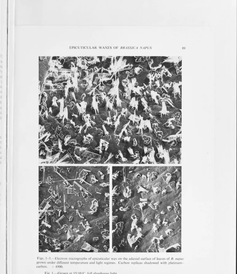
Plate 1.13a illustrates the surface wax characteristic of a plant
grown at 15/10 °C. Wax rods of average length 1.8 µm, and rarely
exceeding 3.4 µm with a base diameter of 0.2-0.6 µm typify this
condition, and platelet type wax was consistently absent. The same wax,
viewed with a scanning electron microscope, is presented in Plate 1.13a
(inset). Wax rods have been shown to have a hollow centre (Johnson
&
Jeffree, 1970) but the use of the term 'tube' which is commonly found in
17
for the structures as they appear in replica form, the term is confusing
when applied to the interpretation of what has been replicated on the
leaf surface itself.
Plate 1.13b illustrates the surface was characteristic of a plant
grown at 18/13 °C. Wax rods of the type observed in Plate 1.13a may be
seen again in conjunction with a series of narrow flat wax platelets.
Quantitatively the ratio of rods:platelets was approximately 2:1.
Plate 1.13c illustrates the surface wax characteristics of a plant
grown at 21/16 °C. In contrast to the previous transition temperature
(18/13 °C) there was here a predominance of complex narrowly branched
open platelets. These generally occurred as a single layer lying
,..._, 0.3 µm above and parallel to the cuticle surface. Wax rods were still
present but in lesser amounts.
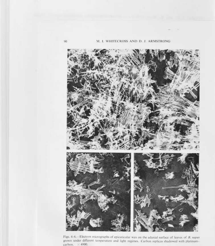
Plate 1.14a illustrates the surface wax characteristic of a plant
grown at 24/19 °C. The wax platelets were more complex than those
observed in the previous temperature (21/16 °C) with fusion of the
primary branching being quite conspicuous. The platelets form an
elevated platform"' 0.3 µm above the leaf surface. Wax rods were not
unconnnon.
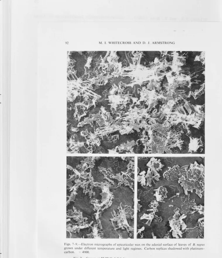
Plates l.14b,c and 1.15a,b illustrate the surface wax
characteristics of plants grown at 27/22 °C, 30/25 °C, 33/28 °C and
36/31 °C respectively. In Plate 1.15b (inset) 36/31 °C wax as viewed by
a scanning electron microscope is presented. A positive correlation
between increasing growing temperature and wax platelet complexity was
observed. Fusion of the primary and secondary branches became so
pronounced with increasing temperature that completely solid wax
platforms were evident at the higher temperatures. In general, only the
outer periphery of such wax platforms showed the typical branching
pattern. The presence of wax rods was quite rare in these higher
temperatures.
(v) Mutant and Chemical Effects on Wax Fine Structure
Plate 1.15c illustrates the surface wax characteristic of the
non-waxy (gl3) mutant grown at 24/19 °C. In contrast to the waxy plant, the
wax was present merely as a smooth largely non-structural layer.
Plate 1.16a,b illustrates the surface characteristics of plants
quantitatively reducing wax formation to a significant degree although
qualitatively the wax still resembled that of the control (24/19 °C,
Plate 1.16c).
18
Dalapon, as well as reducing wax formation quantitatively, altered
the wax qualitatively. The platelet wax formation was almost completely
suppressed while the rod wax formation was reduced to rather short
irregularly-shaped wax blocks.
Plate 1.17a illustrates the surface wax characteristic of a 24/19 °C
grown expanding leaf just prior to two root applications of 2.5 x 10⁻⁴
moles of T.C.A. over a two-day period. Plate 1.17b illustrates the
surface of the same leaf as the control (1.17a) twenty-four hours after
the last herbicide application. An almost complete dewaxing effect,
removing all the wax originally present, was clearly apparent. The only
structural wax then present was that characteristic of a plant treated
with T.C.A. for some duration (Plate 1.16a). Leaves that had fully
expanded prior to the herbicide treatment showed little, if any, tendency
to being dewaxed (Plate 1.17c).
(vi) Effect of Rapid Temperature Changes on Wax Fine Structure
Plate 1.18a,b represents the 15/10 °C and 36/31 °C leaf surface wax
controls respectively.
Plates 1.19 - 1.22 illustrate a series of leaf surface wax
modifications apparent when a 15/10 °C grown plant was transferred to a
36/31 °C temperature regime.
Plate 1.19a. Wax modification was evident within six hours and took
the form of the existing wax rods branching out at"' 90° to their
vertical axes, though fusion into wax platelets was not apparent. Many
of the original wax rods established during the 15/10 °C temperature
regime were consistently observed to have been lost following a low to
high temperature transition.
Plate 1.19b. Within twenty-four hours after the transfer, the
formation of wax platelets, characteristic of high temperatures, had
begun, while branching of some of the original wax rods was still
continuing.
Plate 1.19c. Forty-eight hours after the transfer, the frequency of
wax rods was significantly reduced while the formation of complex
19
Plate 1.20a. Seventy-two hours after the transfer, the wax platelet
formation became even more complex. They became more obviously fused and
markedly increased in coverage of the leaf surface.
Plate 1.20b. Ninety-six hours after the transfer, the modification I
of the wax was so significant that the wax pattern virtually appeared the
same as a 36/31 °C control leaf (Plate 1.18b).
Plate 1.20c. One hundred and twenty hours after the transfer, the
wax modification appeared to be complete, resembling that of a 36/31 °C
control in all respects. No further modification was apparent after this
period.
The above results brought about by a direct transfer from 15/10 °C
to 36/31 °C conditions brought into question the physiological shock
caused to the plant by such a sudden drastic change. Accordingly, it was
decided to programme a gradual increase from the 15 °C to the 36 °C
temperature regime to allow for adaptation.
Plate 1.21a illustrates the wax modification on the leaf surface of
a 15/10 °C grown plant resulting from a programmed temperature increase
at the rate of 0.5 °C per hour for forty-two hours (21 °C range) to 36 °C.
Structurally the wax layer appeared to consist of a massive growth of
long narrow single stranded ribbons, with very little evidence of the
usual upright wax rods. This enormous proliferation of wax due to the
gradual increase in temperature bore little resemblance to the wax as
seen forty-eight hours after a direct transfer from low to high
temperature (Plate 1.19c). In the light of this comparison, it is
apparent that in fact the physiological shock of moving the plant
directly to a 21 °C higher temperature environment had a less stimulatory
effect on wax modification than a steadily increasing temperature.
After growing plants for a substantial period at 15/10 °C,
transferring to the higher temperature, whether suddenly or more
gradually, placed the plants under severe stress. The leaf area and
thickness for the 15/10 °C grown plants was large when compared to the
36/31 °C adapted plants, while the temperature increment for a
transferred plant was considerable. In an attempt to further ascertain
the sensitivity in regard to modification of the leaf wax to increased
temperature, a temperature increment of 12 °C was employed instead of
21 °C. Again, wax modified by a direct and programmed temperature
20
Plates 1.21b and 1.21c illustrate a direct and progrannned temperature
transition to 27/22 °Cover a twenty-four hour period respectively. The
progranuning rate for the latter was 0.5 °C per hour. The wax modification
for the two treatments is qualitatively similar, consisting of initial
wax platelet formation and branching of the pre-existing wax structures.
The programmed treatment has significantly more wax coverage as noted
previously where the temperature increment was 21 °C (Plates 1.19c and
1.21a).
Plate 1.22a,b illustrates the wax observed in thin section of a
plant having undergone a direct transition from 15/10 °C to 27/22 °C for
twenty-four hours. The observed wax modification confirmed the results
obtained in carbon replica studies taking the form of branching wax rods
and platelet formation. Plate 1.22c illustrates the upright typically
non-branched wax rod structure of a 15/10 °C control.
Attempts to obtain a wax modification in the form of wax rods by
transferring a 36/31 °C temperature grown plant to a 15/10 °C temperature
regime were completely unsuccessful. The only alteration of the wax
surface even after a five-day period at 15/10 °C was a partial loss of
the pre-existing wax platelets (Plate 1.23a). Plates 1.23b and 1.23c
represent a 36/31 °C and 15/10 °C control respectively.
DISCUSSION
Variation in growing temperature has been shown to produce an effect
on the cytoplasm and leaf structure generally, while the modification of
the surface wax pattern was quite remarkable. While these variations are
of interest in their own right, no specific correlation was possible
using conventional electron microscopy.
Structurally, the non-waxy (gl3) mutant plant did not differ
significantly from the normal waxy pl ant, in spite of having
substantially reduced wax production. In this and previous studies
(Juniper, 1959a; Dewey
et al.,
1961), herbicidal treatments have beenfound to reduce wax production and modify the wax pattern on the leaf
surface. Fine structural similarities were seen between the wax
herbicidal and high temperature treatments, but these did not correlate
with wax production, which was high at high temperature and low with
herbicides. Suppression of wax formation, whether due to a genetic
mutation or to herbicide treatment, would seem to be specifically related
Macey
&
Barber, 1970a,b), while wax chemistry and/or physical factorswere the cause of the varjations in fine structure of wax due to
different temperatures.
21
Large vesicles, possibly lipoidal in nature, were frequently
observed in association with Golgi bodies. Hallam (1970b) has observed a
similar relationship in
Eucalyptus
and noted an apparent migration of theGolgi in the direction of the cell wall. If such a migration occurred,
the vesicles may have been responsible for the liberation of wax
precursors across the plasmolemma into the cell wall. The application of
an autoradiographic study would have to be employed to confirm this
suggestion.
The ability of a
Brassiaa
plant to begin modifying its structuralwax on the leaf surface in as little as six hours and significantly to
that characteristic of a higher temperature environment in less than one
hundred hours has been readily demonstrated. Carbon replica and thin
section techniques have shown that the qualitative alteration of a rod
type wax to plates was achieved in part by the modification of
pre-existing wax.
The rapid wax generation response brought about by transfers to
higher temperatures was apparently due to the response of an
environmental protective mechanism. No cell structural differences
attributable to the alteration of the wax pattern were evident. It is
suggested that increased growing temperature affected wax production at
the biochemical level, causing a rapid biosynthesis and subsequent
movement of fresh wax to the leaf surface. Extrusion of wax already
synthesised and present in the cell wall-cuticle complex, or a greater
dispersal of a non-crystallised wax solvent at the leaf surface, are
considered to be of little importance in bringing about a wax
modification.
Theoretically it remains possible that transition to a higher
temperature regime could stimulate the production of a wax solvent alone
on the leaf surface, this in turn redissolving the existing wax. A
secondary effect of the higher temperature would then have been to
recrystallise the wax in the characteristic pattern observed. The
practical aspects of such a system seem unnecessarily complex and
22
Conceivably wax modification caused by increased wax production
could be stimulated by a series of temperature-sensitive enzymes which
are progressively activated by increasing temperature. If such a system
did operate, it is apparent that once a particular enzyme was activated,
the lower temperature enzymes might be deactivated since low to high
temperature wax modifications were quite irreversible. This would seem
to be not an unreal situation since decreasing the growing temperature
would not pose the physiological stresses of a temperature increase.
A sudden application of trichloroacetic acid to the root system of
an established plant has been observed to significantly dewax the
expanding waxy leaves already present. This effect can be achieved so
rapidly that it is tempting to suggest that the cuticle-surface wax zone
may be altered such that the retention of the original wax layer is lost.
This alteration may be achieved by the herbicide inhibiting an enzyme
system which produces normal wax precursors but fails to inhibit and
perhaps even enhances the production and subsequent flow of wax solvent
to the leaf surface. This being so, an extrusion of wax solvent on to
the leaf surface alone might dissolve the surface wax attachment zone,
the wax in turn being lost from the leaf surface.
This reasoning may be extended to the observation that fully
expanded leaves do not develop or regenerate large amounts of surface wax
(Hallam, 1970c). The failure of a fully expanded leaf to become dewaxed
by a sudden herbicide treatment could be explained by the cessation of
the wax solvent flow in conjunction with the wax precursor synthesis.
The cessation of wax production in a fully expanded leaf may not be
necessarily due to a discontinuation of wax precursor formation, but
rather of the solvent which mobilises the wax to the leaf surface. If
this were the case, the cell walls and possibly the cytoplasm may be
heavily loaded with wax precursors up to the time of senescence.
Sectioning leaf tissue parallel and perpendicular to the leaf
surface has confirmed the heterogeneous nature of both cell wall and
cuticle. Wax or its innnediate precursors are synthesised in the
epidermal cell (Kolattukudy, 1970b), traverse the plasmolemma either
actively or passively, and move through the cell wall. This study
suggests that in Brassioa at least, the movement through the cell wall is
one of diffusion across a concentration gradient. Although not
the cell wall as suggested by Bolliger (1959). No evidence has been
obtained for movement through specific channels within the cell wall as suggested by Hall (1967b).
23
It is suggested that subsequent movement across the cuticle in that region adjacent to the cell wall occurs primarily by non-localised
diffusion. The cell wall and cutin meshwork would seem to present very
little resistance to diffusion. Subsequent movement in the outer region
of the cuticle presumably occurs through what appears to be a
microchannel system. These microchannels are oriented at various angles
to the leaf surface, one end of an individual channel generally being
\
traceable to the cuticle surface. The outer zone of the cuticle appears
to be a reticulate system of microchannels opening out on to an
exceedingly irregular cuticle surface. The occurrence of microchannels
completely traversing the cuticle appear to be too infrequent to solely support Hall's hypothesis (Hall, 1967a) that wax migrates through the
entire cuticle via channels.
While the results indicate a combined diffusion-microchannel system
for the excretion of leaf waxes in
Brassica,
this is not to say that insome plants migration does not occur by way of anastomosing platelets as
suggested by Hallam (1964).
Inconclusive evidence from the various sectioning techniques
employed suggest that the microchannels terminate at the cuticle surface in a random array of pore-like openings. Methods other than conventional
electron microscopy would have to be employed to substantiate this
Plates 1 .1 - 1. 23
The following plates illustrate the results for waxy Brassica napus
plants unless specifically stated otherwise.
All thin sections were prepared from material fixed with
glutaraldehyde/osmium tetroxide and stained with uranyl acetate and lead
citrate.
Leaf surface wax preparations employed Pt/C shadowing and carbon
replication, except in the case of scanning electron micrographs where
carbon and gold/palladium coatings were employed.
Dimension lines on micrographs represent 1 µm except where indicated
Plate 1.1: Light micrographs illustrating the upper epidermis and
adjoining palisade cells of plants grown at: (a) 15/ 10 °C
(b) 24/ 19 °C
(c) 36/31 °C.
Plate 1.2: Light micrographs illustrating low power characteristics of
the upper leaf surface for plants grown at:
(a) 15/ 10 °C
(b) 24/19 °C
(c) 36/31 °C.
Plate 1.2: Light micrographs illustrating low power characteristics of
the upper leaf surface for plants grown at:
(a) 15/ 10 °C
(b) 24/19
°c
Plate 1.1: Light micrographs illustrating the upper epidermis and
adjoining palisade cells of plants grown at: (a) 15/ 10 °C
EPIDERMIS
3
fflI "' 0
"'O
::c
-<
r-EPIDERMIS
3
"'
"'
0 "'O::c
-<
r-~
ffl
"'
0 r - 4 ~-- -~ "'Or-Plate 1.2: Light micrographs illustrating low power characteristics of
the upper leaf surface for plants grown at:
(a) 15/10
°c
(b) 24/19 °CPlate 1.3 a,b,c: Electron micrographs illustrating the outer epidermal
cell wall fine structure of 15/10 °C grown leaves.
Plate 1.4 a,b,c: Electron micrographs illustrating the outer epidermal
cell wall fine structure of 24/19 °C grown leaves.
Plate 1.5 a,b,c: Electron rnicrographs illustrating the outer epidermal
"t
.
.
••
' r•
'•,.
/
a
·
:(
..-
··~- .:,,0
. \
Plate 1.6 a,b,c: Electron micrographs illustrating the outer epidermal
cell wall fine structure of 36/31 °C grown leaves.
)
b
Plate 1.7 a,b,c: Electron micrographs illustrating the outer epidermal
cell wall fine structure of non-waxy (gl3) mutant
leaves grown at 24/19 °C. Inset illustrates surface
0
Plate 1.8 a,b,c: Electron micrographs illustrating the outer epidermal
cell wall fine structure of 24/19 °C grown leaves after
root applications of dalapon (2,2-dichloropropionic
.y/ ..
~
_,
·:
t ~ r'
, .
..
r
• 'I
:'
.
·e-,,-Y.
a
.
"(.•_,,1 ~-t;··' r ~ ~ ..,,. >
~-;~:
-I
Plate 1.9 a,b,c: Electron micrographs illustrating outer epidermal cell
wall fine structure of 24/19 °C grown leaves after root
applications of T.C.A. (trichloroacetic acid). Inset
