City, University of London Institutional Repository
Citation
:
Mian, A. & Reyes-Aldasoro, C. C. (2015). Quantification of the Effects of Low
Dose Radiation and its Impact on Cardiovascular Risks. Paper presented at the
Quantification of the Effects of Low Dose Radiation and its Impact on Cardiovascular Risks.
This is the accepted version of the paper.
This version of the publication may differ from the final published
version.
Permanent repository link:
http://openaccess.city.ac.uk/11966/
Link to published version
:
Copyright and reuse:
City Research Online aims to make research
outputs of City, University of London available to a wider audience.
Copyright and Moral Rights remain with the author(s) and/or copyright
holders. URLs from City Research Online may be freely distributed and
linked to.
City Research Online:
http://openaccess.city.ac.uk/
[email protected]
© 2015. The copyright of this document resides with its authors. It may be distributed unchanged freely in print or electronic forms.
Abstract
This work describes an algorithm developed to quantify the effects of low dose radiation on the cardiac endothelial cells with the final objective of inferring how radiation may potentially initiate cardiovascular disease in post radiotherapy-treated patients. The effects are investigated by using an in-vitro co-culture cellular matrix, consisting of endothelial cells on a base of fibroblasts, which in time begin to form capillary (tubular) like structures. A range of radiation doses (0.2-16 Gray (Gy)) was applied to different samples and the effects observed. The automatic segmentation is validated against a set of manual segmented images with satisfactory results presenting a correct classification of 0.93; classification is the measure of comparison between two sets of images, specified as a number from 0 to 1, whereby 1 denotes 100% similarity whilst 0 refers to 0% similarity. Measurements related to geometrical parameters were further obtained. It was found during the course of this project, the largest observable change in endothelial cell structure was found after exposure to 0.2 Grays of radiation.
1
Introduction
In the United Kingdom an estimated number of ≥ 331,000 people were diagnosed with cancer in 2011. Of this number, approximately two-thirds receive radiotherapy as a treatment [1]. Patients receiving Mediastinal (chest) radiotherapy for treatment of Breast cancer, Lung cancer, Oesophageal cancer and Hodgkin’s lymphoma are at the highest risk of exposing ionising radiation to the heart [2].
The heart has long been considered one of the few organs to be moderately resistant to radiation induced tissue damage [3]. However, recent epidemiological studies propose evidence to the contrary [4]. So far, current epidemiological statistics have revealed that moderate to low doses of radiation to the heart may possibly result in a substantial increase in cardiovascular associated mortality [5]. Though, the pathogenesis of heart disease from irradiation, as of yet, has not been explored in detail.
There are various techniques used to quantify the effects of radiation. One of these methods consists of irradiating cardiac cells in-vitro and subsequently assessing the effects
Quantification of the Effects of Low Dose
Radiation and its Impact on
Cardiovascular Risks
Atif S. Mian1
C.C Reyes-Aldasoro1
http://staff.city.ac.uk/~sbbk034/
1
of the radiation on the morphological and functional properties of cardiomyocytes and endothelial cells; the cells that form thin layers that cover the inner part of blood vessels [6][7]. Endothelial cells in a co-culture system contained by fibroblasts develop with time, eventually forming tube-like structures similar in appearance to capillaries. Such a system is used in accordance to replicate the in-vivo angiogenic process; hence, it can be used to test inhibitors and activators of angiogenesis [8].
The aim of this work is to investigate the effects of ionising radiation on the cardiac endothelial cells (microvasculature) grown in-vitro and its relation to angiogenesis inhibition. Tube/capillary-like structures are formed in a perfusion (live) co-culture system consisting of endothelial cells and fibroblasts derived from Murinae heart cells. These cells are subjected to various levels of ionising radiation (0, 0.2, 2, 8, and 16 Gy) [n=5×3]; 0 Gy acting as the reference control for this research. Images of the Murinae heart cells were acquired twenty weeks post irradiation. A sequence of segmentation and geometric analysis algorithms will be performed on the obtained co-cultured Murinae heart cell images via MATLAB programming. These algorithms will subsequently allow observational measurements of dosage effects in correlation to time after radiation.
2
Materials and Methods
2.1
Materials
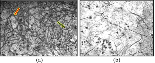
Organotypic cultures (the process of growing cells in a 3-D environment, producing a cellular system that is biochemically and physiologically more resembling in nature to in- vivo tissue as opposed to 2-D culture sets) were stained with lectin to identify the endothelial cells and a representative image at each irradiation dose is shown in Fig. 1. The images are greyscale and with low contrast, and each comprises a cluttered background overlaid with the tube-like structures formed by endothelial cells. There are significant inter-image and intra-image illumination variations. Dead cells or debris were also present, sometimes occluding the tubules. The tubules are the primary target of the analysis (as changes of these structures indicate the progression of angiogenesis), though the debris; biological in nature rather than imaging artefacts, is also relevant.
[image:3.408.72.330.411.516.2](a) (b)
(a) (b) (c)
(d) (e) (f)
[image:4.408.41.377.28.308.2](g)
Figure 2: Graphical description of the algorithm. (a) Original image treated with 8 Gy. (b) Edges are highlighted with the application of 3×3 median filter and un-sharp filter. (c) Binarisation with adaptive thresholding (window size = 35, mean-C = 0.02) and erosion with a disk structural element was applied. (d) Objects smaller than 350 pixels in diameter were removed with morphological operators. (e) Gaps closed by dilation with a disk structural element. (f) Skeleton, branch (yellow-dots) and end points (blue (yellow-dots). (g) The final step was to detect closed meshes (green) as it was assumed the number of meshes to be related with the health of the cellular development.
2.2
Algorithm description
The steps of the algorithm were programmed in MATLAB and the image processing toolbox. These algorithms were designed to assist in separating the relevant elements of the data, i.e., the structure of the tubules, their density per area, size, length and also the geometric distribution; through identification of the number of tubules that go onto form junctions, ones that are isolated within the matrix and furthermore, finding their relative branching angles.
user to create a gold standard for comparison purposes [n=5×3]. The gold standard set was achieved through collectively adding successive masks of the individual tube like structures of each image using the MATLAB function roipoly.
In order to quantify the angiogenic process, the following steps were performed; segmenting the background and debris from the tubules, skeletonising the tubules and from them, determining the branch and end points of the structure, the number of meshes and mesh area (Fig. 2). All these help in quantifying the vessel structure.
3
Results
Classification of the results was achieved by subtracting the automatic segmented images from its equivalent manual set. From the results an average correct classification of 0.93 was obtained, the lowest being 0.85 and the highest being 0.97 (Fig. 3), which are similar to those reported in [5]. The main benefit of the automatic segmentation is time efficiency. Manual segmentation of 15 required approximately 3-4 hours per image, depending on the complexity of the tubules. The automatic segmentation on the other hand took only seconds. However, there is a trade-off between accuracy and time efficiency using automatic segmentation over manual segmentation.
The automatic algorithm found a lower number of meshes as compared to the gold standard, (Fig.4).The number of closed meshes were considered to be one important feature of the angiogenic process. An average reduction of 23% in the number of meshes was observed. This is partly attributed to some of the tubule structures being deleted during the morphological erosion phase necessary to remove the debris. Nonetheless, by applying a suitable dilation phase, some of the mesh structures could be restored.
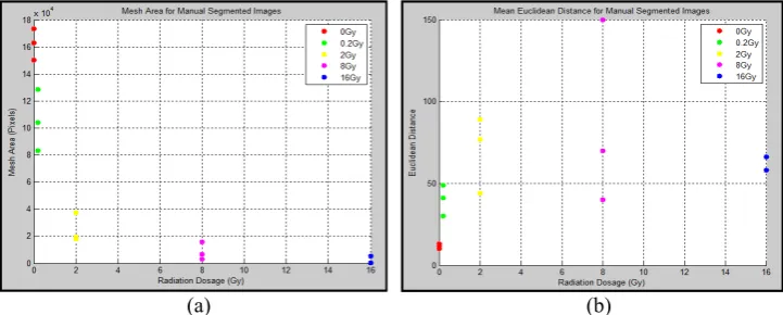
When quantifying the manual set images, an inverse relationship to the radiation dose was witnessed. This trend may be related to the suppression of the signalling protein, vascular endothelial growth factor (VEGF). It has been reported [10] that an increase in ionising radiation reduces the levels of VEGF and its receptor VEGFR-2, which are pivotal in the role of endothelial cell proliferation, hence, a reduction in blood vessel formation. It is interesting to see, from the results below, that the most drastic angiogenic inhibition occurs between 0 and 0.2 Gy whereby a ~20% decrease in mesh area is noticed. This may indicate a prominent rise in VEGF suppression must occur after initial exposure to low range doses of ionising radiation, therefore resulting in the changes recognized [5]. The MATLAB function ‘Spy’ was used to identify the number of meshes within the vessel matrix. This function also produced an adjacency matrix that showed the number of meshes that are neighbouring each other. From this it was found, as the radiation dosage increased so did the distance between the mesh structures within the co-culture system.
(a) (b)
Figure 3: (a) Comparison of manual and automatic classifications against radiation dosage. Correct classification rose amid increase in radiation. It is assumed that as the tubule density decreased, the background had a higher weight in the classification. (b) Number of branch points vs. Radiation dosage. The number of branch points decreased with higher radiation doses.
(a) (b)
Figure 4: Comparison of number of meshes detected for (a) automatic and (b) manual segmentations. It is clear that fewer meshes were detected with the automatic segmentation.
(a) (b)
[image:6.408.30.390.30.180.2] [image:6.408.27.392.231.381.2] [image:6.408.28.389.410.555.2]4
Conclusion
This work presented a segmentation algorithm for the analysis of the effects of irradiation in in-vitro cardiac cells. The algorithm was programmed in Matlab and followed several steps: adaptive thresholding, morphological erosion in conjunction with morphological operators for artefact removal. The average correct classification against the manual segmentation was 0.93. Whilst sequentially reviewing the data, it was noted that lowest dosage of radiation produced the greatest structural change in the tubular structure; suggesting even low doses of radiation exhibit high angiogenic inhibition. Automatic segmentation yielded fairly robust results and may possibly be used for identification and quantification of blood vessel changes in-vitro in place of manual segmentation. In the future we can analyse other metrics like Jaccard score and more sophisticated processing like separation into training and testing sets.
References
[1] C. R. UK, “Cancer Stats: Key Stats,” 02-Apr-2015. [Online]. Available:
http://www.cancerresearchuk.org/cancer-info/cancerstats/keyfacts/cancerstats-key-facts-on-cancer. [Accessed: 01-May-2015].
[2] M. J. Adams, P. H. Hardenbergh, L. S. Constine, and S. E. Lipshultz, “Radiation-associated cardiovascular disease,” Crit. Rev. Oncol. Hematol., vol. 45, no. 1, pp. 55–75, Jan. 2003. [3] S. W. Yusuf, S. Sami, and I. N. Daher, “Radiation-Induced Heart Disease: A Clinical Update,”
Cardiol. Res. Pract., vol. 2011, p. e317659, Feb. 2011.
[4] L. Cella, J. H. Oh, J. O. Deasy, G. Palma, R. Liuzzi, V. D’avino, M. Conson, M. Picardi, M. Salvatore, and R. Pacelli, “Predicting radiation-induced valvular heart damage,” ActaOncol.
Stockh. Swed., pp. 1–9, Mar. 2015.
[5] S. Bowyer, C. Kanthou, and C. C. Reyes-Aldasoro, “Analysis of capillary-like structures formed by endothelial cells in a novel organotypic assay developed from heart tissue.,” presented at the Medical Image Understanding and Analysis, London, UK, 2014, pp. 235–240. [6] G. M. Tozer, S. Akerman, N. A. Cross, P. R. Barber, M. A. Björndahl, O. Greco, S. Harris, S.
A. Hill, D. J. Honess, C. R. Ireson, K. L. Pettyjohn, V. E. Prise, C. C. Reyes-Aldasoro, C. Ruhrberg, D. T. Shima, and C. Kanthou, ‘Blood vessel maturation and response to vascular-disrupting therapy in single vascular endothelial growth factor-A isoform-producing tumors’,
Cancer Res., vol. 68, no. 7, pp. 2301–2311, Apr. 2008.
[7] “CARDIORISK.” [Online]. Available: http://www.cardiorisk.eu. [Accessed: 01-May-2015]. [8] C. C. Reyes-Aldasoro, D. Biram, G. M. Tozer, and C. Kanthou, ‘Measuring cellular migration
with image processing’, Electron. Lett., vol. 44, no. 13, pp. 791–793, Jun. 2008.
[9] C. C. Reyes-Aldasoro, “Retrospective shading correction algorithm based on signal envelope estimation,” Electron. Lett., vol. 45, no. 9, p. 454, 2009.
[10] J. Li, S. Huang, E. A. Armstrong, J. F. Fowler, and P. M. Harari, “Angiogenesis and radiation response modulation after vascular endothelial growth factor receptor-2 (VEGFR2) blockade,”