JOURNALOFVIROLOGY,OCt. 1975,p.937-943
Copyright®1975 American Society for Microbiology Printed in U.S.A.Vol. 16, No. 4
Infantile
Enteritis Viruses:
Morphogenesis and Morphology
IAN H. HOLMES,* BRIAN J. RUCK, RUTH F. BISHOP, AND GEOFFREY P. DAVIDSON
Department ofMicrobiology, University of Melbourne,*and Department of Gastroenterology, Royal Children's Hospital,Parkville,Victoria, Australia
Received for publication 13 May1975
Anewviruswasfoundtobe associatedwithacutegastroenteritis in children. In
duodenalbiopsies, itwasobservedinfecting onlyintestinalepithelial cells,and it
resembled orbiviruses in its morphogenesis. For diagnostic purposes the virus
was readily demonstrated by negative staining of fecal extracts. Two forms of
particleswere seen:double-shelledparticles (70to75nmindiameter) resembling
thoseofreovirusbut withasharper outline,andsingle-shelledparticles (60nmin
diameter) with obvious capsomerstructure andresemblingthose of orbiviruses.
Themorphological resemblance of thishuman virus tothe virusesof "Nebraska"
calfscoursandepizooticdiarrhoea of infant mice isemphasized.
In young children, gastroenteritis or
"in-fantilediarrhoea" is frequently a seriousdisease
requiringhospital treatment tocorrect
dehydra-tion. Despite intensivestudies, aetiologic agents
such as pathogenic entericbacteria or protozoa
can be incriminated in only a minority of
cases(8). We haverecently observedanewvirus
induodenal mucosa(6) andfecal extracts (7, 10)
from a high proportion of young children
hospitalized in Melbourne with acute
gastro-enteritis. The association of this virus with
gastroenteritis in children has been confirmed
in England (15), Canada (23), and the U.S.A.
(17). It has also been demonstrated in limited
surveys of fecal samples from children in
variousother parts of the world (10, 11).
This new viruscan notbe culturedbyroutine
diagnostic procedures. We report here features of its morphogenesis in biopsy specimens of
duodenal mucosa, and of its morphology in
negative-contrast electron microscopy. Whereas
the virus obviously belongs to the family
Re-oviridae, it cannot be placed in either of the
recognized genera (reovirus or orbivirus). We
suggest creation ofa new genus, duovirus. This
new genus could also include morphologically
similar viruses involved in the aetiology of
gastroenteritis incalves("Nebraska"calf scours
virus) and in mice (epizootic diarrhoea of
infant mice, or EDIM virus), and the simian
virusSA-11.
MATERIALS AND METHODS
Clinicalspecimens.Allclinical materials were
ob-tained from childrenadmitted to the Royal Children's Hospital, Melbourne. Clinical details and processing of duodenal biopsy samples for electron microscopy
have been described (6). Virus-containing extracts
were prepared from fecal samples by the method of Bishop et al. (7). Briefly, this involved extraction with trifluor-trichloroethane ("Arklone," Imperial Chemical Industries), precipitation of the virus with 8%polyethylene glycol 6000, and centrifugation of the particles through a layer of 45%(wt/vol)sucrose. The last stage hasbeen modified slightly since the method was published. The deposit obtained after
polyeth-ylene glycolprecipitation is now dissolved in 4 ml of
distilled water. This mixture is layered on to 1 ml of 45%(wt/vol)sucrose in 0.002MTris-hydrochloride
buffer, pH 7.0, and centrifuged for only 75 min at
100,000x g.
Viruses of calf and mouse origin. A sample of calf scours virus waspreparedfromafieldspecimen
supplied by T. A. Mason, Department ofVeterinary
Clinical Science, University of Melbourne, from
Werribee, Victoria. Extraction of the virus from the
calf feces was carried out as described above for humanfecalspecimens.
A sample of EDIM virus was obtained from a
natural outbreak of diarrhoea in infant mice at the John Curtin School of Medical Research, Canberra, bycourtesy ofG. M. Woodroofe.Lengthsofwhole, in-fected gut, including contents, were homogenized
withfluorocarbon, and thenparticles werecentrifuged
through45%sucrose asforhumanfecalspecimens.
Negative-contrast electron microscopy. Virus
suspensions were negatively stained with 2%
po-tassium phosphotungstate at pH7.0 or 6.0, or with 'Ao saturated ammonium molybdate. Specimens were routinely examined andphotographedat a magnifica-tion of x20,000 in a Hitachi HU-11A electron microscope. Magnifications were calibrated against catalasecrystals (31).
RESULTS
Virus morphogenesis observed in human
biopsy specimens. Growthofthisvirusappears
937
on November 10, 2019 by guest
http://jvi.asm.org/
938 HOLMES ETAL.
to be restricted to differentiated intestinal
epithelial cells, and ithas notyetbeen adapted
to growth in any type ofcell culture. Previous
light microscope observations suggested that
celldamage was mostnotablein the duodenum
(5), and our electron microscope observations
on biopsies of duodenal mucosa confirm that
evidence of cytopathology associated withvirus
replication is not difficult to obtain. Even
within the very small areas thus surveyed,
however, infected cells are scattered among
apparently normal neighbors, andnosynchrony
ofinfection isevident. Thus the durationofthe
replicative cycle has not been established, and
the sequence of events in morphogenesis can
only be suggested by analogy with other
mem-bers of the reovirus family which have been
studied under single-cycle growth conditions
(9, 26).
Infected cells are most easily recognized at
the stage illustrated in Fig. 1, in which
num-erous virus particles have accumulated in
cytoplasmic vesicles. The microvilli lining the
apical surface of infected cells may appear
normal or irregular and distorted. The vesicles
containing virus particles are in fact distended
cisternae of the rough endoplasmic reticulum.
Sectioned particleshave an electron dense core
33 nm in diameter enclosed by a moderately
dense layer interpreted ascapsidwith an outer
diameter of 67 to 70 nm. Some particles
lack-ingthe dense core arealso evident in Fig. 1. A
variable proportion (about 10%) of particles
within vesicles were enveloped; their total
diameter was then 87 to 90 nm.
Figure 2 illustrates the very characteristic
appearance ofa massofconvoluted (reticulate)
smooth membrane adjacent to enveloped
particles at the edge of avesicle. The budding
process by which some particles acquire an
envelope when passing into avesicle is clearly
shown inFig.3.
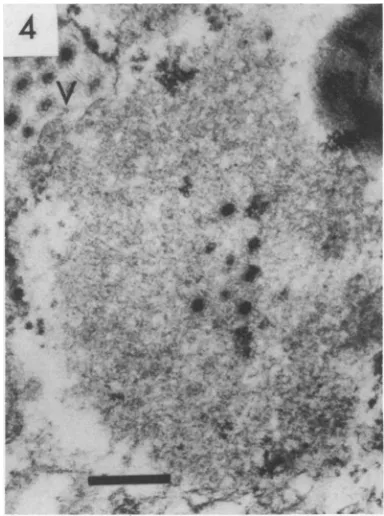
Areas of "viroplasm" or "virus factories"
similar to those observed in reovirus- or
orbi-virus-infected cells were encountered less
fre-quently; one is shown in Fig. 4. These are
moderately electron dense, granular, or finely
fibrillar, and are not membrane bound. Their
relatively uniform texture distinguishes them
from the surrounding cytoplasmic organelles,
butthose observedwere notvery large.
Final dissolution of a necrotic cell is shown
in Fig. 5. Virus particles still within
mem-brane-bound vesiclesarepassing into the lumen
4~~~4
.Ar. .~. ~, + 36-.-#^ .. E
S: , t. ',ffi *' { >
4
Xpiit,9:'u
,
. I',1V
,,7 wib*' 1-vwk--- '^'.!", Iww,s__s. -JvaW.u.&V
FIG. 1. Portion of thecytoplasm ofaduovirus-infected epithelial cellobtained by duodenal biopsyfrom a child with acute gastroenteritis, showing duovirus particles within distended cisternae of the endoplasmic reticulum. Bothnaked (N)andenveloped(E) particlesarepresent.Some particlesof each type appear to lack the usual electron-densecore(arrows).x72,000.Barin thisandsubsequentmicrographsindicates200nm.
J.VIROL.
t
on November 10, 2019 by guest
http://jvi.asm.org/
VIRALENTERITIS OF INFANTS 939
FIG. 2. Detail of the edge of a cisterna containing virus. Enveloped particles are situated near an
[image:3.493.45.436.81.341.2]in-tracytoplasmicmassofconvoluted smooth membranes(R).x72,000.
FIG. 3. Virusparticle budding (arrow) through the membrane of a rough endoplasmic reticulum
cis-terna. Note ribosomes (small arrows) attached to otherregions ofthesamemembrane.x150,000.
A
.>
FIG. 4. "Viroplasm" containing electron dense viruscores in thecytoplasm ofaduodenalepithelial cell. An adjacent cisterna contains complete, un-enveloped virus particles(V).x72,000.
VOL.16,1975
on November 10, 2019 by guest
http://jvi.asm.org/
[image:3.493.44.230.373.633.2] [image:3.493.244.436.375.633.2]940 HOLMES ETAL.
[image:4.493.70.459.78.341.2]_!4t,~
_
FIG. 5. Dischargeof the contents of a necrotic epithelial cell, including membrane-bound groups of duovirus particles, into the lumen (L) of the small intestine. x27,000.
ofthe gut (lower right), whereas an apparently normal adjacent epithelial cell is partly visible below.
Negative-contrast studies. Virus particle
excretion by duovirus-infected children
ap-pears to be maximal during day 3 to 4oftheir
illness; lower concentrations of virus have been
detected up to day 10afteronset ofsymptoms
(10).Theprocedure used for partial purification
and concentration of the virus does facilitate
diagnostic recognitionoftheparticlesby
remov-ing a considerable amount of extraneous
mem-branous and particulate material. Nevertheless
debris of bacterial origin, assorted bacterio-phages, and sometimes adenoviruses are seen, together with variable amounts ofunidentified
fibrillar material. For diagnostic purposes, we
prefer the greater contrast obtained with
phos-photungstate, but ammonium molybdate
re-veals the outer capsid shell of particles more
clearlyand appears preferable forstudies ofthe
fine structure ofthis typeof virus.
Duovirus particles are recognizable by their
regularity, size, and surface structure.
Speci-mens may contain predominantly
double-shelled or single-shelled particles, or a mixture
ofboth(Fig. 6). Particles witha double-shelled
capsid have a diameter of 70 to 75 nm and a
characteristically sharp outline. Arrangements
resembling large ring-shaped capsomers are
sometimes visible (arrow, Fig. 6), but these
may be spaced apart by half their width and
in other cases overlap. A regular arrangement
of hollows surroundedby shared structural units
(as proposed by Vasquez and Tournier
[28]
toexplainreovirus surfacestructure)wouldappear
to us a better description of the general
appearance. Portions ofdisintegratingparticles
appear to consist of a lattice structure, not
separate ring-shaped capsomers (Fig. 7). Very
similar fragmentation of reovirus capsids has
beenreported (2).
Single-shelled particles (60 nm in diameter) exhibitamuch more obvious capsomer structure than cores of reovirus particles; they closely resemble typical orbiviruses.
A small proportion of human virus
prepara-tions contained flattened tubular structures
with a regular surface pattern (Fig. 8). These
were shownto be antigenically related to virus
particles by immunoelectron microscopy (I. H. Holmes, B. J. Ruck, R. F. Bishop, and G. P. Davidson, manuscript in preparation). The
width ofthe tubules varied (54 to 100 nm, mean
70 nm), but the lattice spacing was constant,
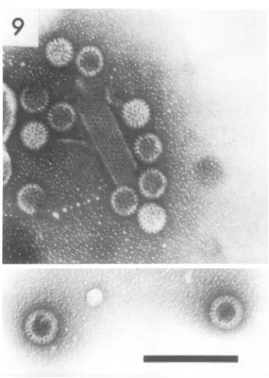
about 10 nm. Similar tubules with a 10-nm lattice spacing were also observed in negatively
stainedpreparations of EDIM virus (Fig. 9) and
J. VIROL.
on November 10, 2019 by guest
http://jvi.asm.org/
VIRAL ENTERITIS OFINFANTS 941
of calfscoursvirus.Variousother"honeycomb"
orlatticework structures ofirregular shape and
different spacings were regularly observed in
fecal extracts, bothinthe presence and absence
ofduovirus, but these latter are believed to be
fragments ofbacterial cell walls.
DISCUSSION
Onthebases of bothmorphologyand
morpho-genesis, the new virus described hereobviously
belongs in the family Reoviridae, in which
there are at present two recognized genera. It
has some features in common with both
reo-viruses and orbiviruses, notably, the type of
viroplasm or "virus factory" produced in
in-fected cells appears identicalineachcase.
Observation of negatively stained particles
aloneemphasizes theresemblanceofthe
double-shelled particles to reovirions (15, 17).
Never-FIG. 6. Negatively stained human fecal extract
containing both double (D) and single-shelled (S)
duovirusparticles. 7Thesurfacestructure oftheouter shell consists of overlapping rather than separate
ring-shapedcapsomers(arrow).Pindicatesa
bacterio-phageparticle.Ammoniummolybdate.x120,000.
7
.1
..;,
IN FIG. 8. Another human fecal extract showing
double-shelled virusparticlesandaflattened tubule
with hexagonally packed subunits (10 nm
center-to-center spacing). Similar tubules have also been observed in preparations of calf scours virus.
x120,000.
FIG. 7. Fragmentation of double-shelled particles observed in a preparation of human duovirus after sonictreatmentandtwocyclesof ultracentrifugation. Note that the fragmentshavea latticestructure,and that no separate capsomers are seen. Ammonium molybdate. x120,000.
7 "7. ..
VOL.16,1975
I
.. I
...t V
..A,
-l'li
4.R
-I:...."e
.j .1
.:.k 1. t
.-q,.,
',,.
"'..i .I,
*'t|*|uOt't
on November 10, 2019 by guesthttp://jvi.asm.org/
[image:5.493.33.227.81.342.2] [image:5.493.242.432.286.551.2] [image:5.493.36.226.413.671.2]942 HOLMES ET AL.
FIG. 9. A group ofEDIM virus particles, mostly single-shelled, and a tubule with the same subunit spacing, 10 nm, as that observed in similar tubules in preparations of human duovirus and calf scours virus. The inset shows two typical double-shelled particles in thesamepreparation. Ammonium molyb-date. x120,000.
theless the particles of the infantile diarrhoea
virus are distinguishable from those of reovirus
by their sharply defined outline (7, 16). This
feature is notable also inpublished micrographs
ofNebraska calfscoursvirus(14) andtwoSouth
African virusesnotknowntobeassociated with
gastroenteritis, SA-11 and the "O" agent (12).
Our crudepreparationsofEDIM virus also
con-tain double-shelled particles with the same
sharply defined outline, althoughthe previously
published electron micrographs of purified
EDIM virus show apparently single-shelled
particles(24).
Single-shelled particles of human duovirus,
like those ofcalfscours virus, EDIM virus, and
SA-11, closely resemble orbivirus particles
rather than the single-shell "core" particles
of reovirus (12, 19, 24, 29). The capsid-related
tubules reported above in human stool extracts
arealmostcertainly analogoustothose observed
within cells infected with EDIM virus (4). In
the negatively stained preparations the tubules
appear to be flattened, since the alternating
clear and fuzzy areas of subunit structure are
believedto arise as Moire patterns from
super-imposition of the upper and lower surfaces,
and they extend to near the edge. Similar
tubules have been observedboth within infected
cells and in negatively stained suspensions
of several orbiviruses, such as bluetongue,
epizootic haemorrhagic disease of deer, and
Tribec and African horse sickness viruses (25,
27). It is probable thatthey are formed by the
aberrant assembly ofviral capsid material and
are analogous to the tubular polyheads of
bacteriophage or the tubules found in
prepara-tions of various icosahedralviruses (3,13).
Morphogenesis observed ininfected duodenal epithelial cells, in particular the accumulation
of enveloped as well as naked particles in
rough endoplasmic reticulum vesicles, sets the
human duovirus apart from the classical
reo-viruses and suggests a possible affinity with
orbiviruses such as Colorado tick fever virus
(26). The same feature has also been reported
in morphogenesis of EDIM and SA-11 viruses
(1, 18).
The results of our analyses ofthe RNA and
polypeptides of calf and human duoviruses (S. M.Rodger, R.D.
Schnagl,
andI.H.Holmes,
manuscript in preparation) suggest a composi-tion broadly similar but distinguishable from thatofeither reovirus or orbiviruses. The close serological relationship between the human gastroenteritis virus, "Nebraska" calf scours
virus, and EDIM virus which has been
es-tablished independently by Flewett et al. (16), Kapikianetal. (17), and
by
our own work, and theimpossibility
of classifying these viruses aseither reoviruses or orbiviruses despite their obvious affinity to both groups, led us to
suggest that they should be
placed
in a newgroup within the family
Reoviridae,
called "duovirus" (10). Thename "rotavirus" has alsobeenputforward forthesamegroup
(16).
So far as is known, in vivo growth of each
of these viruses occurs only in the highly
differentiated cellsofgutepithelium. Thishigh
degree of tissue specificity no doubt accounts
for the difficulty which has been experienced
in attempts to grow these viruses in
conven-tional cell cultures. So far only the calf virus
hasbeensoadapted (21, 30), buttheproduction
of an attenuated oral vaccine for calves (22) is
most encouraging and may foreshadow similar
developments withthe human virus.
ACKNOWLEDGMENTS
We thank all those who have kindly supplied samples, especially R. R. W. Townley, and also Max Murray and
J. VIROL.
on November 10, 2019 by guest
http://jvi.asm.org/
[image:6.493.67.258.80.348.2]VIRAL ENTERITIS OF INFANTS 943
Anneke Veenstra for their excellent technical assistance inprocessing them.
This research was supported by a Lady Latham Re-search Fellowship of the Royal Children's Hospital Re-search Foundation (G.P.D.), the Felton Bequent (R.F.B.), and the National Health and Medical Research Council of Australia (I.H.H., B.J.R.).
ADDENDUM
During the preparation of this manuscript H. Suzuki andT.Konno (TohokuJ.Exp. Med. 115:199, 1975) reported reovirus-like particles in jejunal mucosa of a Japanese infant with acute infectious nonbacterial gastroenteritis. One of their micro-graphs illustrates tubularstructures of approximately the same diameter as virus particles in a section of aninfected cell.
LITERATURE CITED
1. Adams, W. R., and L. Kraft. 1967. Electronmicro-scopic study of the intestinal epithelium of mice infected with the agent of epizootic diarrhea of infant mice(EDIMvirus).Am.J.Pathol. 51:39-60. 2. Amano, Y., S. Katagiri, N. Ishida, and Y. Watanabe.
1971. Spontaneousdegradationof reoviruscapsidinto subunits.J. Virol. 8:805-808.
3. Bancroft, J. B., G. J. Hills, and R. Markham. 1967. A study of the self-assembly process in a small sphericalvirus.Virology31:354-379.
4. Banfield, W. G., G. Kasnic, and J. H. Blackwell. 1968. Further observations on the virus of epizootic diarrhea of infant mice-an electron microscopic
study.Virology36:411-421.
5. Barnes, G. L., and R. R. W. Townley. 1973.Duodenal mucosal damage in 31 infants with gastroenteritis. Arch. Dis.Child. 48:343-349.
6. Bishop, R. F., G. P. Davidson, I. H. Holmes, and B. J. Ruck. 1973. Virus particles in epithelial cells of duodenal mucosa from children with acute
non-bacterialgastroenteritis.Lancet 2:1281-1283. 7. Bishop, R. F., G. P. Davidson, I. H. Holmes, and
B.J.Ruck. 1974.Detection ofa newvirusbyelectron microscopy of faecalextractsfromchildren withacute gastroenteritis. Lancet1:149-151.
8. Cramblett, H. G., P. Azimi, and R. E. Haynes. 1971. The etiology of infectious diarrhea in infancy, with specialreferencetoenteropathogenicE. coli. Ann. N.Y. Acad.Sci.176:80-92.
9. Dales, S. 1973. The structure and replication of
reo-viruses, p. 155-171.In A. J. Dalton and F. Haguenau (ed.), Ultrastructure of animal viruses and
bacterio-phages:anatlas. Academic PressInc.,New York. 10. Davidson, G.P., R. F. Bishop, R. R. W.Townley, I.H.
Holmes, and B. J. Ruck. 1975. Importance ofa new
virus in acute sporadic enteritis in children. Lancet 1:242-245.
11. Editorial. 1975. Rotaviruses ofmanandanimals. Lancet 1:257-259.
12. Els, H. J., and G. Lecatsas. 1972. Morphological studies on simian virus SA 11 and the "related" 0agent.J.Gen.Virol. 17:129-132.
13. Favre, R., E. Boyde laTour, N. Segre, andE. Kellen-berger. 1965. Studies on the morphopoiesis of the
head of phage T-even. 1. Morphological, immunolog-ical, and genetic characterization of polyheads. J. Ultrastruct. Res. 13:318-342.
14. Fernelius, A. L., A. E. Ritchie, L. G. Classick, J. 0. Norman, and C. A. Mebus. 1972. Cell culture adapta-tion and propagation of a reovirus-like agent of calf diarrhea from a field outbreak in Nebraska. Arch. Gesamte Virusforsch. 37:114-130.
15. Flewett, T. H., A. S. Bryden, and H. A. Davies. 1973. Virusparticles ingastroenteritis. Lancet 2:1497. 16. Flewett, T. H., A. S. Bryden, H. Davies, G. N. Woode,
J. C. Bridger, and J. M. Derrick. 1974. Relation between viruses from acute gastroenteritis of children and newborn calves. Lancet 2:61-63.
17. Kapikian, A. Z., H. W. Kim, R. G. Wyatt, W. J. Rodriguez, S. Ross, W. L. Cline, R. H. Parrott, and R. M. Chanock. 1974. Reoviruslike agent in stools: association with infantile diarrhea and development ofserologic tests. Science185:1049-1053.
18. Lecatsas, G. 1972. Electron microscopic and serological studies on simian virus SA 11 and the "related" 0 agent.OnderstepoortJ. Vet. Res. 39:133-137. 19. Luftig, R. B., S. S.Kilham, A. J. Hay, H. J. Zweerink,
and W. K. Joklik. 1972. An ultrastructural study of virions and cores of reovirus type 3. Virology 48: 170-181.
20. Martin, S. A., and H. J. Zweerink. 1972. Isolation and characterization of two types of bluetongue virus particles.Virology 50:495-506.
21. Mebus, C. A., M. Kono, N. R. Underdahl, and M. J. Twiehaus. 1971. Cellculture propagation of neonatal calf diarrhea (scours) virus. Can. Vet. J. 12:69-72. 22. Mebus, C. A., R. G. White, E. P. Bass, and M. J.
Twiehaus. 1973. Immunity to neonatal calf diarrhea virus. J. Am. Vet. Med. Assoc. 163:880-883.
23. Middleton, P. J., M. T. Szymanski, G. D. Abbott, R. Bortolussi, and J. R. Hamilton. 1974. Orbivirus acute gastroenteritis ofinfancy. Lancet 1: 1241-1244. 24. Much, D. H., and I. Zajac. 1972. Purification and
char-acterization of epizootic diarrhea of infant mice virus. Infect. Immun. 6:1019-1024.
25. Murphy, F. A., E. C. Borden, R. E. Shope, and A. Harrison. 1971. Physicochemical and morphological relationships of somearthropod-borneviruses to blue-tongue virus-anewtaxonomic group. Electron micro-scopic studies. J. Gen. Virol. 13:273-288.
26. Murphy, F. A., P. H. Coleman, A. K. Harrison, and G. W. Gary, Jr. 1968. Colorado tick fever virus: an electronmicroscopicstudy.Virology35:28-40. 27. Oellermann, R. A., H. J. Els, and B. J. Erasmus.
1970. Characterization ofAfrican horsesickness virus. Arch.Gesamte Virusforsch. 29:163-174.
28. Vasquez, C., and P. Tournier. 1964. Newinterpretation ofthe reovirusstructure.Virology24:128-130. 29. Welch, A. B. 1971.Purification, morphologv and partial
characterization ofareovirus-like agent associated with neonatalcalf diarrhea.Can. J.Comp.Med. 35:195-202. 30. Woode, G.N.,J.C. Bridger,G.Hall,and M.J. Dennis.
1974. The isolation ofareovirus-like agent associated with diarrhea in colostrum-deprived calves in Great Britain. Res.Vet.Sci. 16:102-105.
31. Wrigley, N. G. 1968. The lattice spacing ofcrystalline catalaseas aninternal standard oflength inelectron microscopy.J.Ultrastruct. Res. 24:454-464.
VOL.16,1975
on November 10, 2019 by guest
http://jvi.asm.org/