0022-538X/82/110475-12$02.00/0
Copyright©1982, AmericanSocietyforMicrobiology
Methylation
State
and DNase
I
Sensitivity
of Chromatin
Containing
Moloney
Murine
Leukemia
Virus
DNA
in
Exogenously
Infected
Mouse
Cells
PAUL-ETIENNE MONTANDON,tFABIENNEMONTANDON,ANDHUNGFANt*
TumorVirology Laboratory, TheSalk Institute, San Diego,California92138 Received 7 May1982/Accepted16July 1982
The nature of Moloney murine leukemia virus (M-MuLV)-specific proviral DNA inexogenously infectedmousecellswas studied. M-MuLV clone A9cells,
NIH-3T3fibroblasts productively infectedwith M-MuLV,wereused. These cells contain 10to15copiesof M-MuLVproviralDNA.Thestateofmethylationof M-MuLV proviral DNA was examined by cleaving A9 cell DNA with restriction endonucleases which have the dinucleotide CpG in their cleavage sequences.
Analysis with suchenzymes, whichrecognized nine different sites in M-MuLV DNA, indicated thatmostifnotallof the M-MuLVprovirusesin A9cellswere
completelyunmethylated. An individual proviralintegrationwasexamined, using
asprobe adjacentsingle-copy cellularsequences.Thesesequences wereobtained fromalambdaphagerecombinant clonecontaininganM-MuLVprovirusfrom the A9 cells. This individual integration also showed no detectable methylation. In contrast, endogenous MuLV-related sequences presentinNIH-3T3 cells before infectionwerelargelymethylated. The configurationof chromatincontaining M-MuLV proviruses was also investigated by digesting A9 nuclei with DNase I,
followed by restriction analysis of the remaining DNA. Endogenous MuLV-related DNAwasinchromatin relativelyresistanttoDNase Idigestion, whereas the majority ofM-MuLV-specific proviruses were in domains of intermediate DNase I sensitivity. Two proviral copies hypersensitive to DNase I digestion
were identified. Analogy to the DNase I sensitivity ofexpressed and
nonex-pressed globin genes suggested that the proviral copies containing DNase I-hypersensitive sitesweretranscribed.
Moloney murine leukemia virus (M-MuLV),
like all retroviruses, replicates via a stranded DNA intermediate (34). The double-stranded DNA molecule is synthesized in the
cytoplasmas alinearmolecule whichiscolinear
with viral RNA and contains terminalrepetitions (the long terminal repeats [LTRs]; 20, 28, 33). ThisDNAis thentransportedtothe nucleus of the cell, where it integrates into the chromo-somal DNA toform theprovirus. Viral RNA is
synthesizedfromproviral DNAby cellularRNA
polymerase II, the enzyme which synthesizes cellular heterogeneous nuclear RNA (hnRNA) and mRNA. The structure and specificity of proviral DNAintegration have been studied in great detail(reviewed by H. E. Varmusand R. Swanstrom [38]). Integration of proviral DNA occurs at many sites within the host chromo-somal DNA. The viral DNA is not permuted
tPresent address: Laboratoire deBiochimie, Universite, 2000Neuchatel, Switzerland.
t Present address: Department of MolecularBiology and Biochemistry,University ofCalifornia, Irvine, CA 92717.
during integration, and thus itcontains LTRsat
either end.
Proviral DNA represents a complete
tran-scription unit. The LTRs contain the signals required for initiation of viral RNA synthesis (15, 36),aswellasforcleavage-polyadenylation
(29, 36). Transcriptionpresumably beginsin the 5'LTR andterminatesin the 3' LTR. Thesize of the initialtranscriptof M-MuLV RNA in infect-ed cells is thesame sizeas maturevirion RNA (12). This provides a very useful system for
studyingtheeffect of adjacentcellsequences on expression of a gene ("position effect"), since integration of the proviral transcription unit oc-curs at multiple sites within the host chromo-somalDNA.
Several lines of evidence indicate that adja-cent host cell DNA sequences or chromatin
configuration can influence the transcriptional
activityof retrovirus proviralDNA.Infection of M-MuLV in embryonic mice has been studied by Jaenischet al. (21). A variety ofpatterns of expression in different tissues was observed
475
on November 10, 2019 by guest
http://jvi.asm.org/
after birth, and the patterns were different for differentproviral integrations. Ringoldetal. (27) studied rat hepatoma cells infected in vitro with murine mammary tumor virus and found that some cells into which murine mammary tumor virus proviral DNA had integrated expressed viral RNA, whereas others did not. We studied the transcriptional activity of M-MuLV proviral DNA in exogenously infected mouse fibroblasts by the technique of DNase I digestion of chro-matin (5). Nuclei from cell lines which contained multiple copies of integrated M-MuLV proviral DNA were digested with DNase I, and the relative sensitivity of M-MuLV-specific DNA was measured by solution hybridization. The results indicated that only a minority of
M-MuLV-specific DNA was preferentially sensi-tive to DNase I digestion. Weintraub and Grou-dine (41) have shown that transcribed genesare preferentially sensitive to DNase I digestion, and our results suggested that the majority of integrated M-MuLV DNA copies in exogenous-lyinfected cells are not transcribed.
In the experiments reported here, a cloned
lineof NIH-3T3cellsproductively infectedwith M-MuLV wasstudied. Thestateofmethylation of the M-MuLVproviralDNAwasinvestigated, since several studies have shown that tran-scribed genes are usually undermethylated (7, 22, 37). In addition, proviral integrations were examined for the presence of sites of DNase I hypersensitivity, which are indicative of tran-scribed genes (31, 44). The results showed that
endogenousMuLV-related DNA wasrelatively
methylated andwascontained within chromatin
resistant to DNase I. In contrast, M-MuLV
proviral copies wereunmethylated andwere in chromatin of intermediate DNase I sensitivity. Also,twoM-MuLVproviruseswerefoundtobe in chromatinwhichwasverysensitivetoDNase Iand thuscontained sites of DNase I hypersen-sitivity.
MATERIALS AND METHODS
Cells.M-MuLV clone A9 cellsare aline of NIH-3T3 cells productively infected with M-MuLV and have been described previously (13). They were grown in tissue culturemonolayersinDulbecco-modifiedEagle
medium supplemented with 10%o calfserum (Irvine Scientific).NIH-3T3cells (35)weregrown in thesame medium.
Cell fractionation and DNase I digestion ofnuclei.
Cells were removed from tissue culture dishes by trypsinization on ice (12). For DNase I digestion experiments, cultures at one-half to three-quarters confluencywere used, whereas completelyconfluent cultureswereusedfor isolation of total high-molecu-lar-weightDNA.Trypsinizedcellswereharvestedby centrifugation(1,000 xgfor 2min)and washed twice in Tris-bufferedsalinecontainingcalcium and magne-sium. Nuclei were isolated essentially according to Weintraub andGroudine(41).Atotal of 2x10⁸to3x 108 cells were suspended in 1 ml of reticulocyte
standard buffer(0.01 M NaCl, 0.01 M Tris-hydrochlo-ride [pH 7.4], 3 mM MgCl2), and Nonidet P-40was
added to 0.5%.After blending in a Vortex mixer for30
s, the nucleiwere harvested by centrifugation (1,000 x g for 2 min) and washed twice with reticulocyte standardbuffer.
For DNase I digestion of nuclei, a modification of the conditions of Weintraub and Groudine was used. Nuclei weresuspended in reticulocyte standard buffer at 25absorbance units at 260 nm per ml anddivided
into severalportions. DNase I (RNase-free; Worthing-ton Diagnostics) was added to different concentra-tions, and the samples were incubated for 20 minat 15°C. After incubation, the reactions were terminated by adjusting the samples to 0.2 MNaCl-0.5%sodium
dodecylsulfate-10 mM EDTA-200 ,ug of pronaseper
ml (preincubated at 37°C to remove residual
nu-cleases). Thesamples were then incubated overnight atroomtemperature or for 1 h at 37°C. High-molecu-lar-weight DNA was then extracted.
DNA extraction. High-molecular-weight DNA was extracted by two phenol-chloroform extractions fol-lowed by two chloroform extractions, as described previously (1). The extracted DNA was precipitated by addition of 2 volumes of isopropanol, harvested by
centrifugation, and suspended in 10 mM
Tris-hydro-chloride(pH7.4)-i mM EDTA.
Restrictionenzyme digestion and gel electrophoresis. DNA samples weredigestedwith various restriction enzymes (Bethesda Research Laboratories or
Boeh-ringerMannheimCorp.) in the recommended buffers at aDNAconcentration of 60 to 120Fg/ml,with the
addition of gelatin to 0.01%. In experiments with
methyl-sensitive enzymes, adenovirus type 2 DNA wasadded tothecellularDNA before digestion, and completeness of digestion was assessed by the pres-enceof the predicted restriction fragments after gel
electrophoresis, staining in ethidium bromide, and
visualizationwith UVlight.
Electrophoresis of DNA in horizontal agarose gels was performedas described previously (1). For gels
whichwerehybridized with radioactive DNA in situ, the thickness of the gels was 3 to 4 mm. After
electrophoresis,gels were stained withethidium
bro-mide, and distribution of DNA was determined by observation under UVlight.
Preparation of labeled DNA probes. 32P-labeled M-MuLV cDNA was prepared asdescribed previously
(14). Calf thymus oligodeoxynucleotide primers were added to the reaction to render the cDNAuniformly
representativeof the viral genome.
Nick-translation of recombinant DNA restriction fragments was performed as described by Rigby et al. (26).
Gelhybridization. For in situhybridizationto agar-osegels (30), thefollowing procedurewasused. The gels were soaked for 30 min in 0.2 N NaOH-0.6 M NaCl, rinsed briefly in water, and then soaked in 0.5 M Tris-hydrochloride (pH 7.5)-0.6M NaCl for 30 min. Afterrinsing the gels in water, they were dried under vacuum and gentle heat on Whatman 3MM filter
paper. Afterdrying, the gels were removed from the paper by placing them in water, and the slightly
rehydrated gels were transferred toplastic food bags forhybridization.
Hybridizations weredone in sealableplasticbags, asdescribedpreviously(1). The gels were first prehy-bridized for 30 min at 68°C with prehybridization
J. VIROL.
on November 10, 2019 by guest
http://jvi.asm.org/
buffer: 6x SSC = 0.9 M NaCl, 0.09 M sodium
cit-rate; 5x Denhardt solution (9); 10 mM TES
[N-tris(hydroxymethyl)methyl-2-aminoethanesulfonic
acid] buffer (pH 7.5), 500 p.g of denatured salmon sperm DNA per ml, 0.1% sodium dodecyl sulfate. After prehybridization the buffer was expelled and labeled DNAprobe was added inhybridization buffer (6xSSC, 1 x Denhardt solution, 10 mM TES [pH 7.5], 50 .gof denatured salmon sperm DNA per ml, 0.1% sodiumdodecyl sulfate),andhybridizationwascarried out at 68°C overnight. Concentration ofthe labeled cDNAwas 0.5 x 10⁶to 1.0x10⁶ cpm/ml.
Afterhybridization, the gels were removed from the food bags and washedbrieflythreetimeswith 2x SSC + 0.1% sodium dodecyl sulfate, followed by two washes with 0.1xSSC+0.1% sodiumdodecylsulfate for 30 min at 45 to 55°C. Afterwashing, damp gels
were sealed between two layers ofcellophane wrap andexposed to X-ray film between two intensifying
screens.Thefilm wassensitizedbypreflashingunless otherwise noted.
RESULTS
Methylation state of M-MuLV proviral DNA. The methylation state of M-MuLV proviral
DNAwasinvestigatedbyusingmethyl-sensitive restriction endonucleases. 5-Methylcytosine is the most commonmethylated basefoundin the DNAofhighereucaryotesandis almost exclu-sively foundin thedinucleotide CpG (18).
Cer-tain restriction endonucleases recognize and cleave a DNA sequence thatcontains CpG,and
methylation of this dinucleotide inhibits cleav-agebymany (25). Fortheseexperiments, such enzymesused were XhoI, Sal, ClaI,and SmaI
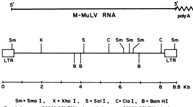
sincethey also cut M-MuLV DNA at least once (17). The sites of cleavage for these enzymes within M-MuLV DNA are shown in Fig. 1. Two
other restriction enzymes were also used,
BamHI and EcoRI. These enzymes recognize
sitesonthe DNA that do notcontain CpG and cleave methylated andunmethylatedeucaryotic
DNAequally well. Also, EcoRIdoes notcleave
within M-MuLV DNA at all (16, 39).
EcoRI
fragments
which contain M-MuLV pro-virus. In the first experiments, the methylationstateof M-MuLV proviral DNA was
investigat-ed by digesting infected and uninfected cell DNA withEcoRI in combination with
methyl-sensitive enzymes. The infected cell line M-MuLV clone A9 (13) was usedforthese
experi-ments; M-MuLV clone A9 cells are NIH-3T3 fibroblasts productively infected withM-MuLV and contain 10 to 15 copies of integrated M-MuLVproviralDNA (2).SinceEcoRP doesnot
cleaveM-MuLV DNA,digestion of infected cell
DNA with
EcoRI
yields eachM-MuLVprovirusin a single restriction fragment,containing both
M-MuLV DNA and adjacent host cell DNA sequences. In Fig. 2 (lanes 1 and 2),
EcoRl-digested uninfectedNIH-3T3cell and A9 DNAs wereseparated according tosize by agarose gel
electrophoresisandblot-transferredtoactivated aminophenylthioether paper (11; B. Seed,
per-sonalcommunication), and restriction fragments
which could hybridize radioactive M-MuLV cDNA wereidentifiedbyautoradiography. The
MuLV-related DNA fragments from the
unin-fected cells (lane 1) represented endogenous
MuLVsresident in the genomesofmice,and the M-MuLV proviral copies acquired by infection
were evident as additional M-MuLV-specific EcoRI fragments in the infected cells (1). A9 cells contain M-MuLVproviralDNA inEcoRI
5
M-MuLV
RNA
C Sm Sm Sm Sm
T
B
2 4 6
3'
polyA
C Sm
LTR
8 8.8 Kb
SmxSmaI, XzXho I, SzSaII, CzClaI, B=BomHI
FIG. 1. Restriction map of M-MuLV DNA. A restriction map of M-MuLV DNAis shown, according to
Gilboa etal. (17). The viral DNA is shown in proviral form, with two copies of the LTRs, and the linear relationshiptogenomic RNA is indicated. Sites for the followingenzymes areindicated:B,BamHI;X,XhoI;S, SalI; C,ClaI;Sm, SmaI.The identification ofthetwoClaI siteswasobtained fromour owndata(notshown)and confirmed inthepublished nucleotidesequenceofM-MuLVDNA(29). Poly A, Polyadenylate.
0
on November 10, 2019 by guest
http://jvi.asm.org/
[image:3.491.83.418.458.644.2]1
2
3
45
6
M.W. M.W
(kb) (kb)
23!7- 2
16
4.5-
[image:4.491.54.239.72.385.2]2.3-RI RI+S RI+X
FIG. 2. Methylation of EcoRI fragments containing M-MuLVproviral DNA. NIH-3T3 and A9cellDNAs
were digested with EcoRI or EcoRI in combination with Sall orwith XhoI (30 tag of DNAper digest). After digestion, the DNAwasresolved by
electropho-resis in a0.6%agarose gel, transferred to activated aminophenylthioesterpaper,andhybridized with
32P-labeled M-MuLV cDNA in hybridization buffer
con-taining 10odextran sulfate (40). cDNA probewasat 106cpm/ml. An autoradiogram(2-day exposure) of the washed blot is shown.Odd-numbered lanesare NIH-3T3 DNA, and even-numbered lanes are A9 DNA. Lanes 1 and2, Digestion with EcoRP; lanes 3 and 4,
digestion with EcoRI and Sall; lanes5and6,digestion
with EcoRI and XhoI. M.W., Molecular weight.
fragmentsof22, 20,18,and 16kilobases(kb; 2) (lane 2). Each of thesefragment sizes consistsof more than a single M-MuLV provirus. Three different 22-kb proviral organizations and two
20-kborganizations have beenisolated from the A9 cellsby recombinant DNA techniques (3).
In Fig. 2, lanes 3 and 4, the same DNA samples were digestedwith EcoRI in combina-tion with Sail. The EcoRIfragments containing integrated M-MuLV provirus were digested by theaddition of Sall (lane 4),but theendogenous MuLV-relatedfragmentsinthe samecellswere unaffected. Thus, at least one Sall site was
unmethylatedin the EcoRIfragmentscontaining
each exogenously acquired M-MuLV provirus,
whereas therewerefew unmethylated SalI sites in EcoRI fragments carrying endogenous
MuLV-related sequences. The resistance to di-gestion of the endogenous MuLV-containing
EcoRI fragments was not duetoalack of Sall sites, since Sall sites have been identified ina
number of endogenous MuLV organizations iso-latedbyrecombinant DNA techniques(H. Fan,
unpublished data; R. Mural et al., personal
communication).
In Fig. 2, lanes 5 and 6, uninfected and infectedcellDNAsweredigestedwith EcoRI in combination with XhoI, and the results obtained weresimilar to those for EcoRI incombination withSail, except thatsomeof thelarger
endoge-nousEcoRI fragments were also digested with XhoI.
These results indicated that all detectable EcoRI fragments carrying integrated M-MuLV provirus contained unmethylatedSall and XhoI sites. Theunmethylated sites might be the SalI and XhoI sites within the viral DNA itself, or they might be inthe adjacent cell DNA on the same EcoRIfragment.Furthermore, methylated M-MuLV proviruses would not have been de-tected if they were contained in EcoRI frag-ments which comigrated with an endogenous MuLV-containing EcoRI fragment. For these reasons, a different approach was taken to mea-sure methylation within the M-MuLV viral se-quences themselves.
Extent of methylation within proviral DNA. The extent ofmethylation in M-MuLVproviral DNA copies in the A9 cells was assessed by comparing the efficiency with which methyl-sensitive and methyl-insensitive enzymes could cleave this DNA. In Fig. 3A, such an experi-ment was performed by cleaving infected and uninfected cell DNA with Sail in combination withXhoI. Each of these enzymes cleave un-methylated M-MuLV DNAat asingle site (see
Fig. 1).Combinedcleavage would releasea 2.2-kbfragment only fromintegrated M-MuLV pro-viruses unmethylated at both sites. For stan-dardization, infected and uninfected cell DNAs were also cleaved with BamHI, which cuts a DNA sequence that does not contain CpG.
Cleavage of infected cell DNA with BamHI releases a 3.0-kb M-MuLV-specific fragment (1), and the amount of this fragment generated
(indicated bytheintensityofhybridization) rep-resents an internal BamHI fragment released from eachproviral integration.
A9 and NIH-3T3 DNAs were digested with SalIplus XhoI,andequalamounts were digest-ed with BamHI (Fig. 3A). After digestion, the DNA fragments were analyzed by gel electro-phoresis andhybridizationinsitutothegelwith
J. VIROL.
on November 10, 2019 by guest
http://jvi.asm.org/
A
1 2 3 4 5
B
1 23 4 6
MW. (kb)
-3.0
-2.2
S+X B
5 6 7 8 9
F: .S
3 -3.25
«i ~~3.0-*
2.7-*. -2.2
b-I.95 *'
-1.75
B+Sm Sm B C
FIG. 3. Methylation atSalI, XhoI, ClaI, andSmaI sitesinproviral DNA. NIH-3T3 and A9 DNAs were digestedwithvariousenzymes orcombinations ofthem(3 igperdigest).ThedigestedDNAwasresolvedby electrophoresis in a0.8% agarose gel andhybridized insitu with M-MuLV cDNA (0.5 x 10⁶ cpm/ml). An
autoradiogramof thewashed gelis shown(4-day exposure). (A)Lanes 1 and 3containNIH-3T3DNA;allothers
contain A9 DNA. Lanes1and2,DigestionwithSalIandXhoI;lanes 3 and4,digestionwithBamHI;lanes 5and 6,digestionofsmalleramountsofA9 DNA withBamHI (1 and 0.3 Fg,respectively). (B)Lanes1, 3,and8, Digestion ofNIH-3T3 DNAwithBamHI plus SmaI (lane 1),withSmaI (lane 3),orwithClaI(lane 8);lanes2, 4, 5,6, 7,and 9,digestion ofA9 DNAwithBamHI plusSmaI(lane2), SmaI(lane 4), BamHI (3,1, and 0.3jigofA9 DNAinlanes5,6, and 7,respectively),orClaI (lane 9).Digestionof bothinfectedanduninfectedcellDNAwith
BamHI yieldedahybridizing fragmentof2.3 kb. Thisfragmentrepresentedaninternalfragmentderived from multiple copies ofendogenousMuLV-related sequences present inallmouse strains (11;R. Mural, personal communication).M.W.,Molecularweight.
radioactive M-MuLV cDNA. Hybridization to
gels in situ was approximately five times as efficientas hybridization toblottransfers when M-MuLVcDNAwasused,andone-fifthto
one-tenthas muchDNAasfor blot transfers(3to6 ,ugof cell DNA per lane) was loadedonto the agarose gels. A 2.2-kb M-MuLV-specific
Sal-plus-XhoIfiagmentwasreleased fromM-MuLV
cloneA9 DNA (lane 2)and notfromNIH-3T3 DNA(lane 1),and theintensity of hybridization for thisfragmentwasonly slightlyless than the
larger3-kb M-MuLV-specificBamHI fragment (lanes4to6). Since the M-MuLV cDNAprobe
used in these experiments was relatively
uni-formly representative of the entire M-MuLV genome, asmaller
fragment
wouldbeexpectedto hybridize proportionately less cDNA. This
thereforesuggestedthat theXhoI andSaiI sites in mostifnotall M-MuLVproviralDNAcopies
wereunmethylated.
AnM-MuLV-specific fragmentof 2.5 kb was alsoevident intheSall-XhoIdouble digest of M-MuLVclone A9 DNA(lane 2). The origin of this fragment is presently unclear. It was M-MuLV specific since digestion of NIH-3T3 DNA did not yield it. However, it did not result from
partial digestion of M-MuLV proviral DNA,
sincenootherSall and XhoI sites besides those
giving risetothe 2.2-kbfragmentare present in M-MuLV DNA. The intensity of hybridization wasalso too great to result from digestion of one M-MuLV provirus which had an XhoI or Sail site in adjacent cell DNA at the appropriate
distancetothe5' side of a provirus. One
possi-bility was that the A9cells were also infected withavariant of MuLV with internal Sail and XhoI sites spaced 2.5 kb apart.
The methylation state of Clal sites in M-MuLVproviralDNAwasinvestigated, using an approach similar to that for the Sall-XhoI double bOMMM WA Od
on November 10, 2019 by guest
http://jvi.asm.org/
[image:5.491.86.419.73.343.2]digests. Digestion of unmethylated M-MuLV DNA with ClaI generates an internal 2.7-kb fragment (Fig. 1). A9 and NIH-3T3 DNAs were digested witheitherClaI (Fig. 3B, lanes 8 and 9) or BamHI(Fig.3B, lanes 5 to7) and analyzed by gel hybridization with M-MuLV cDNA. The expected 2.7-kb M-MuLV-specific fragment in the ClaI-digested A9 DNA was observed (lane 9),and its intensity was also proportional to the intensity of the M-MuLV-specific BamHI frag-ment.Thus, both ClaIsites in the majority of M-MuLVproviral copieswereunmethylated, simi-lar to theSall and XhoI sites.
Theabove experiments indicated that sites at different internal locations in M-MuLV DNA wereunmethylated in the majority of integrated M-MuLVDNA. However, regions that contain signals for regulation of transcription might show differences in the methylation state
de-pendingonwhether or not an individualprovirus is transcribed. Therefore, the methylation state of sites within the M-MuLV LTRs was also
investigated,usingSmaI.SmaIcleaves unmeth-ylated M-MuLV DNAin the LTRs, as well as three times internally. In particular, the SmaI
site in the M-MuLV LTR is within 30 nucleo-tides of the sequence for the 5' terminus of viral RNA. Digestion of NIH-3T3 and A9 DNA with SmaI is shown in lanes 3 and 4 of Fig. 3B. EndogenousMuLV-related DNA from NIH-3T3 cells remained in high-molecular-weight DNA
fragments (lane 3). Since several SmaI sites havebeen mapped in the DNA of endogenous
MuLV-related organizations (Mural, personal communication), this result indicates that
en-dogenous MuLV-relatedDNAismethylated. In contrast, the A9cells yielded major
M-MuLV-specific fragments of5.75 and 1.95kb,aswellas aminor fragment(s) of6.1 to 6.25 kb (lane 4). Examinationofthe location of the SmaI cleav-age sites in M-MuLV DNA suggested that the 5.75-kb fragment resulted from cleavage at the SmaI site in the 5' LTRaswellascleavageatthe SmaI site at6.25 kb on theproviralmap.
Simi-larly, the 1.95-kb fragment likely resulted from cleavage at the SmaI site at6.75 kb and at the SmaI site in the 3' LTR. Anexplanationfor the minor 6.1- to 6.25-kbfragment could bethatthe
SmaIsiteat6.25kbwaspartiallymethylated, so
that cleavage in the 5' LTR yielded slightly
largerfragments whose 3' termini resulted from
cleavage at the SmaI site at 6.6 or 6.75 kb.
Alternatively, this fragment might have been generated by SmaI digestionofavariant virus. In conclusion, the SmaI sites in the LTRs as well as at least someof the internal sites were unmethylated.
To more closely examine theSmaI sites, A9 DNA was digested with SmaI in combination with BamHI. The internal SmaI sites are
con-tained within the 3-kb internal BamHI fragment (see Fig. 1), andcompletedigestion at the SmaI sites would yield a BamHI-SmaI fragment of 2.2 kb, as well as several very small fragments. Methylation at one or more SmaI sites would result in SmaI cleavage of the internal 3-kb BamHIfragment tofragments of2.5and 2.65 kb instead of the 2.2-kb fragment. In lane 2 of Fig. 3B, thecombinedBamHI and SmaI digestion of A9 DNA is shown, andM-MuLV-specific frag-mentsof3.25, 2.2, and 1.75 kb weregenerated.
The3.25-kb fragment was the sizepredicted for cleavage at theSmaIsite in the 5' LTR and the 5'-most internal BamHI fragment (at 3.75 kb),
and the 1.75-kbfragmentwasthesizepredicted
for cleavage at the 3'-most terminal BamHI site (at 7.0kb) and the SmaI site in the 3' LTR. The 2.2-kb fragment derived from cleavage at the BamHI site at 3.95 kb and the SmaIsiteat6.25 kbcomigrated withtheinternalBamHIfragment
derived from endogenous MuLV-related se-quences(lane 1). Sincenobandsat 2.5 or 2.65
kb correspondingto thepredicted BamHI-SmaI fragments were present, these resultsindicated that theSmaI sites at 6.6 and 6.75 kb were not methylated. This supports the hypothesis that the 6.1- to 6.25-kb fragment generated after SmaI digestion ofA9 DNAwasderived froma variant virus. The BamHI-plus-SmaI digestion results also supported the conclusion from di-gestion with SmaI alone: that the SmaI sites in the LTRs and at internal sites were largely unmethylated.
Methylation ofan individual proviral integra-tion. Aproblem involvedwithanalysis of the A9 cellsisthatthey contain10to15 integrated M-MuLV proviruses, and the observations de-scribed above represented the averagesituation
for all of them. Any individual provirus might
havehadamethylationstatedifferent from the average. Therefore, the methylation stateof an individual M-MuLV provirus was investigated. For this, a lambda phage recombinant clone carryingacomplete integrated M-MuLV
provi-rus was used. The clone used (no. 80) was isolated from thesame cloningexperiment that wereportedpreviously (3), and its map is shown inFig. 4A. Whereas the viral sequences in such arecombinant clone wouldhybridizewithall M-MuLVproviruses, single-copy sequences in the adjacent cell DNA in clone 80 would only hy-bridize to the provirus which was cloned. A
HindlIl fragment of clone 80 phageDNAwhich contained 5'adjacent cell DNA as wellas lamb-da phage vector DNA (see Fig. 4A) was
pre-paredandradioactively labeledby
nick-transla-tion. Thisprobewasthen usedforhybridization
with agel of A9 and NIH-3T3 cell DNA cleaved with EcoRI orEcoRIincombination with
Sail,
XhoI, orClaI, similartoFig. 2(Fig. 4B). When
on November 10, 2019 by guest
http://jvi.asm.org/
M-MuLV-CONTAINING CHROMATIN 481
M-MuLV RNA
3'
_z%AN poly A
X S HD
* IeII I
0
HD= Hind Mt,
B 1 2 3 4 5 6 7
"Ia..
::' om_
8
I
-SW- -,
-'it
RI RI+S RI+X RI+C
[image:7.491.53.239.265.481.2]X=XhoI, S= Sol I, RI = Eco RI
FIG. 4. Methylation of the recombinant clone 80 provirus. (A) Anewrecombinant lambda phage clone
(clone 80) containinganintegrated M-MuLVproviral
organization from A9 cellswasisolatedaspreviously described(3). The clone containedacompletecopyof M-MuLVproviral DNA andgaverisetoinfectious
M-M.W. MuLVonDNA infection. Theproviruswascontained
(kb) inan18-kbEcoRI
fragment
andcontainedboth5'and3'
adjacent
cell sequences. A restriction map of the recombinantphage is shown. H, HindIII; S, SalI; X, -18 XhoI.Preliminary data also indicated at leastonesite forClaI in theadjacent cellsequencestothe 5' side of theprovirus. A HindIII restriction fragment contain-ing5' adjacent cell sequences (as well assequences9 fromthe right lambda cloning arm) was isolated by
9 preparativegel electrophoresis followedby recovery
on glass filter paper (6) and was labeled by
nick-translation. The location of thisfragment is shownin theshaded bar. Poly A, Polyadenylate. (B) NIH-3T3 (odd-numbered lanes)and A9(even-numbered lanes) DNAs(3 jig perdigest)weredigestedwithEcoRIor
EcoRI in combination withSall, XhoI, orClaI. The digests were resolved by electrophoresis in a 0.6%
agarose gel and hybridized in situ with the nick-translated 5'adjacent cell probe of (A) (106cpm/ml).
Exposure was for 15 days. Nonspecific binding of labeled DNAtothegelwasgenerally agreater prob-lem whennick-translatedprobeswereusedinstead of
M-MuLV cDNA.M.W., Molecularweight.
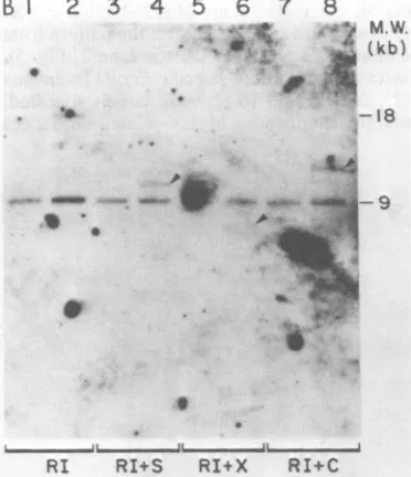
the DNAsweredigested with EcoRI alone, both NIH-3T3 and A9 DNAs showed a 9-kb
frag-ment, and the A9 DNA showedaless abundant fragment of 18 kb (lanes 1 and 2). The A9 cell 18-kb fragment was the same size as the EcoRI fragment containing the proviral DNA inserted into clone 80 andrepresented the provirus from which itwascloned. The 9-kb fragmentpresent
inbothuninfected and infected cells represented the alleles in host cell DNA which did not
receive an M-MuLV integration (the host site) and, as expected, was approximately 8.8 kb smallerthan thefragment into whichan integra-tion occurred. Thegreaterintensity of the 9-kb fragment than the 18-kb fragment in the A9 cells likely reflected the fact that the parental NIH-3T3 cellsareapproximately tetraploid (with four
copies of the host site), and M-MuLV DNA integration only occurred inone of them.
Digestion of the infected and uninfected cell DNAs with EcoRP in combination with SalI, XhoI, and ClaI reduced the size of the 18-kb A9-specific fragmentto10(lane 4), 8.4 (lane 6), and 11 (lane 8) kb, respectively. These sizes were appropriate for cleavage of the clone 80 M-MuLVprovirus in the A9cellsattheSall,XhoI, and ClaI sites within the M-MuLV DNA. The results indicated that the sites for theseenzymes were unmethylated in the clone 80 provirus. Methylationatthemore3' ClaI site couldnotbe tested in thismanner,since theprobe usedwasa
5'adjacentcellprobe, andcleavage atthemore
5' ClaIwascomplete.
The map of clone 80 phage DNA revealed a
A
5'RI HD
I//E///X/LLEm74
HD
XR
-10
HD S RI
..I
-5 5 10 Kb
.* * \
on November 10, 2019 by guest
http://jvi.asm.org/
MONTANDON, MONTANDON, AND
Sall site inadjacent cell DNA very near the 3' end of the provirus, and this site was presum-ably present in the 9-kb EcoRI host fragment. However, this fragment was resistant to Sail
digestion in both infected and uninfected cells, indicating that the host site was methylated in
both uninfectedandinfected cells. Experiments
with a 3' adjacent probe from clone 80 phage
indicatedthatmethylation at this 3' adjacent cell Sall site was maintained even after integration of the unmethylated M-MuLV DNA (not shown).
DNase I
sensitivity
ofM-MuLV proviral DNA in isolated A9 nudei. DNase I digestion of A9 nuclei was used toinvestigatethetranscriptional state of M-MuLV proviruses. Weintraub andGroudine (41) showed thatdigestion of nucleior
chromatin withDNase Ipreferentiallyremoves
transcribedgenes. This observationwas recent-ly extended, and it was found that sites of
DNase I hypersensitivity are generally located
atthe 5' endsof transcribedgenes (23, 31, 44). As a consequence, a hypersensitive DNA re-striction fragmentis rapidly removed from
nu-clei digested withDNase I and generates aseries
of subfragments (19, 42). In addition, domains of intermediate DNase I sensitivity have been found to surround transcribed genes. These re-gions are generally not transcribed, and they lack DNase I-hypersensitive sites (42).
M-MuLV proviruses in DNase I-sensitive do-mains. Investigation of the DNase I sensitivity of M-MuLVprovirusesin theA9 cells is shown in Fig. 5. Nuclei from A9 cells were incubated with variousconcentrations of DNase I, and the DNA was extracted. The extracted DNA was then cleaved with EcoRI and analyzed by gel hybridization withradioactiveM-MuLV cDNA. At high DNase I concentrations (>10
p,g/ml;
lane 10), all ofthe M-MuLV-hybridizing frag-ments weredigested. However, atintermediate
concentrations, differential sensitivity of
M-MuLV-hybridizing fragments wasobserved. In A9nuclei digestedwith5 ,ugof DNase I per ml (lane 9), the endogenous MuLV-related EcoR]
fragmentswerestill present (cf. the pattern from
theuninfectedNIH-3T3 cells in lane 2, Fig. 5), whereas theM-MuLV-specificEcoRI fragments
of22, 20, 18, and 16 kb were largely digested.
Thus,the endogenous MuLV-related sequences
1
2
3
4
5
6
7
8
9
10 11
DNoseI' 0 0.2 0.5 1.0 2.0 5.0 10 20 ig/ml
FIG. 5. DNase Isensitivityof EcoRIfragments containing M-MuLVproviralDNA. Nucleifrom A9 cells
wereisolated and incubated for 20 minat15°Cwith differentconcentrationsof DNaseI,and the DNAwasthen extracted. Forcontrols,DNAwasextracted fromNIH-3T3 and A9nuclei thathadnotbeen incubated at all. A
6-pgg portionofDNA from eachsamplewasdigestedwithEcoRlandanalyzed by gel hybridizationwith M-MuLV cDNAasin thelegendtoFig.3.Exposurewasfor 4days.Lanes1and2,Twodifferentpreparationsof NIH-3T3
DNA;lane 3,control A9 DNA (extracted directly). Lanes 4to11, DNA from A9 nuclei incubatedwith the followingamounts of DNase I:0, 0.2, 0.5, 1.0, 2.0, 5.0, 10, and 20,ug/ml, respectively. Essentially all high-molecular-weight (M.W.)DNAwasdigestedin the lasttwosamples.
M.W. (kb)
_122
f*
-20O
it A
8l
1[.S
=_ 1
on November 10, 2019 by guest
http://jvi.asm.org/
[image:8.491.107.390.349.599.2](which are not transcribed in NIH-3T3 cells; 4) are in the DNase I-resistant configuration, whereas the M-MuLV proviral integrations are in DNase I-sensitive domains. However, due to the large numberofM-MuLV-hybridizing frag-ments in the EcoRI pattern, it was not possible to distinguish whether individual integrations
contained DNase I-hypersensitive sites or not. Therefore, individual integrations were exam-ined.
DNase I
sensitivity
ofindividualintegrations.
In the first approach to examining the DNase Isensitivity of individual M-MuLV integrations, the same DNA samples from the DNase
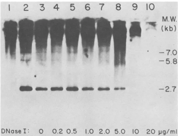
I-digested A9nuclei used in Fig. 5 were digested with ClaI instead of EcoRI and analyzed by electrophoresis and gel hybridization (Fig. 6). The majority of M-MuLV-hybridizing DNA was veryhigh molecular weight and reflected the fact that mostof theClaI sites in cellular DNA are methylated and resistant to cleavage. The inter-nal 2.7-kb ClaI fragment released from all of the M-MuLV proviruses was also evident, as ex-pected. Inaddition, twoM-MuLV-specific frag-mentsof 7 and 5.8 kb were present in undigested A9 nuclear DNA (lane 2). These fragments
pre-sumably represented the 5' portions oftwo M-MuLVproviruseswhich alsocontained
unmeth-ylated ClaI sites in the 5' adjacent cell DNA. This conclusion was supported by the fact that a
nick-translated fragment from the 5' portion of cloned unintegrated M-MuLV DNA (the AvaI fragmentfrom 2.6 to 3.8 kb on theproviral map) hybridized with both fragments (not shown). Bothof these fragments were absent in the DNA samples which had been digested with 1-,ug/ml (lane 6) or higher concentrations of DNase I. A larger fragment (9.5 kb) also present in uninfect-ed cells, and thus containing endogenous MuLV-related DNA, was not digested by 5 ,ug of DNase I per ml. The 5.8- and 7-kb ClaI fragments were more DNase I sensitive than were the M-MuLV-specific EcoRI fragments from the same DNA samples. For instance, in Fig. 5, the 18- and 16-kb M-MuLV-specific EcoRIfragments could still be detected in DNA from nuclei digested with 2 ,ug of DNase I per ml. These results suggested that the 5.8- and 7-kb M-MuLV-specific ClaI fragments contained DNase I-hypersensitive sites. In fact, samples from the higher DNase I digestion
concentra-tionsalso showed subfragments for these provi-ralintegrations(barely visible in Fig. 6, but more visible in autoradiograms which had not been preflashed before exposure of the film). The ClaIdigestions thus also served to identify the DNase I concentrations in which hypersensitive sites were detected in this particular DNase I digestion. These results indicated that many of the M-MuLV proviruses visible in Fig. 5 lack
1
2
3
4
5
6
7
8
9
10
5
il 4 4-_
[image:9.491.104.395.404.627.2]DNaseI:
00.2 0.5
1.02.0
5.0 10 20pg/m1
FIG. 6. ClaI digestion ofDNAfrom DNase I-digested A9 nuclei. ThesameDNAsamples describedin the legendtoFig.5weredigestedwithClaIinstead of EcoRI(6 p.g perdigest) and analyzed by gel hybridization with M-MuLVcDNA. Exposurewasfor 10 days. Lane 1, NIH-3T3 controlDNA;lane2,unincubated A9 control DNA;lanes3to10,DNAfrom the A9 nuclei incubated with0, 0.2, 0.5,1.0, 2.0, 5.0,10,and20,ug ofDNase Iper ml, respectively. M.W., Molecular weight.
M.W.
V
(kb)
-
7.0
-
5.8
-2.7
on November 10, 2019 by guest
http://jvi.asm.org/
484 MONTANDON, MONTANDON, AND FAN
hypersensitive sites and were thus in regions of intermediate DNase Isensitivity.
Asecond approach toexaminingthe DNase I
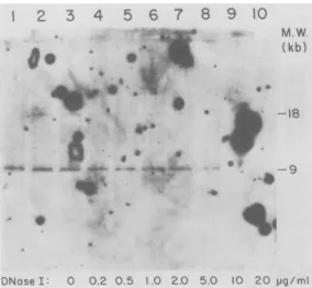
sensitivity of an individualprovirus was to use the 5'adjacentcellprobedescribed inFig.4for the clone 80proviral integration in A9 cells. In Fig. 7 the same DNAsamples as in Fig. 5 and 6 were digested withEcoRIand analyzedby elec-trophoresis and gelhybridization with nick-translated clone 80 5' adjacent cell probe. As in Fig. 4B, the integration at 18 kb and the host site at 9 kb were evident in undigested A9 DNA. Examination of the DNaseI-digestedDNA
sam-ples indicated that the clone 80 integration did notcontainhypersensitivesites,since itwasstill
detectable in the DNA samplewhich had been
digested with 2 ,ug of DNase I per ml. This
integration therefore appearedtobe inadomain
of intermediate DNase I sensitivity. Also indi-cated was that the 9-kbfragment corresponding
totheclone 80 host site was in DNaseI-resistant chromatin.
DISCUSSION
In the results reported here the methylation state and DNase I sensitivity of chromatin con-taining M-MuLV proviral DNA and endogenous
MuLV-related DNA weretested inproductively infected mouse cells. The methylation statewas examined at nine sites distributed throughout the M-MuLV genome. In a cell line which contained 10 to 15 copies of integrated M-MuLV DNA, most if not all of the proviruses were not detectably methylated at these sites, whereas endogenous MuLV-related DNA was methylat-ed.
Chromatincontaining M-MuLVprovirusesor endogenous MuLV-related DNA showed three different types of DNase I sensitivity in these experiments. Endogenous MuLV-related se-quences were in theDNase I-resistant
configu-ration,consistent withtheir nontranscribed state in both infected anduninfected NIH-3T3 cells. In contrast, the integrated M-MuLV proviruses werein domains of greater DNase I sensitivity, since moderate digestion preferentially removed themin comparison to the endogenous MuLV-related sequences. Two individual M-MuLV proviruses were found to be even more sensitive to DNase I digestion than the bulk of the M-MuLV proviruses and may have contained DNaseI-hypersensitivesites. These three class-esof DNase I sensitivity (resistance, intermedi-ate sensitivity, and hypersensitivity) resemble those observed by Weintraub and co-workers
1
2
3
4
5
6
7
8
9
10
O
*^' [image:10.491.108.392.371.633.2]np**
... .t
::. :
js *\.
^ ::
- _.._;j
_
n
_ v
w
_;X$';u. _ _
_:
*as
,vi
M.W.
(kb)
.I
_<
~18
4do
0.4 I .i:-11-.I,
..4 ..,
.,*. .4
I'llo.
'4
ONose : 0 0.2 0.5 1.0 2.0 50 10 20
pg/mi
FIG. 7. DNase Isensitivity of the clone 80proviralintegration. ThesameDNAsamplesdescribed in the legend to Fig. 5 were digested with
EcoRI
and analyzed by gel hybridization, using the nick-translated recombinant clone 80 adjacent cellprobedescribed in thelegendtoFig.4.Numberof the lanesis thesame asin thelegendtoFig.6.Exposurewasfor 18days.M.W., Molecularweight.J.VIROL.
on November 10, 2019 by guest
http://jvi.asm.org/
for chromatin containing the globin genes of
chick cells (31, 42), and analogy with their experiments suggests that only the A9 cell M-MuLVproviral integrations with hypersensitive
sitesmight be transcribed. The suggestion that only M-MuLVproviralintegrations with DNase
I hypersensitive sites are transcribed awaits directtest, perhapsbythe technique ofinvitro
nuclear runoff transcription (42). Previous
ex-periments suggested that only a minority of
integrated M-MuLVproviruses wereexpressed in productively infectedfibroblasts (5, 13).
If the correlation between transcriptional
ac-tivity and DNase I sensitivity proves to be correctfor thissystem, these results suggest that
hypomethylation of exogenously infecting M-MuLV proviral DNA is not correlated with
transcriptional activity. Other factors may be regulating transcription in thesecells.
The putative lackof transcription ofa
major-ity ofthe M-MuLV proviruses inthe A9 cells
was also not due to a defect in theproviral DNA.
Six M-MuLV proviralorganizations have been
isolatedby recombinantDNAtechniques from the A9 cells, at least one ofwhich (clone 80)
lacks DNase I-hypersensitive sites.
Neverthe-less,allof these give rise to infectious M-MuLV upon DNAinfectioninto NIH-3T3 cells athigh efficiency(3; H. Fan and Y. C.Lin, unpublished data).
A general correlation between transcription
and hypomethylation has been documentedfor bothretroviraland host cellular genes (7, 10, 19, 32, 42). In the caseof retroviral genes,
endoge-nously transmitted retroviral copies which are
transcriptionally inactive have been found to be highly methylated (7, 28, 38; see Fig. 4). When
endogenous viral copies are expressed
(some-times as the result of reinfection), the proviral DNA is hypomethylated. Furthermore,
tran-scriptional activation ofavianendogenous retro-viruses (19), aswell as the mouse metallothio-ninegene(8), hasbeen achieved by treatment of
cells with5-azacytidine, an agentwhich blocks DNA methylation. However, the experiments reportedhere differfrom those of van der Putten et al. (38) andGroudineet al. (19) inthat they were performed on cells exogenously infected
with M-MuLV. M-MuLV DNA synthesized during exogenous infection is unmethylated, as shown by the fact that it can be cleaved by
methyl-sensitiverestriction enzymes (17, 39). In eucaryoticcellsthe DNAmethylationpatternis propagated during replication (24, 43). It is thus not surprising that the unmethylated M-MuLV DNAremained unmethylatedduring integration into the host chromosomal DNA. Rather, it is surprising that some exogenously acquired M-MuLVproviral copies may be transcriptionally inactive. The results reported here suggest that
whereashypomethylation of DNA may be nec-essaryfor transcriptional activity,
hypomethyla-tionaloneis notsufficienttocauseit.
Another possible control for the transcription-al activity of exogenously acquired M-MuLV proviruses is the transcriptional state or nature
ofthesurroundinghostchromatin.Inparticular,
proviral DNA might be expressed when integrat-ed into active (DNase I hypersensitive, hypo-methylated) hostchromatin, but not expressed if
integratedinto inactivechromatin.Sucha situa-tion has been suggested for integrated proviral
copiesof murine mammarytumorvirus
integrat-edinto rat HTC cells (14a). Itwill bepossibleto test this when a lambdarecombinant clonefrom
the M-MuLVclone A9 cellscontaininga
provi-ral DNA which is transcribed (DNase I
hyper-sensitive)isidentified. In fact,evidence
consis-tentwiththishypothesiswasfoundin theresults
reported here. For the clone 80integration (lack-ing DNase I-hypersensitive sites), the methyl-ation state and DNase I sensitivity of the
adja-centcellDNAaswellasin the host site without
integratedproviral DNAcouldbe deduced. The hostsite was resistant to DNase Idigestion(Fig.
7), and a Sall site in the host DNA near the
integration remained methylated (Fig. 4B). In the caseofthe two DNase I-hypersensitive M-MuLV integrations identified in Fig. 6, both contained unmethylated ClaI sites in the 5'
adjacent cell DNA relatively close to the inte-gratedprovirus.
ACKNOWLEDGMENTS
Wethank JenniferPrice,Ying-Chih Lin,andJudyeGarfield for technical assistance and the members of the Tumor VirologyLaboratory for valuable advice anddiscussion.
P.-E.M. wassupported byapostdoctoralfellowshipfrom theSwiss National Science Foundation.This workwas sup-ported by Public Health Service grants CA 15747and CA 28616 andcoregrantCA14195,all fromtheNational Cancer Institute.
LITERATURECITED
1. Bacheler,L. T., and H. Fan.1979. Multiple integration sites forMoloney murine leukemia virus inproductively infectedmousefibroblasts. J. Virol. 30:657-667. 2. Bacbeler, L.T., and H. Fan. 1980. Study of integrated
Moloneymurineleukemia virusDNAusing complemen-taryDNAwhich doesnotrecognize endogenous related sequences. J.Virol.33:1074-1082.
3. Bacbeler,L. T., and H. Fan. 1981.Isolation of
recombi-nantDNAclonescarrying completeintegratedproviruses ofMoloneymurine leukemia virus. J. Virol. 37:181-190. 4. Benvenlste,R.E., G. J. Todaro,E.M.Scolnck,andW. P.
Parks. 1973. Partialtranscriptionof murinetype C viral genomes in BALB/ccelllines. J. Virol. 12:711-720. 5. Breindl, M., L.Bacheber,H.Fan,and R.Jaenisch.1980.
Chromatin conformation ofintegrated Moloneyleukemia virus DNA sequencesintissuesof BALB/Mo mice and in virus-infectedcelllines. J. Virol.34:373-382.
6. Chen,C.W., and C.A.Thomas. 1980. Recovery of DNA segments fromagarose gels.Anal.Biochem.101:339-341. 7. Cohen,J. C. 1980.Methylationofmilk-borneand geneti-cally transmitted mousemammarytumorvirusproviral
on November 10, 2019 by guest
http://jvi.asm.org/
AND
DNA. Cell 19:653-662.
8. Compere, S. J., and R. D.Palmlter. 1981. DNA methyl-ation controls the inducibility of the mouse metallothio-nine-1 gene inlymphoid cells.Cell 25:233-240. 9. Denhardt, D. T. 1966. A membrane-filter technique for the
detection of complementary DNA. Biochem. Biophys. Res. Commun.23:641-46.
10. Desrosiers, R., C. Mulder, and B. Fleckenstein. 1979. Methylation of Herpesvirus saimiri DNA in lymphoid tumor cell lines. Proc.Natl.Acad. Sci. U.S.A. 76:3839-3843.
11. Dolberg, D. S., L. T. Bacheler, and H. Fan. 1981. Endoge-nous type C retroviral sequences of mice areorganized in a small number of virus-like classes and have been acquired recently. J. Virol. 40:96-106.
12. Fan, H. 1977. RNA metabolism of murine leukemia virus: size analysis of nuclear pulse-labeled virus-specific RNA. Cell11:297-305.
13. Fan, H., R. Jaenisch, and P. MacIsaac. 1978. Low-multiplicity infection of Moloney murineleukemia virus in mouse cells: effect onnumber of viral DNA copies and virus production inproducer cells. J. Virol. 28:802-809. 14. Fan, H., and I. M. Verma. 1978. Size analysis and
relationship of murine leukemia virusspecific mRNA's: evidence fortransposition ofsequences during synthesis and processing of subgenomic mRNA. J. Virol. 26:468-478.
14a.Feinstein, S., S. Ross, and K. R.Yamamoto.1982. Chro-mosomal position effects determine transcriptional poten-tial of integrated mammary tumor virus DNA. J. Mol. Biol. 156:549-566.
15. Fuhrman, S. A., C. van Beveren, and I. M.Verma. 1981. Identification of a RNApolymerase II initiation site in the long terminal repeat of Moloney murineleukemia virus DNA. Proc. Natl.Acad. Sci.U.S.A. 78:5411-5415. 16. Glanni, A. M., J. R. Hutton, D. Smotkin, and R. A.
Weinberg. 1976. Proviral DNA of Moloney leukemia virus: purification and visualization. Science 191:569-571. 17. Gilboa,E., S. Goff, A.Shields, F. Yoshimura, S. Mitra, and D. Baltimore. 1979. In vitro synthesis of a 9 kbp terminally redundant DNA carrying the infectivity of Moloney murine leukemia virus. Cell 16:863-874. 18. Grippo,P., M.Iaccarino,E.Parlis, and E. Scarano. 1968.
Methylation of DNA indeveloping sea urchin embryos. J. Mol. Biol. 36:195-208.
19. Groudine, M., R. Elsenman, and H. Weintraub. 1981. Chromatin structure ofendogenousretroviral genes and activation by aninhibitor of DNA methylation. Nature (London) 292:311-317.
20. Hsu, T. N.,J.L.Sabran, G. E. Mark, R. V.Guntaka, and J.M. Taylor. 1978.Analysis ofunintegrated avian RNA tumor virusdouble-stranded DNA intermediates. J. Virol. 28:810-818.
21. Jaenisch,R.,D. Jahner, P.Nobis,I.Simon,J.Sohler, K. Harbers, and D. Grotkopp. 1981. Chromosomal position and activation of retroviral genomes inserted into the germ line ofmice. Cell24:519-529.
22. Mandel,J.L., andP. Chambon. 1979. DNA methylation: organ specificvariations in the methylationpatternwithin and aroundovalbumin and other chicken genes. Nucleic Acids Res. 7:2081-2103.
23. McGhee, J. D., W. I. Wood, M. Dolan,J.D.Engel, and G. Felsenfeld. 1981. A 200 base pair region at the 5' end of the chicken adult 0-globin gene is accessible to nuclease digestion. Cell 27:45-55.
24. Poilack, Y., R. Stein, A. Razin, and H. Cedar. 1980. Methylation of foreign DNA sequences in eukaryotic cells. Proc. Natl. Acad. Sci. U.S.A.77:6463-6467. 25. Razin,A., and A. D.Riggs. 1980. DNAmethylation and
genefunction. Science210:604-610.
26. Rigby,P. W. J., M.Dieckmann, C.Rhodes,andP. Berg. 1977. Labeling deoxyribonucleic acid to high specific activity in vitro by nicktranslationwith DNApolymerase I. J.Mol. Biol. 113:237-251.
27. Ringold,G.M.,P. R.Shank, H. E. Varmus,J. Ring,and K.R. Yamamoto. 1979. Integration and transcription of mousemammary tumorvirusDNAinrathepatoma cells. Proc. Natl. Acad. Sci. U.S.A. 76:665-669.
28. Shank,P.R., S. H.Hughes,H.-J. Kung,J.E.Majors,N. Quintrell, R. V.Guntaka, J.M.Bishop,and H. E. Var-mus. 1978. Mapping unintegrated avian sarcoma virus DNA:termini of linear DNA bear 300nucleotides present once or twice in two species of circular DNA. Cell 15:1383-1395.
29. Shinnick, T. M., R. Lerner, and J.G. Sutcliffe. 1981. Nucleotide sequence of Moloney murineleukemia virus. Nature(London) 293:543-548.
30. Shinnick, T. M., E. Lund,0. SmithIes, and F. A. Blattner. 1975. Hybridizationof labeled RNA to DNA in agarose gels.Nucleic Acids Res. 2:1911-1929.
31. Stalder, J., M. Groudine, J. B.Dodgsen,J.D.Engel, and H.Weintraub. 1980. Hemoglobin switching in chickens: evidence fortwoDNase Isensitive chromatin structures. Cell 19:973-980.
32. Sutter, D., and W. Doerfler. 1980. Methylation of integrat-ed adenovirus type 12 DNA sequences in transformintegrat-ed cells is inverselycorrelated with viral geneexpression. Proc. Natl. Acad. Sci. U.S.A.77:253-256.
33. Temin, H. M. 1981.Structure, variation and synthesis of retrovirus long terminal repeat. Cell 27:1-3.
34. Temin, H. M., and D. Baltimore. 1972. RNA-directed DNAsynthesis and RNAtumor viruses. Adv. Virus Res. 17:129-186.
35. Todaro, G.J.,and H.Green. 1963.Quantitative studies of the growth of mouse embryo cells in culture and their development into established lines. J. Cell Biol. 17:299-313.
36. van Beveren, C., J. G. Goddard, A. Berns, and I. M. Verma. 1980.Structure ofMoloney murine leukemia viral DNA:nucleotide sequence of the 5' long terminal repeat andadjacent cellular sequences. Proc. Natl. Acad. Sci. U.S.A.77:3307-3311.
37. van der Pkoeg, L. H. T., and R. A.Flavell. 1980. DNA methylation in the human ,-globin locus in erythroid and nonerythroid tissues. Cell 19:947-958.
38. van der Putten, H., W. Quint,J.vanRaaji,E. R. Maan-dag,1. M. Verma, and A.Berns. 1981.M-MuLV-induced leukemogensis: integration and structure ofrecombinant provirusesin tumors. Cell 24:729-739.
38a.Varmus, H., and R. Swanstrom. 1982. Replication of retroviruses, p. 369-512. In R. Weiss, N. Teich, H. Varmus, and J. Coffin (ed.), RNA tumor viruses. Cold Spring HarborLaboratory, ColdSpringHarbor,N.Y. 39. Verma, I. M., and M. MacKennett.1978. Genome
organi-zation of RNA tumor viruses. II. Physical maps of in vitro-synthesized Moloney murineleukemia virus double-stranded DNA by restriction endonucleases. J. Virol. 26:630-645.
40. Wahl, G. M., M.Stern,andG. R. Stark. 1979. Efficient transfer oflarge DNA fragments from agarose gels to
diazobenzyloxymethyl-paper andrapidhybridization
us-ing dextran sulfate. Proc. Natl. Acad. Sci. U.S.A. 76:3683-3687.
41. Weintraub, H., and M. Groudine. 1976. Chromosomal subunits in active genes have an altered conformation. Science 193:848-856.
42. Weintraub, H., A. Larsen, and M. Groudlne. 1981.
a-Globin-geneswitchingduringthedevelopmentof chicken embryos: expression and chromosome structure. Cell 24:333-344.
43. Wigler,M., D.Levy,andM.Perucho. 1981. The replica-tionof DNAmethylation. Cell 24:33-40.
44. Wu, C.1980. The 5'endsofDrosophilaheat shock genes in chromatin are hypersensitive to DNase I. Nature (London) 286:854-860.
45. Wu, C., and W. GUbert. 1981.Tissue-specificexposure of chromatin structureatthe 5'terminus of the rat preproin-sulin IIgene. Proc. Natl. Acad.Sci. U.S.A. 78:1577-1580.
J.VIROL.
on November 10, 2019 by guest
http://jvi.asm.org/