R E S E A R C H A R T I C L E
Open Access
Contralateral breast cancer can represent a
metastatic spread of the first primary
tumor: determination of clonal relationship
between contralateral breast cancers using
next-generation whole genome sequencing
Sara Alkner
1,2*†, Man-Hung Eric Tang
1†, Christian Brueffer
1, Malin Dahlgren
1, Yilun Chen
1, Eleonor Olsson
1,
Christof Winter
1, Sara Baker
1, Anna Ehinger
1,3, Lisa Rydén
1,4, Lao H. Saal
1, Mårten Fernö
1†and Sofia K. Gruvberger-Saal
1*†Abstract
Introduction:By convention, a contralateral breast cancer (CBC) is treated as a new primary tumor, independent of the first cancer (BC1). Although there have been indications that the second tumor (BC2) sometimes may represent a metastatic spread of BC1, this has never been conclusively shown. We sought to apply next-generation
sequencing to determine a“genetic barcode”for each tumor and reveal the clonal relationship of CBCs.
Methods:Ten CBC patients with detailed clinical information and available fresh frozen tumor tissue were studied. Using low-coverage whole genome DNA-sequencing data for each tumor, chromosomal rearrangements were enumerated and copy number profiles were generated. Comparisons between tumors provided an estimate of clonal relatedness for tumor pairs within individual patients.
Results:Between 15–256 rearrangements were detected in each tumor (median 87). For one patient, 76 % (68 out of 90) of the rearrangements were shared between BC1 and BC2, highly consistent with what has been seen for true primary-metastasis pairs (>50 %) and thus confirming a common clonal origin of the two tumors. For most of the remaining cases, BC1 and BC2 had similarly low overlap as unmatched randomized pairs of tumors from different individuals, suggesting the CBC to represent a new independent primary tumor.
Conclusion:Using rearrangement fingerprinting, we show for the first time with certainty that a contralateral BC2 can represent a metastatic spread of BC1. Given the poor prognosis of a generalized disease compared to a new primary tumor, these women need to be identified at diagnosis of CBC for appropriate determination of treatment. Our approach generates a promising new method to assess clonal relationship between tumors. Additional studies are required to confirm the frequency of CBCs representing metastatic events.
* Correspondence:[email protected];[email protected]
†Equal contributors
1
Division of Oncology and Pathology, Clinical Sciences Lund, Lund University, MV 404-B2, Lund SE-22381, Sweden
Full list of author information is available at the end of the article
© 2015 Alkner et al.Open AccessThis article is distributed under the terms of the Creative Commons Attribution 4.0 International License (http://creativecommons.org/licenses/by/4.0/), which permits unrestricted use, distribution, and reproduction in any medium, provided you give appropriate credit to the original author(s) and the source, provide a link to the Creative Commons license, and indicate if changes were made. The Creative Commons Public Domain Dedication waiver (http://creativecommons.org/publicdomain/zero/1.0/) applies to the data made available in this article, unless otherwise stated. Alkneret al. Breast Cancer Research (2015) 17:102
Introduction
Contralateral breast cancer (CBC) is today treated as a new primary tumor, independent of the first breast can-cer (BC1). However, whether the second tumor (second breast cancer (BC2)) always is a new independent pri-mary breast cancer, or in some cases could represent a metastatic spread from BC1, remains unclear. Support-ing this possibility are recent studies showSupport-ing the lymph node status of BC1 to influence risk of CBC and the time interval between BC1 and BC2 to affect prognosis after CBC [1–4]. If some CBCs are in fact metastatic events, today’s clinical standard of diagnostic work-up and the treatment of patients with CBC as metastasis would need to be revised.
Various characteristics such as X-chromosome inactiva-tion status,p53mutations, partial allelotyping, microsatel-lite instability, and comparative genomic hybridization (CGH) analysis have been used to compare BC1 and BC2 in CBC patients [5–12]. These previous studies suggest that the majority of CBCs appear clonally independent from BC1, although some have features similar enough to the primary tumor that they could potentially represent a metastatic spread [6, 8–10, 13]. However, prior studies utilized methods and techniques that are limited in resolution and were generally not specific enough to dem-onstrate an unequivocal clonal relationship between BC1 and BC2. For example, while a differing X-chromosome inactivation status indicates two distinct progenitor cells, a similar status must be considered noninformative [13]. Finding the samep53mutation in two tumors could indi-cate a similar origin [6]. However thep53gene has muta-tional hotspots, germline mutations are possible, and certain subtypes, such as basal-like tumors, have a greatly elevatedp53mutation rate [14], presenting a risk for mis-classifications. Consequently, neither X-chromosome in-activation status nor p53 mutation analysis can with certainty confirm a clonal relationship [5, 6, 12].
Using array CGH, bilateral cancers have been shown to share genomic imbalances [9, 10]. This could indicate a clonal relationship, but another possibility is that their development in a common milieu and genetic background could lead to pathogenetically similar tumors that are in fact independent cancers. Indeed, a high similarity of allelic imbalances has been seen in simultaneously developing breast cancers in monozygotic twins, and allelotypes of CBCs developing synchronously were found to be more alike than metachronous CBCs [7, 15]. Furthermore, the gene expression-based in-trinsic subtypes of breast cancer can largely be reca-pitulated using DNA copy number variation (CNV) profiles [16], presenting a risk that nonclonal tumors or even those from different patients but of the same intrinsic subtype could appear similar by CNV profiles.
Recent studies have shown that breast cancer genomes are highly disorganized and can harbor tens to hundreds of chromosomal rearrangements [17, 18]. Interestingly, chromosomal rearrangements in breast cancer appear to be highly specific, and thus the set of rearrangements in any given tumor can serve as unique “fingerprint” or “barcode” of that tumor. This is in contrast to somatic point mutations, for which there are recurrent mutations across several genes that may be shared between tumors in unrelated persons. Importantly, it has been shown that extensive clonal diversity exists for point mutations within the same breast tumor mass, in contrast to struc-tural chromosomal changes [19]. Therefore, chromo-somal rearrangements are ideal for studies of clonal origin.
Until recently, however, a lack of feasible and compre-hensive methods to study chromosomal rearrangements has prevented this chromosomal approach from being fully utilized. With the recent advancement of next-generation sequencing, global characterization of chromo-somal rearrangements is now possible. Herein we have characterized the genomes of contralateral breast tumors using whole genome sequencing (WGS) to enumerate chromosomal breakpoints, serving as unique tumor bar-codes, to determine clonal relationships between CBCs. For the first time we show with certainty that CBC can represent a metastatic spread of the first tumor. Given the poor prognosis of a generalized disease compared with a new primary tumor, these women need to be identified at diagnosis of CBC for appropriate determination of treat-ment and diagnostic work-up. We hereby present a prom-ising method for determining the clonal relationship between tumors that may be clinically applicable in the near future.
Methods Patients
handled confidentially according to Sweden’s Personal Data Act.
Whole genome sequencing and data analysis
Using DNA extracted from frozen tumor samples and normal blood samples, whole-genome paired-end Illumina (San Diego, CA, USA) sequencing libraries were generated, sequenced, and aligned to the human reference genome (see Additional file 1).
Sequencing data were analyzed bioinformatically to identify chromosomal rearrangements in each tumor and normal DNA sample (see Additional file 1). Initial filtering removed centromeric, segmentally duplicated re-gions, and intrachromosomal rearrangements <7 kb. Germline rearrangements (both ends matching within ±1 kb in any normal sample) were removed. For every com-parison between two tumors (whether from the same pa-tient or between papa-tients), each rearrangement was classified as either shared or specific to one tumor as fol-lows: rearrangements were considered shared between two tumors if the genomic coordinates for both ends of the rearrangement matched within ±500 base pairs; or to compensate for potential tumor subclonality (in case a re-arrangement was present only in a subclone and therefore represented by fewer reads), each remaining rearrange-ment in a tumor was computationally“looked-up” in the aligned sequence BAM file of the other tumor, and if≥1 read pairs matched within ±1 kb on both ends then the re-arrangement was classified as shared. In addition, all rear-rangements looked up and supported by at least one read pair in the normal samples were removed. The combined shared rearrangement fraction was calculated as the num-ber of shared rearrangements divided by the numnum-ber in the union of all rearrangements found in the compared tumors.
CNV was evaluated from the WGS data. CNV profiles were compared between samples based on windows delineated by the union of their segmentation breaks taking into account both copy number “state” (gain, normal, or loss) and“slope”, corresponding to the differ-ence between the current and previous window’s state (see Additional file 1). Windows with the same state and slope were considered shared. The fraction of shared ab-normal CNV events between two tumors was calculated after excluding windows with a normal diploid state in both tumors.
The rearrangements of each tumor were plotted using Circos [20], and rearrangement and CNV profiles were drawn using standard R graphical libraries [21]. Detailed methods are available in Additional file 1.
PCR validation of rearrangements
Rearrangements were validated with conventional PCR. Using our in-house SplitSeq bioinformatics pipeline, the
local sequence around each breakpoint was retrieved [22]. Rearrangements for validation were randomly selected from all enumerated rearrangements of four patients (Patients 2, 6, 8, and 9). Primers were designed and touchdown PCR was performed on DNA extracted from the BC1 and BC2 (further details in Additional file 1). As germline control, matched normal DNA was used where available (Patient 9) or a normal DNA pool was created from normal lymphocyte DNA extracted from 44 healthy individuals.
Results Clinical features
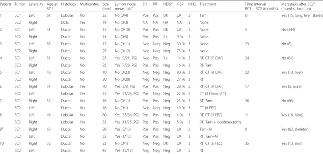
Tumor and treatment characteristics are presented in Table 1. Patient 10 was a known BRCA1 mutation car-rier. Patient 3 had a local recurrence from BC2 7 months after diagnosis of CBC, treated with surgery and endocrine therapy. Patient 1 had a local recurrence after BC1 12 months before diagnosis of BC2, treated with surgery, radiotherapy, and endocrine therapy. None of the other patients had any local/regional recurrences. Patient 4 developed mucosa-associated lymphoid tissue lymphoma 61 months after BC2. To avoid the risk of misdiagnosing metastases past this date, the diagnosis date of the lymphoma was considered the last follow-up date. For Patients 1 and 7, BC2 was diagnosed during endocrine therapy.
Paraffin material was available for both tumors in Patients 3, 4, 5, 6, 7, and 8 and for BC1 in Patient 2. Here histopathological markers have been reevaluated with immunohistochemistry by a pathologist. The cutoff value for estrogen receptor (ER)/progesterone receptor (PR)-positivity was >10 % positive cells. For the remaining tumors these data are from the patient’s chart. For most of these patients (Patients 1, 9, and 10) ER and PR were measured with immunohistochemistry. For Patient 2 BC1 ER and PR were measured in cytosol (ER 140 fmol/mg protein, PR 170 fmol/mg protein; cut-off value for ER/PR-positivity = 25 fmol/mg protein).
Chromosomal rearrangements
WGS was performed on the 10 CBC pairs employing a strategy that combined low sequence coverage paired-end sequencing with larger fragment sizes for improved phys-ical genome coverage. The median sequence coverage was 6.5 (range 1.8–11.2) and the median physical coverage was 14.7 (range 6.9–28.4) (Additional files 1 and 2). The total number of rearrangements detected in each tumor varied widely, from 15 to 256 with a median of 87 per tumor (Fig. 1a, Table 2; all plots in Additional file 3).
We were first interested in ascertaining how often two unrelated tumors from different individuals display iden-tical chromosomal rearrangements because this would
Table 1Clinical characteristics
Patient Tumor Laterality Age at BC1
Histology Multicentric Size (mm)
Lymph node metastasisa
ER PR HER2b Ki67 NHG Treatment Time interval
BC1–BC2 (months)
Metastasis after BC2c
(months, location)
1 BC1 Left 61 Lobular No 32 No (0/4) Pos Pos UK UK 2 Tam 61 Yes (15, lung, liver, skeleton)
BC2 Right DCIS Yes 14 No (0/3) NA NA NA NA 3 None
2 BC1 Left 41 Ductal No 15 No (0/10) Pos Pos UK UK 2 None 5 No (249)
BC2 Right Ductal No 14 No (0/5) Pos Pos 2+ 9 % 3 None
3 BC1 Left 83 Ductal No 17 No (0/11) Neg Neg Neg 30 % 3 None 23 No (8)
BC2 Right Ductal No 25 No (0/12) Neg Neg Neg 75 % 3 None
4 BC1 Left 51 Ductal No 25 Yes (9/21, PG) Neg Pos 3+ 14 % 3 RT, CT (7 CMF) 24 No (61)
BC2 Right Ductal No 25 Yes (7/28, PG) Pos Pos Neg 16 % 3 RT, Tam
5 BC1 Left 43 Ductal No 10 No (0/23) Neg Neg Neg 80 % 3 RT, CT (9 CMF) 22 Yes (13, liver)
BC2 Right Ductal No 20 No (0/20) Neg Neg Neg 27 % 3 RT
6 BC1 Right 51 Lobular Yes 70 Yes (3/8, PG) Pos Pos Neg 28 % 2 RT, CT (9 CMF) 17 Yes (5, brain)
BC2 Left Lobular Yes 12 Yes (23/26, PG) Pos Neg Neg 22 % 2 CT (3 Doxo–2 T)
7 BC1 Right 52 Ductal No 16 No (0/11) Pos Pos Neg 21 % 3 RT, Tam 30 No (66)
BC2 Left Ductal No 16 No (0/1) Neg Neg Neg 40 % 3 CT (6 FEC)
8 BC1 Left 46 Lobular No 80 Yes (33/34, PG) Pos Pos Neg 4 % 3 RT, CT (9 FEC) 11 Yes (16, lung)
BC2 Right Lobular Yes 55 Yes (11/25, PG) Pos Pos Neg 5 % 2 RT, Tam + oophorectomy
9d BC1 Right 63 Ductal No 28 Yes (2/13) Pos Pos Neg UK 2 Tam–AI 0 Yes (62, skeleton)
BC2 Left Ductal No 55 Yes (1/12) Pos Pos Neg UK 3 RT, Tam–AI
10 BC1 Right 32 Ductal No 23 No (0/1) Neg Neg UK UK 3 RT, CT (6 FEC) 35 Yes (13, skin)
BC2 Left Ductal No 45 Yes (12/12) Neg Neg Neg UK 2 RT
AIaromatase inhibitor,BC1first breast cancer,BC2second breast cancer,CMFcyclophosphamide, methotrexate and fluorouracil,CTchemotherapy (cycles and regime used in parentheses),DCISductal carcinoma in situ,Doxodoxorubicin,ERestrogen receptor,FECfluorouracil, epirubicin, and cyclophosphamide,HER2human epidermal growth factor receptor 2,NAnot applicable,Negnegative,NHGNottingham histological grade,
PGperiglandular growth,Pospositive,PRprogesterone receptor,RTradiotherapy,Tdocetaxel,Tamtamoxifen,UKunknown
a
Number of positive lymph nodes/number of investigated nodes in parentheses
b
HER2 determined with immunohistochemistry (Herceptest) where score 0–1 has been classified as negative. For score 2+ and 3+ the individual score is given in the table
c
If the patient developed metastases, the time interval between BC2 and diagnosis of metastasis (months) and the site of the first metastasis is given within parentheses. In patients who do not develop metastases, the follow-up period (months) is given within parentheses
d
Patient 9 was diagnosed with her left and right breast cancer simultaneously (synchronous contralateral breast cancer), hence it cannot be said which tumor was first
Breast
Cancer
Research
(2015) 17:102
Page
4
of
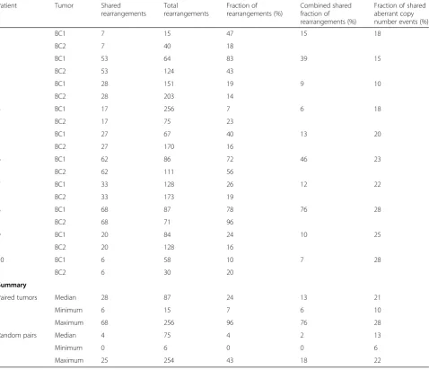
be an indication of overlap occurring by chance or due to incomplete filtering of germline events. For this, all possible pairings of tumors between patients (excluding the true within-patient pairing) were exhaustively com-pared. This comparison showed that 0–25 rearrange-ments (median 4) matched between any two random tumors with a combined shared fraction (i.e., the frac-tion of rearrangements shared between BC1 and BC2 di-vided by the union of all rearrangements detected in both tumors) ranging from 0 to 18 % (median 2 %; Table 2). For the comparisons of matched patient CBCs, on the other hand, the number of shared rearrange-ments between BC1 and BC2 ranged between 6 and 68 (median 28) and the combined shared fraction varied be-tween 6 and 76 % (median 13 %).
Patient 8 was a clear outlier and had the most similar CBCs, with 76 % shared rearrangements between the two tumors (68 shared rearrangements), 19 rearrange-ments unique to BC1, and three rearrangerearrange-ments unique to BC2 (Fig. 1a, Table 2). Patient 8’s extreme degree of similarity of chromosomal rearrangements unequivocally supports BC2 being the result of a metastatic spread from BC1. Furthermore, in a separate study of matched primary and distant metastases using the same experi-mental approach, we have found that the average pri-mary breast cancer shares well over one-half of its chromosomal rearrangements with the distant metasta-sis it seeds (results not shown; Tang and Gruvberger-Saal, manuscript under review). Therefore, the 76 % shared rearrangements between CBCs in Patient 8 is highly consistent with known primary-metastasis pairs.
Seven of the remaining CBC patients had much fewer shared rearrangements between BC1 and BC2 with com-bined shared fractions between 6 and 15 % (median 10 %), most consistent with these CBCs having developed as inde-pendent primary tumors (Fig. 1a, Table 2; Additional file 3). However, Patients 2 and 6 had a rearrangement overlap (39 % and 46 %) between that seen for unmatched randomized tumor pairs and true tumor-metastasis pairs (including Patient 8). The cases of Patients 2 and 6 are highly suspicious for contralateral metastasis, but to classify them with certainty normal DNA samples would be needed to rule out any remaining germline rearrangements. Interestingly, for Patient 1, where BC2 was an in-situ lesion and included in the study as a CBC pair presumably representing two independent le-sions, 15 % of rearrangements matched between the tu-mors. This overlap was considerably lower than the overlap for Patient 8 (76 %) but well within the range between CBC pairs when excluding the three most similar patients (Patients 2, 6, and 8) (6–13 %).
The similarities and differences in chromosomal rear-rangements between tumors are well illustrated in a barcode plot, where all nonredundant identified rear-rangements are arranged and their presence in each tumor is denoted by a line (Fig. 2). For example, the two tumors from Patient 3 were highly eventful yet displayed distinct rearrangement barcodes spread across the whole genome (Figs 1a and 2). Many tumors exhibited localized areas of high density of rearrangements suggestive of one catastrophic shattering event (chromo-thripsis). Chromothripsis is thought to contribute to
P3
A
1 2 3 4 5 6 7 8 9 10 11 12 13 14 15 16 17 18 19 20 21 22 xB
1 5 10 15 20 X
Median ratio
P8
1 2 3 4 5 6 7 8 9 10 11 12 13 14 15 16 17 18 19 20 21 22 x1 5 10 15 20 X
P4
1 2 3 4 5 6 7 8 9 10 11 12 13 14 15 16 17 18 19 20 21 22 x1 5 10 15 20 X
0.5 1 2 3 0.5 1 2 3 0.5 1 2 3
Fig. 1aChromosomal rearrangements for three patients visualized in Circos plots.bCopy number profiles for three patients.Orangedenotes events (rearrangements or copy number traces) specific to BC1,bluedenotes events specific to BC2, andblackdenotes events shared between BC1 and BC2.P3Patient 3,P4Patient 4,P8Patient 8
[image:5.595.59.538.90.305.2]oncogene activation and tumor suppressor loss, and as a consequence is an important driving force for early cancer development and would probably be shared between clonal tumors. For Patient 8 this appears to be true; the two tumors share identical chromothripsis-like rearrangement patterns in two localized hotspots on chromosomes 10 and 11 (Figs 1a and 2). On chromosome 11 alone the two tu-mors share 38 identical intrachromosomal rearrangements, while only five are specific to BC1 and none to BC2. Patient 4, on the other hand, has localized areas on 1q, 6p, and chromosomes 17 and 18 of chromothripsis-like patterns that only appear in BC1, whereas BC2 has specific high-density areas on 6q. Patient 9, with a combined percentage of shared rearrangements of 10 %, has very distinct heavily rearranged areas on 11q and chromosome 12 unique to BC1, while BC2 has areas suggesting chromothripsis on
chromosomes 1, 3, and 11p that are not being shared with BC1, further indicative of their nonclonal origin.
A number of cancer-associated genes (e.g., RB1, RARA,CDKN2A, andPTEN) were found to be involved in the enumerated rearrangements [23], probably con-tributing to tumorigenesis (Additional file 4).
[image:6.595.60.539.101.513.2]Indicative of a suitable reliability of our analysis pipeline, 68 rearrangements out of 86 (79 %) were confirmed by PCR across the breakpoint junction as either specific to BC1, specific to BC2, or shared. However, nine of these re-arrangements (13 %) were also identified in the matched normal DNA, or if unavailable were identified in a pool of 44 normal DNA samples. Therefore, although our pipeline results in a good accuracy of calls based on the sequencing data, owing to the fact that matched normal DNA were unavailable for most patients and
Table 2Information on rearrangement and copy number variation
Patient Tumor Shared
rearrangements
Total
rearrangements
Fraction of rearrangements (%)
Combined shared fraction of rearrangements (%)
Fraction of shared aberrant copy number events (%)
1 BC1 7 15 47 15 18
BC2 7 40 18
2 BC1 53 64 83 39 15
BC2 53 124 43
3 BC1 28 151 19 9 10
BC2 28 203 14
4 BC1 17 256 7 6 18
BC2 17 75 23
5 BC1 27 67 40 13 20
BC2 27 170 16
6 BC1 62 86 72 46 23
BC2 62 111 56
7 BC1 33 128 26 12 22
BC2 33 173 19
8 BC1 68 87 78 76 28
BC2 68 71 96
9 BC1 20 84 24 10 25
BC2 20 128 16
10 BC1 6 58 10 7 28
BC2 6 30 20
Summary
Paired tumors Median 28 87 24 13 21
Minimum 6 15 7 6 10
Maximum 68 256 96 76 28
Random pairs Median 4 75 4 2 13
Minimum 0 6 0 0 6
Maximum 25 254 43 18 22
thus were not sequenced, our results are underestimating to some extent the contribution of rearrangements that are present in the germline.
Copy number variation
CNV profiles were also evaluated using the WGS data (Fig. 1b; Additional file 3). The fraction of shared ab-normal copy number events between CBC pairs ranged between 10 and 28 % (median 21 %; Table 2). When exhaustively comparing all pairings of tumors from dif-ferent patients, the fraction of shared aberrant copy number events ranged between 6 and 22 % (median 13 %), which was only slightly lower than that seen for the matching tumor pairs. Again, the CBCs from Pa-tient 8 had the highest fraction of shared aberrations (28 %) and the CNV profiles appeared similar (Fig. 1b; Additional file 3). Patient 1, with BC2 representing an in-situ lesion, shared 18 % of the aberrant copy number events between the two tumors.
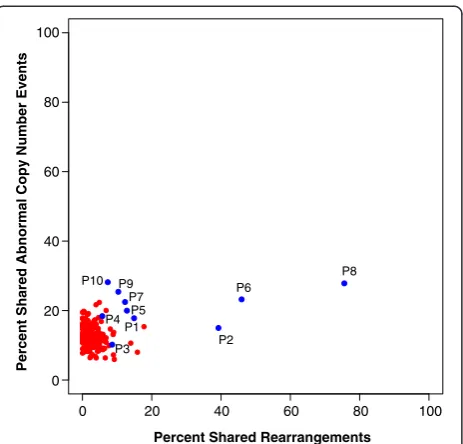
Clonal relationship between contralateral tumors
The analysis of genomic clonal similarities between tu-mors can be visualized in two dimensions (Fig. 3). When looking at chromosomal rearrangements, the majority of the CBC pairs (blue) cluster together just slightly off the cloud of randomly-paired tumors (red) (Patients 2 and 6 showed a somewhat higher similarity as discussed above). Patient 8, on the other hand, is clearly distantly separated from the other CBC pairs (including Patient 1 with an in-situ BC2) and from the randomized pairings.
For the CNV events this separation is not evident and no patient CNV profiles are more similar than some of the tumor pairings from different individuals. Based on the analysis of rearrangements, however, we can con-clude that BC2 from Patient 8 in fact is highly clonally related to BC1.
Discussion
By harnessing the power of WGS-detected tumor-specific chromosomal rearrangement barcodes, we have
Chromosome Color Coding
1 2 3 4 5 6 7 8 9 10 11 12 13 14 15 16 17 18 19 20 21 22 X
Rearranged
Chromosomes
[
1
2
3
4
5
6
7
8
9
10
Patient Tumor
BC1 BC2
BC1 BC2
BC1 BC2
BC1 BC2
BC1 BC2
BC1 BC2
BC1 BC2
BC1 BC2
BC1 BC2
BC1 BC2
All Chromosomal Rearrangements
Fig. 2Tumor genetic barcodes of chromosomal rearrangements for individual tumors. All nonredundant chromosomal rearrangements were included and their presence in each tumor denoted by a line. The rearrangements were plotted in the order of genomic location for the end of the fusion that appears first in the genome. Color coding below indicates which two chromosomes where involved in each rearrangement.BC1 first breast cancer,BC2second breast cancer
[image:7.595.57.540.91.427.2]for the first time conclusively demonstrated that breast cancer can arise as a metastatic clone from a tumor in the contralateral breast. Among the 10 CBC pairs in this study, the Patient 8 CBC pair exhibited 76 % rearrange-ment similarity between the left and right tumors. Our analyses showed that chromosomal rearrangements rarely recurred among randomly paired samples (median shared fraction, 2 %; median shared number, four rangements; Table 2), and hence chromosomal rear-rangements uniquely characterized the tumors. In a separate study of primary-metastasis pairs using the same experimental approach, we have found that the average primary breast cancer appears to share about 50 % or more of its chromosomal rearrangements with the distant metastasis it seeds (Tang and Gruvberger-Saal, manuscript under review). This is highly consistent with the genetic chromosomal rearrangement fingerprint of Patient 8, and thus we can confidently conclude a clonal relationship between BC1 and BC2 for this patient.
However, two patients showed intermediate levels of rearrangement similarity (39 and 46 %). This is higher than that seen in unmatched randomized pairing, but on the lower end of what we have seen for primary-metastasis pairs (including Patient 8). A limitation in our study is the unavailability of matching normal DNA for every CBC patient. WGS analysis of matching nor-mal DNA would have allowed for complete filtering of all germline chromosomal-rearrangement events and CNVs. Although the vast majority of germline events
have been removed by our data filtering steps and by comparison with unmatched normal DNA samples from the Swedish population, matched normal DNA would probably have clarified the clonal relationship in these two uncertain cases. Nevertheless, the purpose of this study was to show that contralateral metastasis does exist and can be reliably detected with newer technolo-gies. Owing to the extreme degree of similarity of chromosomal rearrangements in Patient 8, there is no doubt of the clonal relationship between BC1 and BC2 in this patient. Further studies are needed, however, to determine the prevalence of “metastatic CBC”, optimal diagnostic work-up and treatment. In addition, further comparisons between known primary-metastasis pairs versus unrelated tumor pairs are required to find the op-timal cutoff value for the fraction of shared rearrange-ments indicating clonality, and to make sure that all CBCs actually representing metastatic events are cor-rectly identified.
Clinically, CBC events are today considered to be two separate tumors, and adjuvant treatment is recom-mended on the basis of the clinicopathological charac-teristics of BC1 and BC2 individually. However, since metastatic breast cancer has worse prognosis than and a different treatment regimen from that for localized breast cancer, our finding gives a new important per-spective on the clinical management for women diag-nosed with CBC. A more careful diagnostic work-up is necessary in order to determine an appropriate treat-ment strategy, and we have developed a novel approach to ascertain the metastasis status of a CBC that is afford-able and can be performed in a clinically relevant time frame. In addition, access to normal DNA would not be a problem in the clinic, increasing sensitivity and specifi-city of the analysis.
Whether a patient with a contralateral metastasis has an equally poor prognosis as a patient with distant me-tastases at other sites (lung, liver, brain, etc.) or whether the outcome associated with a contralateral metastasis, if treated correctly, could be more similar to that of a local or regional recurrence are questions that deserve further study. If a contralateral metastasis represents a cancer still prone to thriving mainly in breast tissue, or possibly a local lymphatic spread to the contralateral breast, we may with the right treatment still be able to prevent fur-ther spread and a fatal outcome. If, on the ofur-ther hand, a contralateral metastasis is already a sign of an incurable metastatic disease, treatment may instead be focused on quality of life rather than intense adjuvant therapy.
Among our 10 patients, six developed generalized dis-ease within 5–62 months from BC2. Patient 8, in whom our analysis determined BC2 to be a contralateral metas-tasis, was diagnosed with lung metastases 16 months after BC2. Since she was treated with radiotherapy, tamoxifen,
0 20 40 60 80 100
Percent Shared Rearrangements 0
20 40 60 80 100
Percent Shared Abnormal Copy Number Events
P2 P3
P8
P4 P5
P6 P7
P1 P9 P10
[image:8.595.59.291.87.309.2]and oophorectomy for BC2, the endocrine treatment given could have helped in delaying development of fur-ther metastases. Our study is too small to draw any con-clusions regarding prognosis after contralateral metastasis, but it is an important question with significant clinical im-plications, which needs to be further evaluated.
An important issue is how one may identify patients with a high risk of BC2 representing a metastasis instead of a new primary tumor. Are there clinical indications as to when contralateral metastasis should be suspected and further investigation with WGS is warranted? Patient 8 had a very large BC1 (80 mm), widespread lymph node metastasis, and both tumors were lobular type and had similar hormone receptor, HER2, and Ki67 expression. Further studies will show whether these characteristics could be indicators of a risk for contralateral metastasis. Of note, the Nottingham histological grade of Patient 8 was classified as grade 3 in BC1 and grade 2 in BC2. This is particularly interesting since, using the traditional cri-teria summarized by Chaudary et al. [24], bilateral carcin-omas are considered independent if: the subsequent tumor has an in-situ component; or the lesions are of dis-tinct histological subtypes; or the subsequent cancer has a better degree of differentiation; or there is no evidence of local, regional, or distant metastases from the ipsilateral lesion. Clearly, these clinical criteria do not accurately de-tect the contralateral metastasis in our material. Further-more, for seven of the remaining eight invasive CBC pairs, the CBCs shared the same histological subtype and did not have a better degree of differentiation for BC2 (data on in-situ component unknown) and would consequently be classified as possible metastases. Routine pathological markers do not therefore appear to adequately separate new primaries from contralateral metastases.
Since CBC tumors arise and develop in the same gen-etic and environmental background, and gengen-etic predis-positions such as a germline BRCA1 mutation (also associated with basal-like tumors) are more common [25], it is conceivable that bilateral tumors more often will be of the same intrinsic subtype and may accumu-late similar patterns of mutations and copy number ab-errations, even when they arise as independent tumors. Indeed, while CNV profiles carried some information on genomic similarities in our data, none of the matched pairs had significantly more similar CNV profiles than some unrelated tumors. However, using the rearrange-ment barcodes, even Patient 10, a germlineBRCA1 mu-tation carrier, shared very few rearrangements between BC1 and BC2 and a different clonal origin could be de-termined (Additional files 3 and 4).
Conclusion
By using next-generation sequencing and an analytical strategy based on chromosomal rearrangements, we can
for the first time show strong evidence that some CBCs indeed represent a metastatic spread of BC1. Our ap-proach generated a unique tumor barcode that can as-sess the clonal relationship between tumors. This is a promising new method not only for management of CBCs, but also in a variety of other cancer types where the question of clonality and tumor heterogeneity raises important clinical issues. Although our study had the disadvantage of a lack of matched normal DNA for most patients, this would not be a problem in the clinical set-ting, allowing for complete filtration of germline defects and increasing specificity and sensitivity of the method. Further studies are needed in order to identify the opti-mal cutoff level to with certainty not miss any CBCs ac-tually representing a metastatic event.
CBCs are today treated as two individual tumors, but if BC2 instead represents a metastatic disease state the patient would have a worse prognosis and require a dif-ferent treatment than a new primary breast cancer. With intensified treatment for these patients there may be a possibility to prevent further spread, and avoid develop-ment of a generalized incurable breast cancer.
Additional files
Additional file 1:presents a detailed description of supplemental methods.(DOCX 30 kb)
Additional file 2:is Table S1 presenting sequencing statistics. (DOCX 14 kb)
Additional file 3:is Figure S1 showing chromosomal
rearrangements visualized using Circos and copy number profiles for all 10 CBC patients.Orangedenotes events (rearrangements or copy number traces) specific to BC1,bluedenotes events specific to BC2, andblackdenotes events shared between BC1 and BC2.PPatient. (PDF 1524 kb)
Additional file 4:is Table S2 presenting a list of genes affected by rearrangements and found in the COSMIC Catalogue of somatic mutations in cancer [26].(XLS 116 kb)
Abbreviations
BC1:First breast cancer; BC2: Second breast cancer; CBC: Contralateral breast cancer; CGH: Comparative genomic hybridization; CNV: Copy number variation; ER: Estrogen receptor; PR: Progesterone receptor; WGS: Whole genome sequencing..
Competing interests
The authors declare that they have no competing interests.
Authors’contribution
SA, M-HET, LR, LHS, MF, and SKG-S contributed to study conception and design. All authors contributed to collection and assembly of data. SA, M-HET, CB, MD, YC, EO, CW, AE, LR, LHS, MF, and SKG-S contributed to data analysis and interpretation. SA, M-HET, MF, and SKG-S had main responsibility for manuscript writing, but all authors were involved in drafting the manuscript. All authors read and approved the final manuscript, as well as revised it critically for intellectual content. All authors agree to be accountable for all aspects of the work.
SB contributed to collection and assembly of data, and contributed to data analysis and interpretation. She was also involved in drafting the manuscript, and has read and approved the final manuscript, as well as revised it critically for intellectual content.
Authors’information
SA and M-HET share first author status, and MF and SKG-S share last author status.
Acknowledgements
The authors thank Kristina Lövgren and Carina Forsare for help with construction and evaluation of the tissue arrays, Anders Kvist and Daniel Filipazzi for computing infrastructure support, and Anthony George for technical assistance.
This work was supported by the Swedish Breast Cancer Association (BRO) (2012/4073); the Percy Falk Foundation (2012/4027); the Syskonen Svensson Foundation (2013); Mrs. Berta Kamprad Foundation (2013/13, 2013/14, 2013/ 34); Swedish Cancer Society (2009/1537, 2012/0708, 2013/730); Swedish Research Council (B0262601, 521-2011-3021); Governmental Funding of Clinical Research within National Health Service (2011–2014); King Gustav Vth Jubilee Foundation (124171); Krapperup Foundation (2013/0127); the Gunnar Nilsson Cancer Foundation; BioCARE Research Program (2010, 2012, 2013); and the Anna and Edwin Berger Foundation (2014).
Author details
1Division of Oncology and Pathology, Clinical Sciences Lund, Lund University,
MV 404-B2, Lund SE-22381, Sweden.2Skåne Clinic of Oncology, Skåne
University Hospital Lund, Lund SE-22241, Sweden.3Department of Pathology
and Cytology, Blekinge County Hospital, Karlskrona SE-37185, Sweden.4Clinic
of Surgery, Skåne University Hospital Lund, Lund SE-22241, Sweden. Received: 8 October 2014 Accepted: 1 July 2015
References
1. Alkner S, Bendahl PO, Ferno M, Manjer J, Ryden L. Prediction of outcome after diagnosis of metachronous contralateral breast cancer. BMC Cancer. 2011;11:114.
2. Vichapat V, Garmo H, Holmberg L, Fentiman IS, Tutt A, Gillett C, et al. Patterns of metastasis in women with metachronous contralateral breast cancer. Br J Cancer. 2012;107:221–3.
3. Vichapat V, Gillett C, Fentiman IS, Tutt A, Holmberg L, Luchtenborg M. Risk factors for metachronous contralateral breast cancer suggest two aetiological pathways. Eur J Cancer. 2011;47:1919–27.
4. Vichapat V, Garmo H, Holmqvist M, Liljegren G, Warnberg F, Lambe M, et al. Tumor stage affects risk and prognosis of contralateral breast cancer: results from a large Swedish-population-based study. J Clin Oncol.
2012;30:3478–85.
5. Shibata A, Tsai YC, Press MF, Henderson BE, Jones PA, Ross RK. Clonal analysis of bilateral breast cancer. Clin Cancer Res. 1996;2:743–8. 6. Janschek E, Kandioler-Eckersberger D, Ludwig C, Kappel S, Wolf B, Taucher
S, et al. Contralateral breast cancer: molecular differentiation between metastasis and second primary cancer. Breast Cancer Res Treat. 2001;67:1–8. 7. Imyanitov EN, Suspitsin EN, Grigoriev MY, Togo AV, Kuligina E, Belogubova
EV, et al. Concordance of allelic imbalance profiles in synchronous and metachronous bilateral breast carcinomas. Int J Canc. 2002;100:557–64. 8. Tse GM, Kung FY, Chan AB, Law BK, Chang AR, Lo KW. Clonal analysis of
bilateral mammary carcinomas by clinical evaluation and partial allelotyping. Am J Clin Pathol. 2003;120:168–74.
9. Teixeira MR, Ribeiro FR, Torres L, Pandis N, Andersen JA, Lothe RA, et al. Assessment of clonal relationships in ipsilateral and bilateral multiple breast carcinomas by comparative genomic hybridisation and hierarchical clustering analysis. Br J Cancer. 2004;91:775–82.
10. Brommesson S, Jonsson G, Strand C, Grabau D, Malmstrom P, Ringner M, et al. Tiling array-CGH for the assessment of genomic similarities among synchronous unilateral and bilateral invasive breast cancer tumor pairs. BMC Clin Pathol. 2008;8:6.
11. Regitnig P, Ploner F, Maderbacher M, Lax SF. Bilateral carcinomas of the breast with local recurrence: analysis of genetic relationship of the tumors. Mod Pathol. 2004;17:597–602.
12. Banelli B, Casciano I, Di Vinci A, Gatteschi B, Levaggi A, Carli F, et al. Pathological and molecular characteristics distinguishing contralateral metastatic from new primary breast cancer. Ann Oncol. 2010;21:1237–42. 13. Imyanitov EN, Hanson KP. Molecular pathogenesis of bilateral breast cancer.
Cancer Lett. 2003;191:1–7.
14. Cancer Genome Atlas Network. Comprehensive molecular portraits of human breast tumours. Nature. 2012;490:61–70.
15. Wistuba II, Tomlinson GE, Behrens C, Virmani A, Geradts J, Blum JL, et al. Two identical triplet sisters carrying a germline BRCA1 gene mutation acquire very similar breast cancer somatic mutations at multiple other sites throughout the genome. Genes Chromosomes Cancer. 2000;28:359–69. 16. Bergamaschi A, Kim YH, Wang P, Sorlie T, Hernandez-Boussard T, Lonning
PE, et al. Distinct patterns of DNA copy number alteration are associated with different clinicopathological features and gene-expression subtypes of breast cancer. Genes Chromosomes Cancer. 2006;45:1033–40.
17. Stephens PJ, McBride DJ, Lin ML, Varela I, Pleasance ED, Simpson JT, et al. Complex landscapes of somatic rearrangement in human breast cancer genomes. Nature. 2009;462:1005–10.
18. Hillmer AM, Yao F, Inaki K, Lee WH, Ariyaratne PN, Teo AS, et al. Comprehensive long-span paired-end-tag mapping reveals characteristic patterns of structural variations in epithelial cancer genomes. Genome Res. 2011;21:665–75.
19. Wang Y, Waters J, Leung ML, Unruh A, Roh W, Shi X, et al. Clonal evolution in breast cancer revealed by single nucleus genome sequencing. Nature. 2014;512:155–60.
20. Krzywinski M, Schein J, Birol I, Connors J, Gascoyne R, Horsman D, et al. Circos: an information aesthetic for comparative genomics. Genome Res. 2009;19:1639–45.
21. R Core Team. R: A language and environment for statistical computing. R Foundation for Statistical Computing, Vienna, Austria. 2013. URL http://www.R-project.org/
22. Olsson E, Winter C, George A, Chen Y, Howlin J, Tang MH, et al. Serial monitoring of circulating tumor DNA in patients with primary breast cancer for detection of occult metastatic disease. EMBO Mol Med. 2015 May 18. doi:10.15252/emmm.201404913.
23. COSMIC catalogue of somatic mutations in cancer. Sanger Institute. http://cancer.sanger.ac.uk
24. Chaudary MA, Millis RR, Hoskins EO, Halder M, Bulbrook RD, Cuzick J, et al. Bilateral primary breast cancer: a prospective study of disease incidence. Br J Surg. 1984;71:711–4.
25. Valentin MD, da Silva SD, Privat M, Alaoui-Jamali M, Bignon YJ. Molecular insights on basal-like breast cancer. Breast Cancer Res Treat. 2012;134:21–30. 26. Forbes SA, Beare D, Gunasekaran P, Leung K, Bindal N, Boutselakis H,
Ding M, Bamford S, Cole C, Ward S, Kok CY, Jia M, De T, Teague JW, Stratton MR, McDermott U, Campbell PJ. COSMIC: exploring the world's knowledge of somatic mutations in human cancer. Nucleic Acids Res. 2015 Jan;43(Database issue):D805-11. doi:10.1093/nar/gku1075.
Submit your next manuscript to BioMed Central and take full advantage of:
• Convenient online submission
• Thorough peer review
• No space constraints or color figure charges
• Immediate publication on acceptance
• Inclusion in PubMed, CAS, Scopus and Google Scholar
• Research which is freely available for redistribution