Open Access
Short Report
Characterization of some
Brucella
species from Zimbabwe by
biochemical profiling and AMOS-PCR
Gift Matope
1, Evison Bhebhe
1, John Bwalya Muma*
2, Eystein Skjerve
3and
Berit Djønne
4Address: 1Department of Paraclinical Veterinary Studies, University of Zimbabwe, PO Box MP 167, Mount Pleasant, Harare, Zimbabwe, 2Department of Disease Control, University of Zambia, School of Veterinary Medicine, PO Box 32397, Lusaka, Zambia, 3Department of Food
Safety and Infection Biology, Norwegian School of Veterinary Science, PO Box 8146, Dep, 0033 Oslo, Norway and 4National Veterinary Institute,
PO Box 8156 Dep, N-0033 Oslo, Norway
Email: Gift Matope - [email protected]; Evison Bhebhe - [email protected]; John Bwalya Muma* - [email protected]; Eystein Skjerve - [email protected]; Berit Djønne - [email protected]
* Corresponding author
Abstract
Background: Bovine brucellosis caused by Brucella abortus is endemic in most large commercial and smallholder cattle farms of Zimbabwe, while brucellosis in other domestic animals is rare. The diagnosis of brucellosis is mainly accomplished using serological tests. However, some Brucella spp. have been isolated from clinical cases in the field and kept in culture collection but their biochemical profiles were not documented. We report biochemical profiling and AMOS-PCR characterization of some of these field isolates of Brucella originating from both commercial and smallholder cattle farming sectors of Zimbabwe.
Findings: Fourteen isolates of Brucella from culture collection were typed using biochemical profiles, agglutination by monospecific antisera, susceptibility to Brucella-specific bacteriophages and by AMOS-PCR that amplifies species- specific IS711. The results of the biochemical profiles for B. abortus biovar 1 (11 isolates) and biovar 2 (2 isolates) were consistent with those of reference strains. A single isolate from a goat originating from a smallholder mixed animal farm was identified as B. melitensis biovar 1. The AMOS-PCR produced DNA products of sizes 498 bp and 731 bp for
B. abortus (biovar 1 and 2) and B. melitensis biovar 1, respectively.
Conclusion: We concluded that the biochemical profiles and AMOS-PCR characterization were consistent with their respective species and biovars. B. abortus biovar 1 is likely to be the predominant cause of brucellosis in both commercial and smallholder cattle farms in Zimbabwe.
Background
The Brucellae are small Gram-negative coccobacilli bacte-ria affecting both animals and humans [1,2]. There are nine recognized species; B. abortus, B. melitensis, B. suis, B. canis, B. ovis, B. neotomae, B. ceti, B. pinnipedialis and B.
microti [3-5]. On the basis of phenotypic profiles, some
Brucella spp. are further divided into biovars [3].
Studies of the genome of Brucella spp. have demonstrated the existence of more than 70% homology [6] and based
Published: 22 December 2009
BMC Research Notes 2009, 2:261 doi:10.1186/1756-0500-2-261
Received: 4 September 2009 Accepted: 22 December 2009
This article is available from: http://www.biomedcentral.com/1756-0500/2/261
© 2009 Muma et al; licensee BioMed Central Ltd.
on DNA-DNA hybridisation, a single species, B. melitensis
was once proposed, with the other species being biovars [7]. However, the traditional classification into nine dif-ferent species is still used. This has been further supported by the recent discovery of B. ceti and B. pinnipedialis from marine mammals [5], or B. microti from a common vole
Microtus arvalis [4]. The genomic similarity makes the dif-ferentiation of Brucella spp. difficult, and often a study of biological and physiological characteristics is required [3].
In Zimbabwe, only B. abortus and B. melitensis have been reported to cause animal brucellosis. B. melitensis infec-tion was confirmed in a goat flock, believed to have been translocated from Mozambique [8,9]. Brucellosis in wild-life has been demonstrated by serology [10] and in one instance B. abortus was isolated from a Cape buffalo ( Syn-cerus caffer) [11]. Bovine brucellosis is a problem in some commercial dairy cattle farms, while others have eradi-cated [12]. Previous studies showed higher seroprevelance of between 10-53% in commercial herds in different regions of the country compared to 0-16% in communal (smallholder) cattle [9,12]. The disease continued to be closely monitored by the use of the milk ring test (MRT), serological surveys and bacteriological investigations [12-14]. Consequently, from 1988 to 2006, Brucella spp. iso-lates have been collected from infected herds from differ-ent parts of the country and kept in our laboratories. Although some these isolates have been identified to the species level, the details of their biochemical profiles and biovars have not been documented. The aim of this study was to characterize all Brucella field isolates in our culture collection that originated from both commercial and smallholder cattle farms of Zimbabwe using biochemical profiles and polymerase chain reaction (AMOS-PCR). The PCR assay is based on the repetitive genetic element, the insertion sequence 711 (IS711), that is unique to Brucella
spp. For most Brucella spp., multiple copies of the IS711 occur at a unique species or biovar-specific chromosomal locus [15].
Materials and methods
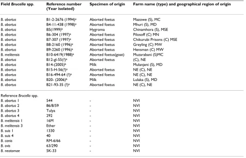
The details of the identity and origin of all the Brucella
spp. isolates, including the reference strains, used in this study are listed in Table 1. Prior to use, all strains from culture collection were stored either as lyophilized or in a -80°C deep freezer. Lyophilized isolates were re-consti-tuted and cultured in tryptone soya broth (TSB) (Oxoid) and subsequently sub-cultured onto Farrell's medium (Oxoid) and assessed for purity on bovine blood agar (Oxoid). All plates were incubated at 37°C under 10% CO2. Isolates were then inoculated into TSB with 5% glyc-erol, frozen and exported to Norway for further character-ization.
For the observation of colonial morphology, Brucella spp. isolates were cultured on Mueller-Hinton agar (Oxoid) and single colonies were examined using a low power ster-eoscopic microscope illuminated by obliquely reflected light as described [3]. The tests for production of urease, catalase, oxidase, H2S and indole; sensitivity to dyes (thionin and basic fuchsin) were carried out as described by Alton and co-workers [3]. Further tests for CO2 require-ment, sensitivity to dyes, lysis by bacteriophages (Tbilisi, Tb; Berkeley, Bk2; Firenze, Fi; Izatnagar, Iz1; R/C) and agglutination by A, M and R monospecific antisera were carried out at the Central Veterinary Laboratory, UK, using the procedures described [3].
The Brucella spp. isolates were grown on Farrell's agar (Oxoid) and incubated for 48 hours at 37°C under 10% CO2. To yield DNA, a few colonies from a pure culture were harvested and suspended in 200 μl of sterile distilled water in Ependorf tubes. A homogeneous suspension was made by stirring with the inoculation loop. Bacterial cells were inactivated by heating the tubes at 100°C for 10 minutes on a heating block (Grant Instruments, UK). To separate the DNA, killed bacterial cells were centrifuged at 15, 700 × g for 10 minutes. The supernatant containing crude DNA template was pipetted into new sterile Ependorf tubes and the sediment discarded. The concen-tration of the extracted crude DNA was measured using a ND-1000 V3.0 spectrophotometer (NanoDrop®
Technol-ogies Inc., USA). The DNA was stored at -20°C until fur-ther tests were carried out.
The AMOS-PCR (B. abortus, B. melitensis, B. ovis and B.
suis-Polymerase Chain Reaction) was done as described previ-ously [15] but with minor modifications of the assay envi-ronment. Briefly, PCR assay reaction mixture consisted of the following: 1 × PCR buffer (Applied Biosystems), 3 mM MgCl2, 200 μM (each) of the four deoxynucleotide tri-phosphates (dNTPs) (Finnzymes Oy, Espoo, Finland), and the 5 sets of primers (0.2 μM each) of B. abortus, B. melitensis, B. ovis, B. suis and IS711-specific primer. One and half units (1.5 U) of AmpliTaq Gold® DNA
with ethidium bromide and photographed using a gene snap camera (Syngene Pvt Ltd, UK).
Results
All 14 Brucella spp. isolates characterized in this study (Table 1) yielded the following results that are typical of the genus; Gram-negative coccobacilli, non-motile, posi-tive for modified Ziehl-Neelsen staining, oxidase and cat-alase production, and negative for indole production (Table 2). Their growth on Mueller-Hinton agar produced colonies that were convex, with entire edges and a smooth shiny consistency. The morphological, growth characteris-tics and biochemical profiles of the field isolates were sim-ilar to their respective reference Brucella species and biovars (data not shown).
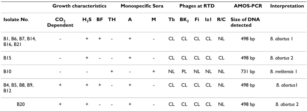
Isolates belonging to the same biovars showed consist-ently similar results, except for their CO2-dependence for growth (Table 3). Regardless of the biovar type, seven of the 13 B. abortus isolates were CO2-independent, while the remaining six strains were CO2-dependent. The B.
abortus isolates were lysed by phages Tb, Fi, Bk2, Iz1 and resistant to R/C. Only one isolate was lysed by the R/C phage. The single B. melitensis isolate was resistant to all phages but showed partial lysis to Bk2 (Table 3). B. abortus
isolates were agglutinated by A-antiserum and B. melitensis
by the M-antiserum, but all were not agglutinated by the R-antiserum (data not shown). Eleven and two of the 13
B. abortus isolates were identified to be biovars 1 and 2, respectively (Table 3).
The Brucella isolates were detected by the AMOS-PCR and produced predicted amplicons of sizes 498 bp and 731 bp for B. abortus and B. melitensis, respectively. Similar DNA products were produced for the reference B. abortus biovar 1 and B. melitensis biovar 1, respectively.
Discussion
[image:3.612.57.560.100.424.2]This paper provides the first detailed biochemical profil-ing and AMOS-PCR characterization of some Brucella spp. isolates from Zimbabwe. The phage sensitivity patterns of all the Brucella spp. isolates were consistent with what has
Table 1: Brucella isolates used in the study and the details of their geographical regions of origin.
Field Brucella spp. Reference number (Year isolated)
Specimen of origin Farm name (type) and geographical region of origin
B. abortus B1-2-2676 (1994)a Aborted foetus Mazowe (S), MC
B. abortus B4-11-438 (1998)a Aborted foetus Mhuri (S), MD
B. abortus B5(1999)b Hygroma Chinamhora (S), MSE
B. abortus B6-304 (1997)a Aborted foetus Pilosoff (C) MN
B. abortus B7-307 (1997)a Aborted foetus Chikurubi Prisons (C) MSE
B. abortus B8-2160 (1996)a Aborted foetus Greyling (C) MW
B. abortus B9-2260 (1996)a Aborted foetus Hensman (C) MW
B. melitensis B10-6419(1988)a Aborted foetus(goat) Muzarabani (S)MC
B. abortus B12-gl-55(?)a Aborted foetus (C), NE
B. abortus B14-(2005)b Milk Mulanjeni (S), MD
B. abortus B15-H-56(?)a Aborted foetus NE (C), NE
B. abortus B16-494-64 (?)a Aborted foetus NE (C), NE
B. abortus B20- (2006)b Milk Lulaka (S), MD
B. abortus B21-93-35 (?)a Aborted foetus NE (C), NE
Reference Brucella spp.
B. abortus 1 544 - NVI
B. abortus 2 86/8/59 - NVI
B. abortus 3 Tulya - NVI
B. abortus 4 292 - NVI
B. melitensis 1 16M - NVI
B. melitensis 3 Ether - NVI
B. suis 1 1330 - NVI
B. suis 4 40 - NVI
B. canis RM-6/66 - NVI
B. ovis 63/290 - NVI
B. neotomae 5K-33 - NVI
aObtained from the Central Veterinary Laboratory bObtained from the University of Zimbabwe
been reported [16]. However, a single isolate of B. abortus
(B15) was lysed by R/C phage and this susceptibility could be indicative of the presence of phage attachment sites which are present in the non-smooth phases of bru-cellae [16]. The B. melitensis (B10) also showed an atypical reaction because it was not lysed by the Iz1 phage which
[image:4.612.55.558.93.281.2]normally lyses smooth strains of this species. However, the examination of single colonies on microscopy by Henry illumination [3] showed no sign of dissociation and none of the isolates were agglutinated by the R-mon-ospecific antiserum which is an indicator of dissociation. The agglutination reaction by monospecific antisera
Table 2: Basic biochemical and metabolic profiles of field Brucella spp. from Zimbabwe.
Brucella isolate Biochemical properties Growth on TSA in the presence of dyes reference no. Cat Oxi aUre Mot Ind MZN T
20 T40 BF20
B1 + + + - - + - - +
B4 + + + - - + - - +
B5 + + + - - + - - +
B6 + + + - - + - - +
B7 + + + - - + - - +
B8 + + + - - + - - +
B9 + + + - - + - - +
B10 + + + - - + + + +
B12 + + + - - + - - +
B14 + + + - - + - - +
B15 + + + - - + - -
-B16 + + + - - + - - +
B20 + + + - - + - -
-B21 + + + - - + - - +
Cat, Catalase; Oxi, Oxidase, Ure; Urea hydrolysis; Mot, Motility test (+ = motile, - = non-motile); Ind, Indole production; MZN, Modified Ziehl Neelsen stain; TSA, Tryptone Soya agar; T20, 20 μl/ml thionin; T40, 40 μl/ml thionin; BF20, 20 μl/ml basic fuchsin; + = positive test result; - = negative test result.
aUrea hydrolysis = All isolates positive within 2 hours of culture
Table 3: Summary of phenotypic characteristics of the field Brucella spp. from Zimbabwea
Growth characteristics Monospecific Sera Phages at RTD AMOS-PCR Interpretation Isolate No. CO2
Dependent
H2S BF TH A M Tb BK2 Fi Iz1 R/C Size of DNA detected
B1, B6, B7, B14, B16, B21
- + + - + - CL CL CL CL NL 498 bp B. abortus 1
B15 - + - - + - CL CL CL CL CL 498 bp B. abortus 2
B10 - - + - + NL PL NL NL NL 731 bp B. melitensis 1
B4, B5, B8, B9, B12
+ + + - + - CL CL CL CL NL 498 bp B. abortus1
B20 + + - - + - CL CL CL CL NL 498 bp B. abortus 2
a All tests carried out by the reference laboratory (VLA), Weybridge, UK.
Isolate No. = Isolate identification DNA: Test by the AMOS PCR
BF = Basic fuchsin at 20 μl/ml (1/50,000 w/v) TH = Thionin at 20 μl/ml (1/50,000 w/v)
Phages: Tb = Tbilisi, BK2 = Berkeley type 2, Fi = Firenze, Iz1 = Izatnagar, R/C = phage lytic for non-smooth species of Brucella
CL = Confluent Lysis PL = Partial lysis NL = No lysis
RTD = Routine test dilution + = positive (yes)
[image:4.612.53.556.432.615.2]showed the predominance of A-specific and M-specific epitopes in our B. abortus and the B. melitensis isolate, respectively. All smooth strains of Brucella may possess either the A, M or both A and M antigenic epitopes on the O chains of the lipopolysaccharides [16].
Although phage typing is used primarily for identification at the nomen species level, some Brucella strains, espe-cially B. melitensis may show deviation from the standard pattern of susceptibility to Bk2, Iz1 and Wb phages [16]. The use of phage typing as a means of differentiating Bru-cella spp. has become less discriminatory as a typing tool because of the discovery of new strains with atypical sen-sitivity patterns [17].
The growth characteristics and the biochemical profiles of the field Brucella spp. isolates (Table 2) were similar to those of the reference strains used in this study. In addi-tion, the results were consistent with what is reported for
Brucella spp. and biovars [2,3,16]. However, the require-ment for CO2 for growth was at variance with reports from literature [3]. Although most strains of B. abortus biovars 1-4 require CO2 for primary isolation, this attribute is
quickly lost on repeated subcultures and such isolates will adapt to growing in atmospheres without CO2 [3,16].
The use of the AMOS-PCR results were consistent with those reported elsewhere [15]. These results confirmed the identity of the Brucella spp. that was obtained using bio-chemical profiles. The IS711 analysis using AMOS-PCR can identify only three B. abortus biovars, 1, 2 and 4; all three biovars of B. melitensis; biovar 1 of B. suis and B. ovis, but the individual biovars within a species are not differ-entiated [15]. Therefore, further DNA fingerprinting methods such as the variable number of tandem repeat analysis (VNTR) [15] could be used to investigate the molecular epidemiology of these Brucella isolates.
Although the B. abortus used in this study originated from five of the eight geographical provinces of Zimbabwe (Table 1), it is difficult to conclude on the spatial distribu-tion due to the limited number of isolates used. These iso-lates could possibly be restricted to one or a few geographical regions of Zimbabwe from where they have spread through movement of infected cattle. A study of more isolates is required to determine the spatial distribu-tion of B. abortus in Zimbabwe. However, the predomi-nance of B. abortus biovar 1 over biovar 2 suggested that it is the major cause of bovine brucellosis in both commer-cial and smallholder cattle farms. Another study which used fewer Brucella isolates from commercial dairy farms reported similar findings [12]. Although B. abortus biovar 2 was also detected and originating from both the com-mercial and smallholder cattle farms, its distribution could be limited to a few isolated areas. Elsewhere in
South Africa biovar 1 had been reported to account for about 90% while biovar 2 contributed 10% of all the B. abortus isolates [18]. South Africa, to a large extent, shares similar geographic, climatic and livestock husbandry sys-tems with Zimbabwe. While it is difficult to explain the reasons for the distribution of these B. abortus biovars in the cattle farming sectors, this could largely be influenced by movement of infected cattle between farms. Some farms often purchase cattle from other farms for the pur-pose of improving the genetics of their herds [9] or in the case of smallholder farms, to restock their herds which are continuously lost due to infectious diseases and lack of adequate grazing, especially during the drought seasons.
Despite the relatively few isolates studied, our results sug-gested that B. abortus biovar 3 and indeed other biovars may be rare in Zimbabwe, but this requires further study.
B. abortus biovar 3 has been infrequently reported in South Africa, East and North Africa, while there seems to be no reports of isolation of the other biovars [2,18]. World wide, in countries where bovine brucellosis is endemic, B. abortus biovar 1 is predominant and B. abortus
biovar 2 occurs less frequently while the other biovars are rare [2,18].
Conclusion
We concluded that the biochemical profiles and AMOS-PCR characterization were consistent with their respective species and biovars. B. abortus biovar 1 is likely to be the predominant cause of brucellosis in both commercial and smallholder cattle farms in Zimbabwe. Further studies are required that will apply DNA-fingerprinting to study dis-tribution patterns of B. abortus biovars in Zimbabwe.
Abbreviations
AMOS-PCR: B. abortus, B. melitensis, B. ovis, B. s
uis-Polymerase Chain reaction; bp: base pairs; DNA: deoxyri-bonucleic acid.
Competing interests
The authors declare that they have no competing interests.
Authors' contributions
Publish with BioMed Central and every scientist can read your work free of charge
"BioMed Central will be the most significant development for disseminating the results of biomedical researc h in our lifetime."
Sir Paul Nurse, Cancer Research UK
Your research papers will be:
available free of charge to the entire biomedical community
peer reviewed and published immediately upon acceptance
cited in PubMed and archived on PubMed Central
yours — you keep the copyright
Submit your manuscript here:
http://www.biomedcentral.com/info/publishing_adv.asp
BioMedcentral
in phenotypic and molecular characterization of isolates, result interpretation, supervision of laboratory work and helped in drafting of the manuscript. All authors have read and approved the final manuscript.
Acknowledgements
This study was fully sponsored by the Norwegian Council for Higher Edu-cation and Development (NUFU), project vote: pro/2002/06. We are grateful to the members of staff at the National Veterinary Institute, Oslo, for their technical assistance and in particular Dr. Arve Lund for his material support and encouragement. Our sincere appreciation goes to Ms L. Perret of Central Veterinary Laboratory, Weybridge, the UK, for further typing of the Brucella isolates.
References
1. WHO/FAO: Brucellosis in humans and animals, World Health Organization and Food and Agricultural Organization, 20 Avenue Appia, 1211 Geneva, Switzerland. 2006:1-66. 2. Quinn PJ, Carter ME, Markey B, Carter GR: Clinical Veterinary
Microbiology, Mosby International Limited, Edinburgh.
1999:261-267.
3. Alton G, Jones LM, Angus RD, Verger JM: Techniques for the bru-cellosis laboratory, Institut National de la Recherche Agronomique, Paris, France. 1988.
4. Scholz HC, Hubalek Z, Sedla ek I, Vergnaud G, Tomaso H, Al Dahouk S, Melzer F, Kampfer P, Neubauer H, Cloeckaert A, Maquart M, Zyg-munt MS, Whatmore AM, Falsen E, Bahn P, Göllner C, Pfeffer MB, Huber B, Busse H, Nöckler K: Brucella microti sp. nov., isolated from the common vole Microtus arvalis. Int J Syst Evol Microbiol 2008, 58:375-382.
5. Foster G, Osterman BS, Godfroid J, Jacques I, Cloeckaert A: Brucella ceti sp. nov. and Brucella pinnipedialis sp. nov. for Brucella strains with cetaceans and seals as their preferred hosts. Int J Syst Evol Microbiol 2007, 57:2688-2693.
6. Clavareau C, Wellemans V, Walravens K, Tryland M, Verger JM, Grayon M, Cloeckaert A, Letesson JJ, Godfroid J: Phenotypic and molecular characterization of a Brucella strain isolated from a minke whale (Balaenoptera acutorostrata). Vet Microbiol 1998, 144:3267-3273.
7. Verger JM, Grimont F, Grimont PAD, Grayson M: Brucella, a mon-ospecific genus as shown by deoxyribonucleic acid hybridiza-tion. Int J Syst Bacteriol 1985, 35:292-295.
8. Madzima WM: Zimbabwe: Bovine brucellosis and brucellosis of small ruminants: Diagnosis, control and vaccination. In Technical series Paris, France: Office International des Epizoties; 1987. 9. Madsen M: The current status of brucellosis in Zimbabwe.
Zimbabwe Veterinary Journal 1989, 20:133-145.
10. Madsen M, Anderson EC: Serologic survey of Zimbabwean wild-life for brucellosis. J Zoo Wildlife Med 1995, 26(2):240-245. 11. Condy JB, Vickers DB: Brucellosis in Rhodesian Wildlife. Journal
of the South African Veterinary Association 1972, 43:175-179.
12. Mohan K, Makaya PV, Muvavarirwa P, Matope G, Mahembe E, Pawandiwa A: Brucellosis surveillance and control in Zimba-bwe: bacteriological and serological investigation in dairy herds. Onderstepoort Journal of Veterinary Research 1996, 63:47-51. 13. Manley FH: Brucellosis in Rhodesia. A report to the Director
of Veterinary Services. Department of Veterinary Services, Harare, Zimbabwe; 1969.
14. Swanepoel R, Blackburn NK, Lander KP: The occurrence, diagno-sis and control of Brucellodiagno-sis in cattle in Rhodesia. Rhodesia (Zimbabwe) Veterinary Journal 1976, 7:24-31.
15. Bricker BJ, Halling SM: Differentiation of Brucella abortus bv. 1, 2, and 4, Brucella melitensis, Brucella ovis, and Brucella suis bv. 1 by PCR. J ClinMicrobiol 1994, 32:2660-2666.
16. Garritty GM, Bell JA, Lilburn T: Family III, Brucellaceae Breed, Murray and Smith 1957, 394 AL. In Bergey's Manual of Systematic BacteriologyVolume II. 2nd edition. Edited by: Brenner DJ, Krieg NR, Staley JT. Springer Science and Business Media, Inc., New York, NY 10013, USA; 2005:370-392.
17. Jahans KL, Foster G, Broughton ES: The characterisation of Bru-cella strains isolated from marine mammals. Vet Microbiol 1997, 57:373-382.