R E S E A R C H A R T I C L E
Open Access
Telomerase expression and telomere length in
breast cancer and their associations with
adjuvant treatment and disease outcome
Lingeng Lu
1, Chong Zhang
1,2, Gongjian Zhu
1,3, Melinda Irwin
1, Harvey Risch
1, Guido Menato
4, Marco Mitidieri
4,
Dionyssios Katsaros
4and Herbert Yu
1*Abstract
Introduction:Telomere length plays important roles in maintaining genome stability and regulating cell replication and death. Telomerase has functions not only to extend telomere length but also to repair DNA damage. Studies have shown that telomerase may increase cancer cell resistance to DNA-damaging anticancer agents; tamoxifen may suppress telomerase expression in breast cancer cells. This study aimed to investigate the role of telomere length and telomerase activity in breast cancer prognosis.
Methods:qPCR and qRT-PCR were used to analyze telomere length and telomerase expression, respectively, in tumor samples of 348 breast cancer patients. Cox regression analysis was performed to examine telomere length and telomerase expression in association with disease-free survival and cause-specific mortality.
Results:Telomere length had no relation to tumor features or disease outcomes. Telomerase expression was detected in 53% of tumors. Larger tumors or aggressive disease were more likely to have telomerase expression. Among patients treated with chemotherapy, high telomerase was found to be associated with increased risk of death (hazard ratio (HR) = 3.15; 95% CI: 1.34 to 7.40) and disease recurrence (HR = 2.04; 95% CI: 0.96 to 4.30) regardless of patient age, disease stage, tumor grade, histological type or hormone receptor status. Patients treated with endocrine therapy had different results regarding telomerase: high telomerase appeared to be associated with better survival outcomes. Telomerase expression made no survival difference in patients who received both chemotherapy and endocrine therapy.
Conclusions:Overall, telomerase expression was not associated with disease outcome, but this finding may be masked by adjuvant treatment. Patients with high telomerase expression responded poorly to chemotherapy in terms of disease-free and overall survival, but fared better if treated with endocrine therapy.
Introduction
Telomeres are repeated sequences of oligonucleotides (TTAGGG) located at the ends of chromosomes, and have important functions in regulating cell replication and maintaining genome integrity [1-3]. The length of telomeres decreases with the number of cell divisions; reduced telomere length carries functional cellular sig-nals. Cells enter senescence or initiate apoptosis when telomere length is reduced to a critical level. Shortened
telomere length often leads to genome instability, result-ing in loss of cell-cycle control, a hallmark of cancer. Tel-omere shortening has been found to be associated with increased risk of several human cancers, including blad-der, lung, esophagus, pancreas, ovary and thyroid [4-8].
Telomerase, a ribonucleoprotein complex composed of telomerase reverse transcriptase (TERT), telomerase RNA (TERC) and dyskerin [9], is a nuclear enzyme that increases the length of telomeres. Using TERC as a tem-plate, TERT adds telomeric repeats to the ends of chro-mosomes [10,11]. While it is important to maintain appropriate telomere length for genome stability, dysre-gulated telomere elongation also makes cells immortal, * Correspondence: herbert.yu@yale.edu
1Department of Epidemiology and Public Health, Yale Cancer Center, Yale
University School of Medicine, 60 College Street, New Haven, CT 06520-8034, USA
Full list of author information is available at the end of the article
which is a feature of cancer cells. Telomerase expression is low during early carcinogenesis, but markedly increases in tumor invasion, preventing cancer cells from entering senescence or apoptosis [12-14]. Com-pared to that in adjacent normal tissues, the activity of telomerase is up-regulated in many types of cancer, including breast [15-17]. Several studies have shown high telomerase activity to be linked to poor prognosis of breast cancer [18-20]. Based on the experimental observations of in vitrocell cultures and in vivoanimal models, telomerase inhibitors can reduce tumorigenicity and suppress breast cancer growth and metastasis [21-23]. Up-regulated telomerase activity and telomere elongation may also increase drug resistance in breast and colorectal cancer cell lines [17,24]. Telomerase inhi-bitors can increase cancer cell death when used in com-bination with DNA-damaging anticancer drugs [25]. However, our understanding of telomerase activity in breast cancer remains limited. The purpose of this study was to investigate the possible effects of telomerase expression and telomere length on breast cancer treat-ment outcomes.
Materials and methods Study patients
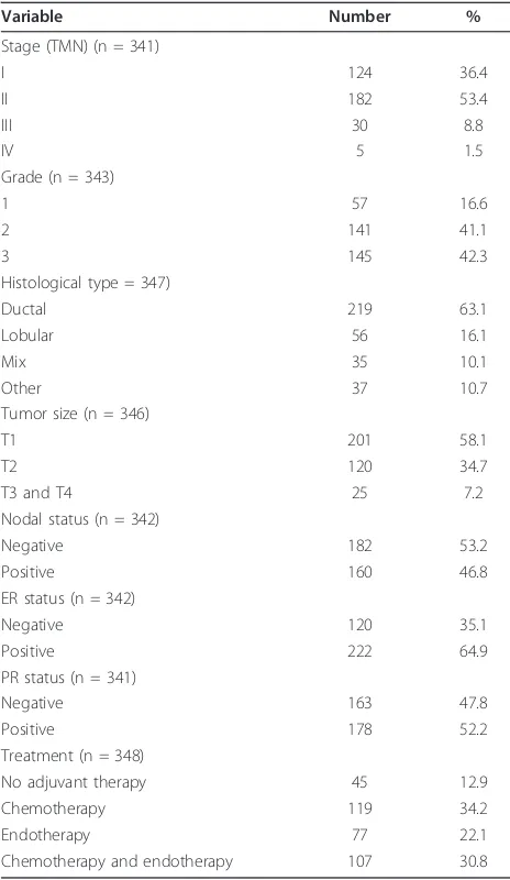
Patients undergoing surgery for primary breast cancer between January 1998 and July 1999 in the University Hospital at University of Turin were recruited to a clinical follow-up study. The study was approved by the university’s ethical review committee, and a written consent form was obtained from each study partici-pant. In total, 348 consecutive patients were enrolled in the study, with the average age of patients at sur-gery being 57 years (range: 23 to 84). Of the patients, 302 were followed from surgery through February 2007. The median follow-up time was 86 months (range: 8 to 108). Clinical and pathological information on these patients was collected from the hospital records and pathology reports (Table 1). The majority of patients in the study were diagnosed with early stage cancer (36.4% stage I and 53.4% stage II). Patients with late stage diseases (stages III and IV) accounted for only 10.3%. Grades 1 (well differen-tiated), 2 and 3 tumors were seen in 16.6%, 41.1% and 42.3% of the patients, respectively. The major histolo-gical tumor type was ductal carcinoma (63.1%), fol-lowed by lobular carcinoma (16.1%). Other histological types were ductal-lobular or tubular-lobular mixed histologies which accounted for 10.7% and 10.1%, respectively. Two hundred one patients (58.1%) had tumors smaller than 2 cm, 120 patients (34.7%) had tumors between 2 cm and 5 cm, and 25 (7.2%) had tumors greater than 5 cm. One hundred sixty patients (46.8%) had lymph node-positive tumors. According to
the 10% assay cutoff, 64.9% of the patients were ER positive, and 52.2% were PR positive.
[image:2.595.304.536.110.510.2]The majority of patients (303 out of 348) received adjuvant therapy. Of these patients, 119 (34.2%) received chemotherapy, 77 (22.1%) had hormonal therapy, and 107 (30.8%) received both chemotherapy and endocrine therapy. Forty-five patients did not receive any adjuvant treatment. Chemotherapy protocols used for the patients included CMF (cyclophosfamide, methotrexate, 5 fluoro-uracil; dose: 600/60/600 mg/mq every three weeks for six cycles), CEF (cyclophosfamide, epirubicin, 5 fluoro-uracil; dose: 600/90/600 mg/mq every three weeks for six cycles), EPI-TAX (epirubicin-paclitaxel; dose: 90/175 mg/mq every three weeks for six cycles), EPI-VNB (epir-ubicin-vinorelbine; dose: 90/30 mg/mq every three weeks for six cycles), DTX-EPI-VNB (doxetaxel-epirubi-cin-vinorelbine; dose: 75/90/30 mg/mq every three Table 1 Clinical and pathological features of breast cancer patients in the study
Variable Number %
Stage (TMN) (n = 341)
I 124 36.4
II 182 53.4
III 30 8.8
IV 5 1.5
Grade (n = 343)
1 57 16.6
2 141 41.1
3 145 42.3
Histological type = 347)
Ductal 219 63.1
Lobular 56 16.1
Mix 35 10.1
Other 37 10.7
Tumor size (n = 346)
T1 201 58.1
T2 120 34.7
T3 and T4 25 7.2
Nodal status (n = 342)
Negative 182 53.2
Positive 160 46.8
ER status (n = 342)
Negative 120 35.1
Positive 222 64.9
PR status (n = 341)
Negative 163 47.8
Positive 178 52.2
Treatment (n = 348)
No adjuvant therapy 45 12.9
Chemotherapy 119 34.2
Endotherapy 77 22.1
weeks for six cycles), and TXT-EPI-VNB (paclitaxel-epirubicin-vinorelbine; dose: 175/90/30 mg/mq every three weeks for six cycles). Tamoxifen was the only endocrine therapy used at that time, and the dose was one 20 mg tablet per day for five years or until disease progression or unacceptable toxicity. By the end of fol-low-up, 55 of the 81 patients who had had recurrences had died. Among the 221 patients without disease pro-gression, 5 had died.
Tissue analysis for telomere length and telomerase expression
Fresh tumor samples were collected from the enrolled patients during surgery. The specimens were snap-fro-zen in liquid nitrogen immediately after resection and stored at -80°C until analysis. All tissue samples were examined by pathologists to confirm tumor content which varied between 80% and 90%. The specimens were pulverized manually in liquid nitrogen; aliquots of the tissue powders (about 100 mg each) were pro-cessed to extract DNA and total RNA separately, fol-lowing conventional phenol-chloroform protocols. RNA samples were quantified and treated with DNase using a commercial kit (Ambion Inc, Austin, TX, USA).
A quantitative PCR (qPCR) developed by Cawthon [26] was used to analyze telomere length. The method compares the quantities of telomere and albumin in a common PCR reaction assuming that both products are amplified with similar efficiency. The primer sequences used were 5’- ACA CTA AGG TTT GGG TTT GGG TTT GGG TTT GGG TTA GTG T (for-ward) and 5’- TGT TAG GTA TCC CTA TCC CTA TCC CTA TCC CTA TCC CTA ACA (reverse) for tel-omere, and 5’-CGG CGG CGG GCG GCG CGG GCT GGG CGG AAA TGC TGC ACA GAA TCC TTG (forward) and 5’- GCC CGG CCC GCC GCG CCC GTC CCG CCG GAA AAG CAT GGT CGC CTG TT (reverse) for albumin. Chromo4™ continuous fluores-cence detector system (MJ Research Inc., Waltham, MA, USA) was used to obtain Ct values for both telo-mere and albumin in each sample. Based on the for-mula 2-(ΔCt), where ΔCt = Ct telomere- Ct albumin, the
relative quantity of telomere versus albumin (T/S) was calculated. qPCR was done in 20 μl solution which mixed 10 μl of 2 × Quantifast SYBR green (Qiagen, Valencia, CA, USA) with telomere primers (900 nM), albumin primers (900 nM), and approximately 20 ng DNA template. The PCR conditions included initial denaturing at 95°C for 15 minutes, followed by 2 cycles of 15 sec at 94°C, and 15 sec at 49°C, and then 26 cycles of 15 sec at 94°C, 10 sec at 62°C, and 15 sec at 73°C with signal acquisition for telomere, 10 sec at 84° C, and 15 sec at 87°C with signal acquisition for
albumin. After PCR amplification, melting curves were analyzed to confirm the size of PCR products. Each sample was run in duplicate. Samples with coefficient of variation >15% were reanalyzed.
Levels of telomerase expression along with an inter-nal control (GAPDH) were determined with real-time reverse-transcriptase PCR (qRT-PCR). The forward and reverse PCR primers for telomerase described by Zhu et al. [27] were 5’- CGT CGA GCT GCT CAG
GTC TT and 5’- AGT GCT GTC TGA TTC CAA
TGC TT, respectively. The primer sequences for GAPDH and the qRT-PCR methods are described else-where [28-30]. Approximately 1.0 μg of DNase-treated total RNA from each sample was used for reverse tran-scription using the Cloned AMV First-Strand cDNA Synthesis kit (Invitrogen™, Carlsbad, CA, USA). qRT-PCR was performed using the Chromo4™ continuous fluorescence detector system. The PCR reaction was carried out in a 20μl solution which contained 0.1 μg of cDNA template plus 10 μl of 2 × Power® SYBR Green PCR Master Mix (Applied Biosystems, Foster City, CA, USA) and a pair of primers at a final con-centration of 100 nM. PCR thermal cycle conditions included initial incubation at 50°C for 2 minutes, dena-turing at 95°C for 10 minutes, and 40 cycles of 95°C for 15 sec and 60°C for 1 minute. Dissociation curves were generated after PCR to determine the size of PCR product. Each sample was tested in duplicate for each gene, and the analysis was repeated for those samples with large variation in their duplicates (coefficient of variation >5%).
Statistical analysis
An expression index (EI) was calculated for telomerase expression using the formula 1,000 × 2 (-ΔCt), where ΔCt = Ct (telomerase)- Ct (GAPDH). In data analysis,
telo-merase expression was analyzed as a categorical vari-able. Patients with undetectable telomerase, Ct
(telomerase) >35, were grouped as low telomerase, and
Results
Telomerase expression and telomere length in breast cancer
Telomerase expression was analyzed in the 336 tumor samples that had sufficient quantities of RNA. Among these, 159 samples had low (undetectable) and 177 had high (detectable) telomerase expression. For the detect-able samples, the average expression was 0.23 EI, and the expression range varied widely from 0.001 to 2,266 EI. The average value of telomere length in 348 tumor samples was 7.6, and the range was between 0.4 and 40.9. When analyzed as continuous variables, no correla-tion was found between telomerase expression and telo-mere length (r = 0.02, P = 0.746). This analysis was repeated for the samples with detectable expression (n = 177) using the log transformed data, and the result remained same. In categorical analysis, patients with detectable telomerase expression seemed more likely to have long telomeres compared to those with undetect-able telomerase, but the difference was not significant (53.1% versus 45.3%,P = 0.152).
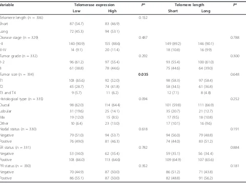
Telomerase expression and telomere length in relation to clinical and pathological features of breast cancer are shown in Table 2. A statistically significant association was observed between telomerase expression and tumor size (P= 0.035); patients with high telomerase expression tended to have larger tumors. Patients with high telomer-ase also seemed more likely to have stage III or IV dis-ease (11.4% versus 9.1%) and grade 3 tumors (44.6% versus 38.8%), but these differences were not statistically significant. No significant associations were found between telomerase expression and histological type, lymph node involvement or hormone receptor status. Telomere length, either analyzed as a continuous or cate-gorical variable, was not associated with any of the clini-cal or pathologiclini-cal features (categoriclini-cal data in Table 2)
Telomerase expression and telomere length in relation to disease outcomes
Table 3 presents telomerase expression and telomere length in relation to disease outcomes. Patients with high telomerase expression had higher risk of disease recurrence and death than those with low expression, but the elevated risks were not statistically significant. Patients with long or short telomere lengths had quite similar survival outcomes, either for disease recurrence or death. Furthermore, these findings did not seem to change substantially after the major clinical and patholo-gical features of breast cancer were adjusted in the ana-lysis, including age at surgery, disease stage, tumor grade, histological type and receptor status.
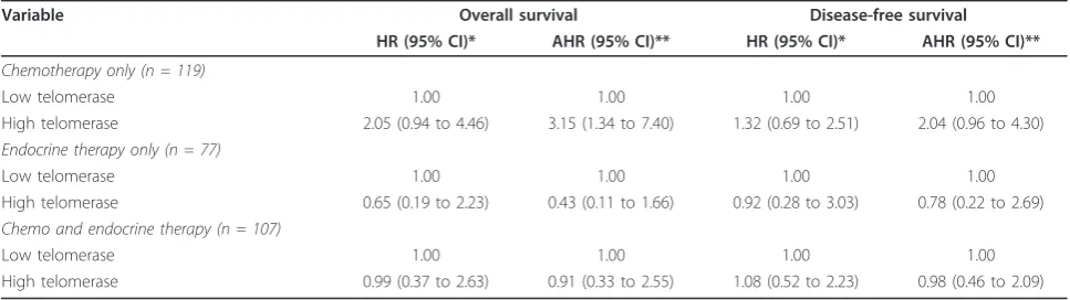
However, since systemic adjuvant treatment may influ-ence survival and interact with telomerase activity in its therapeutic effects, we performed survival analyses
stratified by treatment modality (Table 4). Among patients treated with adjuvant chemotherapy, high telo-merase expression was associated with higher risk of death and disease recurrence in comparison to low expression: adjusted hazard ratios 3.15 (95% CI: 1.34 to 7.40) and 2.04 (95% CI: 0.96 to 4.30), respectively. Inter-estingly, similar results were not observed in patients treated with endocrine therapy. These patients, in con-trast, appeared to have better survival outcomes with high expression of telomerase, though none of the asso-ciations were statistically significant. For patients who received both chemotherapy and endocrine therapy, sur-vival outcomes, either overall sursur-vival or disease-free survival, were similar according to high or low telomer-ase expression. No associations were found between tel-omere length and patient survival when patients were stratified by either adjuvant endocrine therapy or che-motherapy in both univariate and multivariate analyses (data not shown).
Discussion
In this study, we analyzed breast tumors for telomere length and telomerase expression in order to assess their possible effects on disease outcomes. We found no evidence that telomere length was related to any of the major clinical or pathological features of the disease. In addition, we found no indication that telomere length was associated with patient survival outcomes, either disease-free survival or overall survival. We did notice, however, that tumors with longer telomere lengths were more likely to have detectable telomerase expression, although statistically significant correlation between the two was not observed. It has been reported that telo-mere length can be maintained without telomerase through an alternative mechanism, namely alternative lengthening of telomeres (ALT) [31]. Although the mechanisms by which ALT works remain unclear, it is thought that homologous DNA recombination is involved. About 10 to 15% of cancers including some with extremely poor prognosis use ALT to elongate tel-omere length [31]. Additionally, many environmental and host factors including psychological stress, physical activity, oxidative stress, hormones and growth factors have been shown to affect telomere length [32-36].
appeared to have better outcomes if their telomerase was high in the tumors. This discrepancy suggests that telomerase may interfere with adjuvant treatment. It is known that telomerase can maintain genome stability by elongating telomeres in normal cells, but evidence also suggests that telomerase may play different roles in tumors, and the effects are independent of telomere length. The enzyme may facilitate malignant
[image:5.595.58.538.108.466.2]transformation and render cells more resistant to apop-tosis [37-39]. It is found that when telomerase is sup-pressed, short-term telomere integrity is not affected, but the overall chromatin configuration undergoes sig-nificant changes in terms of histone modification. These epigenetic alterations can impair DNA repair capacity and increase cell sensitivity to radiation [40]. Telomer-ase not only promotes DNA damage repair and Table 2 Telomerase expression and telomere length in association with clinical and pathological features of breast cancer
Variable Telomerase expression P* Telomere length P*
Low High Short Long
Telomere length (n = 336) 0.152
Short 87 (54.7) 83 (46.9)
Long 72 (45.3) 94 (53.1)
Disease stage (n = 329) 0.487 0.788
I-II 140 (90.9) 155 (88.6) 149 (89.2) 146 (90.1)
III-IV 14 (9.1) 20 (11.4) 18 (10.8) 16 (9.9)
Tumor grade (n = 332) 0.292 0.300
1-2 96 (61.2) 97 (55.4) 93 (55.4) 100 (61.0)
3 61 (38.8) 78 (44.6) 75 (44.6) 64 (39.0)
Tumor size (n = 334) 0.035 0.648
T1 103 (65.6) 92 (52.0) 98 (58.3) 97 (58.4)
T2 45 (28.7) 74 (41.8) 58 (34.5) 61 (36.8)
T3 and T4 9 (5.7) 11 (6.2) 12 (7.1) 8 (4.8)
Histological type (n = 335) 0.094 0.252
Ductal 98 (62.0) 114 (64.4) 101 (59.8) 111 (66.9)
Lobular 31 (19.6) 25 (14.1) 35 (20.7) 21 (12.7)
Mix 19 (12.0) 15 (8.5) 17 (9.5) 18 (10.8)
Other 10 (6.4) 23 (13.0) 17 (10.1) 16 (9.6)
Nodal status (n = 330) 0.618 0.191
Negative 79 (51.0) 94 (53.7) 94 (56.0) 79 (48.8)
Positive 76 (49.0) 81 (46.3) 74 (44.0) 83 (51.2)
ER status (n = 331) 0.782 0.884
Negative 53 (34.0) 62 (35.4) 59 (35.1) 56 (34..4)
Positive 103 (66.0) 113 (64.6) 109 (64.9) 107 (65.6)
PR status (n = 330) 0.352 0.181
Negative 70 (44.9) 87 (50.0) 86 (51.2) 71 (43.8)
Positive 86 (55.1) 87 (50.0) 82 (48.8) 91 (56.2)
Table 3 Survival outcomes in association with telomerase expression and telomere length in all patients
Variable Overall Survival Disease-free Survival
HR (95% CI)* AHR (95% CI)** HR (95% CI)* AHR (95% CI)**
Telomerase
Low telomerase 1.00 1.00 1.00 1.00
High telomerase 1.46 (0.87 to 2.45) 1.31 (0.78 to 2.22) 1.25 (0.80 to 1.95) 1.21 (0.79 to 1.94)
Telomere length
Short telomere 1.00 1.00 1.00 1.00
Long telomere 0.79 (0.47 to 1.31) 0.83 (0.49 to 1.40) 0.84 (0.55 to 1.31) 0.83 (0.53 to 1.31)
*HR, hazard ratio; 95% CI, 95% confidence interval.
[image:5.595.57.540.612.712.2]facilitates cell survival, but also induces the resistance of cancer cells to anticancer agents that target DNA damage [25]. Our finding of unfavorable survival out-comes in association with high telomerase in patients treated with chemotherapy, but not in those treated with endocrine therapy, may be explained by the possi-bility that telomerase offsets the effect of chemotherapy since most chemotherapeutic drugs target DNA damage to induce cell death. In contrast, endocrine treatment exerts its effect through a mechanism that is completely different from chemotherapy.
If our observations are confirmed, our findings may have clinical implications in selecting adjuvant treatment for breast cancer. Patients with high or detectable telo-merase expression may not be suitable for chemother-apy which targets DNA damage. Adjuvant treatment which combines chemotherapy with endocrine ment may not be appropriate either because the treat-ment effects may cancel each other out. In our study, we observed no difference in survival benefit in this treatment subgroup between those with high and low telomerase expression. Thus, for patients with high telo-merase, endocrine therapy may be the choice of adju-vant treatment. Our speculation that endocrine therapy may be a better choice for breast cancer patients with high telomerase activity is supported by laboratory find-ing that endocrine therapy antagonizes the activity of telomerase in breast cancer. A number of in vitro experiments have shown that both tamoxifen and raloxi-fene can inhibit telomerase expression in several breast cancer cell lines [41-43]. The inhibition of telomerase expression by tamoxifen is dependent on the presence of estrogen receptors [41]. Although our study shows a potential value of telomerase expression in breast cancer adjuvant treatment, these are preliminary findings which need to be confirmed by larger studies with considera-tion of molecular subtypes and endocrine therapy invol-ving regimens other than tamoxifen.
Studies have shown that telomerase is highly active in most types of human cancers including breast cancer, but remains inactive in adjacent normal tissues. Telo-merase inhibitors can suppress the growth and prolifera-tion of tumor cells [17,44,45]. In our study, we found that telomerase expression was detectable in 53% of the tumors. This level of detection appears to be low when compared to some studies. Elkak et al. found that telo-merase expression was detectable in all 116 breast can-cer samples examined [18]. Other studies showed that 74% of breast carcinomas had detectable telomerase [46-48]. Using immunohistological staining, two studies found 72 to 86% of breast cancer samples having detect-able telomerase activity [49,50]. However, there were two studies which found the frequency of detectable tel-omerase very similar to our study [51,52]. Reasons for the inconsistent detection of telomerase expression could be manifold, including different laboratory meth-ods used for analysis and various cell contents in tissue samples. Liu and colleagues reported that normal mam-mary cells expressed higher telomerase than cancer cells [53]. Our findings of no associations between telomerase expression and disease stage or tumor grade were in agreement with those reported by Salhab and colleagues, who also found no association of telomerase with dis-ease stage and tumor grade, although patients with poorly differentiated tumors (grade 3) appeared to have higher expression [19].
Conclusions
[image:6.595.56.540.100.236.2]In summary, we analyzed telomerase expression and tel-omere length in breast cancer and examined the rela-tionships with disease outcomes. The study showed that telomere length was not associated with breast cancer clinical features or patient survival. Telomerase expres-sion appeared to be slightly higher in tumors with longer telomeres as well as in larger tumors or aggres-sive disease. Overall, telomerase expression was not Table 4 Survival outcomes in association with telomerase expression stratified by adjuvant treatment
Variable Overall survival Disease-free survival
HR (95% CI)* AHR (95% CI)** HR (95% CI)* AHR (95% CI)**
Chemotherapy only (n = 119)
Low telomerase 1.00 1.00 1.00 1.00
High telomerase 2.05 (0.94 to 4.46) 3.15 (1.34 to 7.40) 1.32 (0.69 to 2.51) 2.04 (0.96 to 4.30)
Endocrine therapy only (n = 77)
Low telomerase 1.00 1.00 1.00 1.00
High telomerase 0.65 (0.19 to 2.23) 0.43 (0.11 to 1.66) 0.92 (0.28 to 3.03) 0.78 (0.22 to 2.69)
Chemo and endocrine therapy (n = 107)
Low telomerase 1.00 1.00 1.00 1.00
High telomerase 0.99 (0.37 to 2.63) 0.91 (0.33 to 2.55) 1.08 (0.52 to 2.23) 0.98 (0.46 to 2.09)
*HR, hazard ratio; 95% CI, 95% confidence interval.
associated with disease outcome, but this finding may be masked by adjuvant treatment. Patients with high telo-merase expression responded poorly to chemotherapy in terms of disease-free and overall survival, but fared bet-ter if treated with endocrine therapy. High or low telo-merase expression made no difference in survival outcomes when patients received both chemotherapy and endocrine therapy. These findings suggest that telo-merase activity may be a useful factor in determining the choice of adjuvant treatment for breast cancer patients.
Abbreviations
AHR: adjusted Hazard Ratio; ALT: alternative lengthening of telomeres; CEF: cyclophosfamide, epirubicin, 5 fluoro-uracil; CI: confidential interval; CMF: cyclophosfamide, methotrexate, 5 fluoro-uracil; DTX-EPI-VNB: doxetaxel-vinorelbine; EPI-TAX: paclitaxel; EPI-VNB: epirubicin-vinorelbine; ER: estrogen receptor; GAPDH: glyceraldehyde 3-phosphate dehydrogenase; HR: hazard ratio; PR: progesterone receptor; qPCR: quantitative Polymerase Chain Reaction; qRT-PCR: quantitative reverse-transcription PCR; TERT: telomerease reverse transcriptase; TERC: telomerase RNA; TXT-EPI-VNB: paclitaxel-epirubicin-vinorelbine.
Author details 1
Department of Epidemiology and Public Health, Yale Cancer Center, Yale University School of Medicine, 60 College Street, New Haven, CT 06520-8034, USA.2Gansu Provincial Hospital for the Protection of Mother and Baby’s Health, 143 North Street, Lanzhou 730050, China.3Gansu Provincial Academy of Medical Science, Gansu Provincial Tumor Hospital, 2 Xiaoxihu East Street, Lanzhou 730050, China.4Department of Obstetrics and Gynecology, Gynecologic Oncology and Breast Cancer Unit, University of Turin, Via Ventimiglia 3, Turin 10126, Italy.
Authors’contributions
LL, MI, HR, DK and HY participated in the conception and design of the study. LL, CZ, GZ, GM and MM acquired the data. LL, CZ, GZ, MI, HR, DK and HY analyzed and interpreted the data. LL, CZ, GZ, MI, HR, GM, MM, DK and HY prepared and revised the manuscript. All authors read and approved the final version of the manuscript.
Competing interests
The authors declare that they have no competing interests.
Received: 28 February 2011 Revised: 21 April 2011 Accepted: 6 June 2011 Published: 6 June 2011
References
1. Smogorzewska A, de Lange T:Regulation of telomerase by telomeric proteins.Annu Rev Biochem2004,73:177-208.
2. Hug N, Lingner J:Telomere length homeostasis.Chromosoma2006, 115:413-425.
3. von Zglinicki T, Saretzki G, Ladhoff J, d’Adda di Fagagna F, Jackson SP: Human cell senescence as a DNA damage response.Mech Ageing Dev
2005,126:111-117.
4. Capezzone M, Cantara S, Marchisotta S, Filetti S, De Santi MM, Rossi B, Ronga G, Durante C, Pacini F:Short telomeres, telomerase reverse transcriptase gene amplification, and increased telomerase activity in the blood of familial papillary thyroid cancer patients.J Clin Endocrinol Metab2008,93:3950-3957.
5. Broberg K, Bjork J, Paulsson K, Hoglund M, Albin M:Constitutional short telomeres are strong genetic susceptibility markers for bladder cancer.
Carcinogenesis2005,26:1263-1271.
6. Jang JS, Choi YY, Lee WK, Choi JE, Cha SI, Kim YJ, Kim CH, Kam S, Jung TH, Park JY:Telomere length and the risk of lung cancer.Cancer Sci2008, 99:1385-1389.
7. Risques RA, Vaughan TL, Li X, Odze RD, Blount PL, Ayub K, Gallaher JL, Reid BJ, Rabinovitch PS:Leukocyte telomere length predicts cancer risk in Barrett’s esophagus.Cancer Epidemiol Biomarkers Prev2007,16:2649-2655. 8. Mirabello L, Garcia-Closas M, Cawthon R, Lissowska J, Brinton LA,
Peplonska B, Sherman ME, Savage SA:Leukocyte telomere length in a population-based case-control study of ovarian cancer: a pilot study.
Cancer Causes Control2010,21:77-82.
9. Cohen SB, Graham ME, Lovrecz GO, Bache N, Robinson PJ, Reddel RR: Protein composition of catalytically active human telomerase from immortal cells.Science2007,315:1850-1853.
10. Holt SE, Wright WE, Shay JW:Regulation of telomerase activity in immortal cell lines.Mol Cell Biol1996,16:2932-2939.
11. Kelland LR:Overcoming the immortality of tumour cells by telomere and telomerase based cancer therapeutics–current status and future prospects.Eur J Cancer2005,41:971-979.
12. Hashimoto Y, Murakami Y, Uemura K, Hayashidani Y, Sudo T, Ohge H, Fukuda E, Shimamoto F, Sueda T, Hiyama E:Telomere shortening and telomerase expression during multistage carcinogenesis of intraductal papillary mucinous neoplasms of the pancreas.J Gastrointest Surg2008, 12:17-28; discussion 28-29.
13. Gisselsson D, Jonson T, Petersen A, Strombeck B, Dal Cin P, Hoglund M, Mitelman F, Mertens F, Mandahl N:Telomere dysfunction triggers extensive DNA fragmentation and evolution of complex chromosome abnormalities in human malignant tumors.Proc Natl Acad Sci USA2001, 98:12683-12688.
14. Bardeesy N, DePinho RA:Pancreatic cancer biology and genetics.Nat Rev Cancer2002,2:897-909.
15. Healy KC:Telomere dynamics and telomerase activation in tumor progression: prospects for prognosis and therapy.Oncol Res1995, 7:121-130.
16. Stewart SA, Weinberg RA:Telomeres: cancer to human aging.Annu Rev Cell Dev Biol2006,22:531-557.
17. Cerone MA, Londono-Vallejo JA, Autexier C:Telomerase inhibition enhances the response to anticancer drug treatment in human breast cancer cells.Mol Cancer Ther2006,5:1669-1675.
18. Elkak A, Mokbel R, Wilson C, Jiang WG, Newbold RF, Mokbel K:hTERT mRNA expression is associated with a poor clinical outcome in human breast cancer.Anticancer Res2006,26:4901-4904.
19. Salhab M, Jiang WG, Newbold RF, Mokbel K:The expression of gene transcripts of telomere-associated genes in human breast cancer: correlation with clinico-pathological parameters and clinical outcome.
Breast Cancer Res Treat2008,109:35-46.
20. Poremba C, Heine B, Diallo R, Heinecke A, Wai D, Schaefer KL, Braun Y, Schuck A, Lanvers C, Bankfalvi A, Kneif S, Torhorst J, Zuber M, Kochli OR, Mross F, Dieterich H, Sauter G, Stein H, Fogt F, Boecker W:Telomerase as a prognostic marker in breast cancer: high-throughput tissue microarray analysis of hTERT and hTR.J Pathol2002,198:181-189.
21. Hochreiter AE, Xiao H, Goldblatt EM, Gryaznov SM, Miller KD, Badve S, Sledge GW, Herbert BS:Telomerase template antagonist GRN163L disrupts telomere maintenance, tumor growth, and metastasis of breast cancer.Clin Cancer Res2006,12:3184-3192.
22. Joseph I, Tressler R, Bassett E, Harley C, Buseman CM, Pattamatta P, Wright WE, Shay JW, Go NF:The telomerase inhibitor imetelstat depletes cancer stem cells in breast and pancreatic cancer cell lines.Cancer Res
2010,70:9494-9504.
23. Gryaznov SM, Jackson S, Dikmen G, Harley C, Herbert BS, Wright WE, Shay JW:Oligonucleotide conjugate GRN163L targeting human telomerase as potential anticancer and antimetastatic agent.Nucleosides Nucleotides Nucleic Acids2007,26:1577-1579.
24. Kuranaga N, Shinomiya N, Mochizuki H:Long-term cultivation of colorectal carcinoma cells with anti-cancer drugs induces drug resistance and telomere elongation: anin vitrostudy.BMC Cancer2001, 1:10.
25. Tamakawa RA, Fleisig HB, Wong JM:Telomerase inhibition potentiates the effects of genotoxic agents in breast and colorectal cancer cells in a cell cycle-specific manner.Cancer Res2010,70:8684-8694.
26. Cawthon RM:Telomere length measurement by a novel monochrome multiplex quantitative PCR method.Nucleic Acids Res2009,37:e21. 27. Zhu CQ, Cutz JC, Liu N, Lau D, Shepherd FA, Squire JA, Tsao MS:
28. Lu L, Katsaros D, Wiley A, Rigault de la Longrais IA, Puopolo M, Schwartz P, Yu H:Promoter-specific transcription of insulin-like growth factor-II in epithelial ovarian cancer.Gynecol Oncol2006,103:990-995.
29. Lu L, Katsaros D, Wiley A, Rigault de la Longrais IA, Risch HA, Puopolo M, Yu H:The relationship of insulin-like growth factor-II, insulin-like growth factor binding protein-3, and estrogen receptor-alpha expression to disease progression in epithelial ovarian cancer.Clin Cancer Res2006, 12:1208-1214.
30. Mu L, Katsaros D, Wiley A, Lu L, de la Longrais IA, Smith S, Khubchandani S, Sochirca O, Arisio R, Yu H:Peptide concentrations and mRNA expression of IGF-I, IGF-II and IGFBP-3 in breast cancer and their associations with disease characteristics.Breast Cancer Res Treat2009,115:151-162. 31. Cesare AJ, Reddel RR:Alternative lengthening of telomeres: models,
mechanisms and implications.Nat Rev Genet2010,11:319-330. 32. Demissie S, Levy D, Benjamin EJ, Cupples LA, Gardner JP, Herbert A,
Kimura M, Larson MG, Meigs JB, Keaney JF, Aviv A:Insulin resistance, oxidative stress, hypertension, and leukocyte telomere length in men from the Framingham Heart Study.Aging Cell2006,5:325-330. 33. Epel ES, Blackburn EH, Lin J, Dhabhar FS, Adler NE, Morrow JD,
Cawthon RM:Accelerated telomere shortening in response to life stress.
Proc Natl Acad Sci USA2004,101:17312-17315.
34. Barbieri M, Paolisso G, Kimura M, Gardner JP, Boccardi V, Papa M, Hjelmborg JV, Christensen K, Brimacombe M, Nawrot TS, Staessen JA, Pollak MN, Aviv A:Higher circulating levels of IGF-1 are associated with longer leukocyte telomere length in healthy subjects.Mech Ageing Dev
2009,130:771-776.
35. Cherkas LF, Hunkin JL, Kato BS, Richards JB, Gardner JP, Surdulescu GL, Kimura M, Lu X, Spector TD, Aviv A:The association between physical activity in leisure time and leukocyte telomere length.Arch Intern Med
2008,168:154-158.
36. Ludlow AT, Zimmerman JB, Witkowski S, Hearn JW, Hatfield BD, Roth SM: Relationship between physical activity level, telomere length, and telomerase activity.Med Sci Sports Exerc2008,40:1764-1771.
37. Chan SW, Blackburn EH:New ways not to make ends meet: telomerase, DNA damage proteins and heterochromatin.Oncogene2002,21:553-563. 38. Blasco MA, Hahn WC:Evolving views of telomerase and cancer.Trends
Cell Biol2003,13:289-294.
39. Kang HJ, Choi YS, Hong SB, Kim KW, Woo RS, Won SJ, Kim EJ, Jeon HK, Jo SY, Kim TK, Bachoo R, Reynolds IJ, Gwag BJ, Lee HW:Ectopic expression of the catalytic subunit of telomerase protects against brain injury resulting from ischemia and NMDA-induced neurotoxicity.J Neurosci
2004,24:1280-1287.
40. Masutomi K, Possemato R, Wong JM, Currier JL, Tothova Z, Manola JB, Ganesan S, Lansdorp PM, Collins K, Hahn WC:The telomerase reverse transcriptase regulates chromatin state and DNA damage responses.
Proc Natl Acad Sci USA2005,102:8222-8227.
41. Wang Z, Kyo S, Maida Y, Takakura M, Tanaka M, Yatabe N, Kanaya T, Nakamura M, Koike K, Hisamoto K, Ohmichi M, Inoue M:Tamoxifen regulates human telomerase reverse transcriptase (hTERT) gene expression differently in breast and endometrial cancer cells.Oncogene
2002,21:3517-3524.
42. Aldous WK, Marean AJ, DeHart MJ, Matej LA, Moore KH:Effects of tamoxifen on telomerase activity in breast carcinoma cell lines.Cancer
1999,85:1523-1529.
43. Kawagoe J, Ohmichi M, Takahashi T, Ohshima C, Mabuchi S, Takahashi K, Igarashi H, Mori-Abe A, Saitoh M, Du B, Ohta T, Kimura A, Kyo S, Inoue M, Kurachi H:Raloxifene inhibits estrogen-induced up-regulation of telomerase activity in a human breast cancer cell line.J Biol Chem2003, 278:43363-43372.
44. Collins K, Mitchell JR:Telomerase in the human organism.Oncogene2002, 21:564-579.
45. Masutomi K, Yu EY, Khurts S, Ben-Porath I, Currier JL, Metz GB, Brooks MW, Kaneko S, Murakami S, DeCaprio JA, Weinberg RA, Stewart SA, Hahn WC: Telomerase maintains telomere structure in normal human cells.Cell
2003,114:241-253.
46. Mokbel K, Parris CN, Ghilchik M, Williams G, Newbold RF:The association between telomerase, histopathological parameters, and KI-67 expression in breast cancer.Am J Surg1999,178:69-72.
47. Mokbel KM, Parris CN, Ghilchik M, Amerasinghe CN, Newbold RF: Telomerase activity and lymphovascular invasion in breast cancer.Eur J Surg Oncol2000,26:30-33.
48. Mokbel K, Ghilchik M, Williams G, Akbar N, Parris C, Newbold R:The association between telomerase activity and hormone receptor status and p53 expression in breast cancer.Int J Surg Investig2000,1:509-516. 49. Herbert GS, Sohn VY, Mulcahy MJ, Champeaux AL, Brown TA:Prognostic
significance of reactivation of telomerase in breast core biopsy specimens.Am J Surg2007,193:547-550; discussion 550. 50. Kalogeraki A, Kafousi M, Ieromonachou P, Giannikaki E, Vrekoussis T,
Zoras O, Tsiftsis D, Stathopoulos E:Telomerase activity as a marker of invasive ductal breast carcinomas on FNABs and relationship to other prognostic variables.Anticancer Res2005,25:1927-1930.
51. Poremba C, Bocker W, Willenbring H, Schafer KL, Otterbach F, Burger H, Diallo R, Dockhorn-Dworniczak B:Telomerase activity in human proliferative breast lesions.Int J Oncol1998,12:641-648.
52. Murillo-Ortiz B, Astudillo-De la Vega H, Castillo-Medina S, Malacara JM, Benitez-Bribiesca L:Telomerase activity, estrogen receptors (alpha, beta), Bcl-2 expression in human breast cancer and treatment response.BMC Cancer2006,6:206.
53. Liu J, Baykal A, Fung KM, Thompson-Lanza JA, Hoque A, Lippman SM, Sahin A:Human telomerase reverse transcriptase mRNA is highly expressed in normal breast tissues and down-regulated in ductal carcinoma in situ.Int J Oncol2004,24:879-884.
doi:10.1186/bcr2893
Cite this article as:Luet al.:Telomerase expression and telomere length in breast cancer and their associations with adjuvant treatment and disease outcome.Breast Cancer Research201113:R56.
Submit your next manuscript to BioMed Central and take full advantage of:
• Convenient online submission
• Thorough peer review
• No space constraints or color figure charges
• Immediate publication on acceptance
• Inclusion in PubMed, CAS, Scopus and Google Scholar
• Research which is freely available for redistribution