INCIDENCE AND FACTORS INFLUENCING POST OPERATIVE
PAIN FOLLOWING ROOT CANAL TREATMENT IN VITAL
TEETH –A CLINICAL TRIAL
MIC
ROLEAKAGE IN CERAMIC AND COM
Dissertation submitted to
THE TAMILNADU Dr. M.G.R. MEDICAL UNIVERSITY
In partial fulfillment for the Degree of MASTER OF DENTAL SURGERY
BRANCH IV
ACKNOWLEDGEMENTS
I record my deep sense of gratitude to my guide, Dr. I. Anand
Sherwood M.D.S., Ph.D,, Professor & HOD, Department of Conservative
Dentistry and Endodontics, CSI College of Dental Sciences and Research for
the inspiration, keen interest and expert guidance and constant encouragement
throughout the course of this study. His guidance with patience and
suggestions helped the study evolve. And his motivation propelled the
betterment of this dissertation all time.
I extend my sincere thanks to, Dr. Thanvir Mohammed Niazi, M.D.S.,
Principal, CSI college of Dental sciences and Research for his incessant
support and guidance throughout the study period.
My sincere thanks to statistician, Mr. Murugesan for all his statistical guidance and help.
I am thankful to all my batch mates and my juniors, friends for their
help. My special note of thanks to Dr. S. Vanitha, my batchmate and Dr. B.
Divyameena my junior for all the support and making the journey of post
graduation memorable.
My special thanks to all the nurses the department, Menaga, Alagu
My heartfelt thanks to my mother Dr. M. Jeyaratnam Ph.D. and my
father Dr. K. Natarajan Ph.D for their eternal love, understanding, support
and encouragement throughout these years without which, I would not have
reached so far. I extend my thanks to my brother Mr.Vasanth Prasanna M.E,
who helped with the dissertation work. And lots of thanks to my dear daughter,
Mahathi for being a bundle of joy, her angelic stories always took me away
from this bothering world.
Above all, I am thankful to God, for everything, the strength bestowed
upon, for all the goodwill, for the trials and opportunities presented and for all
the blessings showered on me throughout and now making this dissertation
possible.
CONTENTS
S. NO. INDEX PAGE.NO
1. INTRODUCTION 1
2. AIMS AND OBJECTIVES 6
3. REVIEW OF LITERATURE 7
4. MATERIALS AND METHODS 25
5. RESULTS 34
6. DISCUSSION 52
7. SUMMARY 70
8. CONCLUSION 72
9. REFERENCES -
LIST OF TABLES
Table No. TITLE Page No.
1. FREQUENCY DISTRIBUTION OF THE STUDY VARIABLES
39
2. POST OPERATIVE PAIN INCIDENCE AFTER 24 HRS/48 HRS/7 DAYS/15 DAYS
41
3. PAIN INTENSITY VALUE OF POST OPERATIVE PAIN AT 24 HRS/48HRS/7 DAYS/15 DAYS
42
4. POST OBTURATION PAIN INCIDENCE OF THE STUDY VARIABLES
43
5.
CHI SQUARE ANALYSIS-IMPACT OF THE STUDY VARIABLES ON POST OPERATIVE PAIN
INCIDENCE
LIST OF GRAPHS
Graph no. TITLE Page No.
1. PAIN INTENSITY VALUE OF POST OPERATIVE
PAIN AT 24/48 HRS,7/15 DAYS 47
2. POST OPERATIVE PAIN INCIDENCE AFTER
24/48HRS/7DAYS/15DAYS
47
3.
CHI SQUARE ANALYSIS – IMPACT OF THE STUDY VARIABLES ON THE POST OPERATIVE PAIN INCIDENCE
48
LIST OF CHARTS
Chart No. TITLE Page No.
1. FREQUENCY DISTRIBUTION OF TYPES OF MISHAPS 48
2. FREQUENCY DISTRIBUTION OF APICAL SIZE
ENLARGEMENT 49
3.
FREQUENCY DISTRIBUTION OF IMMEDIATE PRE OP
PAIN DURATION 49
4.
FREQUENCY DISTRIBUTION OF IMMEDIATE PRE OP
VALUE 50
5. FREQUENCY DISTRIBUTION OF PRE OP PRESENCE 50
6. FREQUENCY DISTRIBUTION OF PRE OP PAIN VALUE 51
7.
FREQUENCY DISTRIBUTION OF POST OPERATIVE
LIST OF FIGURES
Fig No. TITLE Page No.
1.
RUBBER DAM USED IN ISOLATION 32
2.
TRAY SET UP USED IN THE STUDY 32
3. DIGITAL X -RAY MACHINE 33
4. PARAMETERS USED IN THE STUDY 33
LIST OF ABBREVIATIONS
Sl.No. TITLE
AAE AMERICIAN ASSOCIATION OF ENDODONTISTS
IASP INTERNATIONAL ASSOCIATION FOR THE STUDY OF PAIN
LPS LIPOPOLYSACHARIDE
TRPV1
TRANSIENT RECEPTOR POTENTIAL CATION CHANNEL SUBFAMILY V MEMBER 1
CGRP CALCITONIN GENE- RELATED PEPTIDE
SP SUBSTANCE P
NMDA receptor
N- METHYL –D-ASPARTATE RECEPTOR
AMPA receptors
ἀ-AMINO-3-HYDROXY-5- METHYL -4-ISOXAZOLEPROPIONOC ACID RECEPTOR
EDTA ETHYLENE DIAMINE TETRA ACETIC ACID RVG RADIOVISIOGRAPHY
IOPA INTRAORAL PREIAPICAL RADIOGRAPH EAL ELECTRONIC APEX LOCATOR
1
INTRODUCTION
Root canal treatment has been the widely accepted treatment for irreversible pulpitis. The success rate of primary root canal treatment stands at 86.02 %1. Thousands of root canal procedure are been carried out throughout the world riding on this success wave. This endodontic procedure has its own shortcomings, the most important being postoperative pain that may accompany the procedure. Postoperative pain after endodontic procedures is an undesirable occurrence for both patients and clinicians. Understanding the frequency of occurrence of post endodontic pain, the etiology of factors that may be attributing to post operative pain and the means to manage it, can help the clinician achieve better confidence with patients.
2
“acute exacerbation of asymptomatic pulp or periradicular pathosis after the initiation or continuation of root canal treatment”. This undesirable event may be a result of various factors, including mechanical, chemical and bacterial irritation, procedural errors as over instrumentation and inadequate debridement. The clinician needs to know about the incidence, the conditions that may lead to post operative pain and flare ups and to differentiate them.
Understanding postendodontic pain is of utmost importance because of the huge numbers of endodontic procedure being performed across the globe. Prevention and management of postobturation pain is an integral part of endodontic treatment. Informing patients about expected postendodontic pain and prescribing medications to manage it can increase patient confidence in their dentists, increase patients‟ pain threshold, and improve their attitude toward future dental treatment. According to previously published data, pulp therapy and root canal treatment induce more frequent and more severe postoperative pain than do other dental operative procedures3 4 . This pain may be relieved by being more careful during the endodontic treatment procedure. Each step of RCT must be done with utmost perfection, such as accurate working length determination, disoccluding the opposing teeth, proper cleaning and shaping with adequate sequencing of instruments, optimum use and judicious selection of irrigants and use of magnifying devices, such as dental loupes and endodontic microscopes. Management of postendodontic pain requires the knowledge about the factors contributing to the condition.
3
post operative pain when others reported low rates4. The very high incidence rate of post obturation pain of 82.9% have been reported. the huge differences in the incidence rates may be attributed to the differences in sample size, varied methodologies, cultural and ethnic variations. Incidence of post operative pain after single visit treatments has been studied previously3.
Single visit root canal treatment for vital teeth has become a common practice and offers several advantages, including a reduced flare up rate, decreased number of operative procedures, and no risk of interappointment leakage through temporary restorations. Single visit root canal therapy also has the advantages of convenience, patient acceptance and reduced post operative pain. Single visit endodontics also is less time consuming and more economical.
However, single visit endodontic therapy has its own disadvantages, as no easy access to the apical canal is possible if there is a flare up, clinician fatigue and patient fatigue with extended operating time and no opportunity to place an intracanal medicament62.
In the endodontic treatment, extrusion of microbes and debris is not uncommon during the canal cleaning, shaping or filing procedures, and has been reported to worsen the inflammatory response and cause periradicular inflammation6. This could be one of the reasons leading to postobturation pain.
4
tooth type, arch involved, pre operative pain status, history of pre operative swelling, pulpal status, previous emergency access, number of canals, presence of periapical lesion, skill level of the operator, local anaesthetic agent and technique used, type of irrigant used, shaping techniques involved, working length measurements techniques used, intracanal medicament used, obturation technique and materials used, extent of root canal filling, procedural errors like over instrumention, missed canals and inadequate instrumentation, presence of occulsal contacts, intracanal interappointment dressing extrusion, presence of periapical pathosis, apical debris extrusion, and apical patency during root canal preparation and number of treatment visits4. Postendodontic pain most often occurs during the first 24 to 48 hours after obturation, although it occasionally persists for several days.
There are studies that analyzed the parameters like age, gender, tooth arch, tooth type or location, presence and severity of preoperative pain, pulpal status, presence and size of periapical lesion, number of root canals present, intra-canal irrigant and medicament used, presence of inter-appointment pain, extent of root filling in single visit endodontic procedures 2 4 5 6 18. Post operative pain incidence rates, healing rates, success rates of single visit versus multiple visit root canal procedures have been investigated in some studies.
5
6
AIM
This prospective study aimed to investigate the incidence and factors associated with post operative pain following root canal treatment in teeth with vital inflamed pulp.
OBJECTIVES
To determine the incidence of post operative pain in teeth with vital inflamed pulp following root canal treatment in single visit.
To determine the intensity and duration of post obturation pain following single visit endodontics.
7
REVIEW OF LITERATURE
Routine root canal therapy has been practiced for decades but still there are questions if post obturation pain that follows endodontic treatment is to feared of or if it could be manageable, and when so. A brief review of previous studies presented, on the various ascepts of post endodontic pain helps in understanding and planning the present study.
8
Glennon et al (2004)7 investigated the prevalence of post endodontic pain and the factors affecting the pain experience. Two hundred and seventy two patients and twenty practitioners participated in the study. Pain intensity was recorded on a visual analogue scale. Demographic, medical history, preoperative and intraoperative data was recorded for the study. The prevalence of post preparation pain was high at 64.7% within 48 hours. The severe pain experienced during the day or two after the root canal procedure was less than 10%. Pre operative pain within 24 hours before treatment, pre operative pain more than 24 hours before the procedure, tooth type, pre operative swelling within 24 hours before treatment, systemic steroid therapy, type of irrigant, gender and operator were the important variables identified with post operative pain in the study. But the factors significantly associated, were the presence of pre operative pain, tooth type, systemic steroid therapy and pre operative swelling and among the four variables, pre operative pain within 24 hours before treatment was the most significant factor associated with post preparation pain.
9
history of painful treatment in the orofacial region contributed to post endodontic pain. Factors not significantly affecting the occurrence of post obturation pain after successful endodontic treatment were age; tooth type, history of trauma or surgery generally in orofacial region, intensity of preoperative pain from the tooth, preoperative tenderness to palpation of adjacent soft tissue, effectiveness of anaesthesia during treatment and experience of inter-appointment pain. The presence and duration of preoperative pain lasting for at least 3 months, a positive history of previous chronic pain experience or painful treatment in the orofacial region, and female gender were important prognostic factors associated with persistent pain after successful endodontic treatment and were statistically significant. The most significant factors associated with post operative pain were presence and duration of preoperative pain from the tooth.
10
obturation pain respectively. The single visit patients (54.2%) seemed to experience more pain than did the multiple visit patients (45.9) but the differences were not statistically significant. Teeth with vital pulps reported the lowest frequency of pain (48.8%), while those with nonvital pulps were found to have the highest frequency of pain (50.3%) but the difference was however not statistically significant.
Sathorn et al (2008)10 analyzed the prevalence of post operative pain and flare ups in single visit and multiple visit endodontic treatment in the systematic review. Sixteen clinical studies were reviewed, which were categorized according to periapical status. The results were summarized using L’abbe plots. L’abbe plots of the studies report a prevalence of 3% - 58% of post endodontic pain in single visit and multiple visit rootcanal treatment. The role of preoperative pain in prevalence of post operative pain is emphasized in the study.
11
visits, but patients who underwent a single visit may experienced a slightly higher frequency of swelling. The study suggests there is no evidence to suggest that if single visit or multiple-visit root canal treatment is better than the other and that neither can prevent of short- and long-term complications. The study reports that single visit approach may have a higher frequency of late postoperative pain and the multiple visit approach may present with a lower incidence of short term swelling. The findings of the study reveal that healing in single-visit root canal procedure appears to be slightly more effective than multiple-visit approach, but the difference was not statistically significant.
Risso et al. (2008)2analysed the frequency and intensity of postobturation pain and associated factors in adolescents undergoing one- and two- visit root canal treatment in one hundred and twenty one patients. Gender, age ,spontaneous preoperative pain, percussive preoperative pain, apical periodontitis, Culture obturation, Extrusion of filling material, Group of treatment were the factors studied in the study. Postobturation pain was recorded on a visual analogue scale (VAS) of 0–5. Data were statistically analyzed using multivariate logistic regression. The frequencies of postobturation pain were 10.5% (6/57) in the one-visit group and 23.0% (14/61) in the two-visit group. There were no statistically significant differences between the groups. Postobturation pain was significantly associated with the presence of preoperative pain and a positive culture at the time of obturation.
12
hundred and six patients who were included in the study. Mild, moderate, and severe pain occurred in 31.4, 13.7, and 4.6% of vital teeth, respectively. Postoperative pain occurred in 107 (69.9%) and 106 (69.3%) teeth in the single- and multi-visit treatment groups, respectively. There was no significant difference in postoperative pain between the two groups. The prevalence of postoperative pain did not differ between vital and non-vital teeth. The results of the study indicate that postoperative pain after successful endodontic therapy is mainly related to preoperative pain rather than the clinical or radiographic diagnosis and that postoperative pain was not related to the condition of the pulp. Various factors, such as tooth type and location, the presence and severity of preoperative pain, pulpal status, and the presence and size of a periapical lesion, the number of treatment visits also has a significant effect on postoperative pain.
13
Su et al. (2010)14 analyzed post-endodontic pain between single- and multiple-visit root canal treatments in teeth with infected root canals in the systematic review. Ten randomized control trials were included in the review. The study categorised pain incidence during short-term (up to 72 hours), medium term (7–10 days) and long term (30 days) during the post-obturation period.
This review article reported that the incidence of post-obturation pain was greatest during the first 24 hours, which reduced rapidly and disappeared on the 30th day of observation. In this systemic review, three studies which included three hundred and eighty seven patients were analyzed for short term post-obturation pain. The prevalence of short-term post-obturation pain was significantly lower in single-visit treatment. Patients with single-visit root canal treatment experienced short-term postoperative pain less frequently (26%) than those with multiple-visit root canal treatment (37%). Four studies were analysed for medium-term post-obturation pain. Patients who had single-visit root canal treatment had 7% lower medium-term post-obturation pain incidence than multiple-visit treatment, but the difference was not statistically significant. Two hundred and fifteen patients who participated in a study, were investigated for longer term post endodontic pain (1 month), and no patient reported pain at 30 days. This review article reported that the incidence of post-obturation pain was greatest during the first 24 hours, which reduced rapidly and disappeared on the 30th day of observation.
14
analyzed factors like age, gender and pain prevalence among vital and nonvital groups. In the study, male patients experienced more pain than the female patients, though the numbers was not statistically significant and more pain was experienced by older patients (25 years and above). Group with vital pulp had a lower frequency of postobturation pain (26.0% and 8.6% on 1st and 7th postoperative day, respectively) than those with non-vital pulp (37.8% and 20.6% on 1st and 7th postoperative day, respectively). Mild and moderate pain occurred in 4.34% and 4.34% respectively in teeth with vital pulp and 17.3% and 3.44% in teeth with non-vital pulp respectively. There was no significant difference in postoperative pain between the vital and nonvital groups. Despite the high percentages of postobturation pain reported on the first postobturation day in both groups, after seven days of obturation 91.30% in vital group and 79.31% in non-vital group were free of symptoms. Also, since no postobturation pain persisted to the 30th day in both groups.
15
nonvital teeth (13.7%) than in vital teeth (7.1%). The incidence of postoperative pain in patients with history of preoperative pain was significantly high. The incidence of post operative pain was high in teeth with history of preoperative pain (15.9%) and low (7.1%) in teeth without history of preoperative pain.
Pak ( 2011 )17 analyzed the influence of root canal treatment on pain prevalence and severity in patients receiving root canal treatment in a systemic review. Pretreatment, treatment, and posttreatment pain prevalence and severity data were analyzed in 72 studies for meta-analysis. L’Abbe plots were used to evaluate the influence of root canal treatment on pain prevalence and severity. L’Abbe plots revealed that pain prevalence and severity decreased substantially after treatment. Mean pretreatment, 24- hour posttreatment, and 1-week posttreatment pain prevalence were 81 (28%), 40 (24%), and 11 (14%), respectively. Pretreatment, 24-hour posttreatment, and 1-week posttreatment pain severities, on a 100-point scale, were 54 (24%), 24 (12%), and 5 (5%), respectively. The results derived revealed that pain prevalence and severity decreased substantially after treatment. The author concluded that the pretreatment root canal–associated pain prevalence was high but dropped moderately within 1 day and substantially to minimal levels in 7 days. Pretreatment root canal–associated pain severity was moderate, dropped substantially within 1 day of treatment, and continued to drop to minimal levels in 7 days.
16
post operative pain was studied. The study concludes that the mean incidence of post operative pain was 54.7% at 6 hours post treatment and was 46.4% at 18 hrs post root canal. The incidence of post operative pain in vital teeth was 63.8% ,at 6 hours post treatment and 51.8% ,at 18 hours. Teeth with vital pulp were associated with a higher incidence and intensity of post operative pain compared to teeth with necrotic pulp. Six hours posttreatment, incidence and intensity of post operative pain were higher among patients teeth with vital pulp than in teeth with necrotic pulp. No such relation was found 18 h after treatment. No statistical relation was found between the pulp condition and the type of pain, 6 or 18 h after treatment. Higher levels of post operative pain among women was reported and gender was significantly associated with the intensity of post operative pain but tooth location and the intensity of pain had no statistical significance.
17
A Arias et al (2013)4 studied the incidence, intensity, duration and triggering of post-endodontic pain in five hundred patients in a single visit root canal treatment. The factors investigated are age, gender, medical evaluation, tooth group, location, number of canals, pulp vitality, preoperative pain, periapical radiolucencies, presence of occlusal contacts with antagonist. The pain prevalence was at 47.3%. Most influential factor in predicting the incidence of post-endodontic pain is the absence of occlusal contacts. The factors associated with post endodontic pain in the study are that preoperative pain present, the existence of periapical radiolucencies, teeth with three or more canals. tooth type and presence of previous emergency endodontic treatment. The intensity of post-endodontic pain depended on the type of the tooth and the age of the patient. The duration of post-endodontic pain was predicted by the following factors: age, gender and the presence of radiolucencies. The probability of experiencing moderate or severe pain was higher with increasing age and in mandibular teeth. The probability of pain lasting more than 2 days was increased with age and decreased in males and when a radiolucent lesion was present on radiographs.
18
hours. The incidence of post operative pain in RCT was 9% after 12 hours, which was reduced to 8.6% after 24 hours and drastically to 4% after 48 hours. The degree and frequency of post endodontic pain was more in the presence of pre operative pain (10.5%) than in its absence (0.9%). The factors that contributed to post obturation pain were age, gender, arch and presence of pre operative pain. Female gender, old age, mandibular teeth and pre operative pain were statistically significant and the most important prognostic factors in the study. The status of the tooth was not significant with the intensity and frequency of post obturation pain.
Wong et al (2014)21 studied nonsurgical single-visit versus multiple-visit endodontic treatment in a literature review. Forty seven studies on clinical trials were reviewed. Meta-analysis on the selected studies was performed and the results showed that the postoperative complications and success rates of the single-visit and multiple-visit endodontic treatment were similar. Forest plot applied showed that the post operative complications and the success rates of single-visit and multiple-visit endodontic treatment were not dissimilar. The study concluded there was no significant difference in the success rate of single-visit and multiple-visit treatment.
19
postoperative pain. Mandibular molars had greater moderate pain than maxillary molars. Obturation length was not found to be significantly associated with postoperative pain. Sealer extrusion was not found to be significantly associated with postoperative pain. This study showed that teeth with acute pulpitis had greater moderate pain than teeth with chronic pulpitis, pulp necrosis and apical periodontitis. Teeth presented with preoperative pain more frequently experienced postoperative pain than previously asymptomatic teeth. Female gender, mandibular molar teeth and presence of preoperative symptoms are risk factors associated with postoperative pain in endodontic therapy.
20
tha vital and nonvital samples. This study found that teeth with pre-operative pain increased the risk of post-obturation pain, and post-obturation pain in maxillary teeth after seven days.
C. Keskin et al (2015)24 compared postoperative pain in single and multiple-visit RCT in vital versus non-vital teeth and studied the relationship between postoperative pain and pulp vitality, gender and tooth type. Three hundred patients were analyzed in the study and were recalled at 24, 48 and 72 h after obturation to record post operative pain. The study excluded the influence of preoperative pain on post operative pain. The results of the study show that gender had no significant influence on postoperative pain and molar teeth showed the higher levels of pain but was not statistically significant. There was no statistically significant difference between single visit and multiple visit root canal treatment regarding the incidence of postoperative pain, No significant difference was found regarding postoperative pain between vital and non-vital teeth at 24 and 48 h intervals but there was statistically significant difference at 72 h interval. This finding shows postoperative pain intensity was not affected by pulp vitality. The study concluded that, the intensity of postoperative pain after single or multiple visit root canal treatment does not depend on pulpal vitality, as there was no statistically significant difference.
21
fifty two patients, The Graded Chronic Pain Scale was used to measure pain intensity. 19.5% of the patients reported severe post obturation pain. The factors studied were age, sex, type of tooth, status of the tooth, apical radiolucency, number of visits, presence of swelling, pretreatment pain, pain made worse with stress, dentists speciality training and symptomatic apical periodontitis. The prognostic factors contributing to severe post treatment pain were pretreatment pain, number of days in the past week kept from usual activities due to pain, pain made worse with stress and diagnosis of symptomatic apical periodontitis. The factors that did not contribute to predicting severe postoperative pain were the dentist’s specialty training, the patient’s age and sex, the type of tooth, the presence of swelling, or other pulpal and apical endodontic diagnoses. Factors measured preoperatively were found to predict severe postoperative pain following root canal treatment.
22
in the articles chosen for the study. And the author concludes that there is a strong positive correlation between preoperative and post endodontic pain or post obturation pain.
Nagendrababu & Gutmann (2016)27 analyzed the factors that influence postobturation pain in patients who received single-visit nonsurgical root canal treatment in a systematic review. The review included a literature search performed manually and in Pubmed (Medline) database to identify relevant articles and twenty four articles were identified for the systematic review. Several factors like age, sex, type of teeth, presence of preoperative pain, and absence of periapical radiolucency, administration of prophylactic drug, anesthetic agent, working length determination methods, instrumentation, irrigating system, laser, obturation technique, occlusal contacts, and postoperative drugs were discussed in the review. The review presented that preoperative factors like old age, female gender, mandibular molar teeth, presence of preoperative pain, and absence of periapical radiolucency could be high risk factors for post obturation pain. Various intra operative factors that minimize post obturation pain include administration of prophylactic drugs, type of anesthetic agent, use of radiographic or apex locator working length determination methods, instrumentation, irrigation system, use of lasers, obturation technique, occlusal reduction, and the use of postoperative drugs.
23
The trials included four thousand and three hundred and forty one patients. The study assessed the risk of long term complications like pain, infection, new or persisting or increasing periapical lesions ≥1 year after treatment, short-term pain or flare-up. The study concluded that comparing single-visit with multiple-visit root canal treatment in permanent teeth, single-visit treatment did not significantly increase the risk of short-term postoperative pain, but found single-visit treatment to significantly increase the risk of flare-up. For risk of long-term complications, the study did not find a difference between single-visit and multiple-visit endodontic treatment.
24
higher preoperative pain intensity were female gender, mandible arch and molar tooth also had a higher value of postoperative pain.
M S Gomes et al (2017)30 studied the prevalence of preoperative endodontic pain and the incidence of post operative endodontic pain and attempted to identify the predictors of pre operative pain and post operative pain in a southern Brazilian population. The retrospective study included five hundred and sixty three patients and the pain was recorded on a numeric scale. The analysis was performed using a two tailed tail test. The prevalence of moderate or severe pre operative pain and post operative pain was 44.4% and 3.8% respectively. Moderate or severe pre operative pain was associated with age, group of teeth (premolars and molars), location (mandibular teeth), preoperative swelling, and pulp status and periapical status. And post operative pain was not associated with age, pre operative pain in the study.
25
MATERIALS & METHODS
MATERIALS USED
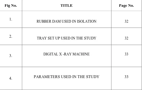
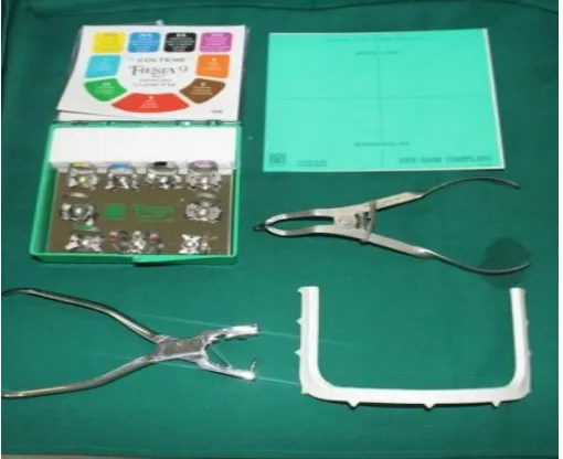
ARMAMENTARIUM : (Fig 1,2,3) 1. Mouth mirror, probe & tweezer
2. Endofrost (Roeko, Germany) 3. DG 16 ( GDC, India)
4. Rubber dam (Hygenic, Roeko, Germany)
5. Local anaesthesia (Lignox 2% A, Warren, India) 6. Bevelled 27 gauge needle gauges
7. EndoPrep RC ( EDTA 10%, Anabond Stedman Pharmaceuticals, India) 8. Round bur ( Mani Co.Japan)
9. Hand K-files (Mani Co. Japan)
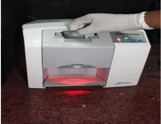
10. RVG imaging system (VistaScan, Durrdental, Germany) 11. Endomotor (NSK Endomate DT, Japan )
12. RaCE rotary files - 6% taper 20/25 size (FKG Dentaire, Switzerland) 13. Root ZX Mini Apex locator (J Morita, Japan)
26
15. Zinc oxide Eugenol sealer (DPI, India ) 16. Paper points ( DiaDent, Korea)
17. Guttapercha points ( DiaDent, Korea)
METHODOLOGY
ETHICS COMMITTEE APPROVAL
This prospective study was approved by the Ethics Committee of CSICDSR (CSICDSR/IEC/0026/2016). All the patients were informed of the aims and design of the study. A written informed consent was obtained from all patients before the clinical procedures, risks were explained and all doubts raised by the patients were clarified.
SAMPLE SIZE CALCULATION
The sample size was derived using G Power Version 3.1.9.2 (Universtät Kiel, Germany). A sample of 317 patients was derived to find a significant difference for effect size variable of 0.25 with alpha error probability of 0.05 using chi squared test.
SUBJECT SELECTION
27
QUESTIONNAIRE
A structured questionnaire was framed which assessed the following parameters including preoperative and intraoperative factors which may contribute to postoperative pain. The postgraduate students who performed the root canal procedures in the study were informed how to record the patient centric and intraoperative information.
INCLUSION CRITERIA
1. The inclusion criteria were the treatment of single tooth with good periodontal
status and completion of treatment in a single sitting. 2. The indication is the teeth with vital pulp with the diagnosis of
symptomatic/asymptomatic irreversible pulpitis.
EXCLUSION CRITERIA
Exclusion criteria is as follows
1. Teeth with presence of necrotic pulp or purulent discharge 2. Patients on corticosteroids, opioids
3. Pregnant women
4. Canal variations like curved canals, calcified canals
28
EVALUATION OF PULP STATUS & RADIOGRAPHIC PRESENTATION
The vitality status of a tooth was determined, testing the tooth to a cold stimulus (Endo Frost, Roeko, Germany) and eliciting an evidence of haemorrage during access cavity preparation. Teeth were assessed with a digital radiograph using a RVG imaging system (VistaScan, Durrdental, Germany). Ostravik periapical index was used to rate the periapical status of the tooth.
DETERMINATION OF PREOPERATIVE PAIN & PREOP MEDICATION
INTAKE
Pre operative pain was the pain assessed if the patient presented with the complaint of pain/discomfort in the concerned tooth 24 hrs prior to treatment and the period before which the pain was present was noted down.
Immediate pre operative pain was recorded as the pain present in the involved tooth within 24 hrs of the root canal procedure being performed and duration of pain was also recorded.
Patients were also enquired about any medication taken within 24 hours prior to root canal procedure.
PARAMETERS INCLUDED
29
OPERATIVE PROCEDURE
30
EVALUATION OF POSTOPERATIVE PAIN
All the patients were prescribed analgesics ( Acelofenac 100 mg and Paracetamol 500 mg) to be taken an hour after the procedure to relieve pain. A postgraduate student unaware of the details of the study called the patients over the telephone and enquired about the pain levels 24 hrs, 48 hrs. On the seventh day and fifteeth day, patients reported back for change of restoration and crown preparation respectively were examined clinically for intensity and type of pain,stimulus of pain and periapical evaluation.
The study used a Likert scale of 0-4 to rate postobturation pain. The pain values utilized in the study as follows,
0-no pain
1-mild pain on mastication and is self relieved
2-moderate persistent pain on mastication and relieved on medication
3-spontaneous persistent pain ,requires higher medication and requires no intervention
4-very severe pain, not relieved by medication and requires intervention
STATISTICAL ANALYSIS
32
ARMAMENTERIUM USED IN THE STUDY
FIG: 1 RUBBER DAM USED FOR ISOLATION
[image:49.612.142.397.174.382.2]
33
[image:50.612.157.430.154.364.2]
FIG 3: DIGITAL X: RAY MACHINE
34
RESULTS
Table 1 depicts the frequency distribution of the study variables included in the study.
35
Instrument fracture incidence was 11 (3.3%) and master cone beyond apex was 10 (3 %). Apical enlargement size of root canals were at 35, 40, 45, 50, 55, 60, 6% 20 and 6% 25 sizes. The apical enlargement size of 6% 25 size was 235 (70.8%) followed by 40 size enlargement and open apex cases had the least value at 2 (0.6%). The incidence of post operative pain observed in the study was at 106 (32.6%).
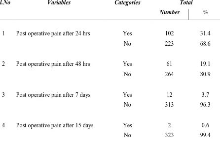
Table 2 shows the post operative pain incidence following root canal treatment at 24 hours, 48 hours,7 days and 15 days. Post operative pain incidence after 24 hours was, 102 (31.4%) of the patients had pain. 61 (19.1%) of the patients had post operative pain after 48 hrs. The post operative pain after 7 and 15 days was at 12 (3.7%) and 2 (0.6%) respectively.
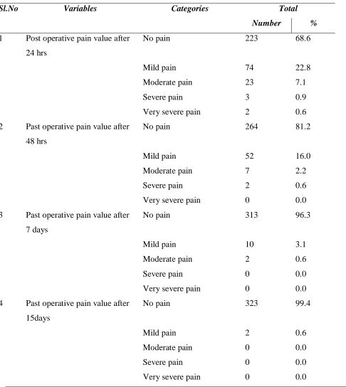
Table 3 depicts that the post operative pain intensity at 24 hours, 48 hours, 7 days and 15 days.
36
Table 4 shows the post operative pain incidence of the study variables.
37
Table 5 depicts the chi square analysis on the impact of post operative pain for the study variables.
The variables immediate pre operative pain status, immediate pre operative pain value, immediate pre operative pain duration, pre operative pain presence, pre operative pain value and intra operative pain were significantly associated with post operative pain (P < 0.05). The other study variables did not show a significant association with post operative pain.
Graph 1 shows the post operative pain value after 24,48 hours, seven days and fifteen days.323 (99.4%) had no pain and 2 (0.6% ) had mild pain at 15 days.
Graph 2 shows the post operative pain incidence at 24,48 hours and seven and fifteen days. According to the study, 106(32.6%) patients had post operative pain.
Graph 3 shows the chi square analysis for the various variables discussed in the study. The graph also depicts shows the significant factors associated with post operative pain.
Chart 1 depicts the frequency distribution of types of mishaps and 10 (3%) of the patients had master cone beyond apex group formed one of the the major ones among the various types of mishaps studied
Chart 2 depicts the frequency distribution of apical enlargement size. 235 (70.8%) of the patients had a apical enlargement size of 6%25 as a major group.
38
Chart 4 depicts the frequency distribution of immediate pre op pain value which shows that 199 (60 %) of the patients had mild pain.
Chart 5 depicts the frequency distribution of pre op pain presence where 223 (61%) patients had pre op pain presence.
Chart 6 depicts the frequency distribution of pre op pain value, 39% and 6% had no pain and severe pain respectively.
Chart 7 depicts the frequency distribution of post operative pain, 106 patients had post operative pain.
39
TABLE 1
FREQUENCY DISTRIBUTION OF KEY EXPLANATORY VARIABLES
Sl.No Variables Categories Total Number %
1 Age 15-20 26 7.8
21-30 106 31.9
31-40 91 27.4
41-50 68 20.5
51-60 29 8.7
61-70 11 3.3
70 plus 1 0.3
2 Gender Male 139 41.9
Female 193 58.1
3 Preop Medication
Yes 115 34.6
No 217 65.4
4 Immediate Preop Pain status
Yes 133 40.1
No 199 59.9
5 Immediate Preop Pain value
No pain 199 59.9
Mild 67 20.2
Moderate 52 15.7
Severe 9 2.7
Very severe 5 1.5
6 Immediate Preop Pain duration
No pain 199 59.9
>1 week 80 24.2 <1week 20 6.0 <10 days 17 5.1 < 1 month 16 4.8
7 Pre op pain presence Yes 203 61.1
no 129 38.9
40
Very severe pain 10 3.0
9 Type of tooth Upper anteriors 44 13.3 Lower anteriors 12 3.6 Upper premolar 50 15.1 Lower premolar 31 9.3 Upper molar 59 17.8 Lower molar 136 41.0
10 Main masticatory tooth Yes 29 8.7
No 303 91.3
11 Intra operative pain Yes 51 15.4
No 281 84.6
12 Mishaps Yes 37 11.1
No 295 88.9
13 Types of Mishaps WL beyond apex 6 1.8 Master cone beyond apex 10 3.0 Obt short of apex 3 0.9 Obt. beyond apex 4 1.2 Instrument fracture 11 3.3
Ledge 2 0.6
Combination of above 1 0.3
None 295 88.9
14 Apical size enlargement 35 size 7 2.1 40 size 41 12.3
45 size 8 2.4
50 size 3 0.9
55 size 8 2.4
60 size 1 0.3
Open apex 2 0.6 6% 20 size 27 8.1 6% 25 size 235 70.8
15 Post operative pain Yes 106 32.6
41
TABLE 2
POST OPERATIVE PAIN INCIDENCE AFTER 24 HRS/48 HRS/7 DAYS/15 DAYS
Sl.No Variables Categories Total
Number %
1 Post operative pain after 24 hrs Yes 102 31.4
No 223 68.6
2 Post operative pain after 48 hrs Yes 61 19.1
No 264 80.9
3 Post operative pain after 7 days Yes 12 3.7
No 313 96.3
4 Post operative pain after 15 days Yes 2 0.6
42
TABLE 3
PAIN INTENSITY VALUE OF POST OPERATIVE PAIN AT 24HRS/48HRS/7DAYS/15DAYS
Sl.No Variables Categories Total
Number %
1 Post operative pain value after 24 hrs
No pain 223 68.6
Mild pain 74 22.8
Moderate pain 23 7.1
Severe pain 3 0.9
Very severe pain 2 0.6 2 Past operative pain value after
48 hrs
No pain 264 81.2
Mild pain 52 16.0
Moderate pain 7 2.2
Severe pain 2 0.6
Very severe pain 0 0.0 3 Past operative pain value after
7 days
No pain 313 96.3
Mild pain 10 3.1
Moderate pain 2 0.6
Severe pain 0 0.0
Very severe pain 0 0.0 4 Past operative pain value after
15days
No pain 323 99.4
Mild pain 2 0.6
Moderate pain 0 0.0
Severe pain 0 0.0
Very severe pain 0 0.0
43
[image:62.612.83.553.140.703.2]
TABLE 4
POST OBTURATION PAIN INCIDENCE OF THE STUDY VARIABLES
Pain incidence Pain incidence
Yes No
Variables Categories Number % Number %
Age 15-20 7 26.9 19 73.1
21-30 36 34 70 66
31-40 28 30.8 63 69.2 41-50 24 35.3 44 64.7 51-60 7 24.1 23 75.9 61-70 3 27.3 8 72.7
Gender Male 43 30.9
96 69.1 Female 63 32.6 130 67.4
Preop Medication Yes 41 35.6
74 64.3
No 65 29.9 152 70
Immediate pre op pain
status Yes 63 47.3
70 52.6
No 42 21.1 157 78.9
Immediate pre op Pain
value No pain 43 21.6
156 78.4
Mild 32 47.7 35 52.2 Moderate 24 46.1 28 53.8 Severe 3 32.3 6 66.7 Very severe 4 80 1 20
Immediate pre op No pain 41 20.6
158 79.9 Less than 1
week 39 48.8 41 51.3 < 1 week 9 45 11 55 <10 days 9 52.9 8 47.1 < 1 Month 5 31.3 11 68.8
Pain incidence
44
Pre op pain presence Yes 77 37.9 126 62.1 No 29 22.5 100 77.5
Pre op pain value No pain 29 22.5
100 77.5 Mild pain 35 33 71 67 Moderate pain 32 41 46 59 Severe pain 4 44.4 5 55.6 Very severe pain 6 60 4 40
Tooth type Upper anterior 12 27.3
32 72.7 Lower anterior 1 8.3 11 91.7 Upper premolar 14 28 36 72 Lower premolar 7 22.6 24 77.4 Upper molar 23 38.9 36 61 Lower molar 49 36 87 64
Main masticatory tooth Yes 12 41.3
17 58.6
No 94 31 209 69
Intra operative pain Yes 31 60.8
20 39.2
No 73 25.9 208 74
Mishaps Yes 14 37.8
20 62.2 No 92 31.2 203 68.8
Types of mishaps WL beyond apex
4 66.7 2 33.3
Master cone
beyond apex 1 10 9 90 Obt short of WL 3 100 0 0
Obturation
beyond WL 3 75 1 25
Instrument
fracture 3 27.3 8 72.7
Ledge 0 0 2 100
Combination 0 0 1 100
none 0 0 295 100
45
Apical size enlargement 35 size 0 0 7 100 40 size 12 29.3 29 70.7
45 size 2 25 6 75
50 size 0 0 3 100
55 size 2 25 6 75
60 size 1 100 0 0
Open apex 1 50 1 50 6% 20 size 5 18.5 22 81.5
46
TABLE 5
CHI SQUARE ANALYSIS
IMPACT OF THE STUDY VARIABLES ON POST OPERATIVE PAIN INCIDENCE
S.No Variable Pearson Chi Square Test
Value DF Significance
1 Age 3.79926 6 0.70382
2 Gender 0.06125 1 0.80452
3 Pre Operative Medication 1.06036 1 0.30301
4 Immediate Pre Operative Pain Status 26.41322 1 0.00000*
5 Immediate Pre Op Pain Value 27.95962 4 0.00001*
6 Immediate Pre Op Pain Duration 27.68335 4 0.00001*
7 Pre Operative Pain Value 12.43350 4 0.01440*
8 Pre Op Pain Presence 8.62811 1 0.00331*
9 Tooth Type 7.61561 5 0.17873
10 Main Masticatory tooth 1.11277 1 0.29148
11 Intra Operative Pain 21.76129 1 0.00000*
12 Mishaps 1.26632 1 0.26046
13 Types Of Mishaps 13.11631 6 0.04123
14 Apical Size Enlargement 10.77652 8 0.21469
47
GRAPH : 1 PAIN INTENSITY VALUE POST OPERATIVE PAIN AT 24/48HRS,7/15DAYS
48
GRAPH 3: CHI SQUARE ANALYSIS IMPACT OF THE STUDY VARIABLES ON POST OPERATIVE PAIN INCIDENCE
CHART 1: FREQUENCY DISTRIBUTION OF TYPES OF MISHAPS
2
6 7 8
9 11 12 6 10 3 4 11 1 2 16.2% 27.0% 8.1% 10.8% 29.7% 2.7% 5.4% 16.2% 27.0% 8.1% 10.8% 29.7% 2.7% 5.4%
16.2% 43.2% 51.4% 62.2% 94.6%
100.0% 1.0% 10.0% 100.0% 0 2 4 6 8 10 12 14
TYPES OF MISHAPS
49
CHART 2: FREQUENCY DISTRIBUTION OF APICAL SIZE ENLARGEMENT
CHART 3: FREQUENCY DISTRIBUTION OF IMMEDIATE PREOP PAIN DURATION
1
2 3
4 5 6
7 8 9
7 41 8 3 8 1 2 27 235 2.1% 12.3%
2.4% 0.9% 2.4% 0.3% 0.6% 8.1%
70.8%
2.1%
12.3%
2.4% 0.9% 2.4% 0.3% 0.6% 8.1%
70.8%
2.1%
14.5% 16.9% 17.8% 20.2% 20.5% 21.1%
29.2% 100.0% 0.0% 10.0% 20.0% 30.0% 40.0% 50.0% 60.0% 70.0% 80.0% 90.0% 100.0% 1 10 100 1000
35 size 40 size 45 size 50 size 55 size 60 size open apex6% 20 size6% 25 size
APICAL SIZE ENLARGEMENT
value Frequency Percent Valid Percent Cumulative Percent
199 60% 80 24% 20 6% 17 5% 16 5%
IMMEDIATE PREOP PAIN DURATION
50
CHART 4: FREQUENCY DISTRIBUTION OF IMMEDIATE PREOP PAIN VALUE
CHART 5: FREQUENCY DISTRIBUTION OF PRE OP PAIN PRESENCE
199, 60% 67, 20%
52, 16% 9, 3%
5, 1% 14, 4%
IMMEDIATE PREOP PAIN VALUE
No pain
Mild
Moderate
Severe
Very severe
203, 61%
129, 39%
PRE OP PAIN PRESENCE
Yes
51
CHART 6 : FREQUENCY DISTRIBUTION OF PRE OP PAIN VALUE
CHART 7: FREQUENCY DISTRIBUTION OF POST OPERATIVE PAIN
39% 32% 23% 3% 3% 6%
PRE OP PAIN VALUE
No pain
Mild Pain
Moderate pain
Severe pain
Very severe pain
0 100 200 300 Y es N o 106 219
POST OPERATIVE PAIN
52
DISCUSSION
Pain is the most common reason for physical consultation in most dental clinics. It is a major symptom in many medical conditions and can interfere with the person’s quality of life and general functioning. A recent survey conducted by the American Association of Endodontists (AAE) suggests that 67% of Americans declared the “fear of pain” as their primary concern regarding root canal procedure31
. Cost of the treatment and pain were the greatest pre-treatment concerns. Anticipated pain has also been demonstrated to be consistently higher than the actual pain experienced during and after the root canal procedure18.
Pain is usually transitory lasting only until the noxious stimuli is removed or the underlying pathology has healed. Endodontic pain arises as a result of the pulp tissue response to any causative agents like dental caries or other irritants like trauma, restorative procedures. The pulp tissue’s bacterial interaction and microbial invasion plays a vital role in pulp inflammation apart from traumatic injuries.
53
Pulpal pain involves peripheral and central sensitization. Pulp nociceptors undergo a process of peripheral sensitization during pulp inflammation, where inflammatory substances are released into the pulp microenvironment. This leads to morphological and phenotypical changes of pulp nociceptors, including sprouting of nerve terminals, increased expression of neuropeptides and awakening of silent nociceptors. Central sensitisation is a pain state characterised by the increased excitability of the projection secondary-order neurons situated in the pars caudalis to peripheral stimulation32.
Pulp nociceptors can be activated and sensitized by bacteria and their by-products directly or indirectly, such as lipopolysaccharide (LPS) is one of the most prominent microbial antigens in endodontic infections, which is recognised by toll-like receptor 4 (TLR-4) expressed in the plasma membrane of odontoblasts32.
54
detected in periradicular lesions. Although some mediators can cause pain by direct effects on sensory nerve fibers, the major inflammatory event responsible for periradicular pain is the increase in vascular permeability and the consequent edema, which lead to the compression of nerve fibers.
Postendodontic pain, our focus of interest is one of the most commonly seen complications of endodontic treatment analysis.
This study involved three hundred and thirty two patients who underwent root canal treatment and seven patients did not report for the follow up. The study involved a questionarire analysis of preoperative, intraoperative and postoperative variables. The post operative pain analysis was done at 24 hrs, 48 hrs, 7 days and 15 days.
The incidence of post operative pain was assessed and the variables were studied for association with postoperative pain. The variables were analyzed by bivariant analysis – the chisquare tests.
Different scales and methods have been used for the assessment of postoperative pain. Likert scale was used in the study to assess the range of pain following root canal treatment. It is a psychometric scale commonly involved in research that employs questionnaires. When responding to a Likert scale, respondents specify their level of agreement or disagreement on a symmetric agree - disagree scale for a series of
statements. Thus, the range captures the intensity of their response for a given item. The
55
The study was restricted to vital teeth. The vitality status of a tooth was determined, testing the tooth to a cold stimulus and having an evidence of haemorrage during access cavity preparation. The treatment was completed in a single visit.
Single visit endodontics is generally indicated for teeth with vital inflamed pulp considering the complications associated, time available for the procedure , clinician’s skills and severity of the symptoms33 34 . The advantages of single visit endodontics include the patient comfort as the number of visits are reduced, economics as the extra cost of multiple visits, use of comparatively less chair side time, fewer materials used which increase the economics to both patient as well as operator, restorative considerations as the immediate placement of coronal restoration ensure effective coronal seal and esthetics, reduced intra appointment pain and flare ups which are usually caused by leakage of the temporary cements50. The success and healing rate of single visit endodontics vs multiple visit endodontics is fairly the same, hence single visit endodontics have been advocated in the treatment procedure10 63.
The following discussion deals with the incidence of post operative pain following root canal treatment, the incidence and intensity of post obturation pain at 24 hours,48 hours,7 days and 15 days and the prognostic and non prognostic factors contributing to the post endodontic pain.
56
The discrepancy in the rate of incidence may be attributed to differences in preoperative status of the tooth, treatment procedures used and severity of the pain, differences in the study methods, geographic and cultural and ethnic variations included in the analysis. Some studies included asymptomatic teeth and reported lower incidence of post operative pain, as preoperative pain was reported a significant finding in many studies7
The reason for the higher incidence and severity of PEP after treatment of teeth with vital pulp may be attributed to the injury of periapical vital tissue during endodontic treatment, which in turn promotes more intensive secretion of inflammatory mediators, such as prostaglandins, leukotrienes, serotonin, histamine, and bradykinin, the pain mediators as well7.
57
neurotransmitters including glutamate and SP from the C fibres leads to activation and upregulation of central receptors for glutamate (NMDA and AMPA receptors) and SP (neurokinin 1 receptor), as well as the release of glutamate in both the trigeminal nuclei and the thalamus32. In addition, central terminals of afferent fibres continue to exhibit increased release of CGRP in the absence of peripheral input from inflamed tissue. Therefore, even if endodontic treatment has removed all peripheral factors contributing to the hyperalgesia, the central mechanism may persist for some time44.
Post operative pain was observed in 24 hours, 48 hours, 7 days and 15 days. The post operative pain intensity was studied as mild, moderate, severe and very severe categories.
The post operative pain incidence at 24 hours, 48 hours, 7 days, 15 days after root canal treatment was at 31.4%, 19.1%, 3.7% and 0.6% respectively (Table 2).
58
The above findings show that post operative pain levels were moderate at 31.4% at 24 hours and the pain levels drops to almost to half the value at 48 hours. At 7 days, the pain levels dropped still further (3.1%) and the pain almost disappeared on the completion of 15th days (0.6%). The post operative pain levels immediate after the root canal procedure is high and it may be caused by ongoing inflammatory processes, apical instrumentation, especially when preexisting periradicular inflammation was present, injection of local anesthetic, pressure from a rubber dam clamp, or discomfort because of prolonged opening of the mouth17.
Study variables studied for association with post obturation pain were age, gender, pre operative medication, pre operative pain value and presence, immediate pre operative pain status, value and duration, intraoperative pain, tooth type, main masticatory tooth, mishaps and type of mishaps and apical enlargement size.
The variables like age, gender, pre operative medication, tooth type, main masticatory tooth, mishaps and type of mishaps and apical enlargement size were not significantly associated with post operative pain.
Age
59
healing and hence postobturation pain6. Age on the increase may be associated with increasing root canal complications like calcifications, pulp stones or the systemic complications that may be associated which would contribute to patient fatigue and compromised healing, which was irrelevant to the study results.
Gender
In this study, females formed the majority of the samples at 58.1% and were associated with pain levels of 32.6%. Gender was not significantly associated with postoperative pain similar to other studies18 21. Various studies reported significant association of gender with post operative pain6 41 42. Females, in these studies were associated with increased pain levels because the incidence of pain is governed by emotional factors and biological differences between genders5, but this was not relevant to the present study.
Pre operative medication
60
prior to treatment procedure43 44. This time period is essential as this closely in accordance to the NSAIDs half-life period.
Tooth type
Upper and lower anteriors, upper and lower premolars and upper and lower molars were included in the study. Lower molars formed the major group in the study sample at 131 patients (41%) and 49 (36%) of them reported post operative pain. Mandibular teeth has been significantly associated with post operative pain as mandible has a dense trabecular pattern, which causes reduced blood flow and localization of infection leading to delayed healing pattern and greater number of canals and complex apical root canal morphology4 6. In the present study tooth type variable is not significantly associated with post operative pain, which is consistent with findings of other studies18 45. Although some studies found significant association of tooth type with postoperative pain6 21 35 46.
Main masticatory tooth
61
where the incidence of post-endodontic pain was lower in the absence (27.7%) than in the presence of occlusion (56.5%) 4.
Mishaps
Endodontic mishaps or procedural accidents are those unfortunate occurrences
that happen during treatment, some owing to inattention to detail, others totally
unpredictable. Over instrumentation may extrude infected material contained in the
canals beyond the apex impeding the healing process of the periapical tissue. Gutta-percha cones, which had been extruded past the apices, have demonstrated the presence of a “biofilm” on the cones49. This “biofilm” allows undisturbed growth of the bacteria
and renders them resistant to the defences of the host and may be responsible for foreign body reactions. The consequences of overfilling can result in infective periapical periodontitis caused by the transport of bacteria beyond the apex and an incomplete cleansing, foreign body reactions, and pain symptoms which are due to irritative stimuli and chronic inflammation even in the absence of radiological evidence50. Mishaps occurred in 11.1% of the pool of patients and among those patients, 37.8% of them
reported post operative pain. The study variable, mishaps was not significantly associated
with post operative pain.
Types of mishaps
Master cone beyond apex, Obturation short of apex, Obturation beyond apex,
Instrument fracture, Ledge, Combinations, WL beyond apex were studied in the study.
Instrument fracture and master cone beyond apex formed the major groups at 3.3% and
62
was not a statistically significant factor contributing to post obturation pain, though
some studies found extent of root filling to be one of the leading factors to postoperative
pain35.
Apical enlargement size
Extrusion of debris lead to irritation and inflammation of periapical tissues. Several factors affect the extrusion of debris includes the irrigation protocol, the final apical size, the time spent on root canal instrumentation and the technique employed for it and the instrument design51. Irritation of periapical tissues results in inflammation and release of many chemical substances which initiate inflammatory responses. The amount of extruded debris and neuropeptides released from C-fibers found in the periodontal ligament, which explains the differences in the severity of postoperative pain experienced by patients52.
Among the various enlargement sizes were used in the study and 6% 25 size remains the leading group at 70.8%. In the present study, the association of apical enlargement size with postendodontic pain is not statistically significant.
The study variables pre operative pain value and presence, immediate pre operative pain status, value and duration, intraoperative pain, were significantly associated with postoperative pain (P < 0.05).
63
Pre operative pain - Pre operative pain was the pain assessed if the patient presented with the complaint of pain/discomfort in the concerned tooth 24 hrs prior to treatment and the period before which the pain was present was noted down.
Immediate pre operative pain - Immediate pre operative pain was recorded as the pain present in the involved tooth within 24 hrs of the root canal procedure being performed and duration of pain was also recorded.
Pre operative pain
Pre operative pain was studied as pre operative pain presence and preoperative pain value. Pre operative pain presence was recorded in the format of yes or no, if the pain was present, patient affirmed the condition, else the patient replied in the negative. Preoperative pain value measures the severity of pain, was recorded as no pain, mild, moderate, severe and very severe pain.
61.1% (203) of the patients reported pre operative pain presence. Among these patients, 37.9% (77) of them had post obturation pain (Table 1). 31.9% (106) of them had mild preoperative pain and of them 33% (35) had post operative pain. 23.5% (78) had moderate pre operative pain and among them 41% (32) had post operative pain. 2.7% (9) had severe pre operative pain and 44.4% had post operative pain. 3% (10) had pre operative pain history and of them 60.1% (6) had post operative pain (Table 4).
64
Pre operative pain and pre operative pain value are statistically significant with post operative pain (P < 0.05) (Table 5) with chi square analysis.
Immediate pre operative pain
Immediate pre operative pain was studied as immediate pre operative pain status, immediate pre operative duration and immediate pre operative value.
Immediate pre operative pain status was recorded in the yes or no format. Immediate pre operative duration was recorded if the patient suffered pain for more than a month, 10 days, a week or less than a week. Immediate pre operative value was measured as mild, moderate, severe and very severe according to the intensity of the symptoms.
Among the 332 patients, 40.1% (133) of the patients had immediate pre operative pain and 47.3% (63) of them had post operative pain (Tables 1, 4).