© 2016. Published by The Company of Biologists Ltd.
αE-catenin-dependent mechanotransduction is essential for proper
convergent extension in zebrafish
Mitchell K.L. Han
1, Esteban Hoijman
2,3, Emily Nöel
2,4, Laurence Garric
2, Jeroen Bakkers
2,5and
Johan de Rooij
1*1Molecular Cancer Research, Center for Molecular Medicine, University Medical Center Utrecht,
Universiteitsweg 100, 3584CG, Utrecht, The Netherlands
2The Hubrecht Institute for Developmental Biology and Stem Cell Research and University Medical
Center Utrecht, Uppsalalaan 8, 3584CT, Utrecht, The Netherlands
3present address: Department of Experimental & Health Sciences, Universitat Pompeu Fabra/PRBB,
Doctor Aiguader 88, 08003, Barcelona, Spain
4present address: Department of Biomedical Science and Bateson Centre, University of Sheffield,
Sheffield, United Kingdom
5Department of Medical Physiology, University Medical Center Utrecht, Yalelaan 50, 3584 CM
Utrecht, the Netherlands
* Corresponding author: Johan de Rooij ([email protected])
Keywords
Summary statement
Cadherin adhesions transduce tension across the junction-actomyosin axis into proportional
biochemical responses via α-catenin. Here we show for the first time how this function of the
Abstract
Cadherin complexes mediate cell-cell adhesion and are crucial for embryonic development. Besides
their structural function, cadherin complexes also transduce tension across the junction-actomyosin
axis into proportional biochemical responses. Central to this mechanotransduction is the stretching of
the cadherin-F-actin-linker α-catenin, which opens its central domain for binding to effectors such as
vinculin. Mechanical unfolding of α -catenin leads to force-dependent reinforcement of
cadherin-based junctions as studied in cell culture. The importance of cadherin mechanotransduction for
embryonic development has not been studied yet. Here we used TALEN-mediated gene disruption to
perturb endogenous αE-catenin in zebrafish development. Zygotic α-catenin mutants fail to maintain
their epithelial barrier, resulting in tissue rupturing. We then specifically disrupted
mechanotransduction, while maintaining cadherin adhesion, by expressing an αE-catenin construct in
which the mechanosensitive domain was perturbed. Expression of either wild-type or
defective α-catenin fully rescues barrier function in α-catenin mutants. Expression of
mechano-defective α-catenin, however, also induces convergence and extension defects. Specifically, the
polarization of cadherin-dependent, lamellipodia-driven cell migration of the lateral mesoderm was
lost. These results indicate that cadherin mechanotransduction is crucial for proper zebrafish
Introduction
During embryonic development, tissue shape and organization is established by a series of complex
cell migrations and rearrangements. These developmental cell-movement processes are largely
dependent on dynamic intercellular adhesions (Martin and Goldstein, 2014). Central to cell-cell
adhesion are the classical cadherins, such as E-Cadherin, which connect cells through homotypic
interactions between their extracellular domains (Leckband and de Rooij, 2014). Through their
cytoplasmic tails, classical cadherins bind p120- and β-catenin, the latter of which in turn associates
with the actin-binder α-catenin. Together they form the cadherin complex which assembles in
structures known as adherens junctions (AJs), which are essential for the maintenance of barriers in
epithelial tissues (Niessen, 2007). Besides in barrier forming AJs, cadherins also cluster in a different
type of cell-cell junction called focal adherens junction (FAJ) that is more dynamic and particularly
apparent during tissue remodeling processes (Huveneers and de Rooij, 2013). By coupling the
actomyosin cytoskeletons of neighboring cells through AJs or FAJs, cadherins mediate the formation
of a contractile network of cells. Thus cadherins are central to many aspects of cell-cell adhesion and
tissue dynamics.
How strongly cadherin-based junctions are involved in tissue shape generation is especially apparent
during gastrulation, the developmental process in which germ layer progenitor cells migrate and
rearrange their cell-cell contacts to achieve tissue morphogenesis. One such movement in zebrafish is
epiboly, the event in which several layers of cells spread and migrate over the yolk cell (Bruce, 2016).
Here the loosely packed deep cells intercalate radially outward into the outer-most epithelial sheet
known as the EVL, thereby pushing their neighbors and contributing to thinning of the deep cell layers
during epiboly progression. Both embryos in which E-cadherin- and α-catenin was knocked down with
anti-sense morpholinos, as well as E-cadherin mutant embryos, show epiboly delay (Kane et al., 2005;
Schepis et al., 2012; Shimizu et al., 2005). This is possibly due to reduced adhesion of the deep cells to
the EVL, which is mediated by cadherin adhesion. Moreover, in these embryos it was observed that
deep cells failed to integrate into more superficial layers and also dropped back into lower layers.
Cadherins have also been implicated in the regulation of cell intercalation in Xenopus (Chen and
Gumbiner, 2006; Marsden and DeSimone, 2003) and Drosophila (Rauzi et al., 2010).
Another, concurrent morphogenetic process in zebrafish embryos in which the importance of cell-cell
junctions is apparent, is convergent extension (CE), which facilitates body axis elongation (Tada and
Heisenberg, 2012; Yin et al., 2009). During CE, the germ layer progenitor cells undergo a combination
of migration and cell intercalation events to converge towards the dorsal midline, while
simultaneously elongating along the anterior-posterior axis (Yin et al., 2008). At the shield stage,
margin as single cells, but subsequently form a highly cohesive cluster of cells that migrates along the
overlying epiblast towards the animal pole, thereby contributing to tissue extension along the
anterior-posterior axis. Both the cohesiveness of the cluster, the migration along the overlying epiblast
(Montero et al., 2005; Ulrich et al., 2005), as well as its directionality (Dumortier et al., 2012) were
shown to be dependent on E-cadherin adhesion. Convergence is mainly driven by the migration of
mesoderm cells to the dorsal midline (Sepich et al., 2005), and is dependent on both E- and N-cadherin
(Arboleda-Estudillo et al., 2010; Warga and Kane, 2007). Furthermore, a BMP-induced gradient of
N-cadherin-dependent cell-cell adhesion was found to regulate the lammelipodial dynamics underlying
the dorsal migration of lateral mesoderm (von der Hardt et al., 2007).
Recent studies show that the cadherin complex not only serves as a structural anchor between cell
adhesions and the actomyosin cytoskeleton, but is also an active site of mechanotransduction:
Increasing tensile forces on cadherin-based junctions induce biochemical changes, which among
others lead to force-dependent reinforcement of the adhesion (Bazellieres et al., 2015; Kannan and
Tang, 2015; le Duc et al., 2010). A key element in cadherin mechanotransduction is α-catenin, which
undergoes a conformational change when under tension. This change alters the conformation and
orientation of central domains in a-catenin (structure papers). The best known consequence of this is
the recruitment of the protein vinculin to tensile cell-cell junctions (Yonemura et al., 2010).
Force-dependent recruitment of vinculin is particularly apparent during tissue remodeling processes, where
dynamic FAJs are formed (Huveneers et al., 2012). Replacement of the mechanosensitive
vinculin-binding-domain in α-catenin by an homologous domain from vinculin (-catenin-VBS), perturbed
vinculin recruitment as well as mechanosensing and junction re-inforcement during epithelial barrier
formation (Twiss et al., 2012). Human umbilical vein endothelial cells (HUVEC) expressing
-catenin-VBS show more severe junction breakage during junction remodeling (Huveneers et al., 2012). It
should be noted that besides vinculin, additional proteins (-actinin, formin, afadin) were mapped to
interact to the perturbed domain in this mechanosensing-defective a-catenin (Kobielak et al., 2004;
Nieset et al., 1997; Pokutta et al., 2002) and could therefore be of equal importance to cadherin
mechanotransduction.
While the importance of the structural role of the cadherin complex has been well established, the
importance of cadherin mechanotransduction during the development of a live organism has not been
investigated. To address this we used zebrafish morphogenesis as a model system. We first generated
αE-catenin-deficient zebrafish using TALEN gene engineering. Homozygous mutant embryos show
defects in epithelial barrier formation and subsequent lethality during somitogenesis. These defects
could be fully rescued by restoring -catenin function through mRNA injection. We then specifically
α-catenin-ΔVBS construct. The resulting embryos showed no barrier defects and survived somitogenesis,
but now exhibited defects in convergent extension-underlying cell migrations that caused severely
perturbed morphogenesis. Zooming in on one of these migration processes by live cell imaging, we
observed that the dorsal migration of the lateral mesoderm cells was reduced. In particular, we found
defects in the polarization of cadherin-dependent lammelipodial adhesion in these cells. Thus,
α-catenin mediated cadherin mechanotransduction is essential for cadherin-dependent cell migration
Results
Generation of
ctnna1
-deficient mutants
To investigate the role of αE-catenin-dependent cadherin mechanotransduction during zebrafish
morphogenesis, we first generated an αE-catenin (ctnna1) deficient mutant. We disrupted the
endogenous ctnna1 locus using TAL-effector nucleases (TALENs) (Cermak et al., 2011) by targeting the
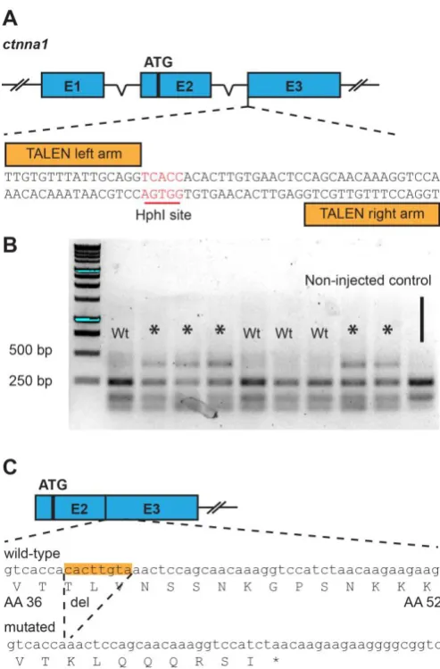
Intron 2-Exon 3 boundary (Fig. 1A). The TALEN cleavage activity was assessed using Restriction
Fragment Length Polymorphism (RLFP) analysis, which is facilitated by the presence of an HphI
restriction enzyme recognition sequence in the cleavage site. The target site was amplified from
genomic DNA of ctnna1 TALEN-injected embryos, and the resulting PCR product was subjected to RFLP
analysis (Fig. 1B). Samples from non-injected controls show bands of approximately 140 bp and 240
bp after HphI digestion, while samples from TALEN-injected embryos may show a third, uncleaved
band at around 400 bp. These uncleaved bands point to the induction of indel mutations which disrupt
the restriction enzyme recognition site. 64 out of 94 embryos injected with the ctnna1 TALEN showed
targeted gene disruption at the endogeneous locus. While no bi-allelic targeting was observed, even
with higher dosages of TALEN mRNA (Data not shown), we can thus conclude that the TALEN is able
to efficiently cleave the endogenous locus at the intended target site.
Next, TALEN-injected embryos were raised to adulthood and then genotyped by fin clipping to identify
potential founders. Of 54 fish screened we could identify 25 fish with somatic ctnna1 mutations.
Individual mutant fish were outcrossed to wild-type fish and the offspring checked for germline
transmission of mutations. While individuals showed different germline transmission rates, of the 87
embryos screened from crosses of potential founders with wild-type, 16 embryos (18%) were found
to be heterozygous for TALEN-generated ctnna1 mutant alleles. TALENs induce double-stranded
breaks at the target site, which due to the error-prone non-homologous end joining mechanism results
in indel mutations (Cermak et al., 2011). Only deletion Δ8 caused a non-sense mutation, leading to a
premature stop codon after Threonine37, which resides 5’ to the β-catenin-binding domain and
actin-binding domain (Fig. 1C). The α-catenin fragment resulting from this mutated allele contains no known
interaction sites and is thus most likely a null mutation. Therefore from this point on, the line carrying
the ctnna1Δ8 allele is designated as ctnna1hu10414 and is the main ctnna1 mutant described further. In
conclusion, we generated an αE-catenin null allele which we used to investigate the effect of loss of
Depletion of zygotic
ctnna1
causes loss of epithelial barrier function
Zygotic ctnna1 mutants show no major defects during cleavage or epiboly stages. In contrast, during
somitogenesis, tissue rupture and subsequent swelling of the yolk was observed, leading to a striking
embryonic lethal phenotype in which the yolk and cells of the embryo proper have undergone lysis
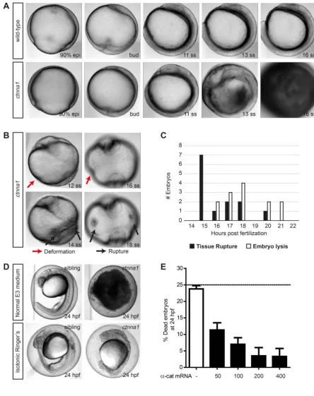
(Fig. 2A, stills from time-lapse sequence, and Movie 1). Prior to embryo lysis, slight embryo
deformations can be visible as indentations compared to the normally spherical morphology of the
embryo at this stage (Fig. 2B – top). In addition, ruptured weak points in the external epiblast can be
observed as black spots. This tissue rupturing did not occur in a fixed location in the embryo, but
occurred in diverse regions in different embryos, such as on the lateral epithelium overlaying the yolk
or near the forming tailbud (Fig. 2B - bottom). Furthermore, not all embryos undergo lysis at the same
time (Fig. 2C), indicating that tissue rupture seemed not restricted to a particular tissue or
developmental process but rather occurs stochastically due to gradual loss of tissue integrity. We
reasoned that tissue rupture is caused by a perturbed epithelial barrier due to loss of functional
adherens junctions. This in turn would increase the osmotic pressure and induce the observed swelling
of the yolk. To test this, we transferred embryos at 50% epiboly stage from regular E3 embryo medium
to isotonic Ringer’s buffer. Under these conditions most mutants did not undergo tissue rupturing,
but survived until after 24 hpf. However, unlike wild-type or heterozygous siblings, the cells of these
ctnna1 mutant embryos show a very rounded morphology at this time point, indicating a lack of
functional adherens junctions (Fig. 2D). By injecting αE-catenin mRNA into offspring of incrossed
ctnna1 heterozygous mutants at the one-cell stage, we were able to rescue the embryo lysis seen in
zygotic ctnna1 homozygous mutants in a dose-dependent manner (Fig. 2E), further confirming that
this phenotype is indeed caused by loss of α-catenin. Rescued ctnna1 embryos survive and look
indistinguishable from non-injected control embryos until at least 24 hpf, after which the outer
epithelium starts to undergo lysis. This is most likely attributed to a gradual loss of injected α-catenin
mRNA and subsequent loss of functional protein. These results demonstrate that ctnna1 is essential
for the maintenance of a functional epithelium, which in turn is crucial for zebrafish embryonic
development.
The mechanosensitive domain of αE-catenin is not necessary for the formation of a
functional epithelial barrier
Tension-induced unfolding of α-catenin is a key element in cadherin mechanotransduction (Twiss et
al., 2012; Yao et al., 2014)}. To interfere with cadherin mechanotransduction in adherens junctions,
we generated an αE-catenin construct lacking mechanosensing capacity. Specifically, we exchanged
highly homologous domain. Coincidentally this domain is obtained from vinculin itself, as it is
α-catenin’s closest homologue (as has previously been described for mammalian α-catenin in
(Huveneers et al., 2012; Twiss et al., 2012)) (Fig. 3A). The mammalian version of this mutant protein
called α-catenin-ΔVBS, was shown to maintain barrier forming capacity in mammalian epithelial cell
culture even though its mechanosensitive capacity (as measured by magnetic twisting cytometry) was
completely lost (Twiss et al., 2012). Moreover, mammalian α-catenin-ΔVBS was shown to be incapable
of binding vinculin even when fully activated (Huveneers et al., 2012). It was concluded from this
previous work that the domain replacement resulted in a structurally intact protein that can function
to link the cadherin complex to the actomyosin cytoskeleton. Conversely, α-catenin-ΔVBS fails to
perform mechanotransduction because binding to key effectors is abolished due to the difference in
primary sequence between the original D3a domain and the replacement.
To characterize zebrafish a-catenin and α-catenin-ΔVBS, we expressed GFP-tagged versions in
α-catenin-deficient DLD1-R2/7 cells (that do not form any cell-cell junctions normally). As shown in
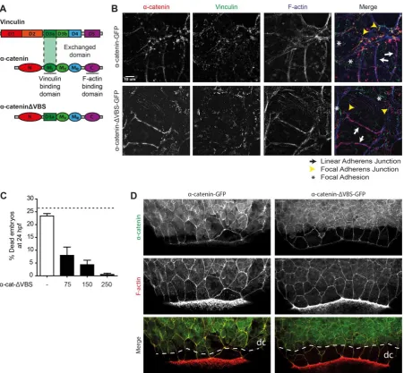
Figure 3B, both zebrafish α-catenin-ΔVBS-GFP and wild-type zebrafish α-catenin-GFP restore junction
formation to a similar level (Fig. 3B – bottom). Furthermore, the mechanosensitive effector vinculin is
recruited to wild-type -catenin-induced junctions. More specifically, vinculin concentrates at those
junctions that display a discontinuous morphology and are oriented perpendicular to the plane of
cell-cell contact (demarcated by yellow arrowheads in Fig. 3B - top). We have previously called these
junctions focal adherens junctions to convey their distinct appearance and association with radially
oriented actin bundles, indicating that tension is applied to these junctions. Conversely, the
-catenin-induced junctions that are continuous within the plain of adhesion and likely experience less tension,
which we call linear adherens junctions (demarcated by white arrows in Fig. 3B), recruited vinculin
less efficiently. This closely mimics the vinculin recruitment to junctions induced by mammalian
-catenin in these cells (Huveneers et al., 2012; Twiss et al., 2012) and this shows that zebrafish a--catenin
is functionally equivalent to mammalian a-catenin both in structural support of cadherin adhesion as
well as in mechanosensing capacity. In contrast to wild-type -catenin-GFP, -catenin-VBS-GFP does
not induce the recruitment of vinculin to focal adherens junctions, as observed by the lack of overlap
in the red (α-catenin) and green (vinculin) channels (Fig. 3B – bottom). This result confirms that also
in zebrafish -catenin-VBS-GFP mechanosensitive capacity is perturbed, while its structural function
in cell-cell adhesion is preserved.
To investigate whether α-catenin-ΔVBS-GFP is able to support junction formation in vivo, we
injected α-catenin-ΔVBS-GFP mRNA in the offspring of incrossed heterozygous ctnna1 mutants. As
shown in Fig. 3C, α-catenin-ΔVBS-GFP is able to rescue the embryonic lethal phenotype, caused by a
that both α-catenin-ΔVBS-GFP can rescue the barrier defect similar to wild-type α-catenin and that
the replacement of the VBS does not impede the maintenance of a functional epithelial barrier
throughout early development. Indeed both α-catenin-GFP and α-catenin-ΔVBS-GFP properly and
similarly localize to cell-cell junctions in the developing embryo. This can be observed in the enveloping
layer (EVL) as well as in the underlying deep cells in the embryos shown in Fig. 3D and Fig S2 . Movie
2 shows additional evidence of junctional localization of α-catenin-GFP and also demonstrates the
difference in dynamics between EVL adherens junctions that are stable and linear and deep cell
adherens junctions that are constantly remodeled and short-lived similar to the FAJs that we have
characterized in 2D cultured mammalian epithelia (see also Movie 5).
To conclude, we have generated an α-catenin-deficient zebrafish mutant as well as functional
rescue constructs, which behave analogously to the mammalian a-catenin constructs previously
described, and that allow us to specifically perturb mechanotransduction without interfering with the
structural role of α-catenin in junctions.
The mechanosensitive domain is essential for proper convergence extension
movements
Because the mutant ctnna1 allele we generated is embryonic lethal in zygotic homozygous mutants,
we could not establish an adult homozygous ctnna1 mutant line. In addition, from the offspring of an
incross of heterozygous ctnna1 mutants, we could not distinguish the 25% homozygous mutants from
their siblings before they started exhibiting the tissue rupture phenotypes described above. This
negatively impacted our ability to analyze rescue experiments. To circumvent this problem, we opted
to use anti-sense transcription-blocking ctnna1 morpholino knockdown. By titrating the right amount
of morpholino (Fig. S1A), we could recapitulate the embryo lysis phenotype observed in ctnna1 zygotic
mutants in almost 100% of the injected embryos. Similar to zygotic mutants, up until early
somitogenesis no severe developmental defects were obvious in the MO-injected embryos. A slight
delay in development is apparent in all injected early stage embryos in Fig. 5A, which is likely caused
by the injection procedure. (See also Fig. S3 for additional images of MO-injected embryos). At later
stage during somitogensis, ctnna1 morphants injected with morpholino undergo embryo lysis just like
zygotic mutants (Fig. 4A, top, still images from time lapse recordings). Embryo lysis could be prevented
by transferring ctnna1 morphants to isotonic Ringer’s buffer at 50% epiboly and cells from the
surviving morphants now exhibited a round morphology, similar to zygotic ctnna1 mutants (Fig. S1B).
Most importantly, we could rescue ctnna1 morphants from embryo lysis by expressing either
α-catenin-GFP (Fig. 4A,B) or α-catenin-ΔVBS (Fig. 4B) using mRNA injection at the 1-cell stage. Together
these results show that ctnna1 morpholino-injected embryos exhibit a similar, αE-catenin-specific
Although ctnna1 morphants rescued with α-catenin-ΔVBS were rescued from embryo lysis, they did
show defects in anterior-posterior axis elongation after gastrulation. α-catenin-ΔVBS-GFP-rescued
embryos often show a shorter body axis than embryos rescued with α-catenin-GFP or wild-type, as
assessed by eye at 1 dpf (Figs. 4C and S4A). For quantification and initial characterization, we grouped
body axis elongation defects in 2 classes: mild and severe. These 2 classes could be clearly
distinguished in α-catenin-ΔVBS rescued embryos but not in non-injected control and only sporadically
in α-catenin-GFP rescued embryos. In the mild phenotype, in addition to the shorter body axis, some
embryos also showed a flatter, wider head, as well as a broader dorsal body axis (Figs. 4C and S4A).
Severely affected embryos show significant retardation in anterior-posterior body axis formation, in
combination with a highly enlarged mediolateral body axis (Fig. 4C). These defects point to impaired
convergent extension movements during gastrulation and have indeed been observed in embryos of
well-established CE regulators such as Wnt/PCP signaling (Topczewski et al., 2001; van Eekelen et al.,
2010). To rule out that these CE defects arose due to non-specific effects of co-injection of ctnna1
morpholino with the mRNAs, we injected either α-catenin-GFP or α-catenin-ΔVBS-GFP mRNA in
embryos from a heterozygous ctnna1 mutant incross. Only injection of α-catenin-ΔVBS-GFP, but not
wild-type α-catenin-GFP, induced defects in body axis elongation (Fig. S4B), further indicating that the
observed defects arose due to the loss of the vinculin binding domain in α-catenin.
To further quantify the extent of CE defects, we measured the angle between the anterior and
posterior ends of embryos at the 5-7 somite stage (12 hpf) (Fig. 5A). Although we did not observe
strong defects in early body development in our visual inspection of ctnna1 mutants (Fig. 2A), ctnna1
morpholino-injected embryos did show an enlarged anterior-posterior angle compared to
non-injected controls (Fig. 5B). We could not rescue this observed phenotype to control levels by
expressing α-catenin-GFP mRNA and therefore conclude that it likely represents a slight delay in
development caused by injection of the morpholino. Importantly, we observed a much more enlarged
anterior-posterior angle in ctnna1 morpholino-injected embryos expressing α-catenin-ΔVBS-GFP.
These embryos also show reduced convergence of the somites to the midline compared to
non-injected control embryos or to ctnna1 morphants expressing α-catenin-GFP (Fig. 5C and Fig. S4C). In
addition, the notochords of more severely affected embryos show a wave-like morphology near the
tailbud, while the most affected embryos the complete structure of the notochord is misformed (Fig.
Blocking cadherin mechanotransduction perturbs directionality of the lateral mesoderm
during dorsal convergence
One major cadherin-dependent cell migration process underlying convergent extension is the dorsal
convergence of the lateral mesoderm during late epiboly stages (Arboleda-Estudillo et al., 2010;
Bakkers et al., 2004; von der Hardt et al., 2007). To test whether the widening of the dorsal bodyplan
in ctnna1 morphant embryos rescued with α-catenin-ΔVBS is correlated with impaired migration of
the lateral mesoderm, we tracked individual mesodermal cells at approximately 90° degrees laterally
from the dorsal midline during 85-90% epiboly stages. Ctnna1 morphants expressing
α-catenin-ΔVBS-GFP show a marked decrease in mesoderm cell migration compared to morphants expressing
wild-type α-catenin-GFP, as shown in representative migration tracks (Fig. 6A,B and Movie 3,4). The
quantification of the movement of these cells in Fig. 6C,D shows that both the net dorsal migration
velocity as well as the total migration speed is significantly lower in α-catenin-ΔVBS-GFP-expressing
morphant embryos in comparison to morphants expressing wild-type α-catenin-GFP. These results
suggest that a reduction in dorsal migration of the lateral mesoderm cells contributes to the
impairment of body axis formation in embryos in which cadherin mechanotransduction is perturbed.
The directional migration of the lateral mesoderm cells was found to be driven by lamellipodial
protrusions. Whereas lamellipodia were found projected in all directions, productive lamellipodia (i.e.
those that coincided with cell body movement) were predominant in the dorsal direction. A
BMP-induced gradient in N-cadherin adhesion was hypothesized to cause the polarized productiveness of
lamellipodia (von der Hardt et al., 2007). Supporting the hypothesis from this previous study, confocal
imaging of lateral mesoderm cells labeled with α-catenin-GFP reveals the rapid formation and
disassembly of cadherin junctions in mesoderm cells during dorsal migration. (Movie 4). To directly
investigate whether blocking cadherin mechanotransduction using α-catenin-ΔVBS also affects
lamellipodia productiveness, we quantified the amount of lammelipodia that coincided with cell body
displacements in either the dorsal or the ventral direction during lateral mesoderm migration at
85-90% epiboly. Similar to what has been reported before for wild-type embryos (von der Hardt et al.,
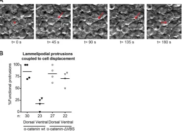
2007), ctnna1 morphants expressing wild-type α-catenin-GFP show approximately 85% functional
lammelipodial protrusions in the dorsal direction (Movie 5), and only 20% in the ventral direction (Fig.
7A,B). In contrast, while morphants expressing α-catenin-ΔVBS show a similar amount of functional
protrusions in the dorsal direction, the amount of functional protrusions in the ventral direction was
strikingly increased to approximately 70%. Interestingly, the total frequency of protrusion formation
was not observed to be different between morphants expressing α-catenin-GFP or
does not alter the formation of lammelipodia per se, but rather perturbs the dorsal polarization of
functional lammelipodial protrusions.
Discussion
The cadherin complex present in adherens junctions is essential for the maintenance of epithelial
tissue barriers. Cadherin complexes also regulate the rearrangements of cell-cell contacts between
neighboring cells in multiple tissues to ultimately achieve proper tissue morphogenesis in the
developing embryo. In this study we show that cadherin complex member αE-catenin is essential for
epithelial barrier functioning and maintaining tissue shape during early morphogenesis. Furthermore,
disruption of its vinculin-binding domain, which has been found to be important for force-induced
junction reinforcement in cell culture, leads to altered lammelipodial polarization and defective dorsal
migration of the lateral mesoderm during dorsal convergence movements. These results indicate that
αE-catenin-dependent cadherin mechanotransduction is essential for the cell migration processes
that underlie zebrafish morphogenesis.
We have used TALEN gene editing technology, targeting the ctnna1 gene at exon 3, to generate a
loss-of-function mutant called ctnna1hu10414 (Fig. 1A). Gene disruption was verified by RLFP analysis and
sequencing. We could not confirm absence of α-catenin protein expression in ctnna1hu10414 mutants
(Data not shown) for lack of suitable antibodies to detect zebrafish α-catenin on Western Blot.
Nevertheless, based on its essential role as an adherens junction component, the resulting epithelial
barrier phenotype is fully consistent with a lack of αE-catenin protein. In analogy, knockdown of
α-catenin in epithelial MDCK cells (Twiss et al., 2012), as well as VE-Cadherin knockdown in endothelial
HUVECs (Pannekoek et al., 2013) induced defects in barrier functioning as measured by Trans
Epithelial/Endothelial Resistance. Many studies exist in which the crucial importance of
cadherin-based cell-cell adhesion formation for epithelial barrier function was reported (Niessen, 2007). In
addition, we were able to rescue the barrier defect by injecting α-catenin mRNA (Fig. 2E) and also
anti-sense ctnna1 morpholino knockdown closely phenocopies the mutant (Fig. 4A). Taken together this
demonstrates that our mutant ctnna1hu10414 is a true functional knockout, and is to our knowledge the
first loss-of-function ctnna1 mutant zebrafish line generated.
Tissue rupturing and subsequent embryo lysis in ctnna1 mutant embryos occurred stochastically
during somitogenesis (Fig. 2A-C). Based on previously-described cadherin mutants and morphants,
earlier severe defects in development would be expected if cells were unable to form cell-cell
junctions from the start of development (Babb and Marrs, 2004; Kane et al., 2005; Shimizu et al.,
phenocopy the mutant. At least 2 related -catenin encoding genes exist in the zebrafish genome
besides E-catenin and it could well be that some functions of E-catenin are rescued by redundancy
with the products from these genes. In a previous study, however, the same morpholino was used
with a 10-fold higher concentration where it did induce epiboly delay (Schepis et al., 2012). Another
explanation for a lack of strong defects in early development, as we derive our mutants by incrossing
heterozygous parents, is that maternal contribution of mRNA and possibly protein provides sufficient
α-catenin to sustain proper morphogenesis during early developmental stages. Tissue rupture is then
observed in locations of the epithelium where the gradual decrease in functional α-catenin due to loss
of zygotic expression has progressed the most. At our used concentration, similarly, the morpholino
may not deplete the maternally contributed α-catenin enough to induce epiboly defects, but may
sufficiently inhibit translation of zygotic ctnna1 transcripts to lead to the same embryo lysis phenotype
also observed with the ctnna1 mutant.
During the rescue experiments, it became clear that expression of both wild-type and α-catenin-ΔVBS
by mRNA injection in ctnna1 mutants/morphants rescued the barrier defect occurring during late
somitogenesis stages. Whereas wild-type αE-catenin-rescued embryos now developed normally until
at least 24 hpf, α-catenin-ΔVBS mRNA injection induced strong defects in convergence extension
already in stages of development that appeared unaffected in mutants of MO-only injected embryos
(Fig 5). We also observed these convergence extension defects upon injection of α-catenin-ΔVBS in wt
embryos in the absence of MO injection, although higher concentrations of mRNA were needed (Data
not shown). This apparent dominant effect of α-catenin-ΔVBS injection is consistent with the notion
that proteins translated from the injected mRNAs gradually replace maternally supplied or redundant,
related α-catenin proteins in the cadherin complex in the developing embryos. Indeed, both wild-type
α-catenin-GFP and α-catenin-ΔVBS-GFP localize to junctions already in early stage embryos (Fig. 3D
shows 8 hpf embryos). The fact that this does not lead to additional defects in the case of wild-type
αE-catenin is expected. In the case of α-catenin-ΔVBS, however, this leads to a dominant perturbation
of the VBS at cell-cell junctions, resulting in a loss of recruitment of vinculin and possibly other
associated proteins. It is very likely that the loss of recruitment of VBS-associated proteins causes the
observed defects in convergence extension. It should be noted that multiple proteins, including
vinculin, -actinin, afadin and formin have been found associate with, or close to, the VBS of -catenin
and the perturbation of each of these or even additional unidentified binding partners could be the
cause of the observed defects.
Convergent extension movements in zebrafish are driven by coordinated migration of mesoderm and
overlaying ectoderm cells towards the dorsal midline and by intercalations of deep cells into the
on functional adherens junctions (Schepis et al., 2012) or E-Cadherin (Babb and Marrs, 2004; Kane et
al., 2005; Shimizu et al., 2005) and it is likely that blocking the catenin-vinculin interaction by
α-catenin-ΔVBS negatively affected adherens remodeling and stabilization. Whether this contributed to
the observed phenotypes, however remains to be investigated. We did find that dorsal convergence
of the lateral mesoderm was strongly reduced when we perturbed cadherin mechanotransduction
using α-catenin-ΔVBS (Fig. 5C and 6). A previous study showed that this coordinated cell migration
process is dependent on E-Cadherin, as this migration is reduced in both E-Cadherin MO knockdown,
as well as E-Cadherin/weg mutant embryos (Arboleda-Estudillo et al., 2010). Another study showed
that this migration is also dependent on N-cadherin, which is negatively regulated by a
ventral-to-dorsal Bmp gradient to generate a reverse gradient in cadherin adhesion from ventral-to-dorsal to ventral regions
(von der Hardt et al., 2007). Dorsal convergence of the paraxial mesoderm was also affected in
N-Cadherin/bib mutants (Warga and Kane, 2007). Our results now show that not only the presence of
cadherin adhesions is needed, but that also their mechanotransduction capacity is essential for the
proper migration of the lateral mesoderm cells towards the dorsal midline.
Besides the above mentioned studies implicating cadherin adhesion in dorsal convergence in zebrafish
embryos, perturbation of myosin IIB was found to disturb convergence in Xenopus embryos (Skoglund
et al., 2008) and it was argued that the failure to generate polarized cytoskeletal forces was the main
cause of the observed defects. It is conceivable that polarized organization of the cytoskeleton
depends also on its anchoring to the cellular cortex, which is mediated for a large part by adhesion
complexes. In Xenopus, both integrin adhesions as well as cadherin adhesions have been implicated
in convergence cell movements (Weber et al., 2011), in zebrafish there is little evidence for
involvement of integrin adhesion. In general it is clear that both integrin as well as cadherin adhesions
generate feedback signals that organize cellular cytoskeletons in response to mechanical interactions
with the extracellular environment (Leckband and de Rooij, 2014). As we have perturbed a-catenin,
which is a cadherin-specific cytoskeletal linker, it is likely that we have directly impacted on the
feedback signals from cadherin adhesion to cytoskeletal organization, which may have impacted
integrin-dependent adhesion and migration processes indirectly (Barry et al., 2015).
Repeating the detailed assessment of mesoderm cell migration by Von der Hardt et al., we find an
increasing number of functional lammelipodial protrusions in the ventral direction, resulting in loss of
migration polarity in α-catenin-ΔVBS-expressing embryos (Fig. 7B). This indicates that the
mechanosensitive domain is important for the polarization of lammelipodial functionality in the dorsal
direction. As discussed above, a likely explanation is that a polarized, force-dependent recruitment of
vinculin and/or another VBS-interacting proteins normally initiates a positive feedback mechanism
lamellipodia preferentially. A gradient of tissue stiffness along the ventral-dorsal axis could establish
such polarization. Whereas the Hammerschmidt work suggests interactions among deep cells within
the same layer orchestrate polarized movements, it is to be expected that cadherin-mediated
interactions between mesoderm cells and other cell layers (epiblast or EVL, or yolk syncytial layer) also
contribute (Carvalho and Heisenberg, 2010). Mechanosensitive feedback, establishing a correlation
between pro-migratory traction generation and substrate stiffness, has been observed for
N-cadherin-based adhesions in a tissue culture model (Ladoux et al., 2010). Related, tension-regulated
morphogenetic processes have been hypothesized for Drosophila border cell migration, in which the
front/back polarity of border cell clusters is maintained through higher tension on E-Cadherin at the
front of the cluster, which in turn polarizes Rac activity in the same direction (Cai et al., 2014), and in
the migration of cells in the prechordal plate, where directionality of the cell collective may depend
on mechanical forces transmitted at E-cadherin contacts (Dumortier et al., 2012). Future studies will
have to determine which cues could contribute to the initial polarization of the lateral mesoderm. The
fact that both BMP signaling as well as cadherin mechanotransduction contribute to mesoderm
convergence underscores the notion that tissue morphogenesis is the combined result of regulation
by chemical and mechanical cues.
In conclusion, the morphogenetic defects induced here by α-catenin-ΔVBS in zebrafish gastrulation
provide direct evidence for the hypothesis that α-catenin-dependent cadherin mechanotransduction
is essential for the proper development of a living embryo. Our experiments furthermore show how
mesoderm cell migration is one process controlled by cadherin mechanotransduction. Future studies
using the tools developed may elucidate additional aspects of zebrafish morphogenesis controlled by
Materials and Methods
Fish Lines and HusbandryThe following fish lines were used: Tupfel Longfin (TL) and ctnna1hu10414. Fish were maintained
according to standard laboratory conditions. Animal experiments were approved by the Animal
Experimentation Committee (DEC) of the Royal Netherlands Academy of Arts and Sciences.
Cell lines and cell culture
DLD1 R2/7 α-catenin-deficient colon carcinoma cells (source) were cultured in high glucose DMEM
containing L-glutamine, supplemented with 10% Fetal Calf Serum and Penicillin/Streptomycin in
standard 10 cm culture dishes. Cells were transfected with zebrafish α-catenin-GFP or
α-catenin-ΔVBS-GFP using X-tremegene9 (Roche) as per manufacturer’s instructions. Cells were tested negative for
mycoplasma infection.
Construction of zebrafish α-catenin-GFP constructs
Zebrafish α-catenin was kindly provided to us by James Nelson. We generated a GFP-tagged version
by generating a PCR fragment containing XhoI/XbaI restriction sites at the terminal ends using the
following primers: forward 5’- GACACTCGAGCCATGACGAGCATTAACACTGCTAACATC-3’ and reverse:
5’- GACATCTAGACGGAGGAGAGTAGATTAGACTTA-3’. We then digested the PCR product with XhoI and
XbaI, and the pEGFP parental vector with SalI and XbaI (clontech), after which we ligated the product,
resulting in ZFα-catenin-GFP x pEGFP. We then subcloned α-catenin-GFP into pCS2 vector for mRNA
transcription. To achieve this we generated a PCR product using the following primers: forward 5’-
CGATTCGAATTCAAGGCCTCGCCACCATGGTGAGCAAG-3’ and reverse 5’-
CGTAATACGACTCACTATAGTTTCAAATACTATCCATAGCTTTGAACTCGCTCAG-3’. The PCR product was
then ligated into pCS2 digested with XhoI and XbaI using Gibson Assembly (NEB), resulting in zebrafish
α-catenin-GFP in pCS2.
We generated zebrafish α-catenin-ΔVBS-GFP by swapping the ΔVBS domains (which originate from
chicken vinculin) with our previously described murine α-catenin-ΔVBS (Huveneers et al., 2012). We
first generated a PCR fragment from our B141 α-catenin-ΔVBS-GFP vector using the following primers:
forward 5’- CGAGGATTCCAGCTTCTCCCAGACTGCAGGTGGAGGAGAGCTGGCATACGCT-3’ and reverse
5’- GTTAGGTTTGGCAGCCAGGGCCAAAGCTGCATTAATCACCTGAGGACACAGCGCTTCTAACTG-3’. We
then digested the zebrafish α-catenin-GFP x pCS2 vector using Bsu36I and NheI restriction sites, after
which we ligated the PCR product using Gibson Assembly, resulting in zebrafish α-catenin-ΔVBS-GFP
Plasmids were linearized with NotI (Promega) after which IVT mRNA was generated using the
mMessage mMachine SP6 kit (Ambion), and purified using the RNeasy mini kit (Qiagen).
Immunohistochemistry and cytoskeletal washout
For immunocytochemistry, DLD1 R2/7 cells were washed 1 time in PBS, 1 time in CSK buffer (300 mM
Sucrose, 0.5% TX-100, 10 mM Pipes pH 7, 50 mM NaCl, 3mM CaCl2, 2 mM MgCl2) and 1 time in PBS
before fixation using 4% paraformaldehyde in PBS for 20 min. After fixation, cells were permeabilized
with 0,4% Triton X-100 in PBS for 5 minutes and blocked in 2% BSA for 1 hour. Phalloidin-415
(Promokine), vinculin mouse primary antibody (hVin1 clone 1:500; Sigma) and Alexa-594 secondary
antibody (Life Technologies) were diluted in 2% BSA and incubated with the cells for 1 hour.
Afterwards, cells were mounted in Mowiol 4–88/DABCO solution (Sigma-Aldrich).
Embryos were fixed in 2% PFA in PBS overnight at 80% epiboly stage. After washing in PBS, embryos
were dechorionated and further washed 3-5 times 5 minutes in PBT (PBS + 0.1% Triton-X100). Embryos
were blocked in PBT +10% Normal Goat Serum for 1 hour, then incubated O/N with
Phalloidin-Alexa594 (Life Technologies). Samples were then washed 4x 30 min in PBT before mounting in 0.3%
agarose in E3 embryo medium.
Generation of ctnna1 TALEN constructs
TALENs targeting the ctnna1 locus were designed using the TALE-NT tool
(https://tale-nt.cac.cornell.edu/node/add/talen) using the guidelines specified in Cermak et al. The chosen optimal
target sequence in exon 3: 5’-
TTGTGTTTATTGCAGGTCACCACACTTGTGAACTCCAGCAACAAAGGTCCA-3’ (binding sites of the Left and Right TALEN arms are underlined) contains a HphI (NEB) restriction
enzyme site (GGTGA). TALENs were generated using the Golden Gate kit (Cermak et al., 2011) in
combination with the obligate heterodimeric FokI pCS2TAL3DD (Addgene, #37275) and pCS2TAL3RR
(Addgene, #37276) backbones (Ota et al., 2013).
TALEN targeting plasmids were linearized with NotI (Promega) after which IVT mRNA was generated
using the mMessage mMachine SP6 kit (Ambion), and purified using the RNeasy mini kit (Qiagen).
Microinjection of morpholinos and mRNA
Translation-blocking morpholinos (MOs) targeting zebrafish ctnna1 were synthesized by GeneTools,
LLC. The MO partially blocked the 5’UTR and ATG-start codon: 5’-TAATGCTCGTCATGTTCCAAATTGC-3’
and is described earlier [Schepis]. Both MOs and mRNA were diluted in nuclease-free water containing
phenol-red. Injection mix was microinjected into the yolk at the 1-cell stage. Rescue experiments were
Genotyping of mutant alleles
To isolate genomic DNA, zebrafish embryos or fin clips were incubated in 50 µl or 100 µl embryo lysis
buffer respectively, for 60 minutes at 60°C. Proteinase K was heat-inactivated by incubating the
samples for 15 min at 95°C. The genomic DNA was used as template to amplify the ctnna1 TALEN
target site. Primers flanking the target site of ctnna1: Alphacat HphI forward, 5’-
GTCTTGCCACCCCTTAATCC-3’ and Alphacat HphI reverse, 5’- CAATCTTCTCGCCTTTCTCC-3’.
To assess ctnna1 mutant alleles, the PCR amplicons were incubated with HphI (NEB) at 37°C for 2 hrs,
resulting in the generation of 245 bp and 140 bp fragments. Successful mutagenesis was detected by
loss of the restriction recognition site and the presence of an undigested band of 385 bp. Mutant
alleles were sequenced using primer Alphacat HphI reverse, 5’- CAATCTTCTCGCCTTTCTCC-3’.
In situ hybridization
Embryos were fixed overnight in 4% paraformaldehyde (PFA) and stored in MeOH. In situ hybridization
experiments were performed as previously described (Noel et al., 2013). Once staining had developed,
embryos were washed several times in phosphobuffered saline with 0.1% tween and fixed overnight
in 4% PFA.
Microscopy and live imaging
Fixed cells were imaged using an inverted research widefield microscope (Eclipse Ti; Nikon) with
perfect focus system, equipped with a 60x 1.49 NA Apochromat total internal reflection fluorescence
(oil) objective lens, a microscope cage incubator (OkoLab), and an EM charge-coupled device (CCD)
camera (Andor Technology) controlled with NIS-Elements Ar 4.0 software. All widefield images, unless
specifically indicated otherwise were background subtracted by ImageJ’s rolling ball (r=20) and
sharpened for display by Fiji/ImageJ’s unsharp mask filter (r=1, weight=0.6).
Fixed embryos were imaged on the same microscope using the Nikon C1 Confocal system. For
time-lapse imaging, embryos were dechorionated at 50% epiboly stage and embedded in 0.3% agarose in
E3 embryo medium. DIC images were taken every 15 minutes on the same microscope using a 10x 0.3
NA Plan Fluor objective lens. Embryo staging was determined by wild-type embryos imaged
simultaneously with mutants or morphants. Live embryos at 12-24 hpf and fixed embryos after
Quantification of body axis elongation
Embryos were imaged from the lateral position at 12 hpf. Images were processed in Fiji (ImageJ) as
follows: The center of the embryo was determined by manually fitting a circle over the yolk. Lines
were drawn from the center to the most anterior position of the head, and most-posterior position of
the tail. The head-tail angle was then assessed using the angle tool.
Imaging dorsal convergence and lammelipodia
Embryos were dechorionated at 50% epiboly stage and embedded in 0.3% agarose in E3 embryo
medium. DIC images of the lateral mesoderm during dorsal convergence (85-90% epiboly) were taken
every 45 seconds on a Nikon Ti microscope using a 20x 0.75 NA Plan Fluor objective lens. Cells residing
in the layer closest to the yolk syncytium were tracked using Fiji/imageJ with the Manual Tracking
plugin. Lateral mesodermal cells from wild-type embryos overexpressing α-catenin-GFP were imaged
during dorsal convergence (85-90% epiboly) using a Leica SP8 confocal microscope. Images were taken
every 45 seconds. Images were sharpened for display by using Fiji/ImageJ’s Gaussian blur filter (r=2)
Acknowledgements
We thank Ewart Kuijk, Stefan Schulte-Merker and James Nelson for reagents. We also thank the
Hubrecht Animal Caretakers for the fish husbandry.
Competing interests
The authors declare no competing financial interests.
Author contributions
E.H., J.B. and J.R conceived and guided the project. M.H. and E.N. designed experiments. M.H.
performed most of the experiments and analyzed the data. L.G. performed the in situs. M.H. and J.R.
wrote the manuscript.
Funding
References
Arboleda-Estudillo, Y., Krieg, M., Stuhmer, J., Licata, N. A., Muller, D. J. and Heisenberg, C. P. (2010).
Movement directionality in collective migration of germ layer progenitors. Curr Biol 20, 161-169.
Babb, S. G. and Marrs, J. A. (2004). E-cadherin regulates cell movements and tissue formation in early
zebrafish embryos. Dev Dyn 230, 263-277.
Bakkers, J., Kramer, C., Pothof, J., Quaedvlieg, N. E., Spaink, H. P. and Hammerschmidt, M. (2004).
Has2 is required upstream of Rac1 to govern dorsal migration of lateral cells during zebrafish gastrulation. Development 131, 525-537.
Barry, A. K., Wang, N. and Leckband, D. E. (2015). Local VE-cadherin mechanotransduction triggers
long-ranged remodeling of endothelial monolayers. J Cell Sci 128, 1341-1351.
Bazellieres, E., Conte, V., Elosegui-Artola, A., Serra-Picamal, X., Bintanel-Morcillo, M., Roca-Cusachs, P., Munoz, J. J., Sales-Pardo, M., Guimera, R. and Trepat, X. (2015). Control of cell-cell forces
and collective cell dynamics by the intercellular adhesome. Nat Cell Biol 17, 409-420.
Bruce, A. E. (2016). Zebrafish epiboly: Spreading thin over the yolk. Dev Dyn 245, 244-258.
Cai, D., Chen, S. C., Prasad, M., He, L., Wang, X., Choesmel-Cadamuro, V., Sawyer, J. K., Danuser, G. and Montell, D. J. (2014). Mechanical feedback through E-cadherin promotes direction
sensing during collective cell migration. Cell 157, 1146-1159.
Carvalho, L. and Heisenberg, C. P. (2010). The yolk syncytial layer in early zebrafish development. Trends Cell Biol 20, 586-592.
Cermak, T., Doyle, E. L., Christian, M., Wang, L., Zhang, Y., Schmidt, C., Baller, J. A., Somia, N. V., Bogdanove, A. J. and Voytas, D. F. (2011). Efficient design and assembly of custom TALEN and
other TAL effector-based constructs for DNA targeting. Nucleic Acids Res 39, e82.
Chen, X. and Gumbiner, B. M. (2006). Paraxial protocadherin mediates cell sorting and tissue
morphogenesis by regulating C-cadherin adhesion activity. J Cell Biol 174, 301-313.
Dumortier, J. G., Martin, S., Meyer, D., Rosa, F. M. and David, N. B. (2012). Collective mesendoderm
migration relies on an intrinsic directionality signal transmitted through cell contacts. Proc
Natl Acad Sci U S A 109, 16945-16950.
Huveneers, S. and de Rooij, J. (2013). Mechanosensitive systems at the cadherin-F-actin interface. J Cell Sci 126, 403-413.
Huveneers, S., Oldenburg, J., Spanjaard, E., van der Krogt, G., Grigoriev, I., Akhmanova, A., Rehmann, H. and de Rooij, J. (2012). Vinculin associates with endothelial VE-cadherin
junctions to control force-dependent remodeling. J Cell Biol 196, 641-652.
Kane, D. A., McFarland, K. N. and Warga, R. M. (2005). Mutations in half baked/E-cadherin block cell
behaviors that are necessary for teleost epiboly. Development 132, 1105-1116.
Kannan, N. and Tang, V. W. (2015). Synaptopodin couples epithelial contractility to
alpha-actinin-4-dependent junction maturation. J Cell Biol 211, 407-434.
Kobielak, A., Pasolli, H. A. and Fuchs, E. (2004). Mammalian formin-1 participates in adherens
junctions and polymerization of linear actin cables. Nat Cell Biol 6, 21-30.
Ladoux, B., Anon, E., Lambert, M., Rabodzey, A., Hersen, P., Buguin, A., Silberzan, P. and Mege, R. M. (2010). Strength dependence of cadherin-mediated adhesions. Biophys J 98, 534-542. le Duc, Q., Shi, Q., Blonk, I., Sonnenberg, A., Wang, N., Leckband, D. and de Rooij, J. (2010). Vinculin
potentiates E-cadherin mechanosensing and is recruited to actin-anchored sites within adherens junctions in a myosin II-dependent manner. J Cell Biol 189, 1107-1115.
Leckband, D. E. and de Rooij, J. (2014). Cadherin adhesion and mechanotransduction. Annu Rev Cell Dev Biol 30, 291-315.
Marsden, M. and DeSimone, D. W. (2003). Integrin-ECM interactions regulate cadherin-dependent
cell adhesion and are required for convergent extension in Xenopus. Curr Biol 13, 1182-1191.
Martin, A. C. and Goldstein, B. (2014). Apical constriction: themes and variations on a cellular
Montero, J. A., Carvalho, L., Wilsch-Brauninger, M., Kilian, B., Mustafa, C. and Heisenberg, C. P.
(2005). Shield formation at the onset of zebrafish gastrulation. Development 132, 1187-1198.
Nieset, J. E., Redfield, A. R., Jin, F., Knudsen, K. A., Johnson, K. R. and Wheelock, M. J. (1997).
Characterization of the interactions of alpha-catenin with alpha-actinin and beta-catenin/plakoglobin. J Cell Sci 110 ( Pt 8), 1013-1022.
Niessen, C. M. (2007). Tight junctions/adherens junctions: basic structure and function. J Invest Dermatol 127, 2525-2532.
Noel, E. S., Verhoeven, M., Lagendijk, A. K., Tessadori, F., Smith, K., Choorapoikayil, S., den Hertog, J. and Bakkers, J. (2013). A Nodal-independent and tissue-intrinsic mechanism controls
heart-looping chirality. Nat Commun 4, 2754.
Ota, S., Hisano, Y., Muraki, M., Hoshijima, K., Dahlem, T. J., Grunwald, D. J., Okada, Y. and Kawahara, A. (2013). Efficient identification of TALEN-mediated genome modifications using
heteroduplex mobility assays. Genes Cells 18, 450-458.
Pannekoek, W. J., Linnemann, J. R., Brouwer, P. M., Bos, J. L. and Rehmann, H. (2013). Rap1 and
Rap2 antagonistically control endothelial barrier resistance. PLoS One 8, e57903.
Pokutta, S., Drees, F., Takai, Y., Nelson, W. J. and Weis, W. I. (2002). Biochemical and structural
definition of the l-afadin- and actin-binding sites of alpha-catenin. J Biol Chem 277, 18868-18874.
Rauzi, M., Lenne, P. F. and Lecuit, T. (2010). Planar polarized actomyosin contractile flows control
epithelial junction remodelling. Nature 468, 1110-1114.
Schepis, A., Sepich, D. and Nelson, W. J. (2012). alphaE-catenin regulates cell-cell adhesion and
membrane blebbing during zebrafish epiboly. Development 139, 537-546.
Sepich, D. S., Calmelet, C., Kiskowski, M. and Solnica-Krezel, L. (2005). Initiation of convergence and
extension movements of lateral mesoderm during zebrafish gastrulation. Dev Dyn 234, 279-292.
Shimizu, T., Yabe, T., Muraoka, O., Yonemura, S., Aramaki, S., Hatta, K., Bae, Y. K., Nojima, H. and Hibi, M. (2005). E-cadherin is required for gastrulation cell movements in zebrafish. Mech Dev 122, 747-763.
Skoglund, P., Rolo, A., Chen, X., Gumbiner, B. M. and Keller, R. (2008). Convergence and extension
at gastrulation require a myosin IIB-dependent cortical actin network. Development 135, 2435-2444.
Tada, M. and Heisenberg, C. P. (2012). Convergent extension: using collective cell migration and cell
intercalation to shape embryos. Development 139, 3897-3904.
Topczewski, J., Sepich, D. S., Myers, D. C., Walker, C., Amores, A., Lele, Z., Hammerschmidt, M., Postlethwait, J. and Solnica-Krezel, L. (2001). The zebrafish glypican knypek controls cell
polarity during gastrulation movements of convergent extension. Dev Cell 1, 251-264.
Twiss, F., Le Duc, Q., Van Der Horst, S., Tabdili, H., Van Der Krogt, G., Wang, N., Rehmann, H., Huveneers, S., Leckband, D. E. and De Rooij, J. (2012). Vinculin-dependent Cadherin
mechanosensing regulates efficient epithelial barrier formation. Biol Open 1, 1128-1140.
Ulrich, F., Krieg, M., Schotz, E. M., Link, V., Castanon, I., Schnabel, V., Taubenberger, A., Mueller, D., Puech, P. H. and Heisenberg, C. P. (2005). Wnt11 functions in gastrulation by controlling cell
cohesion through Rab5c and E-cadherin. Dev Cell 9, 555-564.
van Eekelen, M., Runtuwene, V., Overvoorde, J. and den Hertog, J. (2010). RPTPalpha and PTPepsilon
signaling via Fyn/Yes and RhoA is essential for zebrafish convergence and extension cell movements during gastrulation. Dev Biol 340, 626-639.
von der Hardt, S., Bakkers, J., Inbal, A., Carvalho, L., Solnica-Krezel, L., Heisenberg, C. P. and Hammerschmidt, M. (2007). The Bmp gradient of the zebrafish gastrula guides migrating
lateral cells by regulating cell-cell adhesion. Curr Biol 17, 475-487.
Warga, R. M. and Kane, D. A. (2007). A role for N-cadherin in mesodermal morphogenesis during
Weber, G. F., Bjerke, M. A. and DeSimone, D. W. (2011). Integrins and cadherins join forces to form
adhesive networks. J Cell Sci 124, 1183-1193.
Yao, M., Qiu, W., Liu, R., Efremov, A. K., Cong, P., Seddiki, R., Payre, M., Lim, C. T., Ladoux, B., Mege, R. M., et al. (2014). Force-dependent conformational switch of alpha-catenin controls vinculin
binding. Nat Commun 5, 4525.
Yin, C., Ciruna, B. and Solnica-Krezel, L. (2009). Convergence and extension movements during
vertebrate gastrulation. Curr Top Dev Biol 89, 163-192.
Yin, C., Kiskowski, M., Pouille, P. A., Farge, E. and Solnica-Krezel, L. (2008). Cooperation of polarized
cell intercalations drives convergence and extension of presomitic mesoderm during zebrafish gastrulation. J Cell Biol 180, 221-232.
Yonemura, S., Wada, Y., Watanabe, T., Nagafuchi, A. and Shibata, M. (2010). alpha-Catenin as a
Fig. 1. Generation of ctnna1 mutants using TAL-effector Nucleases (TALENs). (A) Schematic
representation of the endogenous ctnna1 locus which was targeted by TALENs at the intron2-exon 3
boundary. The TALEN arms flank a restriction enzyme recognition site (highlighted in red) used for
screening mutant alleles through restriction fragment length polymorphism (RFLP) analysis. (B) RFLP
analysis of embryos injected with ctnna1-specfic TALENs. Uncleaved bands showing successful TALEN
activity are marked by an asterisk. (C) The sequence of the cttna1 mutant allele and the consequence
Fig. 2. Zygotic ctnna1 mutants show defects in epithelial barrier functioning. (A-B) DIC images from
a time-lapse series of zygotic ctnna1 mutants and wild-type siblings imaged from 90% epiboly until 21
somite stage. Lateral view in (A) shows embryo lysis in zygotic ctnna1 mutants during somitogenesis.
Anterior views of zygotic ctnna1 mutants in (B) show deformation (red arrows) and tissue rupture
(black arrows) occurring before embryo lysis. (C) Quantification of the time of rupture and subsequent
embryo lysis in zygotic ctnna1 mutants. (n=13 embryos from three independent experiments) (D)
Phenotype at 24 hpf of zygotic ctnna1 mutants and siblings grown in regular E3 medium, and embryos
transferred to isotonic Ringer’s buffer at 50% epiboly. (E) Rescue of embryo lysis in ctnna1 mutants
using increasing concentrations of α-catenin-GFP mRNA. The dashed line indicates the expected
mortality of non-injected incrossed heterozygous ctnna1 mutants based on Mendelian genetics. Data
represent three independent experiments, n>120 embryos per condition. Data are represented as the
Fig. 3. The vinculin-binding domain in αE-catenin is not needed for barrier function. (A) Schematic
representation of the structures of α-catenin, vinculin and the exchanged domain in α-catenin-ΔVBS.
The domain containing the vinculin binding site (VBS) was replaced by a homologous domain from
vinculin itself, which does not bind vinculin. (B) Images of fixed α-catenin-deficient DLD1 R2/7 cells
expressing zebrafish α-catenin-GFP (top) or α-catenin-ΔVBS-GFP (bottom) (both depicted in red), and
stained for vinculin (green) and F-actin (blue). Linear Adherens Junction structures are highlighted by
the arrow, Focal Adherens Junctions with the arrowheads, and Focal Adhesions with an asterisk. (C)
Rescue of embryo lysis in ctnna1 mutants using increasing concentrations of α-catenin-ΔVBS-GFP
mRNA. The dashed line indicates the expected mortality of non-injected incrossed heterozygous
ctnna1 mutants based on Mendelian genetics. Data represents three independent experiments,
n>120 embryos per condition. Data is represented as the mean ± s.e.m. (D) Whole-mount
immunostaining of zebrafish embryos at 85% epiboly expressing either catenin-GFP (left) or
α-catenin-ΔVBS-GFP (right)(both depicted in green) and stained for F-actin (red). The dashed white line
Fig. 4. The vinculin-binding domain is essential for proper body axis elongation. (A) DIC images from
a time-lapse image series of embryos injected with 0.84 ng ctnna1 MO (top) rescued with 250 pg
α-catenin-GFP (middle). Embryos were imaged from 80% epiboly until 21 somite stage. (B-D)
Quantification of the mortality (B) and the body axis phenotype (D) of non-injected embryos, embryos
injected with 0.56 ng ctnna1 MO or ctnna1 MO-injected embryos rescued with either 250 pg
α-catenin-GFP or 250 pg α-catenin-ΔVBS-GFP. Data represents three independent experiments, n>70
embryos per condition. Data is represented as the mean± s.e.m. (C) Representative images of
non-injected control embryos (left) and ctnna1 MO-non-injected embryos rescued with catenin-GFP or
ΔVBS-GFP at 24 hpf. Clear morphological differences between GFP and
α-catenin-ΔVBS-GFP-rescued embryos were apparent in anterior-posterior axis extension (top) as well as in the
mediolateral dorsal convergence (middle and bottom). Red lines indicate the width of the dorsal body
Fig. 5. Blocking vinculin recruitment to α-catenin induces convergent extension defects. (A) DIC
images of non-injected embryos, embryos injected with 0.84 ng ctnna1 MO or ctnna1 MO-injected
embryos rescued with either 250 pg α-catenin-GFP or 250 pg α-catenin-ΔVBS-GFP at the 5-7 somite
stage (12 hpf). (B) Quantification of the angle between the anterior-most point in the head, and the
posterior-most point in the tail of the embryos shown in (shown in red in A). Datapoints represent
single embryos from three independent experiments (n>25 embryos per condition). Data is
represented as mean± s.e.m. One-way ANOVA following Tukey's multiple comparison test was used
for comparisons among multiple groups, with *** indicating significance as p<0.0001. (C)
Whole-mount in situ hybridization at 9-11 somite stage of non-injected embryos and ctnna1 MO-injected
embryos rescued with either 250 pg α-catenin-GFP or 250 pg α-catenin-ΔVBS-GFP. MyoD marks the
[image:30.595.173.424.99.360.2]Fig. 6. Loss of cadherin mechanotransduction results in reduced dorsal convergence of the lateral
mesoderm. The dorsal convergence of the lateral mesoderm was imaged using DIC time-lapse
microscopy. Lateral cells were imaged 90°C from the dorsal shield at 85-90% epiboly over 30 min with
45 sec intervals. Embryos were injected with 0.84 ng ctnna1 MO and rescued with either 250 pg
mesodermal cells (n=23) of a single embryo expressing α-catenin-GFP (A) or α-catenin-ΔVBS-GFP (B).
Dorsal to the right and animal pole to the top. Tracks were offset along the dorsoventral axis such that
each track starts at x=0. (C,D) Quantification of the net dorsal migration velocity (C) and the total
migration speed (D) of lateral mesodermal cells. Data represent the mean of n>100 cells from 4
embryos per condition from 3 independent experiments. (***= P<0.001, unpaired two-tailed t-test).
Fig. 7. Altered lammelipodial polarity of the lateral mesoderm after blocking cadherin
mechanotransduction. (A) Representative image sequence of a functional lammelipodial protrusion
(red arrows) coupled to cell body displacement. The red asterisk marks the cell making protrusions,
[image:33.595.71.516.98.660.2]of lammelipodia coupled to cell body displacement in the direction of the protrusion by at least half
the diameter of the cell. The number of cells analyzed (n) for each case is indicated below the x-axis.
These were obtained from 4 embryos from 3 independent experiments and each datapoint in the
Fig.S1. Ctnna1 morpholino induces tissue rupturing
(A) Quantification of the induction of embryo lethality by injection with increasing doses of αE-catenin translation blocking morpholoinos. Data represent three independent experiments, n>120 embryos per condition. Data are represented as the mean +/- s.e.m. (B) Images of non-injected control (top) and ctnna1 MO-injected embryos (bottom) at 24 hpf, incubated in isotonic Ringer’s buffer.
Fig.S2. α-catenin-GFP and α-catenin-∆VBS-GFP localize to EVL and deep cell junctions in developing embryos.
Embryo’s injected with the indicated constructs were fixed at ~50% epiboly and imaged using a confocal at the plane of the EVL and deeper at the indicated z-planes.
Fig.S3. embryos injected with both MO and α-catenin-GFP develop without gross defects in body axis formation. 6 representative, MO- and α-catenin-GFP-injected embryos from the experiment in figure 5B are shown.
Fig.S4. Rescue of ctnna1 morphants/mutants with α-catenin-∆VBS-GFP induces body axis defects.
(A) DIC images of non-injected embryos and 0.84 ng ctnna1 MO-injected embryos rescued with either 250 pg α-catenin -GFP or 250 pg α-catenin-∆VBS-GFP at 24 hpf. (B) Quantificationof the body axis phenotype of embryos from the offspring of heterozygous ctnna1 mutants injected with either α-catenin -GFP or α-catenin-∆VBS-GFP. The dashed line indicates the expected amount of homozygous ctnna1 mutants based on Mendelian genetics. Data represent three independent experiments, >120 embryos per condition and are depicted as the mean +/- s.e.m. (C) DIC images of somites from non-injected control and ctnna1 MO-injected embryos rescued with 250 pg α-catenin-∆VBS-GFP at the 5-7 somite stage (12 hpf), seen from the dorsal side. Red lines indicate the width of the dorsal body embryonic structure.
Movie 1. Timelapse DIC images of wild-type and ctnna1 mutant. Lateral views. 15 min/frame. Playback 15 fps.
Movie 2. Z-planes from a confocal time-lapse showing α-catenin-GFP localization and dynamics in EVL and deep-cells . Images were sharpened for display by using Fiji/ImageJ’s Gaussian blur filter (r=2) and unsharp mask filter (r=2, weight=0.6). 45s/frame. Playback 12 fps.
Movie 3. Dorsal convergence of the lateral mesoderm in ctnna1 morphants expressing α-catenin-GFP.
85-90% epiboly.Cells were manually tracked in Fiji/ImageJ using the Manual Tracking plugin. Dorsal to the right, animal pole to the top. 45s/frame. Playback 10 fps.