Review
Which patellofemoral joint imaging features are associated with
patellofemoral pain? Systematic review and meta-analysis
B.T. Drew
y z
, A.C. Redmond
y z
, T.O. Smith
x
, F. Penny
k
, P.G. Conaghan
y z
* yLeeds Institute of Rheumatic and Musculoskeletal Medicine, University of Leeds, UKzNIHR Leeds Musculoskeletal Biomedical Research Unit, Leeds, UK xSchool of Health Sciences, University of East Anglia, Norwich, UK
kPhysiotherapy Department, Norfolk and Norwich University Hospitals NHS Foundation Trust, Norwich, UK
a r t i c l e i n f o
Article history: Received 30 March 2015 Accepted 9 September 2015
Keywords: Patellofemoral pain Magnetic resonance imaging Systematic review Diagnostic imaging
s u m m a r y
Objectives: To review the association between patellofemoral joint (PFJ) imaging features and patello-femoral pain (PFP).
Design: A systematic review of the literature from AMED, CiNAHL, Cochrane Central Register of Controlled Trials (CENTRAL), MEDLINE, PEDro, EMBASE and SPORTDiscus was undertaken from their inception to September 2014. Studies were eligible if they used magnetic resonance imaging (MRI), computed tomography (CT), ultrasound (US) or X-ray (XR) to compare PFJ features between a PFP group and an asymptomatic control group in people<45 years of age. A pooled meta-analysis was conducted and data was interpreted using a best evidence synthesis.
Results:Forty studies (all moderate to high quality) describing 1043 people with PFP and 839 controls were included. Two features were deemed to have a large standardised mean difference (SMD) based on meta-analysis: an increased MRI bisect offset at 0kneeflexion under load (0.99; 95% CI: 0.49, 1.49) and an increased CT congruence angle at 15kneeflexion, both under load (1.40 95% CI: 0.04, 2.76) and without load (1.24; 95% CI: 0.37, 2.12). A medium SMD was identified for MRI patella tilt and patello-femoral contact area. Limited evidence was found to support the association of other imaging features with PFP. A sensitivity analysis showed an increase in the SMD for patella bisect offset at 0kneeflexion (1.91; 95% CI: 1.31, 2.52) and patella tilt at 0kneeflexion (0.99; 95% CI: 0.47, 1.52) under full weight bearing.
Conclusion:Certain PFJ imaging features were associated with PFP. Future interventional strategies may be targeted at these features.
PROSPERO registration number: CRD 42014009503.
©2015 The Authors. Published by Elsevier Ltd and Osteoarthritis Research Society International. This is an open access article under the CC BY-NC-ND license (http://creativecommons.org/licenses/by-nc-nd/4.0/).
Introduction
Patellofemoral pain (PFP) refers to pain experienced either from the anterior or retro-patellar region and typically occurs in adolescents and younger adults1. Knee pain affects up to 30% of adolescents2with as much as 50% attributed to PFP3. Whilst one in six adults consulting their general practitioner with knee pain will be diagnosed with PFP4. Currently, unfavourable recovery rates in PFP are known to be as much as 40% up to one year following treatment5. The degree of unfavourable recovery is important
given the growing concern that PFP, if not successfully managed, may be a potential precursor to patellofemoral osteoarthritis (PFOA)6.
The exact pathogenesis of PFP remains unknown and thus its management remains inconsistent7. Many factors have been pre-viously associated with PFP, including biomechanical, structural and clinical features7. It is widely believed that abnormalities of the structure and the function of the patellofemoral joint (PFJ) is the underlying cause of PFP8. The prevailing theory is that PFP is caused by abnormal tracking and alignment of the patella leading to irri-tation of richly innervated PFJ structures like subchondral bone, lateral retinaculum or synovium9. The structure of the PFJ has more recently become the subject of increased interest since the PFJ was established as the most common compartment for knee OA10,11. *Address correspondence and reprint requests to: P.G. Conaghan, Leeds Institute
of Rheumatic and Musculoskeletal Medicine, Chapel Allerton Hospital, Chapeltown Rd, Leeds LS7 4SA, UK. Tel: 44-113-3924884; Fax: 44-113-3924991.
E-mail address:p.conaghan@leeds.ac.uk(P.G. Conaghan).
http://dx.doi.org/10.1016/j.joca.2015.09.004
Currently there is a paucity of evidence to support the link between PFP and PFOA12, however, reported similarities in their clinical impairments and functional limitations, such as stair descent, would infer a relationship6. Furthermore, Uttinget al.13reported that over 20% of people undergoing surgery for isolated PFOA recalled experiencing PFP symptoms as an adolescent.
Historically, the PFJ has been visualised using X-rays in a static, non-weight bearing position. Over the last 20 years, imaging has revolutionised the understanding of the knee as a whole14with advances in structure visualisation, kinematic applications and loading capabilities15. More recently, a variety of modern imaging modalities have been used to assess PFJ structure16, but no consensus exists on which of these image modalities should be used or the key features to image.
This systematic review aimed to establish which PFJ imaging features are associated with PFP compared to asymptomatic individuals.
Methods
Protocol and registration
This systematic review was performed using a predetermined protocol in accordance with the PRISMA statement17. The study protocol was registered with PROSPERO, registration number CRD 42014009503.
Search strategy and study selection
A primary electronic search of AMED, CiNAHL, Cochrane Central Register of Controlled Trials (CENTRAL), MEDLINE, PEDro, EMBASE and SPORTDiscus was undertaken from their inception to September 2014. Additionally, a secondary electronic search of unpublished and trial registry databases was performed. This included: OpenGrey, the WHO International Clinical Trials Registry Platform, Current Controlled Trials and the UK National Research Register Archive. The electronic search was complemented by hand searching the references of the retrieved articles. The search terms used for Medline (also used for the other databases) are in
Supplementary Material.
Eligibility criteria
The selection of studies was made using the titles and abstracts, independently screened by two reviewers (BD, FP). Potential studies had the full text retrieved and were screened against the eligibility criteria. Studies were eligible if: (1) they included human participants under 45 years (mean age of participants) diagnosed with PFP; (2) magnetic resonance imaging (MRI), computed to-mography (CT), ultrasound (US) or X-ray (XR) was used to image the PFJ and local structures; (3) a comparison of PFP cases and a healthy control group was provided; (4) they were published in English. For the purposes of this study, PFP was determined using previously published clinical criteria18. Studies that included par-ticipants diagnosed of PFP, anterior knee pain or chondromalacia
patellae were all considered. If a study included participants with arthroscopically confirmed chondromalacia patellae outside the currently accepted clinical presentation of PFP18then these studies were excluded. Studies including other conditions such patella tendinopathy and patella dislocation were also excluded if the PFP could not be analysed separately.
Data extraction was initially piloted by two reviewers (BD, FP) before the formal extraction was undertaken. Two reviewers (BD, FP) then used a standardised, piloted form to extract data regarding study characteristics, participant characteristics, imaging pro-cedures, settings and outcome data results. A third reviewer (TS) was used to resolve disagreements in eligibility, data extraction or quality assessment.
Quality assessment
The methodological quality of the included studies was assessed by the same two reviewers (BD, FP). A modified version of the Down & Black's Checklist19 was used with original 27 items reduced to 17 items as described previously20 (Supplementary
Material), as not all items were applicable for all non-randomised
studies. All included studies were classified using the following quality rating bandings which have been used previously in conjunction with Downs&Blacks checklist21: low (<33.3%), mod-erate (33.4e66.7%) and high (66.8%)22.
Data analysis
Study heterogeneity was assessed using the extraction tables. If there were no heterogeneity between studies in relation to popu-lation, assessment procedure or outcome measurement method, a meta-analysis was conducted to compare between case and control groups for each PFJ feature calculating the standardised mean dif-ference (SMD). SMD was categorised as small (SMD0.2), medium (SMD0.5) and large (SMD0.8)23. Statistical heterogeneity was assessed using I-squared and Chi-squared tests. When I-squared was greater than 20% and Chi-squared less thanP¼0.10, a random-effects model was used. When I-squared was less than 20% and Chi-squared was greater than P ¼ 0.10, a fixed-effect model was adopted. When substantial heterogeneity was present, a narrative synthesis of the literature was presented. Both the narrative syn-thesis and the meta-analysis were interpreted using a best evi-dence synthesis24(Table I25) determined by the results of the risk-of-bias assessment and the methodological quality of the included studies26,27.
Results
Study selection
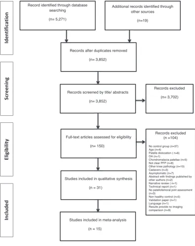
Fig. 1summarizes the results of the search strategy. The search
identified 5,290 papers, with 3,852 after duplications were removed. Following screening of the title and abstract, 3,702 of these were excluded. Subsequent full text assessment identified 46 papers describing 40 studies. Five studies28e38reported the same
Table I
Best evidence synthesis
1.) Strong evidence is provided by generally consistentfindings in multiple high-quality cohort studies.
2.) Moderate evidence is provided by general consistentfindings in one high-quality cohort study and two or more high quality caseecontrol studies or in three or more high-quality caseecontrol studies.
3.) Limited evidence is provided by (general consistent)findings in a single cohort study, in one or two caseecontrol studies or in multiple cross-sectional studies. 4.) Conflicting evidence is provided by conflictingfindings (i.e.,<75% of the studies reported consistentfindings).
5.) No evidence is provided when no studies could be found.
study population in more than one paper. These papers described different outcomes so were analysed independently, although the risk of bias assessment was conducted on only 40 studies to prevent the overestimation of effects.39
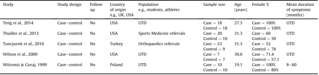
Study characteristics
The study characteristics are presented inTable II. Of the 40 studies included, 22 used MRI28e38,40e56, of whichfive included kinematic MRI41,43,46,55,56, eight used CT57e64, six used US65e70and five used XR60,71e74. The review included 1043 PFP subjects and 839 control subjects. The mean age was 27.0 years (range: 14e40.7 years), with 74.3% women in the case group and 69.0% in the control group. The duration of symptoms was reported in only ten of the 40
studies30,31,37,38,40,47,55,60,63,65,67,73. The duration of symptoms ranged from two47to 168 months63. All studies presented cross-sectional data except for two studies41,50. Pain was established in the PFP cohort most commonly from: reproducible pain in greater than two functional activities28e34,38,40e43,45e47,51,65,66,68,70,75. This
[image:3.595.94.490.67.567.2]Table II
Sample sizes and population characteristics for each included paper
Study Study design Follow
up
Country of origin e.g., UK, USA
Population
e.g., students, athletes
Sample size Age (years)
Female % Mean duration of symptoms (months)
Agliettiet al., 1983 Caseecontrol No USA UTD Case¼53
Control¼150
22 Case¼60.3 Control¼50%
UTD
Bretcher&Powers, 2002a Caseecontrol No USA Orthopaedic referrals Case¼10 Control¼10
34.6 Case¼50% Control¼50%
UTD Bretcher&Powers, 2002b
Botanliogluet al., 2013 Caseecontrol No Turkey UTD Case¼11
Control¼22
29.5 Case¼100 Control¼50%
UTD
Callaghan&Oldham, 2004 Caseecontrol No UK Orthopaedic&Rheumatology referrals
Case¼57 Control¼10
32.6 Case¼61% Control¼60%
34
Chen&Powers, 2014 Caseecontrol No USA Orthopaedic referrals& university students
Case¼20 Control¼20
27 Case¼100% Control¼100%
UTD
Chenet al., 2012 Caseecontrol No Taiwan Orthopaedic referrals Case¼26
Control¼26
27.8 Case¼81% Control¼81%
UTD
Chiuet al., 2012 Caseecontrol 8 weeks Hong Kong UTD Case¼9
Control¼6
33.1 Case¼55.6 Control¼50
UTD
Connollyet al., 2009 Caseecontrol No Canada Sports Medicine Physician referrals
Case¼10 Control¼10
27 Case¼100% Control¼100%
UTD
Draperet al., 2006 Caseecontrol No USA UTD Case¼34
Control¼16
28.8 Case¼64.7% Control¼50%
UTD
Draperet al., 2009 Caseecontrol No USA Orthopaedic&Sports Medicine referrals
Case¼23 Control¼13
29.4 Case¼100% Control¼100%
UTD
Eckhoffet al., 1994 Caseecontrol No USA Failed conservative management
Case¼20 Control¼20
UTD UTD UTD
Farrokhiet al., 2011a Caseecontrol s No USA UTD Case¼10
Control¼10
27.4 Case¼100% Control¼100%
87.6 1 Farrokhiet al., 2011b
Felicioet al., 2011a Caseecontrol No Brazil UTD Case¼19
Control¼20
22.5 Case¼100% Control¼100%
UTD Felicioet al., 2012b
Felicioet al., 2014c
Guzzantiet al., 1994 Caseecontrol No Italy Adolescents Case¼27 Control¼20
14 Case¼77.8 Control¼50
UTD
Haimet al., 2006 Caseecontrol No Israel Military soldiers Case¼61
Control¼25
21.8 Case¼0% Control¼0%
19
Harmanet al., 2002 Caseecontrol No Turkey UTD Case¼17
Control¼10
29.4 Case 0% Controls 0%
UTD
Hoet al., 2014 Caseecontrol No USA UTD Case¼10
Control¼10
25.5 Case¼100% Control¼100%
UTD Hoet al., 2014b
Joensenet al., 2001 Caseecontrol No Denmark Athletes Case¼24 Control¼17
21.6 Case¼37.5 Control¼35.3
UTD
Joneset al., 1995 Caseecontrol No USA Failed conservative management
Case¼40 Control¼10
UTD Case¼UTD Control¼50%
UTD
Kimet al., 2014 Caseecontrol No South Korea Orthopaedic referrals Case¼51 Control¼44
27.4 Case¼47% Control¼50%
UTD
Laprade&Culham, 2003 Caseecontrol No Canada Military Case¼33 Control¼33
30.9 Case¼33.3 Control¼33.3
UTD
Janet al., 2009 Caseecontrol No Taiwan Orthopaedic referrals Case¼54
Control¼54
40.7 Case¼75.9 Control¼75.9
UTD
Metin Cubuket al., 2000 Caseecontrol No Turkey Orthopaedic referrals Case¼42 Control¼40
27 Case¼100% Control¼100%
11
Munetaet al., 1994 Caseecontrol No Japan UTD Case¼60
Control¼19
21 Case¼100 Control¼100
UTD
Palet al., 2013c Caseecontrol No USA University Orthopaedic and Sport Medicine referrals
Case¼37 Control¼15
29.7 Case¼54.1% Control¼53.3%
3e132
Pattynet al., 2011 Caseecontrol No Belgium Hospital Orthopaedic Surgeon referrals
Case¼46 Control¼30
23.3 Case¼54.3 Control¼56.7
17.37 Pattynet al., 2013c
Pinaret al., 1994 Caseecontrol No Turkey UTD Case¼26
Control¼14
29 Case¼78.5 UTD
Powers, 2000b Caseecontrol NAD USA Orthopaedics referrals &university students
Case¼23 Control¼12
27.9 Control¼UTD UTD
Ribeiroet al., 2010 Caseecontrol NAD Brazil UTD Case¼12
Control¼12
22.5 Case¼100% Control¼100%
UTD
Salsich&Perman, 2007 Caseecontrol No USA UTD Case¼21
Control¼21
25 Case¼76.2 Control¼66.7
UTD
Salsich&Perman, 2013 Caseecontrol No USA Multiple sourceseincluding community dwelling population
Case¼27 Control¼29
25.6 Case¼77.8 Control¼65.5
>2
Schootset al., 2013 Caseecontrol No Netherlands Sports medicine&Orthopaedic referrals
Case¼10 Control¼10
29.3 Case¼60% Control¼60%
>6
Schutzeret al., 1986 Caseecontrol No USA UTD Case¼24
Control¼10
19 Case¼91.7 Control¼70
3e168
Souzaet al., 2010 Caseecontrol No USA Orthopaedic referrals &community dwelling population
Case¼15 Control¼15
29.9 Case¼100% Control¼100%
UTD
Taskiranet al., 1998 Caseecontrol No Turkey UTD Case¼10
Controls¼9
27 Case¼100% Control¼88.9
UTD
(continued on next page)
reported intraclass correlation coefficients (ICC) a pooling of the data was available for MRI bisect offset, patella tilt, patellofemoral contact area, with Insall-Salvati ratio and sulcus angle showing mean ICCs of 0.92, 0.85, 0.90, 0.96, 0.82 respectively. Inter-observer reliability data was only presented in seven studies38,41,49,50,59,71,74.
Quality assessment
A summary of the quality assessment results are presented
inTable III. Based on the categorisations used22, 23 studies were
judged as high quality (30e38,40e51,65e67,69,71,73,74), with the remaining 17 studies considered of moderate qual-ity28,29,52e64,68,70,72. The criteria of best performance using the
modified Downs&Black checklist were 1, 2, 3 and 4, which were satisfied by all the included studies. The criteria that the included studies performed most poorly were 9, 10, 11, 15 and 17
(Supplementary Material). Criteria 9, 10 and 15 pertained to the
documentation of population in which participants are recruited. Only half the studies clearly documented from where their par-ticipants were recruited e.g., hospital, military etc. Criterion 11 posed:was an attempt made to blind those measuring the outcome. Only 17.5% (7/40) of the studies we were able to determine whether the person/s interpreting the images were blinded to group allocation. Criteria 17 posed: did the study have sufficient power to detect clinically important effect. Only 17.5% (7/40) of studies40,42,47,48,65,69,71 clearly documented how they calculated
their sample size.
Based on published guidelines77, funnel plots were not employed due to no one feature having more than ten studies and so reducing the likelihood of distinguishing real asymmetry.
Synthesis of results
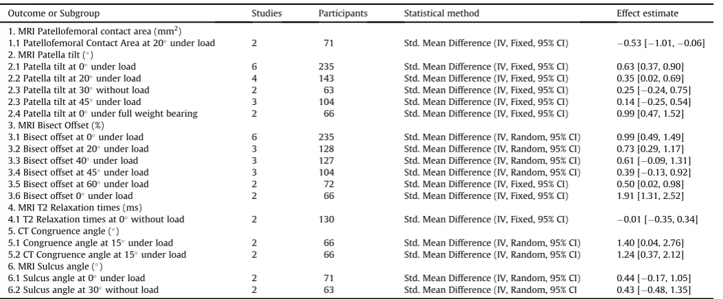
MRI features (patellofemoral contact area, patellar tilt, patellar bisect offset, patellar cartilage T2 relaxation times and sulcus angle) and CT features (congruence angle) were the only imaging features that yielded homogenous data appropriate for meta-analysis. These features are demonstrated schematically inFig. 2. If discrepancies were noted in either the knee loading status, assessments of the imaging feature or knee flexion angle, then features were not considered for meta-analysis. The results of the meta-analyses are displayed inTable IV.
MRI
Of the twenty-two studies that used MRI, sixteen stud-ies30e38,40e51,78 were judged as high quality. Controlling for the knee loading status, assessment of the imaging feature and knee flexion angle, patella bisect offset at 0with load demonstrated the
largest SMD (0.99; 95% CI: 0.49, 1.49; moderate evidence) based on five high quality41,43,46,47,51 and one moderate quality53 study
(Fig. 3). This was the only MRI feature which presented with a large
SMD23. Five other features demonstrated a medium SMD23. These included: patella bisect offset at 20with load (0.73; 95% CI: 0.29, 1.17; limited evidence)41,47,53,67, patella tilt at 0 with load (0.63: 95% CI: 0.37, 0.90; moderate evidence)41,43,46,47,51,53, patella bisect offset at 40 with load (0.61; 95% CI:0.09, 1.31; limited evi-dence)41,47,53, patellofemoral contact area at 20with load (0.53; 95% CI:1.01,0.06; limited evidence)47,50and patella bisect offset at 60with load (0.50; 95% CI 0.02, 0.98; limited evidence)41,53.
A small SMD was found for the pooling of sulcus angle at 0with load (0.44; 95% CI:0.17, 1.05; limited evidence)46,53, sulcus angle at 30 without load (0.43; 95% CI: 0.48, 1.35; limited evi-dence)34,52, patella tilt at 20with load (0.35; 95% CI: 0.02, 0.69; moderate evidence)41,47,50,53, patella tilt at 30without load (0.25; 95% CI:0.24, 0.75; limited evidence)34,52, T2 Relaxation time at 0 with without load (0.01; 95% CI: 0.35, 0.34; limited evi-dence)31,48. The data for patellofemoral joint reaction force (PFJRF) was considered inappropriate for pooling as its outputs were pro-duced via computational modelling, with imaging as only one component. For the data not amenable to pooling, there was limited evidence to support a difference between PFP and a control group with regards to: congruence angle at 2055and 3052without load; T1 value of the lateral patellofemoral cartilage without load48; articular lesions of the patella44; peak PFJRF; and patella cartilage thickness in males49. There was conflicting evidence to support a
difference in patella cartilage thickness in women30,36,45,49and no evidence to support differences in patella tendon morphology54.
US
US was used to assess PFP imaging features in four stud-ies65e67,69. These were all judged as high quality. Pooling of data was not appropriate due to the variety of outcome features ana-lysed and the different assessment techniques used. For the data not amenable to pooling, there was limited evidence, from single studies, to support a difference between PFP and control group in terms of: a reduction in vastus medialis oblique (VMO) contraction ratio and capacity68; an increase in VMO electrical mechanical delay and a reduction in vastus lateralis (VL) delay66; and a differ-ence in VMOfibre angle, insertion level and volume69.
CT
[image:5.595.34.555.76.199.2]CT was employed in eight studies, all of which were judged as moderate quality. Pooling of data was limited for congruence angle57,58,63,64; patella tilt angle57,58,63,64; sulcus angle57,64 since studies either: did not provide adequate data64; it was unclear Table II(continued)
Study Study design Follow
up
Country of origin e.g., UK, USA
Population
e.g., students, athletes
Sample size Age (years)
Female % Mean duration of symptoms (months)
Tenget al., 2014 Caseecontrol No USA UTD Case¼18
Control¼18
27.3 Case¼100% Control¼100%
UTD
Thuilleret al., 2013 Caseecontrol No USA Sports Medicine referrals Case¼20 Control¼10
31.3 Case¼60 Control¼50
UTD
Tuncyureket al., 2010 Caseecontrol No Turkey Orthopaedics referrals Case¼23 Control¼9
31.3 Case¼52 Control¼78
UTD
Wilsonet al., 2009 Caseecontrol No USA UTD Case¼7
Control¼7
30.6 Case¼71.4 Control¼57.1
UTD
Witzonzi&Goraj, 1999 Caseecontrol No Poland UTD Case¼10
Control¼10
19.1 Case¼100% Control¼80%
8e60
Table III
Quality assessment ratings using the Modified Down's and Blacks checklist
Study Q1
(/1) Q2 (/1)
Q3. (/1)
Q4. (/1)
Q5. (/2)
Q6. (/1)
Q7. (/1)
Q8. (/1)
Q9 (/1)
Q10. (/1)
Q11. (/1)
Q12. (/1)
Q13. (/1)
Q14. (/1)
Q15. (/1)
Q16. (/1)
Q17. (/1)
Total % Score
Agliettiet al., 1983 1 1 1 1 1 1 1 0 UTD UTD UTD 1 1 1 UTD 1 0 11 /18 61.1
Botanliogluet al., 2013 1 1 1 1 1 1 1 1 UTD UTD UTD 1 1 UTD UTD 1 0 11 /18 61.1
Bretcher&Powers, 2002 1 1 1 1 1 0 1 1 1 1 UTD 1 1 0 1 0 0 12 /18 66.7
Bretcher&Powers, 2002b
Callaghan&Oldham, 2004 1 1 1 1 2 1 1 1 1 1 UTD 1 1 1 1 1 1 17 /18 94.4
Chen&Powers, 2014 1 1 1 1 1 1 1 1 1 1 UTD 1 1 1 UTD 1 1 15 /18 83.3
Chenet al., 2012 1 1 1 1 1 1 1 1 1 1 UTD 1 1 1 UTD 0 0 13 /18 72.2
Chiuet al., 2012 1 1 1 1 1 1 1 1 UTD UTD 1 1 1 1 1 0 0 13 /18 72.2
Connollyet al., 2009 1 1 1 1 1 1 1 0 1 1 UTD 1 1 1 1 1 0 14 /18 77.8
Draperet al., 2006 1 1 1 1 2 1 1 1 UTD UTD UTD 1 1 1 1 1 0 14 /18 77.8
Draperet al., 2009 1 1 1 1 2 1 1 1 1 1 UTD 1 1 1 1 1 0 16 /18 88.9
Eckhoffet al., 1994 1 1 1 1 0 0 1 0 0 0 UTD UTD 1 1 UTD 0 0 7 /18 38.9
Farrokhiet al., 2011a 1 1 1 1 2 1 1 1 UTD UTD UTD 1 1 1 1 1 0 14 /18 77.8
Farrokhiet al., 2011b /18
Felicioet al., 2011a 1 1 1 1 2 1 1 1 UTD UTD UTD 1 1 1 1 1 0 14 /18 77.8
Felicioet al., 2012b /18
Felicioet al., 2014c /18
Guzzantiet al., 1994 1 1 1 1 1 1 1 0 UTD UTD UTD 1 1 1 UTD 1 0 11 /18 61.1
Haimet al., 2006 1 1 1 1 2 1 1 1 1 1 0 1 1 1 1 1 0 16 /18 88.9
Harmanet al., 2002 1 1 1 1 0 1 0 0 UTD UTD UTD 1 0 1 UTD 0 0 7 /18 38.9
Hoet al., 2014 1 1 1 1 2 1 1 1 UTD UTD UTD 1 1 1 1 1 0 14 /18 77.8
Hoet al., 2014b
Joensenet al., 2011 1 1 1 1 2 0 1 0 1 1 1 1 1 1 1 1 0 15 /18 83.3
Joneset al., 1995 1 1 1 1 0 1 1 0 UTD UTD UTD 1 1 UTD 1 0 0 9 /18 50
Kimet al., 2014 1 1 1 1 1 1 1 1 1 1 UTD 1 1 UTD 1 0 0 13 /18 72.2
Laprade&Culham, 2003 1 1 1 1 2 1 1 1 1 1 1 1 1 1 1 0 1 17 /18 94.4
Janet al., 2009 1 1 1 1 2 1 1 1 1 1 0 1 1 1 0 1 1 16 /18 88.9
Metin Cubuket al., 2000 1 1 1 1 1 1 1 0 UTD UTD UTD 1 1 UTD UTD 0 0 9 /18 50
Munetaet al., 1994 1 1 1 1 1 1 1 0 UTD UTD UTD 1 1 1 UTD 0 0 10 /18 55.6
Palet al., 2013c 1 1 1 1 2 1 1 1 1 1 UTD 1 1 1 1 1 1 17 /18 94.4
Pattynet al., 2012a 1 1 1 1 2 1 1 1 1 1 1 1 1 1 1 1 0 17 /18 94.4
Pattynet al., 2013c
Pinar, 1994 1 1 1 1 1 1 0 0 UTD UTD UTD 1 0 1 UTD 1 0 9 /18 50
Powers, 2000b 1 1 1 1 1 1 1 0 1 1 UTD 1 1 1 1 1 0 14 /18 77.8
Ribeiroet al., 2010 1 1 1 1 1 1 1 1 UTD UTD UTD 1 1 1 1 0 0 12 /18 66.7
Salsich&Perman, 2007 1 1 1 1 1 1 1 1 UTD UTD 1 1 1 1 UTD 1 0 13 /18 72.2
Salsich&Perman, 2013 1 1 1 1 1 1 1 1 UTD UTD 1 1 1 1 UTD 1 1 14 /18 77.8
Schootset al., 2013 1 1 1 1 1 1 1 1 1 1 1 1 1 UTD 0 0 0 13 /18 72.2
Shultzeret al., 1986 1 1 1 1 1 1 0 0 UTD UTD UTD 0 1 UTD UTD 0 0 7 /18 38.9
Souzaet al., 2010 1 1 1 1 2 1 1 1 1 1 0 1 1 1 UTD 1 0 15 /18 83.3
Taskiranet al., 1998 1 1 1 1 1 1 1 0 UTD UTD UTD 1 1 1 UTD 1 0 11 /18 61.1
Tenget al., 2014 1 1 1 1 1 1 1 0 UTD UTD 1 1 1 1 UTD 0 0 11 /18 61.1
Thuilleret al., 2013 1 1 1 1 1 1 1 1 1 1 UTD 1 1 1 0 0 1 14 /18 77.8
Tuncyureket al., 2010 1 1 1 1 1 1 1 1 UTD UTD UTD 1 1 UTD UTD 1 0 11 /18 61.1
Wilsonet al., 2009 1 1 1 1 1 1 1 1 UTD UTD UTD 1 1 1 UTD 0 0 11 /18 61.1
Witzonzi&Goraj, 1999 1 1 1 1 1 1 1 0 UTD UTD UTD 1 1 UTD UTD 1 0 10 /18 55.6
Studies scoring Yes 40 40 40 40 50 37 37 25 17 17 7 38 38 31 18 24 7
Studies scoring Yes % 100 100 100 100 62.3 92.5 92.5 62.5 42.5 42.5 17.5 95 95 77.5 45 60 17.5
UTD¼Unable to detect;Q1:Is the hypothesis/aim/objective of the study clearly described?;Q2:Are the main outcomes to be measured clearly described in the Introduction or Methods section?;Q3: Are the characteristics of the
patients included in the study clearly described?;Q4: Are the interventions of interest clearly described?;Q5: Are the distributions of principal confounders in each group to be compared clearly described?;Q6: Are the main
findings of the study clearly described?;Q7: Does the study provide estimates of the random variability in the data for the main outcomes?;Q8: Have the actual probability values been reported (e.g., 0.035 rather than<0.05) for the
main outcomes except where the probability value is less than 0.001:Q9: Were the subjects asked to participate in the study representative of the entire population from which they were recruited?;Q10: Were the subjects who
were prepared to participate representative of the entire population from which they were recruited;Q11: Was an attempt to blind those measuring the main outcome?;Q12: If any of the results of the study were based on“data
dredging”was this made clear?;Q13: Were the statistical tests used for the main outcomes appropriate?;Q14: Were the main outcome measures used accurate (valid and reliable)?;Q15: Were the case and controls recruited from
the same population?; Q16: Was there adequate adjustment for confounding in the analyses from which the mainfindings were drawn?;Q17: Did the study have sufficient power to detect a clinically important effect?
B.T
.
Drew
et
al.
/
Osteoarthritis
and
Cartilage
2
4
(20
16
)
2
2
4
e
236
whether their participants' knee was loaded or unloaded63or they adopted different measurement techniques for patella tilt angle57. Pooling was appropriate for congruence angle at 15without load and congruence angle at 15 under load. Both features demon-strated a large SMD (1.24; 95% CI 0.37, 2.12; limited evidence)57,58 and (1.40 95% CI: 0.04, 2.76; limited evidence)57,58(Fig. 3). For the data not amenable to pooling there is limited evidence to support a difference between PFP and a control group with regards to: congruence angle at 15 without load58; tibial tubercle rotation
angle at 0 without load59,60; trochlear depth at 15 without load57. Conflicting evidence exists for patella tilt at 15 with load57,58.
XR
[image:7.595.33.560.524.742.2]XR features were assessed infive studies. Of these, three were judged as high quality71,73,74 and two as moderate quality60,72. The following features were considered for meta-analysis: sulcus Fig. 2.Measurement of patella alignment. Line A to B forms the patella width. Line E to F forms a line along the most posterior femoral condyles. Point D is located at the deepest point of the trochlear groove. Point C is the bisecting point of the perpendicular line through the AB line. Line G bisects the sulcus angle to form a zero reference and line H is the projected from the apex of the sulcus angle through the most dorsal part of the patella. A) Bisect offset¼(length of AC/length of BC)100%; B) Congruence angle¼angle formed between G line and H line; C) Patella tilt¼the angle formed by line between AB and EF48.
Table IV
Results of the meta-analysis for all imaging feature amenable to pooling
Outcome or Subgroup Studies Participants Statistical method Effect estimate
1. MRI Patellofemoral contact area (mm2)
1.1 Patellofemoral Contact Area at 20under load 2 71 Std. Mean Difference (IV, Fixed, 95% CI) 0.53 [1.01,0.06] 2. MRI Patella tilt ()
2.1 Patella tilt at 0under load 6 235 Std. Mean Difference (IV, Fixed, 95% CI) 0.63 [0.37, 0.90] 2.2 Patella tilt at 20under load 4 143 Std. Mean Difference (IV, Fixed, 95% CI) 0.35 [0.02, 0.69] 2.3 Patella tilt at 30without load 2 63 Std. Mean Difference (IV, Fixed, 95% CI) 0.25 [0.24, 0.75] 2.3 Patella tilt at 45under load 3 104 Std. Mean Difference (IV, Fixed, 95% CI) 0.14 [0.25, 0.54] 2.4 Patella tilt at 0under full weight bearing 2 66 Std. Mean Difference (IV, Fixed, 95% CI) 0.99 [0.47, 1.52] 3. MRI Bisect Offset (%)
3.1 Bisect offset at 0under load 6 235 Std. Mean Difference (IV, Random, 95% CI) 0.99 [0.49, 1.49] 3.2 Bisect offset at 20under load 3 128 Std. Mean Difference (IV, Random, 95% CI) 0.73 [0.29, 1.17] 3.3 Bisect offset 40under load 3 127 Std. Mean Difference (IV, Random, 95% CI) 0.61 [0.09, 1.31] 3.4 Bisect offset at 45under load 3 104 Std. Mean Difference (IV, Random, 95% CI) 0.39 [0.13, 0.92] 3.5 Bisect offset at 60under load 2 72 Std. Mean Difference (IV, Fixed, 95% CI) 0.50 [0.02, 0.98] 3.6 Bisect offset 0under load 2 66 Std. Mean Difference (IV, Fixed, 95% CI) 1.91 [1.31, 2.52] 4. MRI T2 Relaxation times (ms)
4.1 T2 Relaxation times at 0without load 2 130 Std. Mean Difference (IV, Fixed, 95% CI) 0.01 [0.35, 0.34] 5. CT Congruence angle ()
5.1 Congruence angle at 15under load 2 66 Std. Mean Difference (IV, Random, 95% CI) 1.40 [0.04, 2.76] 5.2 CT Congruence angle at 15under load 2 66 Std. Mean Difference (IV, Random, 95% CI) 1.24 [0.37, 2.12] 6. MRI Sulcus angle ()
angle71e73, congruence angle71e73, Insall-Salvati index72,73 and lateral patellofemoral angle71,74. It was not possible to pool data for any of these XR features however, due to variations in the kneeflexion angle. For the data not amenable to pooling there was limited evidence to support a difference between PFP and a control group with regards to: congruence angle at 45 with load72,74but no evidence at 3571. There was limited evidence to support sulcus angle at 45without load72,74but no evidence to support it at 3073and 3571. There was conflicting evidence for Insall-Salvati index at 30 without load60,72,73 and no evidence for lateral patellofemoral angle at 3571and 4574without load.
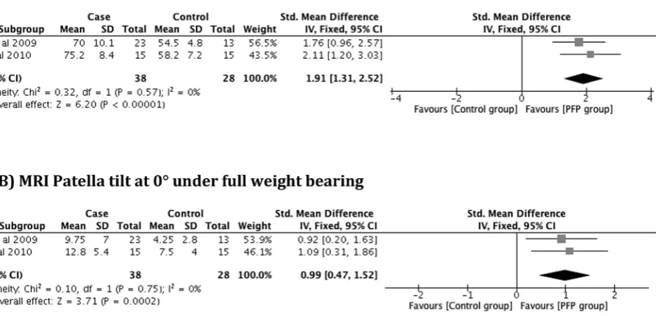
Sensitivity analysis
Two studies included in the meta-analysis41,43 used a full weight-bearing procedure to load the PFJ during imaging. Ana-lysing appropriate features under full weight bearing separately demonstrated a marked increase in the SMD (Fig. 4) of MRI patella bisect offset at 0with load (1.91; 95% CI: 1.31, 2.52; limited evi-dence)41,43and MRI patella tilt at 0with load (0.99; 95% CI: 0.47, 1.52; limited evidence)41,43.
Discussion
The evidence from this review suggested that an increased MRI bisect offset at 0kneeflexion under load and CT-derived congru-ence angle at 15 knee flexion with and without load are both associated with PFP and there is a large SMD as determined from moderate and limited evidence respectively. A medium SMD was identified for the association between PFP and the following MRI features: patella tilt and patellofemoral contact area. Limited evi-dence existed to support the association of PFP with other features of MRI, US, CT and XR.
A previous comprehensive review by Lankhorst et al.79 has
[image:8.595.52.549.73.488.2]provided insight into a broad range of factors associated with PFP (searched up to November 2010). We chose not to restrict inclusion by sample size to improve inclusivity80and together with inclusion of more recent studies, this resulted in over 70% of the current review studies being different from Lankhorstet al.79. Furthermore, by focusing only on imaging-detected features associated with pain, the present review controlled for variables such as imaging modality, kneeflexion angle, and knee loading, known to influence the homogeneity of the imaging outcomes81.
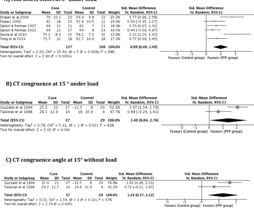
Fig. 3.Forest plots for: A) MRI bisect offset at 0under load; B) CT congruence at 15under load; C) CT congruence angle at 15without load.
Only MRI and CT features demonstrated sufficient homogeneity for appropriate meta-analysis. Bisect offset measured with MRI was most amenable to pooling across a variety of kneeflexion angles demonstrating medium to large SMDs. This is notable as bisect offset has been shown to be the most significant feature in the progression of joint space narrowing over a five year period in adults with symptomatic knee pain aged 70e79 years82. Consid-erable clinical heterogeneity was present in the studies utilising XR and US. Studies using XR reported outcomes with subtle variations in knee flexion angle or assessment techniques that limited the pooling of data. The imaging features used in US were distinctly different and so offered no potential for pooling.
The present review considered loading of the knee as a dichotomous condition, as no consensus exists to the affect of the quantityof loading83. Our sensitivity analysis demonstrated an in-crease in SMD for both patella tilt and bisect offset when MR images were acquired under upright full weight bearing. This is in contrast to previous studies that have shown that bisect offset is more pronounced in the supine position when investigating people with PFP under both supine-loaded and upright full weight bearing conditions76,84. The reason for this disparity is unclear, however, it may be explained by the fact that the previous studies selected people with excessive patella lateralisation, whereas the studies included in the current review likely contained a range of patella alignments. Another possibility is that the control group in the current review demonstrated an averagereductionin bisect offset under full weight bearing, which may also explain the increased SMD.
The concept of‘weight bearing’has been challenged by Har-baughet al.85who suggest that quadriceps activity is the primary determinant of patella position in PFP rather than the axial loading. The full weight bearing studies in this review employed a 0.5T open, upright scanner and thefield strength of 0.5 Tesla (T) may have affected image quality86,87. Full weight bearing conditions also have the potential to elicit pain during the procedure88. In PFP, pain is recognised as having an inhibitory affect on quadriceps89; altering quadriceps activity may influence the validity of the results by affecting patellar orientation85.
This review identified a number of limitations in the literature based on participant selection. Firstly, a number of the included studies30e36,41e43,45,46,52,53,60used all female cohorts, and of these studies only a few selected a matched cohort. Controlling for gender, knee flexion angle and loading of the knee has been advocated because these factors have been reported to influence the PFJ mechanics and the comparisons made81. Furthermore, only half the studies clearly stated the recruitment source of participants e.g., hospital, militaryetc. Extrapolating results taken from a mili-tary or very physically active group and applying them to a more sedentary community dwelling population is likely to affect the external validity. Secondly, the quantification of pain in the PFP cohort was inconsistent. Over two thirds of the included studies selected participants based on reproducible pain with functional activities, however the number of provocative activities required for diagnosis and inclusion varied from one49,53,59,64,73,74 to
five50,55,56. The use of the VAS to quantify pain on provocation ac-tivities was used in six studies30,31,42,43,47,51,53. The duration of symptoms was also poorly reported, with fewer than a quarter of the included studies documenting the duration of PFP, and in these studies the data was presented differently (e.g., mean duration, range of duration). The duration of symptoms is important as this has been shown in PFP to be a predictor of poor long-term out-comes5. The effect of the duration of symptoms in relation to structural imagingfindings is unknown. It is known however, that long term pain will lead to muscle inhibition89and thus there is a probability that a reduction quadriceps strength and activity could influence the PFJ structural features observed.
[image:9.595.75.544.85.311.2]assessment was reported in fewer than half the included studies. Generally the ICCs showed a moderate to high reliability for the MRI variables: bisect offset, patella tilt angle, patellofemoral con-tact area, Insall-Salvati index and sulcus angle, supporting the use of these features in future studies.
Thefindings from a recent international expert consensus group highlight the need for sub-grouping of the PFP population7. The current review demonstrates a number of PFJ imaging features associated with PFP suggesting that these features should be considered as important components of future stratification. In addition, although most of the included studies employed a cross sectional analyses, two studies did employ an interventional pre-post study design41,50. These studies detected a significant change in patellofemoral contact area following strengthening exercise50
and patellofemoral bisect offset and patella tilt following patella bracing41. As these imaging features have been shown to be modifiable it highlights the opportunity of using imaging features clinically as a treatment target.
Limitations of the current review
The nomenclature within the PFP literature is ambiguous, with the condition being referred to historically by a variety of other names92. In the present review, over 20% of the studies used terms differing frompatellofemoral painorpatellofemoral pain syndrome. This makes study selection challenging with selection of the studies based on the description of the condition when more ambiguous terms are used. We attempted to minimise the potential bias in this process by using two reviewers to select studies and a third inde-pendent mediator. Secondly, the small sample sizes used in some of the included studies may influence the validity of the results. Meta-analyses was possible, however, for a number of imaging features thus increasing the overall sample size and improving statistical power93. Thirdly, the cross-sectional nature of the studies means the results from the current review cannot imply causality. To establish this, further research is warranted from prospective cohorts.
Conclusion
This systematic review with meta-analysis suggests that PFP is associated with MRI bisect offset and CT congruence angle analysed at 0knee flexion and 15 kneeflexion respectively; however, a degree of caution in interpretation of this data is advised due to the role of both features being derived from only moderate and limited evidence respectively. It is clear from this systematic review that future studies need to clearly document the specific population in which participants are recruited and to improve reporting of imaging-related issues. The inclusion of two interventional studies demonstrates that imaging features are potentially modifiable and future intervention strategies could be employed to target these features.
Contribution of authors
BD takes responsibility for the integrity of the work as a whole, from inception to thefinished manuscript.
Conception&design: BD, AR, TS, PC. Collection&Assembly of Data: BD, FP, TS.
Analysis&Interpretation of the data: BD, AR, FP, TS, PC. Drafting&final approval of the manuscript: BD, AR, FP, TS, PC.
Conflicts of interest
No conflict of interest were declared.
Acknowledgements
BD is funded by a National Institute for Health Research (NIHR) Clinical Doctoral Research Fellowship (CDRF-2013-04-044). This paper presents independent research funded by the National Institute for Health Research (NIHR). The views expressed are those of the authors and not necessarily those of the NHS, the NIHR or the Department of Health. This work was also supported in part by funding from the Arthritis Research UK Experimental Osteoarthritis Treatment Centre (Ref 20083) and the Arthritis Research UK Centre for Sport, Exercise and Osteoarthritis (Ref 20194).
Supplementary data
Supplementary data related to this article can be found athttp://
dx.doi.org/10.1016/j.joca.2015.09.004.
References
1. Heintjes E, Berger MY, Bierma-Zeinstra SM, Bernsen RM,
Verhaar JA, Koes BW. Exercise therapy for patellofemoral pain
syndrome. Cochrane Database Syst Rev 2003;4:CD003472.
2. Fairbank JC, Pynsent PB, van Poortvliet JA, Phillips H.
Me-chanical factors in the incidence of knee pain in adolescents
and young adults. J Bone Joint Surg Br 1984;66(5):685e93.
3. Molgaard C, Rathleff MS, Simonsen O. Patellofemoral pain
syndrome and its association with hip, ankle, and foot function in 16- to 18-year-old high school students: a single-blind
case-control study. J Am Podiatr Med Assoc 2011;101(3):215e22.
4. Wood L, Muller S, Peat G. The epidemiology of patellofemoral
disorders in adulthood: a review of routine general practice morbidity recording. Prim Health Care Res Dev 2011;12(2):
157e64.
5. Collins NJ, Bierma-Zeinstra SM, Crossley KM, van
Linschoten RL, Vicenzino B, van Middelkoop M. Prognostic factors for patellofemoral pain: a multicentre observational
analysis. Br J Sports Med 2013;47(4):227e33.
6. Crossley KM. Is patellofemoral osteoarthritis a common
sequela of patellofemoral pain? Br J Sports Med 2014;48(6):
409e10.
7. Witvrouw E, Callaghan MJ, Stefanik JJ, Noehren B,
Bazett-Jones DM, Willson JD, et al. Patellofemoral pain: consensus
statement from the 3rd International Patellofemoral Pain Research Retreat held in Vancouver, September 2013. Br J
Sports Med 2014;48(6):411e4.
8. Besier TF, Draper C, Pal S, Fredericson M, Gold G, Delp S,et al.
Imaging and Musculoskeletal Modeling to Investigate the
Mechanical Etiology of Patellofemoral Pain 2011269e86.
9. Davis IS, Powers CM. Patellofemoral pain syndrome: proximal,
distal, and local factors, an international retreat, April 30eMay
2, 2009, Fells Point, Baltimore, MD. J Orthop Sports Phys Ther
2010;40(3):A1eA16.
10. Duncan RC, Hay EM, Saklatvala J, Croft PR. Prevalence of
radiographic osteoarthritiseit all depends on your point of
view. Rheumatol Oxf 2006;45(6):757e60.
11. Hinman RS, Lentzos J, Vicenzino B, Crossley KM. Is
patellofe-moral osteoarthritis common in middle-aged people with
chronic patellofemoral pain? Arthritis Care Res Hob
2014;66(8):1252e7.
12. Thomas MJ, Wood L, Selfe J, Peat G. Anterior knee pain in
younger adults as a precursor to subsequent patellofemoral osteoarthritis: a systematic review. BMC Musculoskelet Disord
2010;11:201.
13. Utting MR, Davies G, Newman JH. Is anterior knee pain a predisposing factor to patellofemoral osteoarthritis? Knee
2005;12(5):362e5.
14. Eckstein F, Mosher T, Hunter D. Imaging of knee osteoarthritis:
data beyond the beauty. Curr Opin Rheumatol 2007;19(5):
435e43.
15. Wenham CY, Conaghan PG. Imaging the painful osteoarthritic
knee joint: what have we learned? Nat Clin Pract Rheumatol
2009;5(3):149e58.
16. Elias DA, White LM. Imaging of patellofemoral disorders. Clin
Radiol 2004;59(7):543e57.
17. Moher D, Liberati A, Tetzlaff J, Altman DG, Group P. Preferred
reporting items for systematic reviews and meta-analyses: the
PRISMA statement. BMJ 2009;339. b2535.
18. van der Heijden RA, Lankhorst NE, van Linschoten R,
Bierma-Zeinstra SM, van Middelkoop M. Exercise for treating patel-lofemoral pain syndrome. Cochrane Database Syst Rev 2015;1:
CD010387.
19. Downs SH, Black N. The feasibility of creating a checklist for
the assessment of the methodological quality both of rando-mised and non-randorando-mised studies of health care in-terventions. J Epidemiol Community Health 1998;52(6):
377e84.
20. Kemp JL, MacDonald D, Collins NJ, Hatton AL, Crossley KM. Hip
arthroscopy in the setting of hip osteoarthritis: systematic review of outcomes and progression to hip arthroplasty. Clin
Orthop Relat Res 2014;473(3):1055e73.
21. Ratcliffe E, Pickering S, McLean S, Lewis J. Is there a
relation-ship between subacromial impingement syndrome and scap-ular orientation? A systematic review. Br J Sports Med
2014;48(16):1251e6.
22. Hootman JM, Driban JB, Sitler MR, Harris KP, Cattano NM.
Reliability and validity of three quality rating instruments for systematic reviews of observational studies. Res Synthesis
Methods 2011;2(2):110e8.
23. Cohen J. 2nd edn.. In: Statistical Power Analysis for the
Behavioral Sciences, xxi Hillsdale, N.J: L. Erlbaum Associates;
1988567.
24. van Tulder M, Furlan A, Bombardier C, Bouter L, G. Editorial
Board of the Cochrane Collaboration Back Review. Updated method guidelines for systematic reviews in the cochrane collaboration back review group. Spine (Phila Pa 1976)
2003;28(12):1290e9.
25. Lievense AM, Bierma-Zeinstra SM, Verhagen AP, van Baar ME,
Verhaar JA, Koes BW. Influence of obesity on the development
of osteoarthritis of the hip: a systematic review. Rheumatol
Oxf 2002;41(10):1155e62.
26. Yusuf E, Kortekaas MC, Watt I, Huizinga TW, Kloppenburg M.
Do knee abnormalities visualised on MRI explain knee pain in knee osteoarthritis? A systematic review. Ann Rheum Dis
2011;70(1):60e7.
27. Drew BT, Smith TO, Littlewood C, Sturrock B. Do structural
changes (eg, collagen/matrix) explain the response to thera-peutic exercises in tendinopathy: a systematic review. Br J
Sports Med 2014;48(12):966e72.
28. Brechter JH, Powers CM. Patellofemoral joint stress during
stair ascent and descent in persons with and without
patel-lofemoral pain. Gait Posture 2002;16(2):115e23.
29. Heino Brechter J, Powers CM. Patellofemoral stress during
walking in persons with and without patellofemoral pain. Med
Sci Sports Exerc 2002;34(10):1582e93.
30. Farrokhi S, Colletti PM, Powers CM. Differences in patellar
cartilage thickness, transverse relaxation time, and deforma-tional behavior: a comparison of young women with and without
patellofemoral pain. Am J Sports Med 2011;39(2):384e91.
31. Farrokhi S, Keyak JH, Powers CM. Individuals with
patellofe-moral pain exhibit greater patellofepatellofe-moral joint stress: afinite
element analysis study. Osteoarthritis Cartilage 2011;19(3):
287e94.
32. Felicio LR, Baffa Ado P, Liporacci RF, Saad MC, De Oliveira AS,
Bevilaqua-Grossi D. Analysis of patellar stabilizers muscles and
patellar kinematics in anterior knee pain subjects.
J Electromyogr Kinesiol 2011;21(1):148e53.
33. Felicio LR, Camargo AC, Baffa Ado P, Bevilaqua-Grossi D.
In-fluence of exercises on patellar height in women with
patel-lofemoral pain syndrome. Acta Ortop Bras 2014;22(2):82e5.
34. Felicio LR, Saad MC, Liporaci RF, Baffa Ado P, dos Santos AC,
Bevilaqua-Grossi D. Correlation between trochlear groove depth and patellar position during open and closed kinetic chain exercises in subjects with anterior knee pain. J Appl
Biomech 2012;28(3):335e42.
35. Ho KY, Hu HH, Colletti PM, Powers CM. Recreational runners
with patellofemoral pain exhibit elevated patella water
con-tent. Magn Reson Imaging 2014;32(7):965e8.
36. Ho KY, Keyak JH, Powers CM. Comparison of patella bone
strain between females with and without patellofemoral pain:
afinite element analysis study. J Biomech 2014;47(1):230e6.
37. Pattyn E, Mahieu N, Selfe J, Verdonk P, Steyaert A, Witvrouw E.
What predicts functional outcome after treatment for
patel-lofemoral pain? Med Sci Sports Exerc 2012;44(10):1827e33.
38. Pattyn E, Verdonk P, Steyaert A, Vanden Bossche L, Van den
Broecke W, Thijs Y,et al. Vastus medialis obliquus atrophy:
does it exist in patellofemoral pain syndrome? Am J Sports
Med 2011;39(7):1450e5.
39. Tramer MR, Reynolds DJ, Moore RA, McQuay HJ. Impact of
covert duplicate publication on meta-analysis: a case study.
BMJ 1997;315(7109):635e40.
40. Pal S, Besier TF, Beaupre GS, Fredericson M, Delp SL, Gold GE.
Patellar maltracking is prevalent among patellofemoral pain subjects with patella alta: an upright, weightbearing MRI
study. J Orthop Res 2013;31(3):448e57.
41. Draper CE, Besier TF, Santos JM, Jennings F, Fredericson M,
Gold GE,et al. Using real-time MRI to quantify altered joint
kinematics in subjects with patellofemoral pain and to eval-uate the effects of a patellar brace or sleeve on joint motion.
J Orthop Res 2009;27(5):571e7.
42. Chen YJ, Powers CM. Comparison of three-dimensional
patel-lofemoral joint reaction forces in persons with and without
patellofemoral pain. J Appl Biomech 2014;30(4):493e500.
43. Souza RB, Draper CE, Fredericson M, Powers CM. Femur
rota-tion and patellofemoral joint kinematics: a weight-bearing magnetic resonance imaging analysis. J Orthop Sports Phys
Ther 2010;40(5):277e85.
44. Joensen AM, Hahn T, Gelineck J, Overvad K,
Ingemann-Hansen T. Articular cartilage lesions and anterior knee pain.
Scand J Med Sci Sports 2001;11(2):115e9.
45. Connolly KD, Ronsky JL, Westover LM, Kupper JC, Frayne R.
Differences in patellofemoral contact mechanics associated with patellofemoral pain syndrome. J Biomech 2009;42(16):
2802e7.
46. Powers CM. Patellar kinematics, part II: the influence of the
depth of the trochlear groove in subjects with and without
patellofemoral pain. Phys Ther 2000;80(10):965e78.
47. Salsich GB, Perman WH. Tibiofemoral and patellofemoral
mechanics are altered at small kneeflexion angles in people
with patellofemoral pain. J Sci Med Sport 2013;16(1):13e7.
48. Thuillier DU, Souza RB, Wu S, Luke A, Li X, Feeley BT. T1rho
imaging demonstrates early changes in the lateral patella in patients with patellofemoral pain and maltracking. Am J
49. Draper CE, Besier TF, Gold GE, Fredericson M, Fiene A,
Beaupre GS, et al. Is cartilage thickness different in young
subjects with and without patellofemoral pain? Osteoarthritis
Cartilage 2006;14(9):931e7.
50. Chiu JK, Wong YM, Yung PS, Ng GY. The effects of quadriceps
strengthening on pain, function, and patellofemoral joint contact area in persons with patellofemoral pain. Am J Phys
Med Rehabil 2012;91(2):98e106.
51. Salsich GB, Perman WH. Patellofemoral joint contact area is
influenced by tibiofemoral rotation alignment in individuals
who have patellofemoral pain. J Orthop Sports Phys Ther
2007;37(9):521e8.
52. Ribeiro Ade C, Grossi DB, Foerster B, Candolo C,
Monteiro-Pedro V. Electromyographic and magnetic resonance imaging evaluations of individuals with patellofemoral pain syndrome.
Rev Bras Fisioter 2010;14(3):221e8.
53. Teng HL, Chen YJ, Powers CM. Predictors of patellar alignment
during weight bearing: an examination of patellar height and
trochlear geometry. Knee 2014;21(1):142e6.
54. Tuncyurek O, Ozkol M, Ozic U, Pabuscu Y. The role of patellar
tendon morphometry on anterior knee pain. Surg Radiol Anat
2010;32(6):539e43.
55. Witonski D, Goraj B. Patellar motion analyzed by kinematic
and dynamic axial magnetic resonance imaging in patients with anterior knee pain syndrome. Arch Orthop Trauma Surg
1999;119(1e2):46e9.
56. Harman M, Dogan A, Arslan H, Ipeksoy U, Vural S. Evaluation
of the patellofemoral joint with kinematic MR fluoroscopy.
Clin Imaging 2002;26(2):136e9.
57. Guzzanti V, Gigante A, Di Lazzaro A, Fabbriciani C.
Patellofe-moral malalignment in adolescents. Computerized tomo-graphic assessment with or without quadriceps contraction.
Am J Sports Med 1994;22(1):55e60.
58. Taskiran E, Dinedurga Z, Yagiz A, Uludag B, Ertekin C, Lok V.
Effect of the vastus medialis obliquus on the patellofemoral
joint. Knee Surg Sports Traumatol Arthrosc 1998;6(3):173e80.
59. Muneta T, Yamamoto H, Ishibashi T, Asahina S, Furuya K.
Computerized tomographic analysis of tibial tubercle position in the painful female patellofemoral joint. Am J Sports Med
1994;22(1):67e71.
60. Metin Cubuk S, Sindel M, Karaali K, Arslan AG, Akyildiz F,
Ozkan O. Tibial tubercle position and patellar height as in-dicators of malalignment in women with anterior knee pain.
Clin Anat 2000;13(3):199e203.
61. Jones RB, Barlett EC, Vainright JR, Carroll RG. CT determination
of tibial tubercle lateralization in patients presenting with
anterior knee pain. Skelet Radiol 1995;24(7):505e9.
62. Eckhoff DG, Montgomery WK, Kilcoyne RF, Stamm ER. Femoral
morphometry and anterior knee pain. Clin Orthop Relat Res
1994;302:64e8.
63. Schutzer SF, Ramsby GR, Fulkerson JP. The evaluation of
patellofemoral pain using computerized tomography. A
pre-liminary study. Clin Orthop Relat Res 1986;204:286e93.
64. Pinar H, Akseki D, Karaoglan O, Genc I. Kinematic and dynamic
axial computed tomography of the patello-femoral joint in patients with anterior knee pain. Knee Surg Sports Traumatol
Arthrosc 1994;2(3):170e3.
65. Callaghan MJ, Oldham JA. Quadriceps atrophy: to what extent
does it exist in patellofemoral pain syndrome? Br J Sports Med
2004;38(3):295e9.
66. Chen HY, Chien CC, Wu SK, Liau JJ, Jan MH. Electromechanical
delay of the vastus medialis obliquus and vastus lateralis in individuals with patellofemoral pain syndrome. J Orthop
Sports Phys Ther 2012;42(9):791e6.
67. Schoots EJ, Tak IJ, Veenstra BJ, Krebbers YM, Bax JG. Ultrasound
characteristics of the lateral retinaculum in 10 patients with patellofemoral pain syndrome compared to healthy controls.
J Bodyw Mov Ther 2013;17(4):523e9.
68. Botanlioglu H, Kantarci F, Kaynak G, Unal Y, Ertan S,
Aydingoz O, et al. Shear wave elastography properties of
vastus lateralis and vastus medialis obliquus muscles in normal subjects and female patients with patellofemoral pain
syndrome. Skelet Radiol 2013;42(5):659e66.
69. Jan MH, Lin DH, Lin JJ, Lin CH, Cheng CK, Lin YF. Differences in
sonographic characteristics of the vastus medialis obliquus between patients with patellofemoral pain syndrome and
healthy adults. Am J Sports Med 2009;37(9):1743e9.
70. Wilson NA, Press JM, Zhang LQ. In vivo strain of the medial vs.
lateral quadriceps tendon in patellofemoral pain syndrome.
J Appl Physiol (1985) 2009;107(2):422e8.
71. Laprade J, Culham E. Radiographic measures in subjects who
are asymptomatic and subjects with patellofemoral pain
syn-drome. Clin Orthop Relat Res 2003;414:172e82.
72. Aglietti P, Insall JN, Cerulli G. Patellar pain and incongruence. I:
measurements of incongruence. Clin Orthop Relat Res
1983;176:217e24.
73. Haim A, Yaniv M, Dekel S, Amir H. Patellofemoral pain
syn-drome: validity of clinical and radiological features. Clin
Orthop Relat Res 2006;451:223e8.
74. Kim TH, Sobti A, Lee SH, Lee JS, Oh KJ. The effects of
weight-bearing conditions on patellofemoral indices in individuals without and with patellofemoral pain syndrome. Skelet Radiol
2014;43(2):157e64.
75. Jan MH, Lin DH, Lin CH, Lin YF, Cheng CK. The effects of
quadriceps contraction on different patellofemoral alignment subtypes: an axial computed tomography study. J Orthop
Sports Phys Ther 2009;39(4):264e9.
76. Draper CE, Besier TF, Fredericson M, Santos JM, Beaupre GS,
Delp SL, et al. Differences in patellofemoral kinematics
be-tween weight-bearing and non-weight-bearing conditions in patients with patellofemoral pain. J Orthop Res 2011;29(3):
312e7.
77. Higgins JPT, Green S, Cochrane Collaboration. In: Cochrane
handbook for Systematic Reviews of Interventions. Cochrane Book Series, xxi. Chichester, England; Hoboken, NJ:
Wiley-Blackwell; 2008649.
78. Feminist methodologies for critical researchers: bridging
dif-ferences. Choice Curr Rev Acad Libr 2006;43(9). 1864e.
79. Lankhorst NE, Bierma-Zeinstra SM, van Middelkoop M. Factors
associated with patellofemoral pain syndrome: a systematic
review. Br J Sports Med 2013;47(4):193e206.
80. Smith TO, Hing CB.“Garbage in, garbage out”ethe importance
of detailing methodological reasoning in orthopaedic
meta-analysis. Int Orthop 2011;35(2):301e2.
81. Besier TF, Draper CE, Gold GE, Beaupre GS, Delp SL.
Patellofe-moral joint contact area increases with knee flexion and
weight-bearing. J Orthop Res 2005;23(2):345e50.
82. Hunter DJ, Zhang YQ, Niu JB, Felson DT, Kwoh K, Newman A,
et al. Patella malalignment, pain and patellofemoral
progres-sion: the Health ABC Study. Osteoarthritis Cartilage
2007;15(10):1120e7.
83. Goudakos IG, Konig C, Schottle PB, Taylor WR, Hoffmann JE,
Popplau BM, et al. Regulation of the patellofemoral contact
area: an essential mechanism in patellofemoral joint
me-chanics? J Biomech 2010;43(16):3237e9.
84. Powers CM, Ward SR, Fredericson M, Guillet M, Shellock FG.
Patellofemoral kinematics during weight-bearing and non-weight-bearing knee extension in persons with lateral
subluxation of the patella: a preliminary study. J Orthop Sports
Phys Ther 2003;33(11):677e85.
85. Harbaugh CM, Wilson NA, Sheehan FT. Correlating femoral
shape with patellar kinematics in patients with patellofemoral
pain. J Orthop Res 2010;28(7):865e72.
86. Cosmus TC, Parizh M. Advances in Whole-body MRI Magnets.
IEEE/CSC & ESAS European Superconductivity News Forum
(ESNF); 2010. 14(October).
87. Gold GE, Besier TF, Draper CE, Asakawa DS, Delp SL,
Beaupre GS. Weight-bearing MRI of patellofemoral joint cartilage contact area. J Magn Reson Imaging 2004;20(3):
526e30.
88. Shapiro LM, Gold GE. MRI of weight bearing and movement.
Osteoarthritis Cartilage 2012;20(2):69e78.
89. Hart JM, Pietrosimone B, Hertel J, Ingersoll CD. Quadriceps
activation following knee injuries: a systematic review. J Athl
Train 2010;45(1):87e97.
90. White LM, Schweitzer ME, Deely DM, Morrison WB. The effect
of training and experience on the magnetic resonance imaging interpretation of meniscal tears. Arthroscopy 1997;13(2):
224e8.
91. Medina LS, Blackmore CC, Applegate K. Evidence-based
Imaging-Improving the Quality of Imaging in Patient Care.
Springer; 2011.
92. Grelsamer RP. Patellar nomenclature: the Tower of Babel
revisited. Clin Orthop Relat Res 2005;436:60e5.
93. Akobeng AK. Understanding systematic reviews and