Identify Host Factors Involved in Viral Amplification
Erik M. Lenarcic,a* Dori M. Landry,aTodd M. Greco,bIleana M. Cristea,bSunnie R. Thompsona
Department of Microbiology, University of Alabama at Birmingham, Birmingham, Alabama, USAa; Department of Molecular Biology, Princeton University, Princeton, New
Jersey, USAb
Eukaryotic RNA viruses are known to utilize host factors; however, the identity of these factors and their role in the virus life
cycle remain largely undefined. Here, we report a method to identify proteins bound to the viral RNA during amplification in cell
culture: thiouracil cross-linking mass spectrometry (TUX-MS). TUX-MS relies on incorporation of a zero-distance cross-linker
into the viral RNA during infection. Proteins bound to viral RNA are cross-linked prior to cell lysis, purified, and identified using
mass spectrometry. Using the TUX-MS method, an unbiased screen for poliovirus (PV) host factors was conducted. All host and
viral proteins that are known to interact with the poliovirus RNA were identified. In addition, TUX-MS identified an additional
66 host proteins that have not been previously described in poliovirus amplification. From these candidates, eight were selected
and validated. Furthermore, we demonstrate that small interfering RNA (siRNA)-mediated knockdown of two of these
unchar-acterized host factors results in either a decrease in copy number of positive-stranded RNA or a decrease in PV translation. These
data demonstrate that TUX-MS is a robust, unbiased method to identify previously unknown host cell factors that influence
vi-rus growth. This method is broadly applicable to a range of RNA vivi-ruses, such as flavivivi-ruses, alphavivi-ruses, picornavivi-ruses,
bun-yaviruses, and coronaviruses.
P
ositive-sense RNA viruses encompass one-third of the viral
genera and include numerous pathogens, such as severe acute
respiratory syndrome (SARS) coronavirus, hepatitis C virus
(HCV), West Nile virus, Chikungunya virus, and western, eastern,
and Venezuelan equine encephalitis viruses. In view of the
signif-icant impact of RNA viruses on human health, considerable effort
has been made toward the development of suitable therapeutics.
Vaccines or combination antivirals are available for several RNA
viruses, such as poliovirus (PV), influenza virus, HCV, and
hu-man immunodeficiency virus (HIV) (
1–5
). Nonetheless, there are
many RNA viruses for which there are no effective treatments or
vaccines, such as dengue virus, West Nile virus, alphaviruses, and
the SARS virus, to name a few. Identification of host proteins
required for viral amplification would help identify possible
tar-gets for antivirals and increase our understanding of the biology of
viral infection. While much is known about the roles of viral
pro-teins in viral amplification, viruses also require cellular host
fac-tors (
6–10
). When viruses enter a cell, they subvert, modify, or
inhibit host cell processes in order to replicate. Identification of
host factors required for viral amplification has been laborious;
however, some studies have demonstrated that host proteins can
and do play a direct role in viral amplification. For example,
in-jection of polioviral and rhinoviral RNA into frog oocytes does not
result in the production of infectious viral particles unless proteins
are also expressed from human mRNAs in the oocytes (
8
,
9
).
Sim-ilarly, rabbit reticulocyte lysate (RRL) translates poliovirus RNA
inefficiently unless human proteins are added (
11–13
).
Drug discovery has traditionally targeted viral proteins to
de-velop antivirals. However, RNA viruses rapidly dede-velop
drug-re-sistant variants in response to monotherapy. This is due to the
error-prone nature of the enzyme that synthesizes the viral
ge-nome, the RNA-dependent RNA polymerase. Misincorporation
of nucleotides during viral replication results in rapid evolution of
the viral genome, while selective pressure by the drug results in an
outgrowth of drug-resistant variants. Therefore, new antivirals or
combination therapies are required. An alternative approach for
developing effective antivirals is to target host proteins that are
required for viral amplification. Host factors may represent a
higher barrier for viruses to evolve resistant variants against. The
major obstacle to this approach is our limited knowledge of which
host factors are required for viral amplification. Therefore, there is
a great need to identify host factors required for RNA viral
repli-cation and to better understand their role in viral amplifirepli-cation. In
particular, host factors that are not essential to the host can be
exploited for development of antivirals.
Among the positive-sense single-stranded RNA viruses that
constitute important human pathogens are enteroviruses from
the
Picornaviridae
family, including poliovirus (PV), enterovirus
71 (EV71), and coxsackievirus B3 (CVB3). These viruses produce
acute infections that can cause debilitating sequelae, such as
pa-ralysis and myocarditis (
14
,
15
). There are effective vaccines
avail-able for PV. However, following eradication of PV and cessation
of vaccination, an antiviral will be necessary to address sporadic
infections or elimination of PV from persistent excretors (i.e.,
people who continue to shed PV throughout their lives, thus
maintaining a worldwide reservoir of PV) (
16
). Vaccines do not
exist for the majority of enteroviruses, and antivirals would be a
Received7 April 2013Accepted24 May 2013
Published ahead of print5 June 2013
Address correspondence to Sunnie R. Thompson, [email protected]. * Present address: Erik M. Lenarcic, Lineberger Comprehensive Cancer Center, University of North Carolina, Chapel Hill, North Carolina, USA.
D.M.L. and T.M.G. contributed equally to this article.
Supplemental material for this article may be found athttp://dx.doi.org/10.1128
/JVI.00950-13.
Copyright © 2013, American Society for Microbiology. All Rights Reserved.
doi:10.1128/JVI.00950-13
on November 7, 2019 by guest
http://jvi.asm.org/
first line of defense in preventing disease progression to more
severe forms.
Poliovirus has a positive-sense single-stranded RNA (ssRNA)
genome encapsidated by viral proteins. The viral genome encodes
a large polyprotein that is proteolytically cleaved by viral
pro-teases. Viral replication occurs in the cytoplasm by synthesis of a
minus-strand RNA that is used as a template for synthesis of the
plus-strand RNA genomes that are packaged. Virions are released
following cell lysis (
17
). Using PV as a model, we have developed a
cell-based method to identify host proteins that bind to viral RNA
during viral infection: thiouracil cross-linking mass spectrometry
(TUX-MS). Briefly, 4-thiouridine (4sU), a zero-distance
cross-linker, is incorporated into viral RNA, and any proteins bound to
the viral RNA are cross-linked to it. Cross-linked RNA-protein
complexes are isolated under denaturing conditions, and the
pro-teins are identified by mass spectrometry. We have demonstrated
the utility of this method by identifying 15 previously known host
proteins plus an additional 66 putative host proteins that bind to
the PV RNA during infection in HeLa cells. Validation of a subset
of these putative host factors in secondary assays demonstrated
that the majority of them affect viral amplification. Furthermore,
we show that knockdown of two host proteins, NONO (non-POU
domain-containing octamer-binding protein) or CNBP (cellular
nucleic acid-binding protein), identified using TUX-MS results in
either a decrease in the number of copies of PV positive-stranded
RNA or a reduction in PV translation. Therefore, TUX-MS is a
robust method to identify host factors for RNA viruses.
MATERIALS AND METHODS
Cells, viruses, and plasmids.Transduction of HeLa cells with uracil phos-phoribosyltransferase (UPRT) gene-containing lentiviruses generated HeLaUPRTcells. Lentivirus was generated by cotransfection of HEK293T
cells with a LNCX plasmid containing myc-tagged UPRT and plasmids expressing tat and vesicular stomatitis virus G protein (VSVG) (a gener-ous gift from Edward Mocarski). HeLaUPRTand 911 (human embryonic
retinoblast) cells were grown at 37°C and 5% CO2in complete Dulbecco’s
modified minimum essential medium (DMEM) with 10% fetal bovine serum (FBS) and penicillin-streptomycin). The HeLaUPRTcells were
sup-plemented with 1 mg/ml G418 to select for the UPRT gene. The Mahoney PV, mengovirus, and adenovirus type 5 were propagated on HeLaUPRT
cells, and titers were determined on either HeLaUPRT(PV) or 911
(adeno-virus) cells. Viral infections were performed in CPBS (phosphate-buffered saline [PBS], 0.1 mg/ml MgCl2and CaCl2) for 1 h. Where indicated, 1 mM
4-thiouracil (4TU) (Sigma-Aldrich) or 10g/ml actinomycin D (Act D)
was added. Then the cells were washed with PBS, and infections were carried out in complete medium for the indicated times. To generate the poliovirus internal ribosome entry site (IRES) dicistronic reporter (pRFPV), the PV IRES (nucleotides [nt] 53 to 739 of the poliovirus type I Mahoney genome) was amplified using the forward and reverse PV_pRF specific primers (Table 1). The PCR product was digested with EcoRI and PciI and ligated to the EcoRI and NcoI sites in the pRF dual-luciferase plasmid, which has been described elsewhere (18). The dicistronic reporter was expressed under the control of the simian virus 40 (SV40) promoter. As an additional control for cap-dependent translation, a re-porter plasmid containing the-galactosidase gene was cotransfected with the IRES construct (19).
Northern blotting.For Northern blotting, 5g of total RNA isolated from UV cross-linked cells using TRIzol (Invitrogen) or 5l of the un-concentrated poly(A) fraction was separated on a denaturing formalde-hyde agarose gel and transferred to a Zeta-probe membrane (Bio-Rad) as described previously (20).
Northwestern blotting.A total of 5⫻106HeLaUPRTcells were either
infected with poliovirus (multiplicity of infection [MOI] of 10) or not (mock infected) at 37°C and 5% CO2in CPBS (phosphate-buffered saline with 0.1 mg/ml MgCl2and CaCl2) with or without 10g/ml actinomycin
D (Sigma) for 1 h. The medium was replaced with DMEM with 10% FBS, with or without 10g/ml actinomycin D and with or without 1 mM 4-thiouracil (Sigma-Aldrich). After 5 h, total RNA was isolated using TRI-zol reagent (Invitrogen). Five micrograms of total RNA was biotinylated with pyridyldithiol-biotin (biotin-HPDP) (Pierce) in TE (10 mM Tris-HCl [pH 7.4], 1 mM EDTA) for 1.5 h at 25°C in the dark. Then RNA was precipitated with isopropanol, centrifuged at 20,000⫻gfor 20 min, re-suspended in RNA loading buffer (1⫻MOPS [morpholinepropanesulfo-nic acid] buffer [pH 7.0] [see below], with 50% formamide, 18% formal-dehyde, and 0.4 mg/ml ethidium bromide) and separated by electrophoresis on a denaturing 6% formaldehyde– 0.8% agarose gel in MOPS (40 mM MOPS, 10 mM NaOAc, 1 mM EDTA [pH 7.0]). The gel was transferred to Hybond N⫹membrane (GE Healthcare) and cross-linked to the membrane using a UV Stratalinker 2400 (Stratagene). Then the blot was blocked (blocking solution: 125 mM NaCl, 17 mM Na2HPO4, 7.3 mM NaH2PO4, 1% sodium dodecyl sulfate [SDS]), probed with
streptavidin-horseradish peroxidase (HRP) (Pierce) (1:10,000 dilution in blocking solution), and washed 2 times with wash I (12.5 mM NaCl, 1.7 mM Na2HPO4, 0.73 mM NaH2PO4, 0.1% SDS) and 2 times with wash II
(10 mM Tris-HCl, 10 mM NaCl, 2.1 mM MgCl2). HRP was detected by
enhanced chemiluminescence (ECL) Western blotting substrate (Pierce) and autoradiography.
TUX-MS.A total of 1⫻108to 2⫻108HeLaUPRTcells were either PV
[image:2.585.44.544.79.234.2]infected or not in the presence of Act D as described above. After 1 h, the medium was replaced with DMEM, 10% FBS, 10g/ml Act D, and 1 mM TABLE 1The gene product and primer designations, accession numbers, and primer sequences used in this study
Gene product or primer
designation Accession no.
Primer sequence (5=¡3=)
Forward Reverse
ACTB NM_001101.3 GCACTCTTCCAGCCTTCC TGTCCACGTCACACTTCATG
GAPDH NM_002046.3 ACATCGCTCAGACACCATG TGTAGTTGAGGTCAATGAAGGG
HNRNPL NM_001533.2 TCAGTGAATCCCGGAACAATCGGT TCCCAGCTCATCGCAGATCTCAAA
RSL1D1 NM_015659.2 AGAGAAGTGGGAGAGCGTGAAACT CAGCAATTGGGATGAAGCCACCAA
XRCC6 NM_001469.3 GTCTTCTGTCCAAGTTGGTCGCTT GGCGATGAAGAAGCAGAGGAAGAA
DDX17 NM_006386.4 CAACAGGATAGGGACATCAGA CACTGCATTCTTTGGCTAAGG
DDX5 NM_004396.3 AGCGTGACTGGGTTCTAAATG AGGAGTTAGGGTAGTCATAATTGATG
NONO NM_001145408.1 AAGAAGAAATGATGCGGCGACAGC TGGGAGGTGCTATGGGCATAAACA
CNBP NM_001127192.1 GCCATCAACTGCAGCAAGACAAGT TTGCACGGGAATGCACAATTGAGG
PVpos V01148.1 ATGTTCCTGTCGGTGCTGTG CACTGTCCTGCTCTGGTTGG
PVneg V01148.1 GCGGGAACACAAAGGCATTC ACTCCTGACAACAACCAGACATC
PV_pRF V01148.1 TACGAATTCACTCCGGTATTGCGGTACCCTTGTACG ATACATGTTGATACAATTGTCTGATTGAAATAACTG
on November 7, 2019 by guest
http://jvi.asm.org/
4-thiouracil. Five hours postinfection (5 hpi) the cells were washed with PBS, and just enough PBS was left on the cells to cover them. The cells were exposed to 365 nm UV for 20 min, followed by lysis (100 mM Tris-HCl [pH 7.5], 500 mM lithium chloride [LiCl], 10 mM EDTA [pH 8], 0.1% lithium dodecyl sulfate [LDS], 1% NP-40, 1% deoxycholic acid [DOC], 5 mM dithiothreitol [DTT], 1⫻cOmplete protease inhibitor cocktail [Roche]). Lysates were incubated with Dynabead oligo(dT)25
beads (Invitrogen) twice. The eluates were concentrated to 10l using Amicon Ultra-0.5 3-kDa columns (Millipore) and treated with 10 ng bo-vine RNase A (Fisher Scientific) per sample. Cross-linking of the [35
S]me-thionine-labeled viral proteins was performed exactly as described, except for the following changes. At 2.5 hpi, the cells were incubated in starvation medium (DMEM minusL-methionine andL-cysteine [Mediatech] plus 10% dialyzed FBS, 10g/ml Act D, and 1 mM 4-thiouracil) for 30 min. Then at 3 hpi, the cells were incubated in starvation medium plus 0.2 mCi/ml [35S]methionine/cysteine (PerkinElmer). At 5 hpi, cells were cross-linked and RNA-protein complexes were isolated using Dynabead oligo(dT)25beads (Invitrogen) (as described above). The proteins were
analyzed by SDS-PAGE and autoradiography.
LC-MS/MS and data analysis.Proteins were digested in solution with trypsin (Promega), separated by reverse-phase nano-liquid chromatogra-phy (nano-LC) using a linear 3-h liquid chromatograchromatogra-phy gradient (Ulti-mate 3000 RSLC; Dionex), and detected online by tandem mass spec-trometry (MS/MS) performed on an electrospray ionization linear trap quadrupole (ESI-LTQ) Orbitrap XL mass spectrometer (Thermofisher Scientific), as described previously (21). Peptide precursors were detected at a resolution of 60,000 and selected for collision-induced dissociation (CID) fragmentation in the LTQ by a data-dependent Top10 method. Thermo RAW data files were processed, and MS2spectra were searched by
Proteome Discoverer/SEQUEST (v. 1.2) against the human subset of UniProt-Swiss-Prot, appended with common contaminants and custom PV protein sequences. Probabilistic calculation of false-positive rates was performed by Scaffold/X! Tandem (v.3.0; Proteome Software) using the Peptide Prophet algorithm (22). Protein identifications were accepted at ⱖ99.0% protein probability, as assigned by the Protein Prophet algorithm (23), with at least 2 unique peptides having⬎90% peptide probability in one biological replicate and at minimum one unique peptide having ⬎90% peptide probability in the other biological replicate. These criteria resulted in an estimated global false discovery rate (FDR) of⬍1% at the protein and peptide level. Spectral counting analyses were performed for proteins withⱖ4 assigned spectra, with a threshold ofⱖ2-fold increase in spectral counts classified as a significant enrichment in PV-infected sam-ples. Gene ontology (GO) classification was performed using either Pro-teinCenter (version 3.7), the BiNGO Cytoscape plugin (24,25) using ex-ternal ontology and gene association annotations (downloaded from www.geneontology.orgin August 2011), or by ClueGO (26). For gene ontology enrichment analysis using ProteinCenter, the reference gene set (35,613 entries) was from the Swiss-Prot database. Functional network analysis was performed and visualized by STRING (www.string-db.org) and Cytoscape, respectively, using default settings, except text mining evidence was disabled and a combined STRING confidence score of⬎0.5 was required to retain functional associations.
siRNA transfections. For PCBP2, hnRNP L, RSL1D1, XRCC6, DDX17, DDX5, NONO, and CNBP (protein designations defined below), HeLaUPRTcells were transfected with either the DS negative scrambled
control (IDT), PCBP2 (E2 siRNA) (27) hnRNP L (Ambion Silencer Select s6741; Applied Biosystems), RSL1D1 (s25187), XRCC6 (s5457), DDX17 (p72/p82 siRNA) (28), DDX5 (s4008), NONO (s9614), NONO_s2 (s9613), or CNBP (s230174) siRNAs using the Neon transfection system (Invitrogen). Two days after transfection, the cells were infected with PV or adenovirus 5 (MOI of 0.1). Alternatively, Silencer Select siRNA against hnRNP U (s6745) or control siRNA was transfected twice at 0 and 3 days and at 5 days. HeLaUPRTcells were infected with PV, mengovirus, or
adenovirus 5 at an MOI of 0.1. At either 6 (poliovirus), 5.5 (mengovirus), or 30 (adenovirus 5) hpi, virus was harvested and titers were determined
using HeLaUPRTcells. Knockdown efficiency was determined at the time
of infection by either Western analysis or quantitative reverse transcrip-tion (qRT)-PCR using iQ SYBR green Supermix (Bio-Rad) and gene-specific primers (Table 1). Total protein was obtained by cell lysis in E1 lysis buffer (50 mM HEPES [pH 7.0], 250 mM NaCl, 0.1% NP-40). Pro-tein was separated by SDS-PAGE, transferred to polyvinylidene difluoride (PVDF) membrane (Millipore), and probed with either anti-PCBP2 (29), mouse anti--actin (sc-47778; Santa Cruz Biotechnology), mouse anti-hnRNP U 3G6 (30), or chicken anti--actin (ab14001; Ambion). Second-ary antibodies were either HRP conjugated (sc-2005; Santa Cruz Biotech-nology) or IRDye 680CW conjugated (LI-COR), and detection was performed by ECL substrate (Pierce) and autoradiography or LI-COR Odyssey, respectively. Cell viability was measured 48 h after siRNA-me-diated knockdown of individual host factors. Briefly, cells were trans-fected via electroporation with 20 nM indicated siRNA and plated either at 5⫻104cells per well in a 24-well plate for RNA isolation or at 8.5⫻103 cells per well in a 96-well plate for cell viability. 48 hpi, qRT-PCR of total RNA isolated using TRIzol (Invitrogen) was performed to measure the efficiency of the knockdown, and cell viability was measured using the Vybrant MTT [3-(4,5-dimethyl-2-thiazolyl)-2,5-diphenyl-2H-tetrazo-lium bromide] assay kit (Invitrogen) according to the manufacturer’s protocol. Cell viability for each knockdown was reported relative to neg-ative-control siRNA (set to 100%;n⫽3).
Real-time qPCR.Total RNA was extracted from cells using TRIzol (Ambion). One microgram of RNA was used to generate cDNA with Moloney murine leukemia virus (MMLV) reverse transcriptase (Pro-mega) as described by the manufacturer using a PV-specific primer, either PVpos reverse or PVneg reverse, for the plus or minus strand of poliovi-rus, respectively (Table 1). qPCR was performed using iQ SYBR green Supermix (Bio-Rad) and complementary strand-specific primers (PVpos forward and reverse or PVneg forward and reverse) (Table 1). Copy num-ber was calculated using a standard curve generated from known quanti-ties ofin vitro-transcribed plus- or minus-strand poliovirus RNA.
Immunoprecipitation.At 5 hpi, cells were harvested and resuspended in 1 ml of FA lysis buffer (50 mM HEPES KOH [pH 7.5], 140 mM NaCl, 0.1% [wt/vol], sodium deoxycholate, 1% Triton X-100, 1 mM EDTA, 1⫻ cOmplete protease inhibitors, EDTA free [Roche Applied Science], 1 l/ml RNasin [Promega]) on ice for 15 min. Lysates were clarified by centrifugation (15000⫻g, 10 min, 4°C). RNA coimmunoprecipitations (co-IP) were carried out essentially as described previously (31). Briefly, lysates were precleared with 75l of a 50% protein A-Sepharose bead (Sigma-Aldrich) in TE (10 mM Tris-Cl [pH 8.0], 1 mM EDTA) for 1 h at 4°C. The beads were removed via centrifugation (4,000⫻g, 1.5 min, 4°C). Ten percent of the precleared lysate was set aside for the input. Aliquots of the remaining precleared lysate were incubated overnight at 4°C rotating with 1g polyclonal antibody to NONO (ab70335; AbCam), 1g mono-clonal c-Myc antibody (SC40; Santa Cruz Biotechnology), or no antibody. Protein A-Sepharose beads (25l of a 50% slurry in TE) were rotated for 90 min at 4°C. Beads were pelleted by centrifugation (4,000⫻g, 1.5 min, 4°C) and washed with FA lysis buffer, FA500 lysis buffer (FA lysis buffer with 500 mM NaCl), with LiCl buffer (10 mM Tris-Cl [pH 8.0], 250 mM LiCl, 0.5% [wt/vol] sodium deoxycholate, 0.5% NP-40, 1 mM EDTA, 1⫻ cOmplete protease inhibitors, 1l/ml RNasin), and with TE plus 1l/ml RNasin. Then coimmunoprecipitates were eluted with 100l of RIP elu-tion buffer (10 mM Tris-Cl [pH 8.0], 1 mM EDTA, 1% [wt/vol] SDS, 1 l/ml RNasin). NaCl was brought up to 300 mM and 20g of proteinase K (Sigma-Aldrich) for input, supernatant, and coimmunoprecipitate and incubated at 42°C for 1 h and then at 65°C for 15 min. The coimmuno-precipitates were pelleted (4,000⫻g, 1.5 min, 4°C), and RNA was ex-tracted using TRIzol (Invitrogen). cDNA was generated from equivalent amounts (1/10 of starting material) input, supernatant, and coimmuno-precipitate using MMLV reverse transcriptase (Promega) and random hexamers. cDNA (2.5l) was amplified by PCR with PV-specific primers (32) (PVpos forward and reverse;Table 1) and visualized by ethidium bromide on an agarose gel.
on November 7, 2019 by guest
http://jvi.asm.org/
Translation assays.HeLaUPRTcells were transfected with scrambled
control, NONO, CNPB, or PCBP2 siRNAs as described above and plated at 5⫻104cells/well in a 24-well plate. At 48 h post-siRNA transfection, the
cells were cotransfected with both the pRFPV plasmid and the -galacto-sidase reporter plasmid using Lipofectamine 2000 (Invitrogen) according to the manufacturer’s protocols with 0.4g of each plasmid per well. Twenty-four hours later (72 h post-siRNA transfection), the cells were washed once with PBS and lysed in 100l of Tropix lysis solution (100 mM potassium phosphate [pH 7.8], 0.2% Triton X-100 [Applied Biosys-tems]).Renillaand firefly luciferase activities were measured using 4l of lysate and the dual-luciferase kit (Promega) according to the manufactur-er’s protocols.-Galactosidase activity was measured using the Galacto-Light Plus kit (Applied Biosystems) and the manufacturer’s protocols. All activities were measured using an FB 12 luminometer (Berthold), all as-says were measured in duplicate, and experiments were performed in triplicate. IRES activity was normalized to-galactosidase activity and expressed as a percentage of that of the scrambled control. Knockdown efficiency was determined as described previously.
Nucleotide sequence accession numbers.Sequences were submitted to the Swiss-Prot database under the following accession numbers: RSL1D1 (ribosomal L1 domain-containing protein 1), O76021; CNBP (cellular nucleic acid-binding protein), P62633; XRCC6 (X-ray repair cross-complementing protein 6), P12956; hnRNP U (heterogeneous nu-clear ribonucleoprotein U), Q00839; DDX5 (probable ATP-dependent RNA helicase), P17844; NONO (non-POU domain-containing octamer-binding protein), Q15233; DDX17 (probable ATP-dependent RNA heli-case DDX17), Q92841; hnRNP L (heterogeneous nuclear ribonucleopro-tein L), P14866; PCBP2 [poly(rC)-binding proribonucleopro-tein 2], Q15366; PV (poliovirus), P03300; and mengovirus, P12296.
RESULTS
Viral RNA can be exclusively labeled in infected cells.
Cells that
stably express uracil phosphoribosyltransferase (UPRT) from
Toxoplasma gondii
will incorporate 4-thiouridine (4sU) into all
newly synthesized RNA (
Fig. 1A
, lane 1) when 4-thiouracil (4TU)
is present in the medium (
33
). Briefly, UPRT converts 4TU into
UMP, which is then converted by cellular kinases to thiouridine
triphosphate (4sUTP). The cellular and viral polymerases can use
4sUTP as a substrate during RNA synthesis, resulting in
incorpo-ration of 4sU into newly synthesized RNAs. This analog only
dif-fers from uracil by the exchange of the 4-keto oxygen atom for a
sulfur atom.
When HeLa
UPRTcells were incubated with 4TU, all newly
syn-thesized RNA incorporated 4sU, as seen by the heterogeneous
distribution of mRNA detected by Northwestern analysis (
Fig. 1A
,
top; lane 1). If cells were infected with PV for 5 h, an additional
viral RNA band appears at 7.5 kb, corresponding to newly
synthe-sized viral RNA (lane 2). Addition of actinomycin D (Act D),
which inhibits cellular transcription, but not the viral polymerase
results in labeling of only the viral RNA (lane 3) (
34
). Therefore,
4sU can be exclusively incorporated into PV RNA. If 4TU was
FIG 1The TUX-MS method. (A) 4-Thiouracil can be exclusively incorpo-rated into poliovirus RNAin vivo. HeLaUPRTcells were incubated with actino-mycin D (Act D), poliovirus, or 4TU (30 min after addition of Act D or PV) as indicated. Total RNA was isolated and cross-linked to a thio-reactive biotin (lanes 1 to 5), separated on a denaturing formaldehyde-agarose gel, transferred to a membrane, and probed with streptavidin-HRP to detect thio-containing RNAs (top). Total RNA was visualized by ethidium bromide staining of the gel (bottom) prior to transfer to the membrane. (B) Diagram of the TUX-MS method. HeLaUPRTcells are mock infected or PV infected in the presence of 4TU and Act D. 4sU is exclusively incorporated into PV viral RNA. UV cross-linking of bound proteins to the thio-containing viral RNA (represented as dark gray balls or lines) is shown. Proteins that are bound to the non-thio-containing mRNA are not cross-linked (represented as light gray balls or lines). Poly(A) RNA is isolated using oligo(dT)25magnetic beads under dena-turing conditions. The RNA is degraded with RNase A releasing the proteins
from the complex. The proteins are trypsinized, and the peptides are identified by LC-MS/MS. (C) Prior to RNase A digestion, mock-infected (lanes M) and PV-infected samples are normalized to one another based on levels of cellular transcripts. RNA from mock- and PV-infected cells was isolated either by TRIzol or by the TUX-MS method, and either equal micrograms (total mRNA) or equal volumes [poly(A) selected] of RNA were subjected to-actin Northern analysis. Band intensities were quantified on a PhosphorImager, and relative mRNA levels are indicated. Similar results were obtained with␣ -tu-bulin (data not shown). (D) Cells were infected with PV in the presence or absence of 4TU. At 8 hpi, virus was harvested and the titer was determined by plaque assay.
on November 7, 2019 by guest
http://jvi.asm.org/
[image:4.585.73.255.72.586.2]omitted from the medium or the RNA was not labeled with biotin,
then no RNA was detected on the Northwestern blot (
Fig. 1A
, top,
lanes 5 and 6), despite the presence of equal amounts of RNA in all
lanes (
Fig. 1A
, bottom). This demonstrates that this labeling
method is specific. PV RNA is visible as a single-stranded form and
a double-stranded replication intermediate (
Fig. 1A
, bottom,
lanes 2, 3, 5, and 6) (
35
). However, only the single-stranded RNA
appears on the Northwestern blot from the infected cells (
Fig. 1A
,
lanes 2 and 3) but was not present in the mock-infected cells (
Fig.
1A
, lane 4), since the reactive sulfur participates in the hydrogen
bonds required for base pairing in the double-stranded form,
making it inaccessible for biotin labeling. Importantly,
incorpo-ration of 4TU into the viral RNA had no effect on viral
amplifica-tion (
Fig. 1D
).
Isolation and identification of proteins that interact with
vi-ral RNA using TUX-MS.
Incorporation of 4sU into RNA
func-tions as a zero-distance cross-linker upon exposure to long-wave
UV light. Cross-linking ensures that only proteins that are bound
to the viral RNA under physiological conditions in the cell will be
isolated. This reduces potential background from proteins that
nonspecifically bind to viral RNA after the loss of spatial
organi-zation during cell lysis. In addition, protein-protein cross-linking
is very inefficient at long UV wavelengths in the absence of
pho-toreactive cross-linkers (
36
).
To identify proteins that bind directly to the viral RNA during
infection, cells were either mock infected or PV infected in the
presence of Act D and 4TU (
Fig. 1B
). Then 5 h postinfection (hpi),
cells were irradiated to cross-link proteins bound to the
4sU-con-taining RNA prior to cell lysis. Since PV RNA is polyadenylated,
cross-linked RNA-protein complexes were isolated from lysates
using oligo(dT)
25magnetic beads under denaturing conditions.
This purification resulted in the isolation of both viral RNA and
cellular mRNAs. However, since Act D was present, only the viral
RNA incorporated 4sU (
Fig. 1A
, lane 3) and thus cross-linked to
proteins. The mock-infected sample served to identify nonspecific
coisolated proteins. We found that the PV-infected samples
pre-pared from oligo(dT)
25-based capture consistently yielded more
cellular mRNAs than uninfected cells. However, RNA extracted by
TRIzol consistently yielded equivalent cellular mRNA levels,
sug-gesting that recovery of RNA using the lysis conditions that are
compatible with oligo(dT)
25-based isolation was biased (i.e., more
efficient) toward PV-infected cells (
Fig. 1C
). The most likely
ex-planation is that this difference is due to the significant cytopathic
effect (CPE) by 5 hpi in PV-infected cells, which leads to a more
efficient lysis of the PV-infected cells under these conditions.
Therefore, equal amounts of mock-infected and infected samples
were used based on the levels of cellular mRNA determined by
Northern analysis (
Fig. 1C
, lanes 3 and 4). This normalization
allowed comparison of relative protein abundance in mock-
ver-sus PV-infected samples using label-free quantitative mass
spec-trometry. Proteins were released from the purified RNA-protein
complexes by digestion with RNase A, digested in solution with
trypsin, and identified using liquid chromatography-tandem
mass spectrometry (LC-MS/MS). Two PV-infected samples were
prepared independently for TUX-MS analysis and were found to
be highly reproducible (see Data Set S1 in the supplemental
ma-terial).
Identification of viral proteins that interact with the
poliovi-rus RNA.
The PV-infected samples yielded an average of 75
unique peptides (129 assigned spectra) for the PV type 1
polypro-tein, representing 46% sequence coverage (
Fig. 2A
). The PV RNA
encodes a single polyprotein that is proteolytically cleaved into
mature proteins. However, several precursor (uncleaved) proteins
(such as 3CD) have functions in the viral life cycle independent of
the fully processed proteins (3C
proand 3D). Mature and precursor
proteins can be distinguished by mass spectrometry if peptides are
identified that have a tryptic cleavage on one end and viral
pro-tease cleavage on the other or the peptides clearly bridge a viral
FIG 2PV proteins identified by TUX-MS. (A) The PV genome encodes a polyprotein that is cleaved by viral proteases into functional precursors and mature proteins. The PV polyprotein is shaded to represent the location of the identified peptides (gray) that were detected by TUX-MS in samples from PV-infected cells. The cleavage of the precursor and mature PV proteins is shown below. The PV proteins that are shaded gray were experimentally iden-tified in panel B. (B) Detection of PV proteins that cross-link to the viral RNA in cells. Cells were mock infected (lanes 1 to 4) or PV infected (lanes 5 to 8) in the presence of Act D and 4TU. Proteins were pulse-labeled with [35 S]methio-nine for 2 h prior to cross-linking at 5 hpi, and cells were lysed. Mock-infected (lane 4) and PV-infected (lane 5) lysates were purified using oligo(dT)25 mag-netic beads, RNase A treated, separated by SDS-PAGE, and visualized by autoradiography (lanes 4 and 5). Increasing amounts of whole-cell [35 S]me-thionine-labeled lysates were obtained from mock-infected (lanes 1 to 3) and PV-infected (lanes 6 to 8) cells. PV proteins detected in the infected whole-cell lysate are indicated (right); asterisks indicate the PV proteins that cross-linked to the viral RNA (lane 5).
on November 7, 2019 by guest
http://jvi.asm.org/
[image:5.585.331.508.67.454.2]cleavage site, respectively. Therefore, the presence of mature viral
proteins was experimentally determined by [
35S]methionine
la-beling prior to cross-linking to the viral RNA. Since PV infection
shuts down translation of host mRNAs, the only proteins that are
[
35S]methionine labeled are viral proteins (
Fig. 2B
, compare lanes
1 to 3 with 6 to 8). The viral proteins that cross-linked to PV RNA
are VP0, VP3, VP1, 2C, 3CD, 3C
pro, and 3D
pol(
Fig. 2B
). 3B (VPg),
the genome-linked protein, was most likely run off the bottom of
the gel due to its small size (
⬍
3 kDa). However, a single peptide
was detected by LC-MS/MS that corresponded to a region within
3B (data not shown). Nearly all of these proteins or their
precur-sors have been shown to bind to PV RNA
in vitro
(
29
,
37–42
).
Importantly, TUX-MS identified peptides that cover all of the
viral proteins expected to interact with the viral RNA and
con-firmed their association with the PV RNA in cell culture.
Identification of cellular proteins that interact with the PV
RNA.
Aside from viral proteins, several cellular proteins are
known to act as viral RNA-binding host factors for PV and related
viruses in the
Picornaviridae
family (
43–45
). All of the 15 known
host proteins were identified using TUX-MS (
Table 2
). These
served as positive controls, which validated the TUX-MS
method and demonstrated that it is robust. In addition,
pro-teins that are involved in protein synthesis, such as initiation
factors (eukaryotic initiation factors eIF4G, eIF4A, eIF3, and
eIF4H), elongation factors (eIF5A and eEF2), and ribosomal
proteins (S10, S2, S3, S14, S26, S5, S9, L22, L18, L3, L4, L6, L7,
and L8) were also detected (see Data Set S1 in the supplemental
material), as would be expected for viral RNAs that were being
actively translated.
[image:6.585.39.553.78.376.2]The primary goal of using TUX-MS was to identify host factors
not previously described to be involved in PV amplification.
Therefore, in an unbiased fashion, proteins identified by TUX-MS
were evaluated by spectral count enrichment as a measure of
rel-ative protein abundance in PV- versus mock-infected samples. In
total, 82 cellular proteins (see Data Set S1 in the supplemental
material) passed strict inclusion criteria (see Materials and
Meth-ods). Then, we applied several bioinformatics strategies to
evalu-ate the ability of TUX-MS to enrich for RNA-interacting host
factors and also to uncover potential novel protein functions or
pathways involved in PV amplification. The respective gene
on-tology (GO) terms and protein family (Pfam) domains
repre-sented by these 82 host proteins were statistically compared to GO
terms represented by the background human genome (see
Mate-rials and Methods). We found a significant overrepresentation of
nucleic acid-binding (79/82) and RNA processing (50/82) gene
ontology terms and the RRM1 (RNA recognition motif 1) domain
(39/80) (
Fig. 3C
), suggesting that these proteins have RNA
bind-ing functions, as would be expected for proteins identified by the
TUX-MS method. However, using TUX-MS we identified less
than 2.6% of the cellular proteins with RNA binding capability,
TABLE 2Known picornaviral host factors identified by TUX-MS
Protein name Designation Accession no.a
Fold increase (no. of spectra)b
Cellular localizationc
Function(s)
Reference Cellulard Virale
Nucleolin NCL P19338 50 (50) N/C RNA binding Translation 79
Lupus La protein SSB P05455 11 (11) N RNA binding Translation 13
Interleukin enhancer-binding factor 2 (NF45)
ILF2 Q12905 3.9 (16) N/C Transcriptional
regulation
Translation, inhibition 80
ATP-dependent RNA helicase A DHX9 Q08211 3.3 (56) N/C RNA helicase Replication 81
Splicing factor, arginine/serine-rich 3 (SRP20)
SFRS3 P84103 3.3 (20) N RNA binding Translation 82
Interleukin enhancer-binding factor 3 (DRBP76)
ILF3 Q12906 2.1 (65) N/C Transcriptional
regulation
Translation, inhibition 80
Heterogeneous nuclear ribonucleoprotein K
HNRNPK P61978 2.1 (54) N/C RNA binding Replication 56
Poly(rC)-binding protein 2 PCBP2 Q15366 1.9 (21) N/C RNA binding Translation, replication
83
Poly(rC)-binding protein 1 PCBP1 Q15365 1.7 (22) N/C RNA binding Replication 60
Heterogeneous nuclear ribonucleoproteins C1/C2
HNRNPC P07910 1.6 (28) N RNA binding Replication 84
Cold shock domain-containing protein E1 (UNR)
CSDE1 O75534 1.2 (32) C RNA binding Translation 85
Polypyrimidine tract-binding protein 1
PTBP1 P26599 1.2 (25) N RNA binding Translation 86
Far upstream element-binding protein 2
KHSRP Q92945 1.1 (28) N/C RNA binding Translation 87
Polyadenylate-binding protein 1 PABPC1 P11940 0.8 (55) N/C Poly(A) binding
Translation 88
Proliferation-associated protein 2G4 (ITAF45)
PA2G4 Q9UQ80 N/A (3) N/C RNA binding Translation 89
aUniProt-SwissProt accession number. b
Average fold increase in total spectra for PV-infected sample versus mock-infected control.
cCellular localization is indicated as nuclear (N) or cytoplasmic (C). N/C, proteins known to shuttle between the nucleus and the cytoplasm. d
General cellular functions of the protein.
eStage of the virus life cycle in which the protein is active.
on November 7, 2019 by guest
http://jvi.asm.org/
demonstrating the selectivity for this method. Furthermore, the
isolated proteins have a diverse range of abundances, ranging
from less than 1 ppm to over 9,000 ppm; therefore, isolation was
not based on protein abundance. Sixteen of the 82 PV-enriched
proteins are translation factors, ribosomal proteins, or are known
to be involved in PV amplification. The remaining 66 host
pro-teins (
Table 3
) have not previously been implicated in enterovirus
amplification, but 63 are known or predicted to contain conserved
nucleic acid binding domains. Notably, 23 proteins were
exclu-sively detected in the samples isolated from the PV-infected cells
(
Table 3
; INF), while 43 proteins were enriched in the RNP
com-plexes isolated from PV-infected cells at least 2-fold over
mock-infected cells using spectral counting analysis. Half of the known
host factors, such as PCBP2, did not meet these strict inclusion
criteria. This implies that there may be additional potential host
factors in the complete data set (see Data Set S1) that did not pass
the spectral counting-based enrichment threshold. Nevertheless,
we opted for a more stringent filtering to focus on those host
factors that are most predominantly associated with viral RNA.
The vast majority of the known host factors (
Table 2
) are either
hnRNPs (heterogeneous ribonucleoproteins), nuclear proteins,
or proteins that shuttle between the cytoplasm and the nucleus
(
43
,
44
) (
Fig. 3A
, left). Interestingly, a similar trend was observed
for host factors identified by TUX-MS. Over half of the proteins
identified using TUX-MS (36/65) can localize to either the nucleus
or cytoplasm, while the remainder are predominantly localized to
the nucleus (
Fig. 3A
, right). (One protein did not have a known
localization.) Although PV replicates in the cytoplasm, PV
infec-tion of cells results in the cytoplasmic accumulainfec-tion of a variety of
shuttling and nonshuttling nuclear proteins, such as nucleolin
(
46
), La (
13
,
47
), Sam68 (
48
), hnRNP A1, hnRNP K, hnRNP C
(
49
), SRp20 (
50
), and PTB (
51
). Therefore, the fact that most of
the proteins identified by TUX-MS are nuclear is not surprising
and demonstrates a common feature with the known host factors
(
Fig. 3A
, left). Given this commonality, known and putative host
factors (
Tables 2
and
3
) were further examined for shared
func-tional relationships. Using the STRING database of global protein
interaction networks, both experimental evidence and
computa-tional prediction evidence were utilized to assemble funccomputa-tional
associations, including direct protein-protein interaction data,
curated pathways, and homology-based predictions (
52
). As
illus-trated in
Fig. 3B
, STRING analysis of 81 host factors (66 putative
and 15 known from
Tables 3
and
2
, respectively) resulted in a
single functional network composed of 54 host factors. Notably,
14 out of 15 known host factors (nodes outlined in bold) were in
the network, which also contained the majority of putative host
factors identified by TUX-MS. Within this network, we observed
an interconnected core set of 25 proteins, the majority of which
were host factors annotated to ribonucleoprotein and
spliceo-some complexes (
Fig. 3B
and
D
). Additionally, functional GO
clustering analysis of host factors found an enrichment in the
cod-ing region instability determinant (CRD)-mediated mRNA
stabil-ity complex (
Fig. 3D
). Specifically, we identified four out of
the five characterized members of this complex, DHX9, YBX1,
SYNCRIP, and hnRNP U, which is a cytoplasmic RNP that has
been shown to control c-Myc stability (
53
). Overall, bioinformatic
evaluation of the proteins identified by TUX-MS provided
multi-ple lines of computational evidence, including protein domain
and function analysis, which support their potential role as
cellu-lar host factors involved in PV amplification.
FIG 3Functional analysis of known and putative host factors reveals shared cellular functions in nucleocytoplasmic shuttling, spliceosome assembly, and pre-mRNA editing. (A) Venn diagrams were constructed based on the subcel-lular localization(s) of known (left) and TUX-MS (right) host factors classified by gene ontology annotation. (B) STRING functional association network representing 54/81 host proteins (seeTables 1and2). Nodes are labeled with the proteins’ primary gene names. Bold node outlines indicate proteins previ-ously identified as PV host factors (Table 1). Node colors represent a range of average spectral count fold enrichment (1.0- toⱖ5-fold) in PV-infected versus mock-infected samples (n⫽2). Red nodes correspond to host factors detected only in PV-infected samples. Node shape corresponds to its cellular localiza-tion: circles, nucleus; diamonds, cytoplasm; squares, both. Functional associ-ations were retained with a combined score of⬎0.5. Associations represented by multiple lines of evidence were collapsed to a single edge. (C) Proteins identified as unique or enriched in PV-infected samples (n⫽82) were up-loaded to ProteinCenter (version 3.7). Functional overrepresentation was de-termined versus the Swiss-Prot reference gene set (35,613 entries). The most statistically significant terms for molecular function (GO MF), biological pro-cesses (GO BP), and Pfam domains are indicated as percentages of annotated genes. RRM_1, RNA recognition motif, RNP-1. FDR-correctedPvalues were 1.8e⫺60, 5.7e⫺39, and 2.7e⫺47, respectively. (D) Proteins from panel C were analyzed by ClueGO functional clustering. The network highlights functional clusters of ribonucleoprotein, spliceosomal, and CRD-mediated mRNA sta-bility complexes.
on November 7, 2019 by guest
http://jvi.asm.org/
[image:7.585.42.284.78.462.2]TABLE 3 The 66 TUX-MS-identified host factors that have not been previously implicated in the enteroviral life cycle a Protein name Designation Accession no. b Mol mass (kDa) No. of unique peptides c Total no. of spectra d Fold incorporated e Gene ontology f Cellular localization g Relative abundance h Interaction Reference(s) Virus i IRES j Non-POU domain-containing octamer-binding protein NONO Q15233 54.2 14 21 INF RRM N ⫹⫹⫹⫹ HIV-1, HDV c-Myc 90–92 Nucleolar RNA helicase 2 DDX21 Q9NR30 87.3 15 17 INF RBP N ⫹⫹⫹ BDV 93 Ribosomal L1 domain-containing protein 1 RSL1D1 O76021 55.0 13 15 INF RBP N/C ⫹⫹⫹ Ribonucleases P/MRP protein subunit POP1 POP1 Q99575 114.7 12 14 INF RBP N/C ⫹⫹ Myb-binding protein 1A MYBBP1A Q9BQG0 148.9 12 12 INF DBP N/C ⫹⫹⫹ SAFB-like transcription modulator SLTM Q9NWH9 117.2 8 11 INF RRM N ⫹⫹ KH domain-containing, RNA-binding, signal transduction-associated protein 1 KHDRBS1 Q07666 48.2 10 11 INF RBP N ⫹⫹⫹⫹ HIV-1 94 TATA-binding protein-associated factor 2N TAF15 Q92804 61.8 4 10 INF RRM N/C ⫹⫹⫹ Cellular nucleic acid-binding protein (ZNF9) CNBP P62633 19.5 7 9 INF RBP N/C ⫹⫹⫹⫹ JCV ODC 95–97 Putative rRNA methyltransferase NOP2 NOP2 P46087 89.3 8 9 INF RBP N ⫹⫹ Fragile X mental retardation syndrome-related protein 1 FXR1 P51114 69.7 7 7 INF RBP N/C ⫹⫹⫹ Fragile X mental retardation syndrome-related protein 2 FXR2 P51116 74.2 4 7 INF RBP N/C ⫹⫹ X-ray repair cross-complementing protein 5 XRCC5 P13010 82.7 6 7 INF DBP N/C ⫹⫹⫹⫹ X-ray repair cross-complementing protein 6 XRCC6 P12956 69.8 5 6 INF DBP N/C ⫹⫹⫹⫹ PDGF2, VEGF 98 Heterogeneous nuclear ribonucleoprotein L-like HNRPLL Q8WVV9 60.1 4 5 INF RRM N ⫹⫹ RNA-binding protein 28 RBM28 Q9NW13 85.7 5 5 INF RRM N/C ⫹⫹ Bcl-2-associated transcription factor 1 BCLAF1 Q9NYF8 106.1 5 5 INF DBP N/C ⫹⫹⫹ Histone H2B type 1-D HIST1H2BD P58876 13.9 2 5 INF DBP N ⫹⫹⫹⫹⫹ RNA-binding protein EWS EWSR1 Q01844 68.5 4 5 INF RRM N/C ⫹⫹⫹ HCV HCV 99 Splicing factor, arginine/serine-rich 5 SFRS5 Q13243 31.3 3 4 INF RRM N ⫹⫹⫹ Importin subunit alpha-2/karyopherin alpha 2 KPNA2 P52292 57.9 4 4 INF RBP N/C ⫹⫹⫹⫹ A-kinase anchor protein 8 AKAP8 O43823 76.1 4 4 INF DBP N/C ⫹⫹ Nucleolysin TIA-1 isoform p40 TIA1 P31483 43.0 3 10 INF RRM N/C ⫹⫹ RNA-binding protein FUS FUS P35637 53.4 12 26 25.5 RRM N/C ⫹⫹⫹ Polyubiquitin-B UBB P0CG47 25.8 7 25 25 N/C ⫹⫹⫹⫹ Splicing factor, proline-and glutamine-rich (PSF) SFPQ P23246 76.1 15 21 21.0 RRM N ⫹⫹⫹⫹ HDV c-Myc 92 , 100 Zinc finger protein 326 ZNF326 Q5BKZ1 65.7 10 12 11.5 DBP N ⫹⫹⫹ Zinc finger RNA-binding protein ZFR Q96KR1 117.0 10 11 11.0 RBP N/C ⫹⫹ Zinc finger protein 638 ZNF638 Q14966 220.6 15 18 8.8 RRM N/C ⫹ Heterogeneous nuclear ribonucleoprotein U-like protein 2 HNRNPUL2 Q1KMD3 85.1 15 21 7.0 RBP N ⫹⫹⫹ Heterogeneous nuclear ribonucleoprotein G RBMX P38159 42.3 18 35 5.8 RRM N ⫹⫹⫹ Ras GTPase-activating protein-binding protein 2 G3BP2 Q9UN86 54.1 5 6 5.5 RRM C ⫹⫹⫹ rRNA 2 = -O -methyltransferase fibrillarin FBL P22087 33.8 5 6 5.5 RBP N ⫹⫹⫹ Transcriptional activator protein Pur-alpha PURA Q00577 34.9 5 5 5.0 DBP N/C ⫹⫹⫹ RNA-binding protein 4 RBM4 Q9BWF3 40.3 8 9 4.3 RRM N/C ⫹⫹⫹ Probable ATP-dependent RNA helicase DDX17 DDX17 Q92841 72.4 23 50 4.1 RBP N ⫹⫹⫹ Heterogeneous nuclear ribonucleoprotein R HNRNPR O43390 70.9 22 65 4.0 RRM N/C ⫹⫹⫹⫹ Zinc finger CCCH-type antiviral protein 1 ZC3HAV1 Q7Z2W4 101.4 11 12 4.0 RBP N/C ⫹⫹⫹ RNA viruses 65 Heterogeneous nuclear ribonucleoprotein U (SAF-A) HNRNPU Q00839 90.5 55 138 3.8 RBP N/C ⫹⫹⫹⫹ Heterogeneous nuclear ribonucleoprotein L HNRNPL P14866 64.1 25 102 3.5 RRM N/C ⫹⫹⫹⫹ HDV, HSV, HCV HCV, Cat-1 67–69 , 91 , 101 RNA-binding protein 39 RBM39 Q14498 59.4 4 4 3.5 RRM N/C ⫹⫹⫹ Splicing factor 3B subunit 4 SF3B4 Q15427 44.4 3 4 3.5 RRM N ⫹⫹ Heterogeneous nuclear ribonucleoprotein A0 HNRNPA0 Q13151 30.8 9 11 3.5 RRM N ⫹⫹⫹⫹ Protein FAM98A FAM98A Q8NCA5 55.4 3 4 3.5 ⫹⫹⫹ Peptidyl-prolyl cis - trans-isomerase A PPIA P62937 18.0 3 4 3.5 N/C ⫹⫹⫹⫹⫹ Probable ATP-dependent RNA helicase DDX5 DDX5 P17844 69.1 28 51 3.4 RBP N ⫹⫹⫹ HCV, IAV 66 , 102 Heterogeneous nuclear ribonucleoprotein D0 (AUF1) HNRNPD Q14103 38.4 17 31 3.4 RRM N/C ⫹⫹⫹⫹ Ubiquitin-associated protein 2-like UBAP2L Q14157 114.5 9 14 3.4 DBP N ⫹⫹⫹
on November 7, 2019 by guest
http://jvi.asm.org/
La-related protein 1 LARP1 Q6PKG0 123.5 9 10 3.3 RBP N/C ⫹⫹ Serine/arginine-rich splicing factor 10 SFRS13A O75494 31.3 5 7 3.3 RRM N/C ⫹⫹ Splicing factor, arginine/serine-rich 7 SFRS7 Q16629 27.3 8 13 3.3 RRM N ⫹⫹⫹⫹ HSV-1 103 RNA-binding protein 47 RBM47 A0AV96 64.1 5 6 3.0 RRM N ⫹ Serine/arginine-rich splicing factor 2 SFRS2 Q01130 25.5 3 9 3.0 RRM N ⫹⫹⫹ RNA-binding protein Raly RALY Q9UKM9 32.5 5 6 3.0 RRM N ⫹⫹⫹ Splicing factor, arginine/serine-rich 9 SFRS9 Q13242 25.5 6 9 2.8 RRM N ⫹⫹⫹ Nuclease-sensitive element-binding protein 1 YBX1 P67809 35.9 17 51 2.6 RBP N/C ⫹⫹⫹⫹ DEN c-Myc 92 , 104 Splicing factor U2AF 65-kDa subunit U2AF2 P26368 53.5 6 8 2.5 RRM N ⫹⫹⫹⫹ Nucleolysin TIAR TIAL1 Q01085 41.6 13 18 2.5 RRM N/C ⫹ Heterogeneous nuclear ribonucleoprotein D-like HNRPDL O14979 46.4 13 29 2.4 RRM N/C ⫹⫹⫹⫹ NRF 105 , 106 Heterogeneous nuclear ribonucleoprotein A3 HNRNPA3 P51991 39.6 25 43 2.4 RRM N ⫹⫹⫹⫹ Serine/arginine-rich splicing factor 1 (ASF-1, SF2-P33) SFRS1 Q07955 27.7 11 19 2.3 RRM N/C ⫹⫹⫹⫹ HDV 91 Heterogeneous nuclear ribonucleoprotein U-like protein 1 HNRNPUL1 Q9BUJ2 96.0 15 23 2.3 RBP N ⫹⫹⫹ Adenovirus 107 Transformer-2 protein homolog beta TRA2B P62995 33.7 6 9 2.3 RRM N ⫹⫹⫹ Serine/arginine-rich splicing factor 6 SFRS6 Q13247 39.6 8 11 2.2 RRM N ⫹⫹⫹ Heterogeneous nuclear ribonucleoprotein Q SYNCRIP O60506 69.6 30 64 2.2 RRM N/C ⫹⫹⫹⫹ HCV HCV, BiP 108–110 Plasminogen activator inhibitor 1 RNA-binding protein SERBP1 Q8NC51 45.0 17 22 2.2 RBP N/C ⫹⫹⫹⫹ aThe mass spectroscopy was performed on two poliovirus-infected samples, and the data presented represent an average of results between those data se ts. The data sets were highly reproducible (see Data Set S1 in the supplemental
material). bUniProt-SwissProt
accession number. cAverage number of unique peptides identified by MS. dAverage total number of spectra that were assigned to that protein from the MS analysis. eAverage fold increase in total spectra for PV-infected versus mock-infected control. For proteins only detected in the PV-infected samples, fold ch ange is indicated as “infinite” (INF). fMost of the identified host factors are RNA binding proteins (RBP) or have RNA recognition motifs (RRMs), which are indicated in the gene ontology colum n. Some are DNA binding proteins (DBP). Blank table cells indicate they are not known to bind either RNA or DNA to date. gCellular localization is indicated as nuclear (N) or cytoplasmic (C). N/C, proteins known to shuttle between the nucleus and the cytoplasm. hProtein abundances according to the Protein Abundance Across Organisms Database (pax-db.org). Abundances are expressed relative to  -actin (5,120 ppm). ⫹⫹⫹⫹⫹ represents the range of 9,999 to 1,000 ppm. Stepwise 10-fold decreases down to 0.99 to 0.1 ppm ( ⫹ ) are indicated. iViruses whose life cycle has been shown to be impacted in some manner by the host factor. HCV, hepatitis C virus; HDV, hepatitis D virus; BDV, Borna disea se virus; JCV, JC virus; HSV, herpes simplex virus; IAV, influenza A virus; DEN, dengue virus. jHost factors that have been implicated as an IRES-trans -acting factor (ITAF) for other IRESs are indicated. ODC, ornithine decarboxylase; PDGF2, platelet-derived growth factor 2; VEGF, vascular endoth elial growth factor; NRF, nuclear respiratory factor.
on November 7, 2019 by guest
http://jvi.asm.org/
siRNA knockdown of host factors identified by TUX-MS
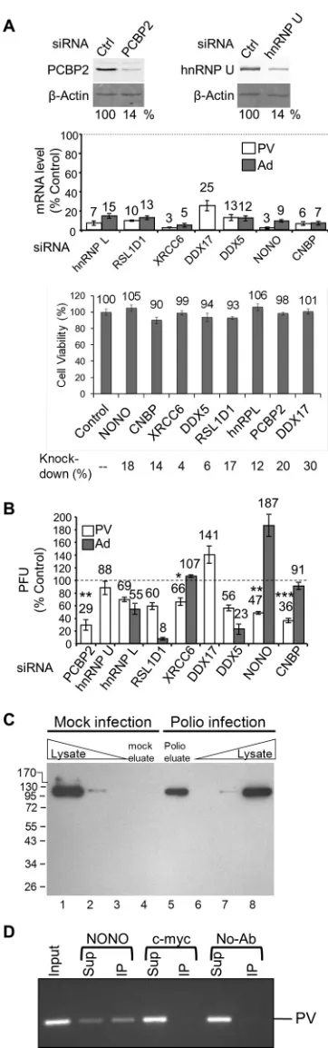
af-fects poliovirus amplification in cells.
To determine what effect,
if any, the identified host factors had on PV amplification in cell
culture, the host factors were knocked down using siRNAs (
Fig.
4A
), and the effects on viral amplification were determined.
Spe-cifically, following siRNA knockdown of a single host factor, cells
were infected with PV at a low multiplicity of infection (MOI) of
0.1, and the titer of the virus was determined after a single round of
amplification at 6 hpi. PCBP2 [poly(rC) binding protein 2] was
used as a positive control in our knockdown assay since it has been
shown to be involved in both translation and replication of PV
(
54
,
55
), although siRNA knockdowns have not been previously
demonstrated for PCBP2. Knockdown of PCBP2 (
Fig. 4A
, left) led
to a decrease in viral amplification by 71% (
Fig. 4B
). To determine
how reliable the TUX-MS method is for identification of host factors
required for PV infection, eight host factors were chosen for
experi-mental validation from
Table 3
. Host factors were chosen throughout
the list, with three identified exclusively in the PV sample and five
demonstrating an enrichment in the PV sample. For these proteins,
we observed efficient knockdown, without an impact on cell viability
(
Fig. 4A
, bottom). Knockdown of heterogeneous nuclear
ribonucle-oprotein L (hnRNP L), ribosomal L1 domain-containing protein 1
(RSL1D1), X-ray repair cross-complementing protein 6 (XRCC6),
probable ATP-dependent RNA helicase DDX5, non-POU
domain-containing octamer-binding protein (NONO), or cellular nucleic
acid binding protein (CNBP) resulted in a decrease in PV titers (
Fig.
4B
, white bars). The greatest effects on PV amplification (64% and
53% decreases) were observed when CNBP or NONO was knocked
down, respectively (
Fig. 4B
). Multiple cycle amplification assays that
measured either the number of plaques or the size of the plaques
when CNBP was knocked down yielded similar results (data not
shown). DDX17 was the only protein examined that resulted in an
increase in viral titers, suggesting that this protein inhibits PV
ampli-fication. The heterogeneous nuclear ribonucleoprotein U (hnRNP
U) was the only host factor tested that did not have a significant effect
on PV amplification, suggesting that the virus either does not require
it, or there is another host factor with a redundant function (such as
the hnRNPU-like proteins in
Table 3
). Nevertheless, it was confirmed
by Western analysis that hnRNP U specifically cross-links to RNA in
PV-infected cells (
Fig. 4C
). This specificity demonstrates that it
in-deed interacts with viral RNA and is not merely an abundant
nonspe-cific binding protein.
Investigation of adenovirus amplification indicates
specific-ity of host factors for poliovirus infection.
To test whether
FIG 4Depletion of host proteins found to interact with PV RNA has an impact on PV amplification. HeLa cells were transfected with either control or specific siRNAs as indicated. (A) Quantitative Western analysis of PCBP2 and hnRNP U knockdown. (Top) A-actin Western blot is shown as a loading control. (Middle) qRT-PCR of mRNA levels following knockdowns normal-ized to both-actin and GAPDH (glyceraldehyde-3-phosphate dehydroge-nase) mRNA levels. (Bottom) Cell viability was measured using an MTT assay 48 h following knockdown by the indicated siRNAs; the amount of mRNA
remaining following knockdown as determined by qRT-PCR is indicated. (B) Knockdown cells were infected at an MOI of 0.1 with PV (white bars) or adenovirus 5 (dark gray bars), and the virus titer after a single round of am-plification (6 or 30 hpi, respectively) was determined by plaque assay. (C) Cells were mock infected (lanes 1 to 4) or PV infected (lanes 5 to 8) in the presence of Act D and 4TU. Cross-linking with long-wavelength UV light was per-formed at 5 hpi prior to cell lysis. RNA-protein complexes were isolated using oligo(dT25) magnetic beads, and then RNA was degraded with RNase A and the proteins were separated by SDS-PAGE (lanes 4 and 5) along with total protein whole-cell lysates from mock-infected (lanes 1 to 3) and poliovirus-infected (lanes 6 to 8) cells. hnRNP U was detected by Western analysis. (D) PV RNA immunoaffinity purifies with NONO. PV-infected HeLaUPRTcells were harvested at 5 hpi and subject to immunoprecipitation with NONO, c-myc, or no antibody (No-Ab). RNA was isolated from input, supernatant (Sup), and immunoprecipitation (IP) and detected by reverse transcription and PCR. Shown is a representative result (n⫽3).
on November 7, 2019 by guest
http://jvi.asm.org/
[image:10.585.72.253.71.648.2]knockdown of the host factors impaired the overall cellular fitness
for viral amplification, the effects of siRNA knockdown on
ade-novirus amplification were determined. PV uses an internal
ribo-somal entry site (IRES) to translate a long polyprotein that is
pro-teolytically cleaved into precursor and mature proteins. Since
many of the known PV host factors are IRES
trans
-acting factors,
adenovirus was chosen because it is a DNA virus that is not known
to contain an IRES. Effects on adenovirus amplification were
de-termined in cells depleted for the host factors that demonstrated a
decrease in poliovirus titers. Similar to PV, adenovirus titers were
decreased for hnRNP L, RSL1D1, and DDX5 (
Fig. 4B
, dark gray
bars), suggesting that either these proteins are essential and their
depletion decreases the overall fitness of the cell, or they are
re-quired for amplification of PV and adenovirus. Interestingly,
NONO depletion results in an 87% increase in adenovirus titers,
while CNBP or XRCC6 depletion does not significantly affect the
ability of the cells to amplify adenovirus (
Fig. 4B
, dark gray bars).
This suggests that NONO may function to inhibit adenovirus
am-plification.
NONO directly binds to PV RNA to promote PV
amplifica-tion.
Identification of NONO by the TUX-MS method, which
uses a zero-distance cross-linker, suggests that NONO is directly
bound to the viral RNA. To confirm this, we performed
immuno-affinity purification of NONO from PV-infected cells and used
reverse transcription (RT)-PCR to detect coisolation of PV RNA.
We found that PV RNA was specifically coisolated with
NONO antibody, but not with c-myc antibody or no
anti-body (
Fig. 4D
). Taken together, these data suggest that NONO
directly interacts with PV RNA to enhance PV amplification.
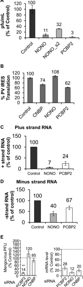
Host factors accelerate viral amplification.
It is worth noting
that virus harvested from cells depleted of a host factor did not
alter the plaque morphology or size following a single cycle of
replication (data not shown). Our studies show that knockdown
of factors identified by TUX-MS triggers a 2- to 3-fold decrease in
viral titers at 6 hpi, in agreement with previously reported
de-creases in viral amplification for other host factors (
32
,
56
). In
particular, the effects of knocking down CNBP and NONO on
viral titers at 6 hpi are consistent with effects on viral amplification
observed for La, PTB, hnRNP K, and hnRNP C1/C2 (
32
,
56–58
).
As 6 hpi represents a late stage of infection, we next asked if
assess-ment of the impact on virus titers at an earlier time point of
infec-tion would reveal delays in virus producinfec-tion. For this earlier time
point, we elected to focus on two proteins, the uncharacterized
factor, NONO, and the positive control, PCBP2. Indeed, we
ob-served a greater reduction in PV titers at the earlier time point of
infection, as NONO and PCBP2 knockdowns triggered a 10- to
30-fold decrease in titers at 4 hpi. (
Fig. 5A
). We confirmed that at
FIG 5Knockdown of NONO or CNBP affects PV replication or translation but not mengovirus amplification. (A). HeLaUPRTcells were transfected with scrambled control, NONO, or PCBP2 siRNAs. Forty-eight hours posttrans-fection of the siRNA, the cells were infected with PV at an MOI of 0.1. Virus was harvested at 4 hpi, and the titer was determined by plaque assay on He-LaUPRTcells. (B) HeLaUPRTcells were transfected with scrambled control, NONO, CNBP, or PCBP2 siRNAs, and 48 h posttransfection, the cells were transfected with a dicistronic reporter plasmid containing the PV IRES in the intercistronic region and a control plasmid expressing a cap-dependent -ga-lactosidase. Twenty-four hours later, luciferase and-galactosidase activities were determined. The amount of mRNA remaining following knockdown was determined by qRT-PCR (CNBP, 9.9%; NONO, 30%; and PCBP2, 23%). IRES-driven translation was normalized to-galactosidase activity and ex-pressed as a percentage of the control siRNA. (C and D) HeLaUPRTcells were transfected with scrambled control, NONO, or PCBP2 siRNA. Forty-eight hours posttransfection, the cells were infected with PV at an MOI of 0.1.
qRT-PCR was used to determine the number of plus-strand (C) and minus-strand (D) RNAs at 4 hpi. Copy numbers were determined using anin vitro -tran-scribed template of a known amount (plus strand, control, 7.7⫻109, NONO, 5.4⫻108, and PCBP2, 1.4⫻109copies/ng RNA; minus strand, control, 3.0⫻ 104, NONO, 1.3⫻104, and PCBP2, 2⫻104copies/ng RNA). (E) Knockdown cells were infected at an MOI of 0.1 with mengovirus (MV), and the virus titer after a single round of amplification (5.5 hpi) was determined by plaque assay (left). mRNA levels after qRT-PCR of mRNA following knockdown were nor-malized to-actin (right). The same results were obtained by normalization to GAPDH mRNA levels. qRT-PCR and titer results are percentages relative to the control siRNA (represented by the horizontal dotted lines). Errors bars are standard errors fornⱖ3. ThePvalue for viral amplification compared to the control siRNA is indicated. **,P⬍0.005.
on November 7, 2019 by guest
http://jvi.asm.org/
[image:11.585.91.236.72.574.2]4 hpi, two independent siRNAs that target NONO resulted in a
decrease in PV amplification (
Fig. 5A
). Consistent with these
ob-servations, if the titer of virus is determined on cells following
NONO or CNBP knockdowns, then there is a corresponding
de-crease in plaque number and size (data not shown). Altogether,
these results demonstrate that in the absence of the host factors
there is a delay in viral production.
Knockdowns of NONO or CNBP distinctly affect PV
trans-lation and PV RNA replication.
In order to determine if
knock-down of NONO or CNBP had an effect on PV translation, a
dicis-tronic reporter assay was used to measure PV IRES activity.
Knockdown of NONO had no effect on PV IRES activity (
Fig. 5B
).
In contrast, knockdown of PCBP2 and CNBP reduced PV IRES
activity by 38% and 26%, respectively. This finding is consistent
with previous reports that demonstrated that PCBP2 is required
for efficient PV IRES activity (
54
,
58–60
). Since NONO had no
effect on PV translation, we next tested whether knockdown of
NONO impacted the synthesis of PV plus- or minus-strand RNA.
NONO knockdown triggered a 10-fold decrease in
positive-stranded RNA (
Fig. 5C
) and a 2-fold decrease in minus-strand
RNA (
Fig. 5D
). These results demonstrate a more significant effect
on viral replication for NONO than the previously known host
factor, PCBP2. Taken together, these results suggest that NONO,
while not affecting viral translation, plays a role in enhancing PV
RNA replication.
Knockdown of NONO does not have an effect on mengovirus
amplification.
To determine whether knockdown of host
fac-tors that were shown to have an effect on poliovirus can also
affect a distantly related picornavirus, we performed a
single-round amplification experiment using mengovirus.
Mengovi-rus belongs to the genus
Cardiovirus
and contains a type II
IRES, similar to encephalomyocarditis virus (EMCV), which
means that the ribosome is recruited to the start codon. This is
in contrast to the type I IRES of polioviruses, which involves
the ribosome scanning to a downstream start codon (
61
).
PCBP2 knockdown has a significant effect on mengovirus
am-plification (
Fig. 5E
). While it is known that PCBP2 is not
re-quired for mengovirus translation (
59
), it may still play a role
in mengovirus replication, since it is known to be involved in
PV replication. To our knowledge, this is the first
demonstra-tion that PCBP2 is required for mengovirus amplificademonstra-tion. In
contrast, knockdown of NONO or CNBP did not significantly
affect amplification of mengovirus (
Fig. 5E
). This suggests that
these host factors may be specific to poliovirus or within the
Enterovirus
genus. Taken together, these data support a
signif-icant role for CNBP and NONO in PV amplification.
DISCUSSION
Using the TUX-MS method, we identified 66 previously unknown
host factors that bind to PV RNA following infection, in addition
to confirming all of the previously reported host factors. Further
validation of eight of the novel host factors revealed that all but
one played roles as enhancers or inhibitors of PV amplification.
For the one exception, hnRNP U, our validation results
neverthe-less demonstrated that, while not having an effect on PV
amplifi-cation, hnRNP U was specifically associated with PV RNA during
infection. Knockdown of NONO or CNBP resulted in a decrease
in PV amplification at 6 hpi equivalent to those of other known
host factors, such as PCBP2 (
Fig. 4
), La, PTB, and hnRNP C1/C2
(
32
,
57
,
58
). Importantly, this effect was accentuated at an earlier
time point of infection for NONO, suggesting that NONO
accel-erates virus production. Further analysis of CNBP and NONO
revealed that they are required for efficient translation and
plus-strand RNA synthesis, respectively. Immunoprecipitation of
NONO revealed that it specifically bound to the PV RNA. Taken
together, these results demonstrate that TUX-MS is a highly
effec-tive method for host factor identification with a low false-posieffec-tive
rate.
Most of the host factors that we validated were enhancers of PV
amplification, with two exceptions—an inhibitor (DDX17) and a
specific RNA binding protein that did not affect viral
amplifica-tion (hnRNP U). DDX17 is a binding partner and cofactor for zinc
finger CCCH-type antiviral protein 1 (ZAP), which was also
de-tected by TUX-MS (
Table 3
) (
62
). ZAP is an antiviral protein that
targets the RNA of retroviruses, filoviruses, and alphaviruses for
degradation (
63–65
) and potentially poliovirus (
Table 3
). The
majority of the assayed host factors appear to be enhancers of PV
amplification, such as the DEAD box RNA helicase DDX5. DDX5
has been shown to participate in RNA replication during HCV
infection (
66
). hnRNP L, XRCC6, CNBP, and RSLID1 have either
been shown to enhance other IRESs or be involved in translation
(
67–70
). NONO, which we found had a role in viral replication,
has two RNA recognition motifs and a coiled-coiled protein
inter-action domain (
71
,
72
). It is known to be involved in a number of
nuclear functions and forms monomers and heteromers to bind to
double-stranded DNA (dsDNA), single-stranded DNA (ssDNA),
and RNA (
73
,
74
). Interestingly, NONO is known to form a complex
with SFPQ (splicing factor, proline- and glutamine-rich) and
Matrin-3 (MATR3) (
73
,
75
), which were also identified by TUX-MS
(see Data Set S1 in the supplemental material).
Although poliovirus is a cytoplasmic replicating virus, most of
its known host factors are either nuclear or cycle between the
nucleus and the cytoplasm. The relocalization of known host
fac-tors from the nucleus to the cytoplasm during poliovirus infection
has been observed for a number of host factors (
13
,
46–51
,
76
). PV
infection results in degradation of several nucleoporins, which
disrupts nuclear-cytoplasmic transport pathways (
49
,
77
). This is
associated with a number of predominately nuclear host proteins
being relocalized to the cytoplasm either due to retention in the
cytoplasm, due to binding to the PV RNA to proteins, or because
import is impaired (
50
,
78
). Therefore, the fact that the majority of
the proteins identified by TUX-MS are either nuclear or cycle
between the nucleus and the cytoplasm is entirely consistent with
the known host factors and relocalization of proteins from the
nucleus to the cytoplasm during viral infection.
Since all eight of the host proteins we tested were validated, this
suggests that TUX-MS is a robust assay to identify host proteins
that bind the viral RNA during infection in cell culture. There are
a number of reasons that likely contribute to the low false-positive
rate and the efficient discovery of all known and 66 novel host
factors. First, the cross-linking was performed prior to cell lysis,
and therefore cross-links were established prior to the disruption
of cellular compartmentalization. Second, the RNA-protein
com-plexes were isolated under denaturing conditions, reducing the
possibility that nonspecific RNA binding proteins would remain
associated with the RNA during isolation. Third, the use of a mock
control allowed for the elimination of potential false positives that
may either bind to host RNA or the resin under denaturing
con-ditions. There is a possibility that some factors are present by
nonspecific association with viral RNA, while not binding to host
on November 7, 2019 by guest
http://jvi.asm.org/
RNA in the mock samples. However, the successful validation of
selected uncharacterized factors suggests that TUX-MS has a low
false-positive rate and is a valuable methodology with the
poten-tial to provide us the biological picture of PV in infected cells.
Fourth, the RNA levels were normalized prior to RNase digestion,
ensuring that mock-infected and infected samples were
compara-ble with respect to the RNA-protein complexes. Taken together,
this suggests that TUX-MS can be used to identify additional host
factors with a low false-positive discovery rate even for a
well-studied virus, such as poliovirus.
Although we identified all of the known host factors, several
did not meet our stringent criteria using spectral counting
analysis (e.g., PCBP2); these criteria were selected to identify
new potential candidates with relatively high confidence.
Spec-tral counting analysis was utilized for relative quantification
for TUX-MS as it is computationally facile, well suited to detect
large differences in relative abundance, and can be readily
in-tegrated into most proteomics workflows. Yet, one
disadvan-tage is the lack of sensitivity for proteins that generate low
spectral counts, either due to low abundance or poor ability to
be detected by MS. As a consequence, we expect a subset of
excluded proteins to be false negatives, and so cannot eliminate
the possibility that additional host factors are present in the
complete data set (see Data Set S1 in the supplemental
mate-rial). On the other hand, it is also likel