Mimic Phosphorylation of a

C1 Protein
Encoded by TYLCCNB Impairs Its
Functions as a Viral Suppressor of RNA
Silencing and a Symptom Determinant
Xueting Zhong,
aZhan Qi Wang,
aRuyuan Xiao,
aLinge Cao,
aYaqin Wang,
aYan Xie,
aXueping Zhou
a,bState Key Laboratory of Rice Biology, Institute of Biotechnology, Zhejiang University, Hangzhou, Chinaa; State
Key Laboratory for Biology of Plant Diseases and Insect Pests, Institute of Plant Protection, Chinese Academy of Agricultural Sciences, Beijing, Chinab
ABSTRACT
Phosphorylation of the

C1 protein encoded by the betasatellite of
to-mato yellow leaf curl China virus (TYLCCNB-

C1) by SNF1-related protein kinase 1
(SnRK1) plays a critical role in defense of host plants against geminivirus infection in
Nicotiana benthamiana
. However, how phosphorylation of TYLCCNB-

C1 impacts its
pathogenic functions during viral infection remains elusive. In this study, we
identi-fied two additional tyrosine residues in TYLCCNB-

C1 that are phosphorylated by
SnRK1. The effects of TYLCCNB-

C1 phosphorylation on its functions as a viral
suppressor of RNA silencing (VSR) and a symptom determinant were investigated
via phosphorylation mimic mutants in
N. benthamiana
plants. Mutations that
mimic phosphorylation of TYLCCNB-

C1 at tyrosine 5 and tyrosine 110
attenu-ated disease symptoms during viral infection. The phosphorylation mimics
weak-ened the ability of TYLCCNB-

C1 to reverse transcriptional gene silencing and to
suppress posttranscriptional gene silencing and abolished its interaction with
N.
benthamiana
ASYMMETRIC LEAVES 1 in
N. benthamiana
leaves. The mimic
phos-phorylation of TYLCCNB-

C1 had no impact on its protein stability, subcellular
localization, or self-association. Our data establish an inhibitory effect of
phos-phorylation of TYLCCNB-

C1 on its pathogenic functions as a VSR and a
symp-tom determinant and provide a mechanistic explanation of how SnRK1 functions
as a host defense factor.
IMPORTANCE
Tomato yellow leaf curl China virus (TYLCCNV), which causes a severe
yellow leaf curl disease in China, is a monopartite geminivirus associated with
the betasatellite (TYLCCNB). TYLCCNB encodes a single pathogenicity protein,

C1 (TYLCCNB-

C1), which functions as both a viral suppressor of RNA silencing
(VSR) and a symptom determinant. Here, we show that mimicking
phosphoryla-tion of TYLCCNB-

C1 weakens its ability to reverse transcriptional gene silencing, to
suppress posttranscriptional gene silencing, and to interact with
N. benthamiana
ASYMMETRIC LEAVES 1. To our knowledge, this is the first report establishing an
in-hibitory effect of phosphorylation of TYLCCNB-

C1 on its pathogenic functions as
both a VSR and a symptom determinant and to provide a mechanistic
explana-tion of how SNF1-related protein kinase 1 acts as a host defense factor. These
find-ings expand the scope of phosphorylation-mediated defense mechanisms and
con-tribute to further understanding of plant defense mechanisms against geminiviruses.
KEYWORDS
TYLCCNB-

C1,
Nicotiana benthamiana
, SnRK1, geminivirus, host defense
factor, posttranscriptional gene silencing, protein phosphorylation, transcriptional
gene silencing
Received24 February 2017Accepted17 May
2017
Accepted manuscript posted online24 May
2017
CitationZhong X, Wang ZQ, Xiao R, Cao L,
Wang Y, Xie Y, Zhou X. 2017. Mimic phosphorylation of a βC1 protein encoded by TYLCCNB impairs its functions as a viral suppressor of RNA silencing and a symptom determinant. J Virol 91:e00300-17.https://doi .org/10.1128/JVI.00300-17.
EditorAnne E. Simon, University of Maryland,
College Park
Copyright© 2017 American Society for
Microbiology.All Rights Reserved.
Address correspondence to Yan Xie, [email protected], or Xueping Zhou, [email protected].
X.Z. and Z.Q.W. contributed equally to this work.
crossm
on November 7, 2019 by guest
http://jvi.asm.org/
T
he
Geminiviridae
are a diverse family of plant viruses that are able to infect food and
cash crops, causing serious crop failure and economic losses worldwide (1–5). The
family
Geminiviridae
is currently divided into nine genera,
Becurtovirus
,
Begomovirus
,
Curtovirus
,
Eragrovirus
,
Mastrevirus
,
Topocuvirus
,
Turncurtovirus
,
Capulavirus
, and
Grablo-virus
, based on genome organization, host range, and insect vector (6).
Whitefly-transmitted begomoviruses, with more than 200 species, constitute the largest genus
of geminiviruses (4, 7). The genomes of begomoviruses can be either monopartite or
bipartite (with genomic components referred to as DNA-A and DNA-B), with a length
of approximately 2.7 kb (1, 8, 9). Over evolutionary time, the monopartite
begomovi-ruses have acquired two classes of satellite molecules, known as betasatellite (DNA

)
and alphasatellite (DNA1), which act to decrease host resistance, move virus particles,
and enhance infection (5, 10). The first DNA satellite was identified with tomato leaf curl
virus (ToLCV) (11); since then, many monopartite begomovirus species have been
frequently found to be associated with betasatellites, and the number of
begomovi-ruses associated with betasatellites is continuously increasing (5).
Tomato yellow leaf curl China virus (TYLCCNV), which causes a severe yellow leaf
curl disease in tomato and tobacco in China, is a monopartite begomovirus in
associ-ation with a betasatellite (TYLCCNB) (12, 13). The genome of TYLCCNB is approximately
half the size of that of its helper virus TYLCCNV and is required for induction of leaf curl
disease in plants (13). It encodes a single pathogenicity protein, TYLCCNB-

C1, which
functions as a viral suppressor of RNA silencing (VSR) and a symptom determinant
(13–15). Our previous studies have shown that in both
Nicotiana
(host) and
Arabidopsis
(nonhost) plants, overexpression of TYLCCNB-

C1 produces virus-like symptoms,
in-cluding leaf curling, vein swelling, and blistering of leaves (13–15). TYLCCNB-

C1 also
upregulates an endogenous RNA silencing suppressor
Nicotiana benthamiana
calmodulin-like protein
gene (
Nbrgs-CaM
), and Nbrgs-CaM regulates RNA silencing and promotes
geminivirus infection by repressing the expression of
RNA-dependent RNA polymerase 6
(
RDR6
) and promoting
N. benthamiana
suppressor of gene silencing 3 (NbSGS3)
deg-radation via the autophagy pathway (16, 17). TYLCCNB-

C1 is also involved in
sup-pressing transcriptional gene silencing (TGS) (18), which serves as a robust defense
strategy against geminiviruses (19). Furthermore, TYLCCNB-

C1 interacts with
ASYM-METRIC LEAVES 1 (AS1) to alter leaf development in
Arabidopsis
(14). TYLCCNB-

C1 can
also repress the jasmonic acid (JA)-mediated plant defenses against the whitefly,
thereby accelerating its population increase (20). Therefore, it is necessary to clarify the
molecular mechanisms underlying the pathogenesis of TYLCCNB-

C1.
Studies during the past decade have led to significant advances in our
understand-ing of plant defenses against geminivirus infections. Host plants have developed at
least four different defense strategies to deal with geminivirus infection. The first line
of defense is posttranscriptional gene silencing (PTGS)-mediated RNA interference
(RNAi) that can aggressively decrease geminivirus transcripts (4, 16, 17, 19, 21).
RNA-dependent DNA methylation (RdDM)-mediated TGS also functions as a defense
mech-anism against geminivirus chromatin (5, 22–24). Recently,
R
gene-mediated resistance
has been proposed as another essential defense strategy against geminiviruses. For
example, the begomovirus nuclear shuttle protein (NSP)-interacting kinase 1 (NIK1) is
able to activate antiviral immunity against cabbage leaf curl virus (CaLCuV) (25, 26).
Besides the above-mentioned defense mechanisms, host factors such as SNF1-related
protein kinase 1 (SnRK1) have also been involved in defense against geminiviruses
(27–29).
Protein kinases phosphorylate serine (Ser), threonine (Thr), or tyrosine (Tyr) residues
of target proteins to alter their enzymatic activity, cellular localization, and interaction
with other proteins, as well as other biochemical properties (28). Protein
phosphory-lation plays crucial roles in signal transduction in developmental and environmental
responses and is vital in modulating plant-pathogen interactions. Plant SnRK1, which is
homologous to AMPK and SNF1 in animals and
Saccharomyces cerevisiae
, respectively,
acts as a central integrator of metabolic homeostasis in plants and a critical regulator
of diverse stress responses triggered by viral, bacterial, and fungal infections or even
Zhong et al. Journal of Virology
on November 7, 2019 by guest
http://jvi.asm.org/
herbivores (29, 30). Recently we and others have shown that TYLCCNB-

C1 and
begomovirus AL2/C2 proteins are targets for plant SnRK1, which plays critical roles in
defense against geminivirus infections (27, 28). Overexpression of SnRK1 in
N.
bentha-miana
plants attenuates virus symptoms and decreases viral DNA accumulation,
whereas knockdown of SnRK1 results in increased susceptibility to infection (27, 31).
SnRK1 is upregulated by TYLCCNB and phosphorylates TYLCCNB-

C1 at Ser-33 and
Thr-78 (27).
Here, we show that two Tyr residues in TYLCCNB-

C1 are also phosphorylation
targets for SnRK1 and that mutations in four residues of TYLCCNB-

C1 that mimic
phosphorylation impair its pathogenic functions as a VSR and a symptom determinant.
Phosphorylation of TYLCCNB-

C1 weakened its ability to reverse TGS and suppress
PTGS and abolished its interaction with
N. benthamiana
AS1 (NbAS1). Our findings give
insights into the molecular mechanisms of host phosphorylation of TYLCCNB-

C1,
which appears to be a critical defense strategy against geminivirus infection, and may
provide practical strategies for viral disease management.
RESULTS
Identification of two novel Tyr phosphorylation sites of TYLCCNB-

C1.
Our
previous study showed that Ser-33 and Thr-78 in TYLCCNB-

C1 are two important
functional phosphorylation sites phosphorylated by SnRK1. A double-phosphorylation
mimic aspartate (D) mutant, TYLCCNB-

C1-S33D/T78D (

C1-2D), shows delayed and
attenuated disease symptoms and lower levels of viral DNA accumulation in
systemi-cally infected leaves when infected with TYLCCNV isolate Y10 and its associated
TYLCCNB (Y10A/

) (27). However, TYLCCNB-

C1 protein with alanine (A) substitutions
for Ser-33 and Thr-78 (

C1-S33A/T78A [

C1-2A]) retained low (37% to 28% of the
wild-type [WT] TYLCCNB-

C1 protein [

C1-WT]) but measurable phosphorylation
signals (27), suggesting that other potential SnRK1 phosphorylation sites exist in
TYLCCNB-

C1.
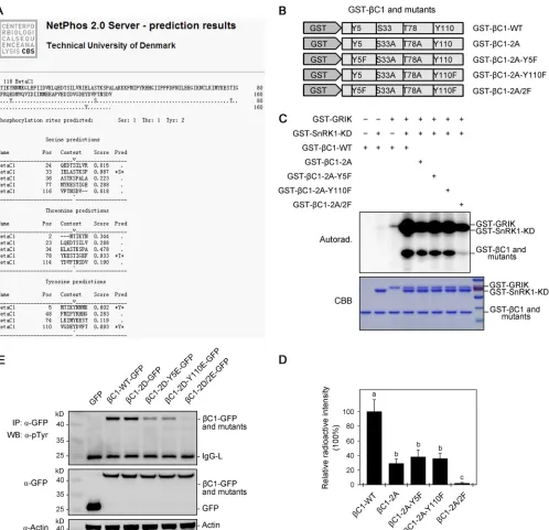
To test whether additional residues within TYLCCNB-

C1 are phosphorylated by
SnRK1, we analyzed the primary amino acid sequence of TYLCCNB-

C1 using the
NetPhos 2.0 server (
http://www.cbs.dtu.dk/services/NetPhos
). The analysis revealed
that Tyr-5 and Tyr-110 are putative phosphorylation sites, in addition to Ser-33 and
Thr-78 (Fig. 1A). To experimentally confirm the bioinformatic prediction, we mutated
Tyr-5 and Tyr-110 to phenylalanine (F), alone or in conjunction with

C1-2A, to
generate triple mutants (

C1-2A-Y5F and

C1-2A-Y110F) or a quadruple mutant (

C1-2A-Y5F/Y110F [

C1-2A/2F]) (Fig. 1B). Remarkably,

C1-2A/2F exhibited little if any
autoradiographic signal, and

C1-2A-Y5F and

C1-2A-Y110F exhibited reduced signals
compared with

C1-WT (Fig. 1C and D). Furthermore, we also mutated Tyr-5 and
Tyr-110 to glutamate (E), alone or in conjunction with

C1-2D, to generate triple
mutants (

C1-2D-Y5E and

C1-2D-Y110E) or a quadruple mutant (

C1-2D-Y5E/Y110E
[

C1-2D/2E]) and examined Tyr-5 and Tyr-110 in TYLCCNB-

C1 in
N. benthamiana
plants by immunoprecipitation, followed by antibody against phosphorylated tyrosine
as described previously (32). As shown in Fig. 1E, compared with

C1-WT,

C1-2D-Y5E
and

C1-2D-Y110E exhibited reduced immunologic signals and

C1-2D/2E exhibited
little if any signal. These results suggest that Tyr-5 and Tyr-110 are actual Tyr
phos-phorylation sites in the TYLCCNB-

C1 protein that are phosphorylated by SnRK1.
Mutation of Tyr-5 and Tyr-110 of TYLCCNB-

C1 to mimic phosphorylation
attenuates virus symptoms.
As our previous study had shown that mutants of
TYLCCNB-

C1 that mimic phosphorylation of Ser-33 and Thr-78 can slow down
devel-opment of Y10A/

-induced disease symptoms (27), we tested whether Tyr-5 and
Tyr-110 of TYLCCNB-

C1 are also involved in symptom development during Y10A/

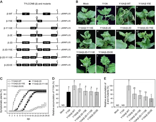
infection. To do this, we mutated Tyr-5 and Tyr-110 to glutamate (E), either alone or
together, to generate phosphorylation mimic infectious clones (

-Y5E,

-Y110E, and

-2E) (Fig. 2A). Wild-type
N. benthamiana
plants were agroinfiltrated with wild-type
TYLCCNB (

-WT) or the phosphorylation mimics in combination with their helper virus,
TYLCCNV (Y10A). As shown in Fig. 2B, iii, plants inoculated with Y10A/

-WT developed
on November 7, 2019 by guest
http://jvi.asm.org/
disease symptoms characterized by severe curling of leaves and twisted shoots at 21
days postinoculation (dpi). In contrast, plants inoculated with Y10A/

-Y5E, Y10A/

-Y110E, or Y10A/

-2E displayed moderate leaf curling without shoot twisting (Fig. 2B, iv
to vi). It is interesting that
N. benthamiana
plants inoculated with Y10A/

-Y110E
showed upward curling, which is opposite to the downward curling induced by
FIG 1Tyr-5 and Tyr-110 phosphorylation sites of TYLCCNB-C1. (A) Prediction of potential phosphorylation sites in the TYLCCNB-C1 protein sequence using NetPhos 2.0. (B) Schematic presentation ofC1-WT and mutant TYLCCNB-C1 proteins. (C) Tyr-5 and Tyr-110 of TYLCCNB-C1 are phosphorylated by SnRK1in vitro. Thein vitrokinase assay was performed using GST-SnRK1-KD (kinase domain) as a kinase, GST-GRIK as a kinase to active SnRK1-KD, and wild-type or mutant TYLCCNB-C1 proteins as substrates. Phosphorylation was analyzed by autoradiography (top), and the protein loading was shown by Coomassie brilliant blue (CBB) staining (bottom). (D) The radioactive signals shown in panel C were quantified with Quantity One Software (Bio-Rad). The data are shown as means and SD of three biological replicates. Means with different letters are significantly different (Tukey’s test;P⬍
0.05). (E) Tyr-5 and Tyr-110 of TYLCCNB-C1 are phosphorylated by SnRK1in vivo. Phosphorylation of Tyr-5 and Tyr-110 on wild-type or mutant TYLCCNB-C1–GFP proteins immunoprecipitated fromN. benthamianaleaves. IP was performed with an antibody to GFP (IP:␣-GFP), and Tyr-5 and Tyr-110 were analyzed by WB with an antibody against phosphorylated tyrosine (WB:␣-pTyr). Inputs of GFP and wild-type or mutant TYLCCNB-C1–GFP proteins are shown by WB (middle gel). WB analysis was carried out using an antibody to actin for a loading control (bottom gel). The experiment was repeated three times with similar results.
Zhong et al. Journal of Virology
on November 7, 2019 by guest
http://jvi.asm.org/
[image:4.585.46.544.68.549.2]Y10A/

-WT (Fig. 2B, v). These results suggest that Tyr-5 and Tyr-110 are associated with
the pathogenicity of TYLCCNB-

C1.
To investigate the additive effects of S33D/T78D and Y5E/Y110E on TYLCCNB-

C1
pathogenicity, we further mutated Tyr-5 and Tyr-110 to glutamate, alone or together
with the double mutation of

C1-S33D/T78D (

-2D), to generate infectious clones
containing triple (

-2D-Y5E and

-2D-Y110E) or quadruple (

-2D-Y5E/Y110E [

-2D/2E])
phosphorylation mimic mutants (Fig. 2A). At 21 dpi, plants agroinfiltrated with Y10A/

-2D-Y5E or Y10A/

-2D-Y110E showed only slight leaf curling without shoot twisting
(Fig. 2B, viii and ix). Moreover,
N. benthamiana
plants inoculated with Y10A/

-2D/2E did
not display any disease symptoms up to 21 dpi (Fig. 2B, x). These results suggest that
phosphorylation of Tyr-5 and Tyr-110 has an additive effect with phosphorylation of
Ser-33 and Thr-78 to weaken TYLCCNB-

C1 pathogenicity.
We also investigated the infection course of Y10A associated with

-WT or its
phosphorylation mimic mutants in wild-type
N. benthamiana
plants, as described
FIG 2Effects of mutants in phosphorylation sites of TYLCCNB-C1 on virus infection. (A) Schematic representation of the infectious clones of-WT and the phosphorylation mimic mutants,-Y5E,-Y110E,-2E,-2D,-2D-Y5E,-2D-Y110E, and-2D/2E, used in panel B. (B) Symptoms observed in wild-typeN. benthamianaplants agroinoculated with infectious clones of TYLCCNV (Y10A), alone or associated with WT or mutant TYLCCNB, at 21 dpi. Mock indicates plants agroinoculated with an empty-vector control (pBINPLUS). (C) Time course of infection in wild-typeN. benthamianaplants inoculated with Y10A in association with WT or mutant infectious clones. The values represent the percentages of systemically infected plants at different dpi. The data are given as means⫾SD of three biological replicates. (D and E) Relative accumulation levels of Y10A (D) and TYLCCNB or its phosphorylation mimic mutants (E) in agroinoculated plants. Viral accumulation was determined by qPCR at 21 dpi, as described for panel B. The values represent viral DNA accumulation relative to levels in control groups (N. benthamianaplants infected with Y10A/-WT), the values of which are set to 100%. The data are shown as means⫾SD of three biological replicates. Means with different letters are significantly different (Tukey’s test;P⬍0.05). ND, not detectable.
on November 7, 2019 by guest
http://jvi.asm.org/
[image:5.585.41.546.72.466.2]previously (27, 33). For Y10A/

-WT inoculation, disease symptoms started to appear at
3 or 4 dpi, with all the plants displaying typical symptoms at 8 to 10 dpi (Fig. 2C). In
contrast, symptom appearance was delayed for plants coinoculated with infectious
clones containing phosphorylation mimic Y10A/

-2D, Y10A/

-2D-Y5E, or Y10A/

-2D-Y110E, which was especially obvious in the quadruple mutant Y10A/

-2D/2E (Fig. 2C).
We further compared viral DNA accumulation in systemically infected leaves of plants
infected with Y10A associated with

-WT or its phosphorylation mimic mutants using
quantitative PCR (qPCR). As shown in Fig. 2D, there was no significant difference in the
accumulation of helper virus DNA (Y10A) in plants infected with the Y10A/

-WT or
Y10A/

phosphorylation mimic mutants, suggesting that phosphorylation of
TYLCCNB-
C1 has little or no impact on the accumulation of its helper virus. However,
accumu-lation of TYLCCNB (

) was lower in plants coinoculated with infectious clones
contain-ing phosphorylation mimic Y10A/

-2D-Y5E, Y10A/

-2D-Y110E, or Y10A/

-2D/2E than
that detected in plants infected with Y10A/

-WT (Fig. 2E). Together, these results
suggest that Tyr-5 and Tyr-110 of TYLCCNB-

C1 are functional phosphorylation sites,
and mutations that mimic phosphorylation at Tyr-5 and Tyr-110 attenuate virus
infec-tion symptoms.
To further validate our results for the phosphorylation mimics of TYLCCNB-

C1 from
the infectious clones and to determine whether the reduced virus symptoms were
related to the stability of the TYLCCNB-

C1 protein, the open reading frames (ORFs) of

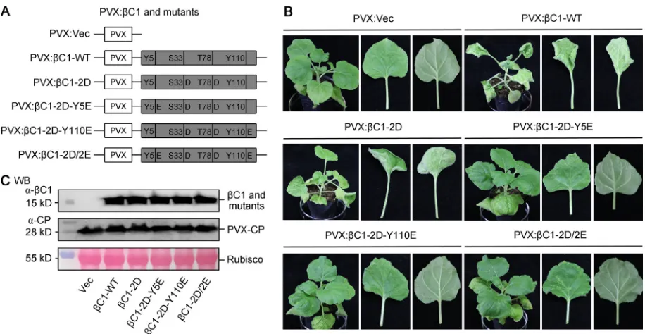
C1-WT or its phosphorylation mimic mutants were cloned into a potato virus X
(PVX)-based vector (34) (Fig. 3A). As shown in Fig. 3B, the phosphorylation mimic
mutations of TYLCCNB-

C1 weakened symptoms in
N. benthamiana
plants inoculated
with the PVX-based constructs. The triple (PVX:

C1-2D-Y5E or PVX:

C1-2D-Y110E) and
quadruple (PVX:

C1-2D/2E) phosphorylation mimic mutants greatly abated the leaf
curl and petiole elongation associated with wild-type PVX:

C1-WT (Fig. 3B). These
observations are consistent with the results obtained using infectious clones.
Further-more, we measured the accumulations of wild-type and mutant TYLCCNB-

C1 proteins
FIG 3Effects of phosphorylation mimic mutants of TYLCCNB-C1 on symptom modulation and protein stability. (A) Schematic representation of PVX expression constructs of wild-type and phosphorylation mimic mutants (PVX:C1-WT, PVX:C1-2D, PVX:C1-2D-Y5E, PVX:C1-2D-Y110E, and PVX:C1-2D/2E) of TYLCCNB-C1 used in panel B. Empty PVX vector (PVX:Vec) was used as the negative control. (B) Symptoms of wild-typeN. benthamianaplants agroinfiltrated with different PVX expression constructs at 10 dpi. Mock indicates plants agroinfiltrated with the control construct PVX:Vec. (C) WB analysis of wild-type and phosphorylation mimic mutants of TYLCCNB-C1 inN. benthamianaplants shown in panel B. Total soluble proteins were extracted from systemically infected leaves. The PVX coat protein (CP) was used as a loading control. The experiments were repeated three times with similar results.
Zhong et al. Journal of Virology
on November 7, 2019 by guest
http://jvi.asm.org/
[image:6.585.41.499.69.306.2]in wild-type
N. benthamiana
plants, as shown in Fig. 3B, by Western blotting (WB). As
shown in Fig. 3C, accumulations of the wild-type and mutant TYLCCNB-

C1 were very
similar in
N. benthamiana
plants. Collectively, these data suggest that mimic
phosphor-ylation of TYLCCNB-

C1 has no apparent impact on its protein stability and that the
weakened virus symptoms observed are not a consequence of reduced stability of
TYLCCNB-

C1 protein.
Subcellular localization and self-interaction of phosphorylation mimic mutants
of TYLCCNB-

C1.
To gain insight into how phosphorylation of TYLCCNB-

C1
attenu-ates disease symptoms during virus infection, we determined the subcellular
localiza-tion of phosphorylalocaliza-tion mimic mutants of TYLCCNB-

C1 (

C1-2D and

C1-2D/2E). The
ORFs of

C1-2D and

C1-2D/2E were cloned by translational fusions with green
fluorescent protein (GFP) at the N or C terminus of GFP and expression of the chimeric
proteins under the control of a CaMV 35S (35S) promoter (35S:

C1-2D-GFP,
35S:GFP-
C1-2D, 35S:

C1-2D/2E-GFP, and 35S:GFP-

C1-2D/2E).
N. benthamiana
leaves
agroin-filtrated with 35S:GFP, 35S:

C1-WT-GFP, or 35S:GFP-

C1-WT were used as controls.
N.
benthamiana
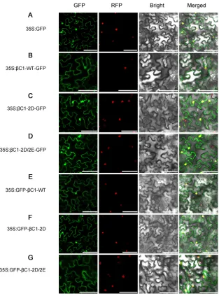
epidermal cells transiently expressing GFP only (35S:GFP) showed
fluo-rescence throughout the cells (Fig. 4A). In agreement with our previous observations,
diffuse fluorescence was detected in both the cytosol and nucleus in leaf cells
express-ing

C1-WT-GFP and GFP-

C1-WT (Fig. 4B and E), indicating that

C1-WT is localized to
both compartments (35). Similarly, fluorescence was also detected in the cytosol and
nucleus in leaf cells expressing

C1-2D-GFP, GFP-

C1-2D,

C1-2D/2E-GFP, and
GFP-
C1-2D/2E (Fig. 4C and F, and D and G). These results, therefore, suggest that mutants
that mimic phosphorylation of TYLCCNB-

C1 have no effect on subcellular localization
of TYLCCNB-

C1.
It has been shown that oligomerization of TYLCCNB-

C1 is critical to its function as
a pathogenicity determinant (35). We next determined whether phosphorylation
mim-ics of TYLCCNB-

C1 affect its oligomerization, using a bimolecular fluorescence
com-plementation (BiFC) assay as described previously (36).
N. benthamiana
leaves were
coagroinfiltrated with constructs designed to express

C1-WT,

C1-2D, and

C1-2D/2E
fused at their N or C termini with the N- or C-terminal portions of a yellow fluorescent
protein (YFP) (2YN and 2YC). Leaves were photographed 48 h after coinfiltration using
a confocal laser scanning microscope.
N. benthamiana
leaves coagroinfiltrated with the
empty vectors 2YN and 2YC were used as a negative control (Fig. 5A). Consistent with
our previous observation, YFP fluorescence was detected in the cytosol and nucleus in
N. benthamiana
epidermal cells expressing 2YN:

C1-WT and 2YC:

C1-WT (Fig. 5B),
indicating oligomerization of

C1-WT. Similarly, YFP fluorescence was detected in the
cytosol and nucleus in
N. benthamiana
epidermal cells expressing 2YN:

C1-2D and
2YC:

C1-2D or 2YN:

C1-2D/2E and 2YC:

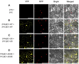
C1-2D/2E (Fig. 5C and D). This suggests that

C1-2D and

C1-2D/2E are still able to form multimeric complexes even when mimic
phosphorylated. Together, these data suggest that mutations that mimic
phosphory-lation of TYLCCNB-

C1 do not affect the ability of TYLCCNB-

C1 to oligomerize.
Mimic phosphorylation of TYLCCNB-

C1 affects its ability to reverse
estab-lished methylation-mediated TGS.
Our previous study showed that TYLCCNB-

C1
suppresses methylation-mediated TGS in
N. benthamiana
plants during Y10A/

infec-tion (18). We next asked whether phosphorylainfec-tion mimic mutants of TYLCCNB-

C1
affect its ability to reverse established methylation-mediated TGS. To determine this, we
used transgenic
N. benthamiana
16-TGS plants, which contain a GFP transgene that is
downstream of a transcriptionally silenced 35S promoter (37, 38). 16-TGS seedlings
were inoculated with Y10A alone or in conjunction with

-WT or the phosphorylation
mimic mutants (

-2D and

-2D/2E). Consistently, the 16-TGS seedlings infected with
Y10A alone or with

-2D and

-2D/2E showed very slight disease symptoms, while
severe disease symptoms were observed in seedlings infected with Y10A/

-WT at 14
dpi (Fig. 6A). Under UV light, GFP fluorescence was obviously visible in veins and
petioles of symptomatic leaves of 16-TGS plants infected with Y10A/

-WT at 14 dpi.
However, 16-TGS seedlings infected with Y10A alone or with

-2D and

-2D/2E did not
display any GFP fluorescence (Fig. 6A). As expected, qPCR and WB analyses showed that
on November 7, 2019 by guest
http://jvi.asm.org/
the lack of visible fluorescence in 16-TGS plants infected with Y10A/

-2D or Y10A/

-2D/2E was due to a deficiency in the accumulation of GFP mRNA and protein (Fig. 6B
and C). These results suggest that the phosphorylation mimic mutants of TYLCCNB-

C1
are unable to reverse established methylation-mediated TGS in
N. benthamiana
plants.
To further confirm the effect of phosphorylation mimics of TYLCCNB-

C1 on
dem-ethylation, a routine chop-PCR (39) was employed to determine the methylation status
of the 35S promoter in 16-TGS plants infected with Y10A alone or with

-WT or its
phosphorylation mimic mutants (

-2D and

-2D/2E). Total nucleic acids were extracted
from vascular tissue and digested with a methylation-sensitive endonuclease, HinfI, or
a methylation-dependent endonuclease, McrBC, and then subjected to PCR. Levels of
PCR product derived from the 35S promoter in 16-TGS seedlings infected with Y10A/

-WT were much lower than those detected in plants infected by Y10A/

-2D or
FIG 4Subcellular localization ofC1-WT and phosphorylation mimic mutants (C1-2D andC1-2D/2E) of TYLCCNB-C1. (A) Localization of GFP fluorescence in epidermal cells ofN. benthamiana. (B and E) Localization of GFP fluorescence fromC1-WT protein fused to the N- and C termini of GFP, respectively. (C and F) Localization of GFP fluorescence fromC1-2D protein fused to the N- and C termini of GFP, respectively. (D and G) Localization of GFP fluorescence fromC1-2D/2E protein fused to the N- and C termini of GFP, respectively. The cells were photographed 48 h after infiltration using a confocal laser scanning microscope. RFP-histone 2B (RFP) was used as a marker for the nucleus. GFP, GFP fluorescence; RFP, RFP fluorescence; Bright, bright-field images; Merged, merged images. Bars, 50m.
Zhong et al. Journal of Virology
on November 7, 2019 by guest
http://jvi.asm.org/
[image:8.585.50.363.69.492.2]Y10A/

-2D/2E when using the endonuclease HinfI. In contrast, restriction with the
endonuclease McrBC produced the opposite outcome (Fig. 6D). These data indicate
that the 35S promoter is methylated during infection of 16-TGS plants with Y10A/

-2D
or Y10A/

-2D/2E. Taken together, these results suggest that phosphorylation mimic
mutants of TYLCCNB-

C1 are not able to reverse established methylation-mediated
TGS in
N. benthamiana
plants.
Mimic phosphorylation of TYLCCNB-

C1 weakens its suppression of PTGS.
Our
previous studies have also shown that TYLCCNB-

C1 is involved in suppressing PTGS in
N. benthamiana
plants during Y10A/

infection, thus counteracting RNAi-based
antivi-ral responses (15–17). We further examined whether phosphorylation mimics of
TYLCCNB-

C1 affect its ability to suppress PTGS. For this purpose, we used a transient
silencing suppression assay based on GFP transgenic
N. benthamiana
16c plants (16,
40). In this assay,
Agrobacteria
containing a binary vector designed to transiently
express sense GFP (35S:GFP) and
Agrobacteria
harboring a candidate suppressor gene
were coinfiltrated into leaves of 16c plants.
Agrobacteria
containing an empty pCHF3
vector (pCHF3:Vec) and
Agrobacteria
containing tomato bushy stunt virus (TBSV) P19
ORF (P19) (41, 42) were used as negative and positive controls, respectively.
Consis-tently, leaves of 16c seedlings coinfiltrated with
Agrobacteria
containing 35S:GFP and

C1-WT (pCHF3:

C1-WT) elicited relatively strong green GFP fluorescence as a
conse-quence of suppression of GFP RNA silencing (Fig. 7A). However, leaves coinfiltrated with
Agrobacteria
harboring a phosphorylation mimic mutant (pCHF3:

C1-2D or pCHF3:

C1-2D/2E) and 35S:GFP displayed very faint GFP fluorescence, similar to the negative
control (Fig. 7A), indicating that the GFP RNA was degraded. As anticipated, qPCR and
WB analyses verified that lower fluorescence observed in leaves of 16c plants
coinfil-trated with the phosphorylation mimic mutants (pCHF3:

C1-2D and pCHF3:

C1-2D/
2E), together with 35S:GFP, was due to reduced accumulation of GFP mRNA and protein
(Fig. 7B and C). Collectively, these results suggest that mimic phosphorylation of
TYLCCNB-

C1 weakens its suppression of PTGS in
N. benthamiana
plants.
FIG 5Self-interaction of C1-WT and phosphorylation mimic mutants (C1-2D and C1-2D/2E) of TYLCCNB-C1 in epidermal cells ofN. benthamianaby BiFC.N. benthamianaleaves were coagroinfiltrated with 2YN and 2YC empty vectors (A), 2YN:C1-WT and 2YC:C1-WT (B), 2YN:C1-2D and 2YC:C1-2D (C), or 2YN:C1-2D/2E and 2YC:C1-2D/2E (D). The cells were photographed 48 h after coinfiltration using a confocal laser scanning microscope. RFP-histone 2B (RFP) was used as a marker for the nucleus. YFP, YFP fluorescence; RFP, RFP fluorescence; Bright, bright-field images; Merged, merged images. Bars, 50m.
on November 7, 2019 by guest
http://jvi.asm.org/
[image:9.585.51.364.70.323.2]Phosphorylation mimics of TYLCCNB-

C1 weaken PTGS suppression at the
level of Nbrgs-CaM.
We next investigated how phosphorylation mimics of
TYLCCNB-
C1 weaken PTGS suppression in
N. benthamiana
plants. Our previous study
demon-strated that TYLCCNB-

C1 is able to upregulate
Nbrgs-CaM
to repress expression of
RDR6
and to suppress PTGS (16). We therefore determined whether phosphorylation
mimics of TYLCCNB-

C1 affect the upregulation of
Nbrgs-CaM
using qPCR. As shown in
Fig. 8A,
Nbrgs-CaM
was upregulated
⬃
6-fold in response to

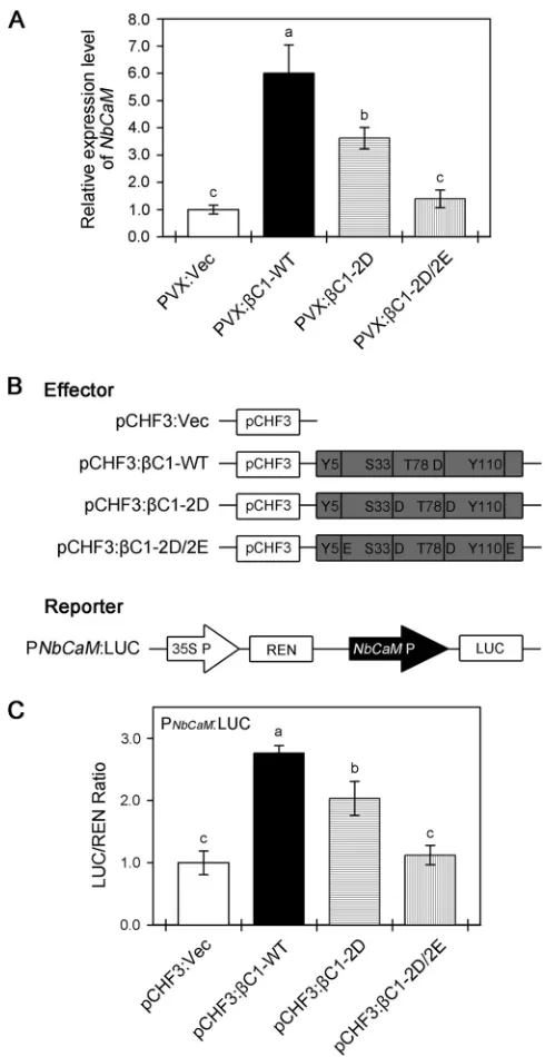
C1-WT. The
phosphory-lation mimic mutants (

C1-2D and

C1-2D/2E) were deficient in their ability to
up-regulate
Nbrgs-CaM
, with

C1-2D exhibiting an
⬃
2-fold reduction and

C1-2D/2E a
FIG 6Mimicking phosphorylation of TYLCCNB-C1 impacts its ability to reverse TGS of aGFPtransgene and suppress cytosine methylation. (A) TransgenicN. benthamiana16-TGS plants were agroinfiltrated with TYLCCNV (Y10A) alone or in conjunction with-WT or the phosphorylation mimic mutants (-2D and-2D/2E), and the plants were photographed under white light or UV light at 14 dpi. 16-TGS plants agroinoculated with Y10A alone were used as negative controls. (B) qPCR analysis ofGFPmRNA accumulation in systemically infected leaves shown in panel A. The level of gene expression was normalized to that ofNbACT2; the values represent relativeGFPmRNA accumulation compared with mRNA from 16-TGS plants infected with Y10A/-WT (100%). The data are shown as means and SD of three biological replicates. Means with different letters are significantly different (Tukey’s test;P⬍
0.05). (C) Western blot assay of GFP protein accumulation in systemically infected leaves shown in panel A. Coomassie brilliant blue (CBB) staining of the large subunit of RubisCO was used as a loading control. (D) Analysis of DNA methylation at the 35S promoter using chop-PCR. Genomic DNA was extracted from the systemically infected leaves shown in panel A. Samples from 16-TGS plants agroinoculated with Y10A alone were used as the negative control. The experiments were repeated three times with similar results.
Zhong et al. Journal of Virology
on November 7, 2019 by guest
http://jvi.asm.org/
[image:10.585.39.404.71.490.2]complete loss of upregulation. These data suggest that mutants that mimic
phosphor-ylation of TYLCCNB-

C1 reduce its ability to upregulate
Nbrgs-CaM
. In parallel, transient
transcriptional activation of the
Nbrgs-CaM
promoter by

C1-WT or its phosphorylation
mimic mutants (

C1-2D and

C1-2D/2E) was examined using a dual-luciferase system
in agroinfiltrated
N. benthamiana
leaves. The ORFs of

C1-WT and its phosphorylation
mimic mutants (

C1-2D and

C1-2D/2E) were cloned into the binary vector pCHF3 to
serve as effectors. A 1.5-kb (relative to the ATG at bp
⫹
1) fragment containing the
Nbrgs-CaM
promoter was cloned into pGreenII0800-LUC upstream of firefly luciferase
(LUC) to serve as a reporter, with 35S promoter-driven
Renilla
luciferase (REN) as the
internal control (Fig. 8B). Consistently, coexpression of

C1-WT with the LUC reporter
significantly increased the LUC/REN ratio (Fig. 8C), indicating that

C1-WT functions as
a transcription activator of
Nbrgs-CaM
. However, compared with

C1-WT, coexpression
of

C1-2D or

C1-2D/2E with the LUC reporter showed reduced LUC/REN ratios (Fig.
8C), indicating that they had partially or totally lost the capacity to upregulate
Nbrgs-CaM
. Taken together, these results suggest that mimic phosphorylation of
TYLCCNB-
C1 weakens its suppression of PTGS, most likely at the level of Nbrgs-CaM.
Interaction of TYLCCNB-

C1 with NbAS1 is abolished in phosphorylation
mimic mutants.
It has been shown that TYLCCNB-

C1 interacts with AS1 to alter leaf
development and phenocopies virus-induced disease symptoms in transgenic
Arabi-dopsis
expressing the pathogenicity determinant TYLCCNB-

C1 (14). In our earlier
experiments, we demonstrated that phosphorylation mimic mutants of TYLCCNB-

C1
attenuate virus infection symptoms in
N. benthamiana
plants (Fig. 2). We asked whether

C1-WT also interacts with AS1 in tobacco plants to induce disease symptoms and if
this interaction is impaired for its phosphorylation mimic mutants (

C1-2D and

C1-FIG 7Mimicking phosphorylation of TYLCCNB-C1 impairs its ability to suppress PTGS. (A) Suppression of PTGS of
GFPin leaves of transgenicN. benthamiana16c plants. Leaves of 16c plants were coagroinfiltrated with constructs harboring GFP (35S:GFP) and either a pCHF3 vector control (pCHF3:Vec), TBSV P19 (P19), wild-type TYLCCNB-C1 (pCHF3:C1-WT), or phosphorylation mimic mutants (pCHF3:C1-2D and pCHF3:C1-2D/2E). The agroinfiltrated leaves were photographed under UV light at 6 dpi. (B) qPCR analysis ofGFPmRNA accumulation in agroinfiltrated leaf patches shown in panel A. The level of gene expression was normalized to that ofNbACT2; the values represent relativeGFPmRNA accumulation compared to mRNA from 16c plants infected with P19 (100%). The data are shown as means and SD of three biological replicates. Means with different letters are significantly different (Tukey’s test;
P⬍0.05). (C) Western blot assay of GFP accumulation in agroinfiltrated leaf patches shown in panel A. CBB staining of the large subunit of RubisCO was used as a loading control. The experiment was repeated three times with similar results.
on November 7, 2019 by guest
http://jvi.asm.org/
[image:11.585.42.404.74.329.2]FIG 8Diminished suppression of PTGS by phosphorylation mimic mutants of TYLCCNB-C1 occurs at the level of Nbrgs-CaM. (A)Nbrgs-CaMmRNA levels in wild-typeN. benthamianaplants agroinfiltrated with PVX:C1-WT, PVX:C1-2D, or PVX:C1-2D/2E at 7 dpi. An empty PVX vector (PVX:Vec) was used as the negative control. The level of gene expression was normalized to that ofNbGAPDH, and the values represent relativeNbrgs-CaMmRNA accumulation levels compared to the mRNA level inN. benthamiana
plants infected with PVX:Vec (1.0). The data are shown as means⫾SD of three biological replicates. Means with different letters are significantly different (Tukey’s test;P⬍0.05). (B) Schematic diagram showing the constructs used in the transient transcriptional activity assay in panel C. The ORFs ofC1-WT and phosphorylation mimic mutants (C1-2D andC1-2D/2E) were cloned into the binary vector, pCHF3, to serve as effectors. Empty pCHF3 vector (pCHF3:Vec) was used as the negative control. A 1.5-kb (relative to the ATG at bp⫹1) promoter ofNbrgs-CaMwas used in the transient transcriptional activity assay. (C) TheNbrgs-CaMpromoter can be activated byC1-WT, but not by the phosphorylation mimic mutant C1-2D/2E. The PNbCaM:LUC reporter was coagroinfiltrated with the indicated effector constructs. The
LUC/REN ratio represents the PNbCaM:LUC activity relative to the internal control (REN driven by the 35S
promoter). The data are shown as means⫾SD of three biological replicates. Means with different letters are significantly different (Tukey’s test;P⬍0.05).
Zhong et al. Journal of Virology
on November 7, 2019 by guest
http://jvi.asm.org/
[image:12.585.82.327.69.544.2]2D/2E). To investigate this, we amplified the
AS1
gene of
N. benthamiana
(
NbAS1
) and
examined whether

C1-WT and its phosphorylation mimic mutants (

C1-2D and

C1-2D/2E) interact with NbAS1 using coimmunoprecipitation (CoIP) assays in
N.
benthamiana
plants.
N. benthamiana
leaves were coinfiltrated with
Agrobacteria
capa-ble of expressing NbAS1 and

C1-WT or its phosphorylation mimic mutants. Leaves
were harvested 36 h after coinfiltration, and proteins were extracted and analyzed by
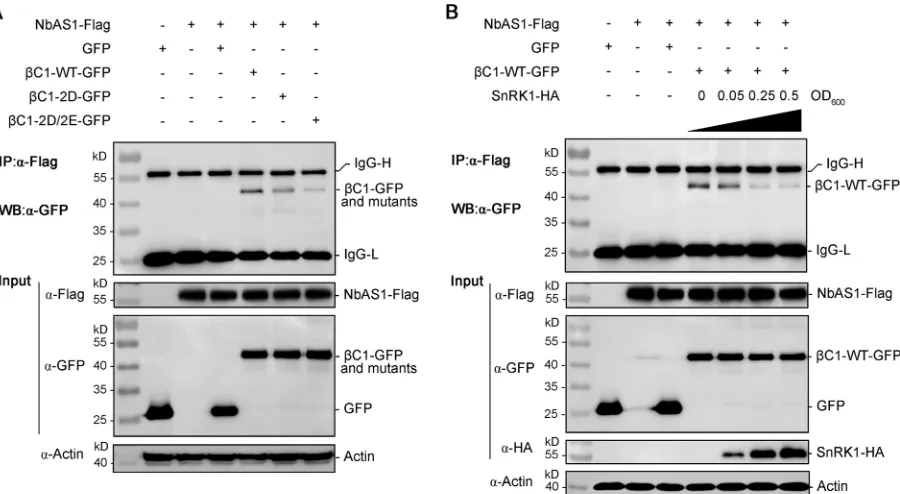
CoIP. As shown in Fig. 9A,

C1-WT interacted strongly with NbAS1 in
N.
benthami-ana
leaves, whereas the interactions between NbAS1 and

C1-2D or

C1-2D/2E
were greatly decreased. These data indicate that

C1-WT is associated with AS1 in
tobacco plants and that phosphorylation mimics of TYLCCNB-

C1 weaken its
interaction with AS1.
To further confirm the above-mentioned result and to determine the effect of
phos-phorylation of TYLCCNB-

C1 on its interaction with NbAS1, we introduced SnRK1, which is
known to phosphorylate TYLCCNB-

C1 (27). Consistently, overexpression of SnRK1
abol-ished the interaction between TYLCCNB-

C1 and NbAS1 in a dose-independent manner
in
N. benthamiana
leaves (Fig. 9B). Together, these results suggest that phosphorylation
of TYLCCNB-

C1 largely abolishes its interaction with NbAS1 to attenuate virus
infec-tion symptoms in
N. benthamiana
plants.
DISCUSSION
Protein phosphorylation is a common and important posttranslational modification
that can alter protein function via addition of phosphate groups to a target protein
through the actions of various kinases (43). Geminivirus proteins, including the
bego-movirus NSP, movement protein, AC3, AC4, BC1, BV1 and capsid protein, the curtovirus
C4 and AL2/C2 proteins, and the satellite

C1 protein, are also substrates of host
FIG 9Mimicking phosphorylation of TYLCCNB-C1 weakens its interaction with NbAS1. (A)In vivointeractions ofC1-WT and phosphorylation mimic mutants (C1-2D andC1-2D/2E) with NbAS1 inN. benthamiana. (Top)N. benthamianaleaves were coagroinfiltrated with the indicated expression constructs, CoIP was performed with an antibody to Flag (IP:␣-Flag), and the proteins were analyzed by WB with an antibody to GFP (WB:␣-GFP).C1-WT,C1-2D, andC1-2D/2E coimmunoprecipitated with NbAS1. (Middle-two gels) Inputs of NbAS1-Flag andC1-WT-GFP, C1-2D-GFP,C1-2D/2E-GFP, or GFP are shown by WB. (Bottom) WB analysis was carried out using an antibody to actin for a loading control. (B) Interaction of TYLCCNB-C1 and NbAS1 is largely abolished by SnRK1. (Top)N. benthamianaleaves were coagroinfiltrated with the indicated expression constructs in conjunction with different concentrations (OD600) of SnRK1, as indicated. CoIP was performed with an antibody to Flag (IP:␣-Flag), andC1-WT was analyzed using WB with an antibody to GFP (WB:␣-GFP). (Middle three panels) Inputs of NbAS1-Flag,C1-WT-GFP, and SnRK1-HA by WB. (Bottom) WB analysis was carried out using an antibody to actin for a loading control. (A and B) Experiments were repeated three times with similar results.
on November 7, 2019 by guest
http://jvi.asm.org/
[image:13.585.47.497.71.318.2]protein kinases during plant-virus interactions (27, 28, 44–49). It is well established that
host Ser/Thr phosphorylation of plant viral proteins plays vital roles in attenuation of
symptom development and efficiency of viral infection (28, 48–52). Consistent with this
view, our previous work has shown that phosphorylation at Ser-33 and Thr-78 of
TYLCCNB-

C1 by SnRK1 significantly attenuates its pathogenesis during Y10A/

infec-tion (27). Furthermore, overexpression of SnRK1 in
N. benthamiana
plants attenuates
virus symptoms and decreases viral DNA accumulation during Y10A/

infection,
whereas knockdown of SnRK1 results in the reciprocal effects (27). However, little is
known about the importance of Tyr phosphorylation of geminivirus proteins, with the
exception that Tyr phosphorylation of a pomovirus MP impairs its cell-to-cell
move-ment in plants (53). In this study, we identified two Tyr residues in TYLCCNB-

C1 that
are phosphorylated by SnRK1 (Fig. 1) and found that a quadruple mutant of
TYLCCNB-
C1 that mimics phosphorylation is defective as a pathogenicity determinant (Fig. 2B
and 3B). Mimicking phosphorylation of TYLCCNB-

C1 also weakened its ability to reverse
TGS (Fig. 6A) and to suppress PTGS (Fig. 7A) and abolished its interaction with NbAS1 (Fig.
9). Together with our previous data (27), these results suggest that phosphorylation of
TYLCCNB-

C1 by SnRK1 is an important defense strategy against Y10A/

infection. The
results of our study significantly expand the scope of phosphorylation-mediated host
defense against geminiviruses by implicating Tyr phosphorylation in the process.
Although phosphorylation of TYLCCNB-

C1 was reported to attenuate viral
infec-tion in our previous study (27), how the phosphorylainfec-tion of TYLCCNB-

C1 impacts its
pathogenic function remained to be investigated. The present study showed that a
quadruple mutant that mimicked phosphorylation of TYLCCNB-

C1 does not induce
disease symptoms associated with the betasatellite (Fig. 2B and 3B). Functional studies
indicated that this phenomenon was not a result of decreased protein stability or
altered subcellular localization of the TYLCCNB-

C1 phosphorylation mimics (Fig. 3, 4,
and 5). Earlier studies showed that TYLCCNB-

C1 functions as a VSR to reverse
methylation-mediated TGS (18) and to suppress PTGS (15–17). We therefore
per-formed experiments to test whether phosphorylation of TYLCCNB-

C1 affected its
ability to function as the VSR. We found that mimicking phosphorylation of
TYLCCNB-
C1 impairs its function as a VSR at the level of both TGS and PTGS (Fig. 6 and 7).
Furthermore, mimicking phosphorylation of TYLCCNB-

C1 weakened its ability to
upregulate
Nbrgs-CaM
, an endogenous suppressor of RNA silencing (Fig. 8).
In addition to functioning as a VSR, TYLCCNB-

C1 acts as a symptom determinant
(13, 14). In both
Nicotiana
and
Arabidopsis
, plants overexpressing TYLCCNB-

C1 display
virus-like symptoms, such as leaf curling, petiole elongation, and twisted shoots (13,
14). In transgenic
Arabidopsis
, TYLCCNB-

C1 interacts with AS1 to alter leaf
develop-ment, which largely phenocopies symptoms observed in virus-infected tobacco plants
(14). More importantly, the results presented here, together with previous observations
(27), clearly indicate that phosphorylation of TYLCCNB-

C1 is able to attenuate disease
symptoms of TYLCCNB-

C1, which led us to speculate that phosphorylation of
TYLCCNB-

C1 may affect its ability to function as a symptom determinant at the level
of interacting with AS1. CoIP experiments showed that

C1-WT and NbAS1 interact
in
vivo
, whereas the interactions between phosphorylation mimic mutants of
TYLCCNB-
C1 and NbAS1 were greatly decreased (Fig. 9A). This suggests that mimicking
phos-phorylation of TYLCCNB-

C1 impairs its association with NbAS1. We also found that
transient overexpression of SnRK1 abolished the interaction between

C1-WT and
NbAS1 in a dose-independent manner in
N. benthamiana
leaves (Fig. 9B), in accordance
with our previous observation that constitutive expression of SnRK1 in transgenic
plants slows down the development of virus-induced symptoms (27). We interpret this
to show that phosphorylation of TYLCCNB-

C1 negatively regulates symptom
devel-opment as a consequence of an inability to interact with NbAS1. In fact, AS1 has been
well characterized as a MYB domain transcription factor that regulates a set of genes
involved in leaf development in a complex with the LATERAL ORGAN BOUNDARIES
domain transcription factor AS2 in
Arabidopsis
(54, 55). AS1 has also been reported to
be involved in immune responses via selective suppression of JA-responsive genes (14,
Zhong et al. Journal of Virology
on November 7, 2019 by guest
http://jvi.asm.org/
56, 57). The precise mechanisms by which a TYLCCNB-

C1–AS1 complex regulates leaf
development and/or immune responses should be investigated in the future.
In conclusion, we have identified two novel Tyr phosphorylation sites of
TYLCCNB-
C1 that appear to be targets for phosphorylation and have shown that
phosphory-lation mimic mutations of TYLCCNB-

C1 impaired its function as a pathogenicity
determinant by weakening its ability to reverse TGS and to uppress PTGS and
abolish-ing its interaction with NbAS1 in
N. benthamiana
leaves. To our knowledge, this is the
first report establishing an inhibitory effect of phosphorylation of TYLCCNB-

C1 on its
pathogenic functions as both a VSR and a symptom determinant and to provide a
mechanistic explanation of how SnRK1 acts as a host defense factor. These findings
expand the scope of phosphorylation-mediated defense and contribute to further
understanding of plant defense mechanisms against geminiviruses.
MATERIALS AND METHODS
Plant material and growth conditions. Wild-type transgenic GFP 16c (58) and 16-TGS (37)N. benthamianalines were used in this study. All experimental plants were grown in an insect-free chamber at 25°C with a 16/8-hour (light/dark) photoperiod. Plants at the 4- to 6-leaf stage were used for the experiments, as described previously (18).
Site-directed mutagenesis and plasmid construction.Site-directed mutagenesis of TYLCCNB-C1 was performed using overlapping PCR as described previously (59). A Tyr residue at position 5 was changed to Phe (F) (TAC to TTT) or Glu (E) (TAC to GAA), a serine residue at position 33 was changed to Ala (A) (TCA to GCA) or Asp (D) (TCA to GAT), a Thr residue at position 78 was changed to Ala (A) (ACA to GCA) or Asp (D) (ACA to GAT), and a Tyr residue at position 110 was changed to Phe (F) (TAT to TTT) or Glu (E) (TAT GAA). Construction of infectious clones containing TYLCCNB-C1 mutants was performed as described previously (12). To construct plasmids producing recombinant glutathioneS-transferase (GST)-tagged proteins, the ORFs ofC1-WT and the mutants were cloned into the BamHI-XhoI sites of the pGEX-4T-3 vector (GE). The GST-GRIK and GST-SnRK1-KD constructs were described previously (27, 60). To generate the PVX expression constructs, ORFs ofC1-WT and its mutants were cloned into the AscI-SalI sites of the PVX vector pGR106 (34). For transient expression of WT and mutantC1 proteins, ORFs were cloned into the KpnI-BamHI sites of the binary vector pCHF3 (61). Plasmids containing 35S:GFP and P19 constructs used for protein subcellular localization and the BiFC assay were constructed as previously described (16, 18, 35). For dual-luciferase assays, a 1.5-kb (relative to the ATG at bp⫹1) promoter ofNbrgs-CaMwas cloned into the XhoI-NcoI sites of pGreenII0800-LUC (62) to generate the PNbCaM:LUC reporter construct. To determine phosphotyrosine in wild-type and mutant TYLCCNB-C1
proteins, ORFs were cloned into the KpnI-BamHI sites of a binary vector, 35SGFP. For CoIP assays, ORFs of WT and mutant TYLCCNB-C1 were cloned into the KpnI-BamHI sites of the binary vector 35SGFP, and ORFs of NbAS1 and SnRK1 were cloned into the SacI-BamHI sites of 35SFlag or the KpnI-BamHI sites of 35SHA binary vectors. All the primers used in these experiments are available upon request, and all constructs were confirmed by sequencing.
Recombinant protein production and kinase assay. Recombinant proteins were produced in
Escherichia colistrain BL21(DE3) induced with 0.5 mM isopropyl-D-thiogalactoside (IPTG) for 6 h at 20°C. GST-fused proteins were purified using glutathione resin (GE) according to the manufacturer’s instruc-tions.In vitrokinase assays were performed as described previously (27).
Viral inoculation and agroinfiltration.For inoculation and TGS experiments,Agrobacterium tume-facienscultures carrying infectious clones were infiltrated into N. benthamianaleaves as described previously (18). For recombinant PVX vectors expressing WT or mutant TYLCCNB-C1, eachA. tumefa-ciensculture was adjusted to an optical density at 600 nm (OD600) of 0.8 before infiltration intoN.
benthamianaplants. Transient silencing suppression assays were performed as described previously (16, 40). For protein subcellular localization, BiFC, dual-luciferase reporter assays, immunoprecipitation (IP), and CoIP,A. tumefacienscultures were used at an OD600of 1.0 unless otherwise stated.
Infectivity test and viral DNA accumulation.The course of viral infection was monitored as described previously (27, 33). Total nucleic acids were extracted from systemically infected leaves using a cetyltrimethylammonium bromide (CTAB)-based method (63). Viral DNA accumulation was measured using qPCR as described previously (26). Relative viral DNA accumulation levels were calculated by the comparative threshold cycle (CT) method (64). TheN. benthamiana 25S nuclear rRNAgene (Nb25SrRNA)
was used as the endogenous control (65). The reactions were performed in triplicate, and the results were averaged.
RNA extraction and qPCR analysis.Total RNA was extracted from samples using an RNAprep Pure Plant kit (Tiangen), and cDNA was reverse transcribed from 1.0g of total RNA using a ReverTra Ace qPCR reverse transcription (RT) kit (Toyobo). Relative quantification of gene expression by qPCR was performed as described previously (66). Relative expression levels were calculated by the comparativeCT
method (64).NbACTIN2RNA was used as the endogenous control. The reactions were performed in triplicate, and the results were averaged.
Protein extraction, WB analysis, IP, and CoIP.Protein extraction and WB analysis were performed as described previously (66). For IP and CoIP assays, 1.0 g of agroinfiltratedN. benthamianaleaf tissue was collected for each combination and homogenized in IP buffer (50 mM Tris-HCl, pH 7.5, 150 mM NaCl, 1 mM EDTA, 0.6% Triton X-100, 10% glycerol, 5 mM dithiothreitol [DTT], 1 mM phenylmethylsulfonyl
on November 7, 2019 by guest
http://jvi.asm.org/
fluoride [PMSF], 100M MG132 with complete protease inhibitor cocktail [Roche]) and centrifuged twice at 13,000 rpm at 4°C for 15 min. The supernatant was incubated with 30l protein G agarose (Millipore) and 1g anti-Flag (␣-Flag) antibody (Sigma) at 4°C for 4 h with gentle shaking and then washed six times with 1 ml of washing buffer (50 mM Tris-HCl, pH 7.5, 200 mM NaCl, 1 mM EDTA, 0.2% Triton X-100, 10% glycerol, 5 mM DTT, 1 mM PMSF). Immunoprecipitated proteins were analyzed by WB analysis using an anti-pY99 (Santa Cruz) or an␣-GFP (Sigma) antibody.
Protein subcellular localization and BiFC assay.N. benthamianaleaves were infiltrated withA. tumefacienscultures harboring the designated constructs, 0.5-cm2leaf explants were harvested approx-imately 48 h postinfiltration, and GFP fluorescence was examined by confocal microscopy as described previously (18). BiFC experiments were performed as described previously (18, 36). YFP fluorescence was observed and photographed 48 h postinfiltration using confocal microscopy. The nucleus was located using a nuclear-localized red fluorescent protein (RFP)-histone 2B (67).
Chop-PCR.Total nucleic acids were extracted from systemically infected leaves using a Hi-DNA secure plant kit (Tiangen). Chop-PCR was performed as described previously (39, 68). Briefly, 1g total nucleic acids was digested with a sensitive restriction endonuclease, HinfI, or a methylation-dependent restriction endonuclease, McrBC (NEB), in a 20-l reaction mixture according to the manu-facturer’s recommendations. Undigested samples were treated in the same way but without adding the enzymes. After digestion, PCR was performed using 2l of the digested DNA as the template in a 20-l reaction mixture and using the 35S promoter-specific primer pairs (35SP-F, 5=-AAGGYAAGTAATAGAGA TTGGAG-3=, and 35SP-R, 5=-CACCTTCCTTTTCCACTATCTTCAC-3=), and the PCR products were separated by electrophoresis on a 1.5% agarose gel.
Dual-luciferase reporter assays.Transient dual-luciferase assays inN. benthamianawere performed as described previously (62, 69). LUC and REN were assayed by dual-luciferase assay (Promega) as previously described (70, 71). Six biological replicates were measured for each sample.
Statistical analysis.The data shown were calculated as the mean⫾standard deviation (SD) for at least three independent experiments. Differences in the mean values were assessed using the statistical software data processing system (DPS) v7.05 (72), followed by Tukey’s test. Values were considered significantly different at aPvalue of⬍0.05.
ACKNOWLEDGMENTS
This work was supported by grants from the National Natural Science Foundation of
China (31390422) and the Postdoctoral Science Foundation of China (2015M581946).
REFERENCES
1. Jeske H. 2009. Geminiviruses. Curr Top Microbiol Immunol 331:185–226. 2. Lefeuvre P, Martin DP, Harkins G, Lemey P, Gray AJ, Meredith S, Lakay F, Monjane A, Lett JM, Varsani A, Heydarnejad J. 2010. The spread of tomato yellow leaf curl virus from the Middle East to the world. PLoS Pathog 6:e1001164.https://doi.org/10.1371/journal.ppat.1001164. 3. Navas-Castillo J, Fiallo-Olivé E, Sánchez-Campos S. 2011. Emerging virus
diseases transmitted by whiteflies. Annu Rev Phytopathol 49:219 –248. https://doi.org/10.1146/annurev-phyto-072910-095235.
4. Hanley-Bowdoin L, Bejarano ER, Robertson D, Mansoor S. 2013. Geminiviruses: masters at redirecting and reprogramming plant pro-cesses. Nat Rev Microbiol 11:777–788. https://doi.org/10.1038/ nrmicro3117.
5. Zhong X, Wang ZQ, Xiao R, Wang Y, Xie Y, Zhou X. 2017. iTRAQ analysis of the tobacco leaf proteome reveals that RNA-directed DNA methyl-ation (RdDM) has important roles in defense against geminivirus-betasatellite infection. J Proteomics 152:88 –101. https://doi.org/10 .1016/j.jprot.2016.10.015.
6. Varsani A, Roumagnac P, Fuchs M, Navas-Castillo J, Moriones E, Idris A, Briddon RW, Rivera-Bustamante R, Murilo Zerbini F, Martin DP. 2017.
CapulavirusandGrablovirus: two new genera in the family Geminiviri-dae. Arch Virol 162:1819 –1831. https://doi.org/10.1007/s00705-017 -3268-6.
7. Miozzi L, Napoli C, Sardo L, Accotto GP. 2014. Transcriptomics of the interaction between the monopartite phloem-limited geminivirus to-mato yellow leaf curl Sardinia virus andSolanum lycopersicumhighlights a role for plant hormones, autophagy and plant immune system fine tuning during infection. PLoS One 9:e89951.https://doi.org/10.1371/ journal.pone.0089951.
8. Zhou X. 2013. Advances in understanding begomovirus satellites. Annu Rev Phytopathol 51:357–381. https://doi.org/10.1146/annurev-phyto -082712-102234.
9. Brown JK, Zerbini FM, Navas-Castillo J, Moriones E, Ramos-Sobrinho R, Silva JC, Fiallo-Olivé E, Briddon RW, Hernández-Zepeda C, Idris A, Malathi VG, Martin DP, Rivera-Bustamante R, Ueda S, Varsani A. 2015. Revision of
Begomovirustaxonomy based on pairwise sequence comparisons. Arch Virol 160:1593–1619.https://doi.org/10.1007/s00705-015-2398-y.
10. Nawaz-ul-Rehman MS, Fauquet CM. 2009. Evolution of geminiviruses and their satellites. FEBS Lett 583:1825–1832.https://doi.org/10.1016/j .febslet.2009.05.045.
11. Dry IB, Krake LR, Rigden JE, Rezaian MA. 1997. A novel subviral agent associated with a geminivirus: the first report of a DNA satellite. Proc Natl Acad Sci U S A 94:7088 –7093.https://doi.org/10.1073/pnas.94.13 .7088.
12. Zhou X, Xie Y, Tao X, Zhang Z, Li Z, Fauquet CM. 2003. Characterization of DNAbeta associated with begomoviruses in China and evidence for co-evolution with their cognate viral DNA-A. J Gen Virol 84:237–247. https://doi.org/10.1099/vir.0.18608-0.
13. Cui X, Tao X, Xie Y, Fauquet CM, Zhou X. 2004. A DNAbeta associated with tomato yellow leaf curl China virus is required for symptom induc-tion. J Virol 78:13966 –13974. https://doi.org/10.1128/JVI.78.24.13966 -13974.2004.
14. Yang JY, Iwasaki M, Machida C, Machida Y, Zhou X, Chua NH. 2008.C1, the pathogenicity factor of TYLCCNV, interacts with AS1 to alter leaf development and suppress selective jasmonic acid responses. Genes Dev 22:2564 –2577.https://doi.org/10.1101/gad.1682208.
15. Cui X, Li G, Wang D, Hu D, Zhou X. 2005. A begomovirus DNAbeta-encoded protein binds DNA, functions as a suppressor of RNA silencing, and targets the cell nucleus. J Virol 79:10764 –10775.https://doi.org/10 .1128/JVI.79.16.10764-10775.2005.
16. Li F, Huang C, Li Z, Zhou X. 2014. Suppression of RNA silencing by a plant DNA virus satellite requires a host calmodulin-like protein to repress RDR6 expression. PLoS Pathog 10:e1003921. https://doi.org/10.1371/ journal.ppat.1003921.
17. Li F, Zhao N, Li Z, Xu X, Wang Y, Liu SS, Wang A, Zhou X. 2017. A calmodulin-like protein suppresses RNA silencing and promotes gemi-nivirus infection by degrading SGS3 via the autophagy pathway in
Nicotiana benthamiana. PLoS Pathog 13:e1006213. https://doi.org/10 .1371/journal.ppat.1006213.
18. Yang X, Xie Y, Raja P, Li S, Wolf JN, Shen Q, Bisaro DM, Zhou X. 2011. Suppression of methylation-mediated transcriptional gene silencing by C1-SAHH protein interaction during geminivirus-betasatellite infection. PLoS Pathog 7:e1002329.https://doi.org/10.1371/journal.ppat.1002329.
Zhong et al. Journal of Virology
on November 7, 2019 by guest
http://jvi.asm.org/
19. Raja P, Wolf JN, Bisaro DM. 2010. RNA silencing directed against gemini-viruses: post-transcriptional and epigenetic components. Biochim Bio-phys Acta 1799:337–351.https://doi.org/10.1016/j.bbagrm.2010.01.004. 20. Zhang T, Luan JB, Qi JF, Huang CJ, Li M, Zhou XP, Liu SS. 2012. Begomovirus-whitefly mutualism is achieved through repression of plant defences by a virus pathogenicity factor. Mol Ecol 21:1294 –1304. https://doi.org/10.1111/j.1365-294X.2012.05457.x.
21. Akbergenov R, Si-Ammour A, Blevins T, Amin I, Kutter C, Vanderschuren H, Zhang P, Gruissem W, Meins F, Jr, Hohn T, Pooggin MM. 2006. Molecular characterization of geminivirus-derived small RNAs in differ-ent plant species. Nucleic Acids Res 34:462– 471. https://doi.org/10 .1093/nar/gkj447.
22. Raja P, Sanville BC, Buchmann RC, Bisaro DM. 2008. Viral genome methylation as an epigenetic defense against geminiviruses. J Virol 82:8997–9007.https://doi.org/10.1128/JVI.00719-08.
23. Raja P, Jackel JN, Li S, Heard IM, Bisaro DM. 2014.Arabidopsis double-stranded RNA binding protein DRB3 participates in methylation-mediated defense against geminiviruses. J Virol 88:2611–2622.https:// doi.org/10.1128/JVI.02305-13.
24. Butterbach P, Verlaan MG, Dullemans A, Lohuis D, Visser RG, Bai Y, Kormelink R. 2014. Tomato yellow leaf curl virus resistance byTy-1
involves increased cytosine methylation of viral genomes and is com-promised by cucumber mosaic virus infection. Proc Natl Acad Sci U S A 111:12942–12947.https://doi.org/10.1073/pnas.1400894111.
25. Santos AA, Lopes KV, Apfata JA, Fontes EP. 2010. NSP-interacting kinase, NIK: a transducer of plant defence signaling. J Exp Bot 61:3839 –3845. https://doi.org/10.1093/jxb/erq219.
26. Zorzatto C, Machado JP, Lopes KV, Nascimento KJ, Pereira WA, Brus-tolini OJ, Reis PA, Calil IP, Deguchi M, Sachetto-Martins G, Gouveia BC, Loriato VA, Silva MA, Silva FF, Santos AA, Chory J, Fontes EP. 2015. NIK1-mediated translation suppression functions as a plant antiviral immunity mechanism. Nature 520:679 – 682. https://doi.org/10.1038/ nature14171.
27. Shen Q, Liu Z, Song F, Xie Q, Hanley-Bowdoin L, Zhou X. 2011. Tomato SlSnRK1 protein interacts with and phosphorylatesC1, a pathogenesis protein encoded by a geminivirus -satellite. Plant Physiol 157: 1394 –1406.https://doi.org/10.1104/pp.111.184648.
28. Shen W, Dallas MB, Goshe MB, Hanley-Bowdoin L. 2014. SnRK1 phos-phorylation of AL2 delaysCabbage leaf curl virusinfection inArabidopsis. J Virol 88:10598 –10612.https://doi.org/10.1128/JVI.00761-14. 29. Hulsmans S, Rodriguez M, De Coninck B, Rolland F. 2016. The SnRK1
energy sensor in plant biotic interactions. Trends Plant Sci 21:648 – 661. https://doi.org/10.1016/j.tplants.2016.04.008.
30. Emanuelle S, Doblin MS, Stapleton DI, Bacic A, Gooley PR. 2016. Molec-ular insights into the enigmatic metabolic regulator, SnRK1. Trends Plant Sci 21:341–353.https://doi.org/10.1016/j.tplants.2015.11.001.
31. Hao L, Wang H, Sunter G, Bisaro DM. 2003. Geminivirus AL2 and L2 proteins interact with and inactivate SNF1 kinase. Plant Cell 15: 1034 –1048.https://doi.org/10.1105/tpc.009530.
32. Macho AP, Schwessinger B, Ntoukakis V, Brutus A, Segonzac C, Roy S, Kadota Y, Oh MH, Sklenar J, Derbyshire P, Lozano-Durán R, Malinovsky FG, Monaghan J, Menke FL, Huber SC, He SY, Zipfel C. 2014. A bacterial tyrosine phosphatase inhibits plant pattern recognition receptor activa-tion. Science 343:1509 –1512.https://doi.org/10.1126/science.1248849. 33. Carvalho CM, Santos AA, Pires SR, Rocha CS, Saraiva DI, Machado JP,
Mattos EC, Fietto LG, Fontes EP. 2008. Regulated nuclear trafficking of rpL10A mediated by NIK1 represents a defense strategy of plant cells against virus. PLoS Pathog 4:e1000247.https://doi.org/10.1371/journal .ppat.1000247.
34. Lu R, Malcuit I, Moffett P, Ruiz MT, Peart J, Wu AJ, Rathjen JP, Bendah-mane A, Day L, Baulcombe DC. 2003. High throughput virus-induced gene silencing implicates heat shock protein 90 in plant disease resis-tance. EMBO J 22:5690 –5699.https://doi.org/10.1093/emboj/cdg546. 35. Cheng X, Wang X, Wu J, Briddon RW, Zhou X. 2011.C1 encoded by
tomato yellow leaf curl China betasatellite forms multimeric complexes in vitro and in vivo. Virology 409:156 –162.https://doi.org/10.1016/j.virol .2010.10.007.
36. Yang X, Baliji S, Buchmann RC, Wang H, Lindbo JA, Sunter G, Bisaro DM. 2007. Functional modulation of the geminivirus AL2 transcription factor and silencing suppressor by self-interaction. J Virol 81:11972–11981. https://doi.org/10.1128/JVI.00617-07.
37. Buchmann RC, Asad S, Wolf JN, Mohannath G, Bisaro DM. 2009. Gemi-nivirus AL2 and L2 proteins suppress transcriptional gene silencing and
cause genome-wide reductions in cytosine methylation. J Virol 83: 5005–5013.https://doi.org/10.1128/JVI.01771-08.
38. Li F, Xu X, Huang C, Gu Z, Cao L, Hu T, Ding M, Li Z, Zhou X. 2015. The AC5 protein encoded by Mungbean yellow mosaic India virus is a pathogenicity determinant that suppresses RNA silencing-based antivi-ral defenses. New Phytol 208:555–569. https://doi.org/10.1111/nph .13473.
39. Qian W, Miki D, Zhang H, Liu Y, Zhang X, Tang K, Kan Y, La H, Li X, Li S, Zhu X, Shi X, Zhang K, Pontes O, Chen X, Liu R, Gong Z, Zhu JK. 2012. A histone acetyltransferase regulates active DNA demethylation in Arabi-dopsis. Science 336:1445–1448.https://doi.org/10.1126/science.1219416. 40. Johansen LK, Carrington JC. 2001. Silencing on the spot. Induction and
suppression of RNA silencing in the Agrobacterium-mediated transient expression system. Plant Physiol 126:930 –938.
41. Qu F, Morris TJ. 2002. Efficient infection ofNicotiana benthamianaby Tomato bushy stunt virus is facilitated by the coat protein and main-tained by p19 through suppression of gene silencing. Mol Plant Microbe Interact 15:193–202.https://doi.org/10.1094/MPMI.2002.15.3.193. 42. Qiu W, Park JW, Scholthof HB. 2002. Tombusvirus P19-mediated
sup-pression of virus-induced gene silencing is controlled by genetic and dosage features that influence pathogenicity. Mol Plant Microbe Interact 15:269 –280.https://doi.org/10.1094/MPMI.2002.15.3.269.
43. Cohen P. 2002. The origins of protein phosphorylation. Nat Cell Biol 4:E127–E130.https://doi.org/10.1038/ncb0502-e127.
44. Wege C, Jeske H. 1998. Abutilon mosaic geminivirus proteins expressed and phosphorylated in Escherichia coli. J Phytopathol 146:613– 621. https://doi.org/10.1111/j.1439-0434.1998.tb04763.x.
45. Hipp K, Rau P, Schafer B, Pfannstiel J, Jeske H. 2016. Translation, modi-fication and cellular distribution of two AC4 variants of African cassava mosaic virus in yeast and their pathogenic potential in plants. Virology 498:136 –148.https://doi.org/10.1016/j.virol.2016.07.011.
46. Hipp K, Schäfer B, Kepp G, Jeske H. 2016. Properties of African cassava mosaic virus capsid protein expressed in fission yeast. Viruses 8:E190. https://doi.org/10.3390/v8070190.
47. Florentino LH, Santos AA, Fontenelle MR, Pinheiro GL, Zerbini FM, Baracat-Pereira MC, Fontes EP. 2006. A PERK-like receptor kinase inter-acts with the geminivirus nuclear shuttle protein and potentiates viral infection. J Virol 80:6648 – 6656.https://doi.org/10.1128/JVI.00173-06. 48. Piroux N, Saunders K, Page A, Stanley J. 2007. Geminivirus pathogenicity
protein C4 interacts with Arabidopsis thaliana shaggy-related protein kinase AtSKeta, a component of the brassinosteroid signalling pathway. Virology 362:428 – 440.https://doi.org/10.1016/j.virol.2006.12.034. 49. Kleinow T, Nischang M, Beck A, Kratzer U, Tanwir F, Preiss W, Kepp G,
Jeske H. 2009. Three C-terminal phosphorylation sites in the Abutilon mosaic virus movement protein affect symptom development and viral DNA accumulation. Virology 390:89 –101.https://doi.org/10.1016/j.virol .2009.04.018.
50. Waigmann E, Chen MH, Bachmaier R, Ghoshroy S, Citovsky V. 2000. Regulation of plasmodesmal transport by phosphorylation of tobacco mosaic virus cell-to-cell movement protein. EMBO J 19:4875– 4884. https://doi.org/10.1093/emboj/19.18.4875.
51. Stork J, Panaviene Z, Nagy PD. 2005. Inhibition of in vitro RNA binding and replicase activity by phosphorylation of the p33 replication protein of Cucumber necrosis tombusvirus. Virology 343:79 –92.https://doi.org/ 10.1016/j.virol.2005.08.005.
52. Champagne J, Laliberte-Gagne ME, Leclerc D. 2007. Phosphorylation of the termini of Cauliflower mosaic virus precapsid protein is important for produc