(TLR7) and TLR8
Soroush T. Sarvestani,aMichelle D. Tate,bJessica M. Moffat,c,dAshley M. Jacobi,eMark A. Behlke,eAlistair R. Miller,b
Simone A. Beckham,aClaire E. McCoy,aWeisan Chen,fJustine D. Mintern,gMeredith O’Keeffe,hMatthias John,i* Bryan R. G. Williams,a Michael P. Gantiera
Centre for Cancer Research, Monash Institute of Medical Research, Monash University, Clayton, Victoria, Australiaa
; Centre for Innate Immunity and Infectious Diseases, Monash Institute of Medical Research, Monash University, Clayton, Victoria, Australiab
; Department of Microbiology and Immunology, The University of Melbourne, Parkville, Victoria, Australiac
; The Walter and Eliza Hall Institute of Medical Research, Parkville, Victoria, Australiad
; Integrated DNA Technologies Inc., Coralville, Iowa, USAe ; Department of Biochemistry, School of Molecular Science, La Trobe University, Bundoora, Victoria, Australiaf
; Department of Biochemistry and Molecular Biology, The University of Melbourne, Parkville, Victoria, Australiag
; Centre for Immunology, Burnet Institute, Melbourne, Victoria, Australiah
; Roche Kulmbach GmbH, Kulmbach, Germanyi
RNA-specific adenosine deaminase (ADAR)-mediated adenosine-to-inosine (A-to-I) editing is a critical arm of the antiviral re-sponse. However, mechanistic insights into how A-to-I RNA editing affects viral infection are lacking. We posited that inosine incorporation into RNA facilitates sensing of nonself RNA by innate immune sensors and accordingly investigated the impact of inosine-modified RNA on Toll-like receptor 7 and 8 (TLR7/8) sensing. Inosine incorporation into synthetic single-stranded RNA (ssRNA) potentiated tumor necrosis factor alpha (TNF-␣) or alpha interferon (IFN-␣) production in human peripheral blood mononuclear cells (PBMCs) in a sequence-dependent manner, indicative of TLR7/8 recruitment. The effect of inosine incorpora-tion on TLR7/8 sensing was restricted to immunostimulatory ssRNAs and was not seen with inosine-containing short double-stranded RNAs or with a deoxy-inosine-modified ssRNA. Inosine-mediated increase of self-secondary structure of an ssRNA resulted in potentiated IFN-␣production in human PBMCs through TLR7 recruitment, as established through the use of a TLR7 antagonist andTlr7-deficient cells. There was a correlation between hyperediting of influenza A viral ssRNA and its ability to stimulate TNF-␣, independent of 5=-triphosphate residues, and involving Adar-1. Furthermore, A-to-I editing of viral ssRNA directly enhanced mouse Tlr7 sensing, when present in proportions reproducing biologically relevant levels of RNA editing. Thus, we demonstrate for the first time that inosine incorporation into immunostimulatory ssRNA can potentiate TLR7/8 acti-vation. Our results suggest a novel function of A-to-I RNA editing, which is to facilitate TLR7/8 sensing of phagocytosed viral RNA.
E
arly detection of viral infection is a critical step in the initiation of innate and adaptive antiviral responses, ultimately resulting in viral clearance. This detection relies on the selective sensing of viral components such as viral RNA and involves specific innate immune sensors, including Toll-like receptors (TLRs) and reti-noic acid gene induced I (RIG-I)-like receptors. Critically, sensing by these receptors must discriminate between self and nonself RNA to protect the host in homeostatic conditions. This is achieved, for example, through the specific detection of 5= -triphosphate residues present on viral RNAs, which facilitate RIG-I activation (1). In addition, restriction of RNA-detecting TLRs, including TLR3, TLR7, and TLR8 (TLR3/7/8), to the endo-somal compartment helps safeguard contact with endogenous RNAs. Nonetheless, such compartmentalization of TLR3/7/8 does not restrict sensing of phagocytosed self-RNAs. Natural modifica-tions of self RNA help to dampen aberrant detection of RNA from phagocytosed cells, as seen with pseudouridine and 2=-O-methyl nucleoside incorporation in rRNA—which represents the bulk of cellular RNA—and the 5=cap of mRNAs inhibiting RNA sensing by TLR7 (2–4) and melanoma differentiation-associated gene 5 (MDA-5) (4).RNA editing is a posttranscriptional modification of RNA, converting specific adenosine (A) residues into inosine (I) resi-dues (referred to as A-to-I editing). It is critical to development (5), and alteration in editing profiles is associated with several pathologies (6). Recent genome-wide RNA studies indicate that
RNA editing is a widespread event, affecting up to 22,000 sites across the human transcriptome (7). A family of enzymes known as RNA-specific adenosine deaminases (ADARs) carry out A-to-I RNA editing. Owing to their specific interaction with double-stranded RNA (dsRNA) regions, editing of RNA by ADARs is limited to RNA regions with high secondary structure (at least 15 to 20 bp long [8]), such asAlu-containing sequences (7).
ADAR-1, one of the two active mammalian deaminases, pos-sesses a type I interferon (IFN)-inducible variant (ADAR-1L or p150), which is induced following detection of viral infection (9). ADAR-1L is an established component of the antiviral response to certain viral infections, and viral products have evolved to block its activity (9). By virtue of its ability to associate with adenosine, cytidine, and uridine through non-Watson-Crick wobble base
Received10 June 2013 Accepted22 October 2013 Published ahead of print13 November 2013
Address correspondence to Bryan R. G. Williams, bryan.williams@monash.edu, or Michael P. Gantier, michael.gantier@monash.edu.
* Present address: Matthias John, Moderna Therapeutics, Cambridge, Massachusetts, USA.
B.R.G.W. and M.P.G. are co-senior authors.
Copyright © 2014, American Society for Microbiology. All Rights Reserved.
doi:10.1128/JVI.01571-13
on November 7, 2019 by guest
http://jvi.asm.org/
pairing, inosine incorporation into double-stranded regions of viral RNA directly affects the encoded protein sequences (10). ADAR-1L-deficient mouse embryonic fibroblasts display height-ened replication of paramyxoviruses (such as measles virus) and orthomyxoviruses (such as influenza A virus), providing direct evidence of the antiviral activity of RNA editing through ADAR-1L for these viruses (5). However, there is no clear under-standing of how A-to-I editing exerts its antiviral effect on such viruses. Reports suggest that inosine-rich single-stranded RNAs (ssRNAs) can potentiate TLR3 sensing (11,12), through the mod-ulation of RNA secondary structures (11). These findings indicate that in addition to its direct effect on viral coding sequences, A-to-I editing could be used by the cell to facilitate innate immune recognition of foreign RNA.
Endosomal TLR7/8 are specialized in the detection of foreign ssRNAs (13) and are critical to the mounting of an effective innate and adaptive immune response against several viruses (14–16). TLR7 and TLR8 recognize different RNA sequences on the basis of specific motifs (17). In humans, TLR7 is predominantly expressed in plasmacytoid dendritic cells (pDCs), which is related to its abil-ity to induce high levels of IFN-␣(13); TLR8, on the other hand, is restricted mostly to monocytes/macrophages, explaining its abil-ity to drive high tumor necrosis factor alpha (TNF-␣) production (13). Conversely, mouse Tlr7 is expressed in both pDCs and monocytes and is essential to RNA sensing, while mouse Tlr8 is dispensable (13). As such, it is well accepted that IFN-␣ produc-tion is indicative of TLR7 recruitment and that producproduc-tion of TNF-␣reflects TLR8 engagement following ssRNA transfection in human peripheral blood mononuclear cells (PBMCs) (13). In mice, however, both TNF-␣and IFN-␣production relate to Tlr7 activation (13). We and others have previously established that TLR7/8 sensing of ssRNAs is strongly dependent on the propen-sity of ssRNAs to form inter- and intramolecular secondary struc-tures, with the limitation that perfect dsRNA molecules are less stimulatory than imperfect ones (13, 18, 19). Whether or not inosine incorporation in RNA can modulate TLR7/8 sensing has not been characterized to date.
Here, we investigate the impact of inosine incorporation into ssRNAs on the activation of TLR7/8. We establish that inosine incorporation in ssRNA can increase TLR7/8 sensing, in a se-quence-specific manner. Our findings provide evidence that A-to-I editing of structured viral RNAs can act to facilitate their sensing by TLR7/8.
MATERIALS AND METHODS
Cell isolation and culture.Blood was collected from healthy male donors in heparin-treated tubes. PBMCs were purified with Ficoll-Paque plus (GE Healthcare) as previously reported (20). PBMCs were plated at 200,000 per well of a 96-well plate in 150l of RPMI 1640 plusL -glu-tamine medium (Invitrogen Corporation), complemented with 1⫻ anti-biotic/antimycotic and 10% fetal bovine serum (FBS) (Invitrogen Corpo-ration), referred to as complete RPMI. The cells were incubated for 4 h at 37°C in 5% CO2atmosphere before stimulation by ssRNAs and dsRNAs. ImmortalizedTlr7⫺/⫺bone marrow macrophages (BMMs) and
wild-type (wt) BMMs were generated using the J2 retrovirus encoding v-rafand v-mycas previously described (19) and were maintained in 80% Dulbec-co’s modified Eagle medium (DMEM) supplemented with 1⫻antibiotic/ antimycotic and 10% FBS (referred to as complete DMEM), with 20% L-929 cell-conditioned medium. Human acute monocytic leukemia (THP-l) cells were grown in suspension in complete RPMI. Mouse LA-4 lung epithelial cells were grown in F-12K nutrient mixture, Kaighn’s
mod-ification (Gibco). THP-1,Tlr7⫺/⫺, andTlr7wt/wtBMMs were passaged 1:3 or 1:5 every 2 or 3 days, respectively. For THP-1 experiments, 80,000 phorbol myristate acetate (PMA)-activated cells were primed for 6 h with 100 U/ml IFN-␥(106U/ml; Chemicon) before stimulation with ssRNAs, as previously described (21,22).
Isolation of BMMs.For stimulation of primary mouse BMMs with ssRNAs, bone marrow extraction and differentiation were carried out following standard procedures (23). Briefly, femurs from wild-type C57BL/6 mice were flushed with complete DMEM, and cells were plated in complete DMEM supplemented with 20% L-929 cell-conditioned me-dium in T75-treated flasks for 6 days at 37°C in a 5% CO2atmosphere. On day 6, the cells were collected, plated at 80,000 cells per well of a 96-well plate in 150l DMEM complete, and incubated overnight at 37°C in 5% CO2atmosphere prior to stimulation on day 7.
Isolation of murine plasmacytoid dendritic cells.Splenic DCs were isolated from age-matched wild-type andTlr7-deficient (24) spleens, as previously reported (25). pDCs were further sorted from splenic DCs by flow cytometry, based on the expression of CD11cintCD45RA⫹Ly6c⫹
(26).
Synthetic RNAs for stimulation of cells.ssRNAs, dsRNAs, and small interfering RNAs (siRNAs) were synthesized as ssRNAs by Integrated DNA Technologies (IDT) with high-pressure liquid chromatography (HPLC) purification and resuspended in duplex buffer (100 mM potas-sium acetate, 30 mM HEPES [pH 7.5], DNase/RNase-free H2O) to 80 M. Annealing of dsRNAs (seeFig. 2) was performed as previously re-ported (20,22). ss/dsRNAs and purified viral RNAs were transfected with dioleoyl trimethylammonium propane (DOTAP; Roche) and pure RPMI in biological triplicate as previously described (19–22). The ratios of DOTAP to ssRNA (at 80M) were 3.74g/l of ssRNA (seeFig. 1,2,3, and6D) and 7.78g/l of ssRNA (seeFig. 6BandC). Following stimu-lation, the samples were incubated overnight for 16 to 18 h before cyto-kine measurement by enzyme-linked immunosorbent assay (ELISA). Gardiquimod (chemical TLR7 agonist), Pam3CSK4 (TLR2/1 agonist), CL075 (mouse TLR7 and human TLR8 chemical agonist), and R848 (hu-man TLR7/8 chemical agonist) were used as positive controls. Sequences of nonimmunostimulatory ss41 RNA (ss41-N) and 2=O-methyl (2=Ome) RNA (AMO NC) were previously reported (19,27). A DOTAP-only (re-ferred to as “Mock”) control was used in all experiments to control for the effect of DOTAP. The control ligands were purchased from Invivogen and were directly added to medium to a final concentration of 1 to 2g/ml (Gardiquimod, CL075, and R848) or 100 ng/ml (Pam3CSK4). Stimula-tions were carried out in biological triplicate in all experiments.
Detection of cytokines.Human TNF-␣and mouse TNF-␣were mea-sured using BD OptEIA ELISA sets according to the manufacturer’s in-structions (BD Biosciences). Human and mouse IFN-␣detection was carried out by sandwich ELISA as previously reported (21,22,26). Te-tramethylbenzidine substrate (Sigma-Aldrich) was used for quantifica-tion of the cytokines on a Fluostar Optima (BMG Labtech) plate reader.
SYBR green melt curve assay.The SYBR green melt curve assay relies on the ability of SYBR green to fluoresce up to 1,000-fold more intensely when intercalated in the minor groove of two strands of nucleic acids and is adapted from studies of DNA melt curves (28). Approximately 2g of ssRNA (4l of 80M ssRNA) diluted in 10l duplex buffer was mixed with 10l of Power SYBR green PCR Master Mix (Applied Biosystems) and analyzed on a Bio-Rad iQ5 iCycler. Following denaturation of ssR-NAs for 4 min at 95°C, the samples were cooled to 20°C using a minimal ramp (to allow formation of secondary structures) and kept for 1 min at 20°C prior to being subjected to the melting curve (from 20°C to 95°C— 151 steps of 0.5°C increments, 10 s per step). For each melt curve, relative fluorescence units (RFU) measured were corrected for background RFU at 95°C and reported to the fluorescence measured at 20°C.
Isolation of influenza virus RNA.Type A influenza virus strains A/PR/8/34 (H1N1) (PR8) and A/Brazil/11/78 (H1N1) (BRA) were grown in 10-day-old embryonated hens’ eggs using standard procedures (29). Cells from allantoic fluid were pelleted at 14,000 rpm for 30 min at 4°C,
on November 7, 2019 by guest
http://jvi.asm.org/
and the RNA was extracted from the cell pellet using the RNeasy Minikit (Qiagen) according to the manufacturer’s instructions. RNA concentra-tions were quantified using a Nanodrop (Thermo Scientific). For RNA from infected cells, a T-75 flask of LA-4 cells was infected with strain PR8 or with a PR8 virus lacking the NS1 gene (referred to as PR8-⌬NS1 or ⌬NS1; a kind gift from A. Garcia-Sastre [30]) for 24 h (multiplicity of infection of 10). RNA was extracted with the RNeasy Minikit (Qiagen). For calf intestinal phosphatase treatment, 3g of total RNA was treated with 5 units of Antarctic phosphatase (New England BioLabs) in the pres-ence of 20 units of RNaseOUT (Invitrogen Corporation) for 2.5 h at 37°C. RNA was subsequently column purified using the innuPREP microRNA kit (Analytik Jena). RNA integrity was analyzed using the Agilent 2100 Bioanalyzer with the RNA 6000 Nano kit (Agilent Technologies). For PBMC stimulation experiments,⬃250 ng of viral RNA (seeFig. 4) or 0.8 to 3.2g of total cellular RNA (seeFig. 5) was mixed with 21l of DOTAP and supplemented with pure RPMI up to 150l. Following incubation for 5 to 10 min, 50l of the resultant mix was added to each well, to give a final 200l per well of a 96-well plate.
RT-qPCR.Viral RNA (50 ng) was reverse transcribed into cDNA us-ing the High-Capacity cDNA archive kit (Applied Biosystems) accordus-ing to the manufacturer’s instructions. Reverse transcription quantitative real-time PCR (RT-qPCR) was carried out with the SYBR GreenER qPCR SuperMix for iCycler instrument (Invitrogen Corporation) on a Bio-Rad iQ5 iCycler. Amplification of viral genomes was carried out using specific primers for a normal and hyperedited 100-bp region of both PR8 and BRA strains (within theHAgene). These regions were inferred from previous reports of influenza virus hyperedited sequences (31). Amplicons were sequence verified and used to generate standard curves for the quantifi-cation of gene expression. Relative editing was calculated by normalizing hyperedited RNA levels to normal RNA levels. The sequences of the primer pairs used are described inTable 1.
Ion Torrent RNA sequencing using the fusion PCR method. Cycle-limited PCR using specific Ion Torrent compatible primers (see DS primer inTable 1) was carried out on the viral cDNA previously used for RT-qPCR, using Platinum PCR SuperMix High Fidelity (Applied
Biosys-tems) according to the manufacturer’s instructions. The primers included degenerated bases to allow for equal amplification of edited targets. Hyperedited amplicons were purified by gel migration and Agencourt AMPure XP Reagent (Applied Biosystems), before normalization and pooling (BRA and PR8 samples were pooled). RNA sequencing was car-ried out using one 318 Ion Torrent chip with 200-bp chemistry according to the manufacturer’s protocol. RNA sequencing was carried out in re-verse orientation to the genomic viral RNA. We obtained a total of 1,029,246 reads and 1,202,254 reads for the PR8 and BRA samples, respec-tively.
Bioinformatic analysis of Ion Torrent sequencing.Sequences were aligned to reference viral sequencesCY020293.1andEF467821.1(GenBank) using Torrent Suite 2.2 software (Life Technologies). Identification of the number of mutations (A-to-G, C-to-U, etc.) for each read was computed using a proprietary Perl script that utilized the Bio::DB::Samperl module (available fromhttp://search.cpan.org/⬃lds/Bio-SamTools/lib/Bio/DB/Sam .pm). Critically, we noticed that inclusion of primer sequences strongly biased the occurrences of A-to-G mutations relative to genomic viral RNA, and we therefore excluded primer regions in our analyses.
[image:3.585.42.540.76.336.2]Adar-1 RNA interference of LA-4 cells by reverse transfection.A highly potent siRNA targeting the three main three-splice variants of mouseAdar-1(NM_001038587,NM_001146296, andNM_019655) was selected through high-throughput screening of 24 siRNA duplexes (IDT). A 0.75-l volume of mouse Adar-1 Dicer-substrate siRNA (D-siRNA) or a targeting noncontrol (NC5) D-siRNA resuspended in duplex buffer to 250M was mixed to 750l of Opti-MEM (Invitrogen Corporation), prior to being added to 50l of Lipofectamine RNAiMax (Invitrogen Corporation) diluted in 750l Opti-MEM for 10 min. One T-75 flask of LA-4 cells was split to seed two T-75 flasks in 15 ml complete medium. The D-siRNA-RNAiMax mix was added to each flask, resulting in a final vol-ume of 16.5 ml, with⬃11 nM siRNA. The cells were incubated for 16 h prior to rinsing and infection with PR8-⌬NS1 for another 24 h and RNA extraction. The sequences of Adar-1 and NC5 D-siRNAs are given in Table 1.
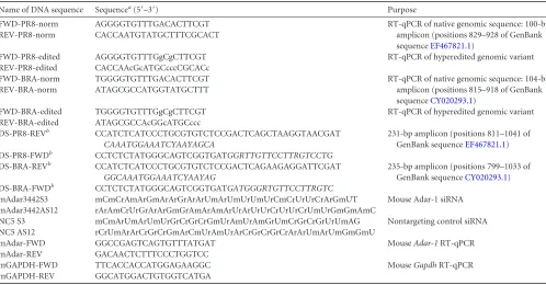
TABLE 1DNA oligonucleotides used in the study
Name of DNA sequence Sequencea(5=–3=) Purpose
FWD-PR8-norm AGGGGTGTTTGACACTTCGT RT-qPCR of native genomic sequence: 100-bp
amplicon (positions 829–928 of GenBank sequenceEF467821.1)
REV-PR8-norm CACCAATGTATGCTTTCGCACT
FWD-PR8-edited AGGGGTGTTTGgCgCTTCGT RT-qPCR of hyperedited genomic variant
REV-PR8-edited CACCAAcGcATGCcccCGCACc
FWD-BRA-norm TGGGGTGTTTGACACTTCGT RT-qPCR of native genomic sequence: 104-bp
amplicon (positions 815–918 of GenBank sequenceCY020293.1)
REV-BRA-norm ATAGCGCCATGGTATGCTTT
FWD-BRA-edited TGGGGTGTTTGgCgCTTCGT RT-qPCR of hyperedited genomic variant
REV-BRA-edited ATAGCGCCAcGGcATGCccc
DS-PR8-REVb CCATCTCATCCCTGCGTGTCTCCGACTCAGCTAAGGTAACGAT
CAAATGGAAATCYAAYAGCA
231-bp amplicon (positions 811–1041 of GenBank sequenceEF467821.1)
DS-PR8-FWDb CCTCTCTATGGGCAGTCGGTGATGGRTTGTTCCTTRGTCCTG
DS-BRA-REVb CCATCTCATCCCTGCGTGTCTCCGACTCAGAAGAGGATTCGAT
GGCAAATGGAAATCYAAYAG
235-bp amplicon (positions 799–1033 of GenBank sequenceCY020293.1)
DS-BRA-FWDb CCTCTCTATGGGCAGTCGGTGATGATGGGRTGTTCCTTRGTC
mAdar3442S3 mCmCrAmArGmArArGrArArUmArUmUrUmUrCmCrUrUrCrArGmUT Mouse Adar-1 siRNA
mAdar3442AS12 rArAmCrUrGrArArGmGrAmArAmArUrArUrUrCrUrUrCrUmUrGmGmAmC
NC5 S3 mCmArUmArUmUrGrCrGrCrGmUrAmUrAmGrUmCrGrCrGrUrUmAG Nontargeting control siRNA
NC5 AS12 rCrUmArArCrGrCrGmArCmUrAmUrArCrGrCrGrCrArArUmArUmGmGmU
mAdar-FWD GGCCGAGTCAGTGTTTATGAT MouseAdar-1RT-qPCR
mAdar-REV GACAACTCTTTCCCTGGTCC
mGAPDH-FWD TTCACCACCATGGAGAAGGC MouseGapdhRT-qPCR
mGAPDH-REV GGCATGGACTGTGGTCATGA
aUppercase, DNA base; A-to-G and T-to-C mutations are in lowercase; the sequence-specific regions are in italics; m, 2=OMe modification; r, RNA base. b
Ion Torrent sequencing primers; sequencing is from the reverse primer (gives antigenomic sequence).
on November 7, 2019 by guest
http://jvi.asm.org/
Statistical analyses.Statistical analyses were carried out using Prism 5 (GraphPad Software Inc.). Two-tailed unpairedttests with Welch’s cor-rection were used from a minimum of six biological values (from a min-imum of two independent experiments). Significance was noted as fol-lows: *,Pⱕ0.05; **,Pⱕ0.01; ***,Pⱕ0.001; ns, nonsignificant.
RESULTS
Inosine incorporation in ssRNA can potentiate RNA sensing by immune cells.We and others have previously reported that al-though directly dependent on the presence and proportion of uri-dine residues and associated motifs, TLR7/8 sensing of ssRNAs could be modulated by the propensity of RNAs to form inter- and intramolecular secondary structures (18,19,22,32). To investi-gate the potential effect of inosine incorporation into ssRNAs on innate immune sensing, we initially studied the immunostimula-tory profiles of a set of 25-nucleotide (nt) synthetic RNAs. These included five inosines dispersed throughout the center of the oli-gonucleotide (ssIA), a central stretch of four consecutive inosines (ssIB), and a central stretch of four consecutive uridines (ssU) (Fig. 1A). The human TLR8-specific chemical agonist CL075 (also known as 3M002) was used as a positive control. Human PBMCs stimulated with increasing concentrations of liposome-com-plexed ssRNAs showed a strong increase in TNF-␣production with ssIA and ssU, while an increase was seen only at 600 nM with ssIB, compared to the native ssN sequence (Fig. 1B). However, IFN-␣levels were not increased with ssIA or ssIB (Fig. 1B), sug-gesting that the inosine modification of ssIA primarily affected TLR8 sensing (13). In addition, the ability of ssIA to induce greater immunostimulation than ssN was also observed in TLR8-respon-sive human monocytic THP-1 cells (22) (Fig. 1C) but was absent in mouse BMMs, which lack the Tlr8 response (22) (Fig. 1D). CL075, which activates Tlr7 in mouse, was used as a positive con-trol. Collectively, these results suggest that selective inosine incor-poration into ssRNAs could potentiate sensing by TLR8 in the context of this ssRNA sequence.
The effect of inosine incorporation on TLR7/8 sensing is re-stricted to immunostimulatory ssRNAs.Our observation that ssIA exerted increased TLR8 recruitment compared to ssIB indi-cated that the position of inosine residues within the ssRNA se-quence was critical in their effect on TLR8 sensing and that poten-tiation of immune stimulation did not relate to the presence of the inosine residues alone. To confirm this hypothesis, we investi-gated the impact on cytokine production of five inosines dispersed throughout the center of an oligonucleotide devoid of uridine residues (ss41-L), which does not activate TLR7/8 (19,33) (Fig. 2A). ss41-L and its inosine variant, ss41-L-I, failed to significantly induce any TNF-␣(Fig. 2B) or IFN-␣(data not shown) produc-tion in human PBMCs, even at high dose. On the other hand, mFOLD structuralin silicoprediction of ssN used in the experi-ment illustrated inFig. 1indicated that this oligonucleotide was prone to a high degree of self secondary structure (Fig. 2C, right). Inosine and its DNA variant, deoxy-inosine, can form wobble base pairing with adenosine, cytidine, and uridine. We therefore spec-ulated that some of the effects of inosine incorporation in ssIA and ssIB could be due to modifications of secondary structures. To assess this hypothesis, we next studied TLR7/8 recruitment by ssN, ssIA, ssIB, and ssU duplexed to the complementary sequence of ssN (resulting in dsN, dsIA, dsIB, and dsU) (Fig. 2C, left). Exper-iments in human PBMCs showed no increase in TNF-␣or IFN-␣ production with either dsIA or dsIB compared to ssN (Fig. 2D),
directly implicating formation of secondary structures in the ef-fect of inosine incorporation in ssIA and ssIB. To determine whether structural variations, independent of inosine residues, were responsible for increased immune stimulation, we studied the immunostimulatory activity of a variant of ssIA synthesized with DNA deoxy-inosine residues substituted for inosine residues (ssdIA). Unlike ssIA, ssdIA failed to increase TNF-␣/IFN-␣
pro-**
C
5′ GCGCCGAGGUGAAGUUCGAGGGCGA 3′
5′ GCGCCIAIIUIAAIUUCGAGGGCGA 3′
0
200 400 600 2000 4000 6000 8000
10000
ns
TNF-α (pg/ml) 60 nM
300 nM
600 nM
0
200 400 600 800 1000 1200 1400
IFN-α (pg/ml) 5′ GCGCCGAGIIIIAGUUCGAGGGCGA 3′
5′ GCGCCGAGUUUUAGUUCGAGGGCGA 3′
ssN ssIA
ssU ssIB
ssN ssIA ssIB ssU
B
**
**
***
***
ssNssIAMock Medi
um CL07
5
0 50 100 150 200 250 300 350 1000 1200 1400
ssNssIAMock Mediu
m CL07
5
0 200 400 600 800 2500 3000 3500
TN
F
-α
(p
g
/m
l)
TN
F
-α (p
g
/m
l) ns
D
ns**
FIG 1Inosine incorporation into ssRNA can increase cytokine production by human immune cells. (A) Sequence details of the synthetic small RNAs used. Ribo-inosine and uridine variations from the native sequence ssN are high-lighted in bold. (B) The indicated ssRNAs were transfected with DOTAP at the indicated doses in human PBMCs and incubated overnight. Cytokine produc-tion was measured by specific ELISA. Data shown are averaged from two independent experiments in three blood donors, in biological triplicate. Un-pairedttests comparing the ssRNA to ssN, for the same concentration, are shown. (C) Human THP-1 cells were activated with PMA overnight and primed for 6 h with IFN-␥prior to rinsing and stimulation with 600 nM the indicated ssRNAs complexed with DOTAP. Following a further 16 h of incu-bation, the supernatants were collected and assayed for TNF-␣levels. Data shown are averaged from three independent experiments in biological tripli-cate. (D) Primary mouse BMMs were stimulated overnight with 600 nM the indicated ssRNAs complexed with DOTAP, and murine TNF-␣levels were measured. Data shown are averaged from two independent experiments in biological triplicate. Unpairedttests are shown in panels C and D. ssU (B) and CL075 (C and D) were used as positive controls for TLR7/8 recruitment. Stan-dard errors of the means (SEM) are shown in panels B to D.
on November 7, 2019 by guest
http://jvi.asm.org/
[image:4.585.302.540.70.445.2]duction in human PBMCs (Fig. 2E), underlining the critical role for ribo-inosine moieties in the effect of ssIA. Collectively, these results suggested that unlike uridine residues, ribo-inosine resi-dues did not directly activate TLR7/8 but rather potentiated sens-ing of uridine motifs through modulation of inter- and intramo-lecular secondary structures.
Inosine-mediated structural modification of ssRNA modu-lates TLR7 sensing.In order to further ascertain the ability of inosine incorporation to modulate TLR7/8 sensing through the modulation of ssRNA structure, we investigated the effect of a single inosine mutation on the secondary structure and immune activity of a previously characterized immunostimulatory ssRNA (19). Relying on the correlation between mFOLD-predicted (34) secondary structures of ssRNAs and the fluorescence of ssRNAs with the nucleic acid intercalant SYBR green (data not shown), we first demonstrated that inosine incorporation into B-406-AS (giv-ing B-406-AS-i), result(giv-ing in the formation of an I·C base pair, robustly increased the strength of the intramolecular secondary structure of this ssRNA, as revealed by the higher energy (temper-ature) required to decrease SYBR green fluorescence by 80% (Fig. 3AandB, compare B-406-AS and B-406-AS-i). Importantly, the wobble I·C pair in B-406-AS-i was less potent than the G·C pair in B-406-AS-1 in its ability to modulate intramolecular secondary structure, as revealed by the SYBR green melt curve assay (Fig. 3A andB, compare B-406-AS-i and B-406-AS-1). In agreement with a role for secondary structure in the modulation of TLR7/8 signal-ing (19), the I·C base pair of B-406-AS-i robustly increased the immunostimulatory activity of this ssRNA in human PBMCs compared to the native B-406-AS sequence, but not as potently as the G·C pair of B-406-AS-1, which conferred greater secondary structure (Fig. 3C, compare B-406-AS, i and B-406-AS-1). Importantly, in the context of the B-406-AS ssRNA, inosine modification potentiated IFN-␣rather than TNF-␣production, suggesting a preferential effect of inosine on TLR7 (22). To deter-mine the direct involvement of TLR7 in the effect of B-406-AS-i, we assessed the ability of B-406-AS-i to induce immune stimula-tion when cotransfected with an RNA containing 2=O-methyl (2=Ome) groups, given that 2=Ome-modified RNAs act as TLR7 antagonists (Fig. 3D) (3). 2=Ome RNA cotransfection entirely ab-lated B-406-AS-i-induced IFN-␣production, consistent with the requirement of TLR7 in the sensing of the inosine modification. Nonetheless, 2=Ome RNA cotransfection did not ablate TNF-␣ production by B-406-AS-i, suggesting residual TLR8 sensing, nor did it prevent IFN-␣production by the TLR9 ligand CpG DNA (Fig. 3D). Similarly, the requirement for TLR7 in the sensing of B-406-AS-i was confirmed using pDCs fromTlr7⫺/⫺mice, which lacked IFN-␣production in response to B-406-AS-i but not the Tlr9 agonist (Fig. 3E). Taken together, these results demonstrate that selective inosine incorporation into ssRNA can potentiate TLR7 RNA sensing, through its impact on inter- and intramolec-ular secondary structures.
Increased inosine content in viral RNA correlates with in-creased TNF-␣production in human PBMCs.The data thus far suggested that inosine incorporation into structured short immu-nostimulatory ssRNA had the potential of increasing TLR7/8 sensing, depending on the sequence modified or the localization of inosine modification(s). To determine the biological signifi-cance of these results, we next investigated the impact of inosine incorporation into influenza virus A (ssRNA virus) RNA on its immunostimulatory activity. Relying on RT-qPCR amplification
B
TNF-α (pg/ml)
0
1000 2000 3000 4000
Medium CL075 Mock ss41-L-I ss41-L
5′GCCGGACAGAAGAGAGACGC 3′
5’GCCGIAIIGAIGIGAGACGC 3’ ss41-L
ss41-L-I
ssN [
Δ
G = -6.40]
G G C C G A G G U
G A A
G
U
U
C G
A G G
G
C
G
C A
5’ 3’
C
y
tok
ine (
p
g/
m
l)
dsN dsIA dsIB dsU Mock Medi
um CL075 0
500 1000 1500 2000 2500 3000 10000 12500 15000
TNF-α IFN-α
ns
**
**
5′ GCGCCGAGGUGAAGUUCGAGGGCGA 3′
[image:5.585.66.264.61.556.2]5′ GCGCCIAIIUIAAIUUCGAGGGCGA 3′
||||||||||||||||||||||||| 3’GGCGCGGCUCCACUUCAAGCUCCCGCU 5’ dsN
||||| | | || ||||||||||| 3’GGCGCGGCUCCACUUCAAGCUCCCGCU 5’ dsIA
C
5′ GCGCCGAGIIIIAGUUCGAGGGCGA 3′ |||||||| ||||||||||||| 3’GGCGCGGCUCCACUUCAAGCUCCCGCU 5’ dsIB
D
C
y
to
k
ine (
p
g/
m
l)
ssN ssdI
A
ssU Mock Medi
um CL07
5 0
200 400 600 800 1000 1200
TNF-α IFN-α
E
FIG 2Inosine content of ssRNA does not have a direct positive impact on TLR7/8 sensing. (A) Sequence details of the synthetic small RNAs used. Ribo-inosines are shown in bold. (B) The indicated ssRNAs were transfected with DOTAP to a final concentration of 600 nM in human PBMCs. Cytokine pro-duction was measured by specific ELISA following overnight incubation. Data shown are averaged from five independent experiments, in biological tripli-cate. (C) Sequence details of the synthetic dsRNAs used (left) and mFOLD-predicted structure of ssN (right) at 37°C (34). I·C base pairs are highlighted with black dots. The free energy (⌬G, in kcal/mol), indicative of the strength of the secondary structure of ssN, is given. (D and E) Double-stranded duplexes of ssN variants (D) and deoxy-inosine variant of ssIA (ssdIA) (E) were trans-fected with DOTAP to a final concentration of 600 nM in human PBMCs. Cytokine production was measured by specific ELISA following overnight incubation. Data shown are averaged from two independent experiments in three blood donors (D) or representative of two independent experiments (E), in biological triplicate. Unpairedttests comparing the dsRNA to dsN are shown. SEM is shown in panels B, D, and E.
on November 7, 2019 by guest
http://jvi.asm.org/
of a known hyperedited (inosine-rich) fragment of influenza virus AHAgene (see Materials and Methods and reference 31), we screened RNA purified from the cells of the allantoic fluid of in-dividual virally infected embryonated hens’ eggs and identified samples with a greater proportion of hyperedited RNAs, for two different strains of influenza virus A of the H1N1 subtype (A/PR/ 8/34 [PR8] and A/Brazil/11/78 [BRA]) (Fig. 4AandB). Notewor-thy, this RNA contained only a small proportion of cellular RNA (revealed by 18S and 28S ribosomal RNAs; data not shown), sug-gesting enrichment of viral RNA. For this reason, this RNA is referred to as “viral RNA” in the rest of this discussion. We next assessed the ability of this viral RNA with various levels of editing to activate TLR7/8 in human PBMCs; the chemical agonists Gardiquimod and CL075, specific to human TLR7 and 8, respec-tively, were used as positive controls. Stimulation of human PBMCs with liposome-complexed viral RNA revealed a correla-tion between the content of hyperedited RNAs and the ability of the viral RNA to induce TNF-␣, in that RNA samples from both viruses with more hyperedited RNAs displayed greater ability to induce TNF-␣production [Fig. 4AandB, compare PR8 (1) to PR8 (2) and BRA (1) to BRA (2)]. However, there was no signifi-cant effect on IFN-␣production, indicating a preferential role of hyperedited content on TLR8 activity (Fig. 4AandB).
A recent report suggested that up to 1% of the influenza A viral RNA produced by infected chicken fibroblastsin vitrowas affected by A-to-I editing (31). To assess further the inosine content of the viral RNA samples studied above, analyses with Ion Torrent tar-geted RNA sequencing were carried out. Inosine (I) is changed to guanosine (G) during reverse transcription, allowing for the iden-tification of A-to-I editing sites (as A-to-G mutations on the viral genomic strand or U-to-C mutations on the antigenomic strand). In agreement with Suspene et al., this analysis revealed the pre-dominant A-to-G/U-to-C mutation of viral RNA across all other mutations, with sequencing reads with at least two A-to-G/U-to-C mutations representing up to 0.8% of all reads for PR8 (Fig. 4C). The antigenomic RNA (replication template) was also edited (as shown by U-to-C mutations), confirming previous observations (31). Noteworthy, C-to-U mutations were over three times more abundant than the other mutations for BRA, indicating possible enriched C-to-U editing (data not shown). Specific analysis of A-to-G frequency of mutations revealed that while⬃0.4% of all reads contained two or more A-to-G mutations, the proportion of reads with three or more A-to-G mutations was⬃10-fold lower (⬃0.04 to 0.06%) (Fig. 4D). Nonetheless, a third of the reads with at least three mutations had six mutations, suggesting that these transcripts were hyperedited (Fig. 4D). Collectively, these results suggest a direct impact of inosine incorporation on the immuno-stimulatory activity of influenza A viral RNA.
RNA from influenza-infected cells activates TLR7/8, inde-pendent of 5=-triphosphate.Mammalian cellular RNAs such as ribosomal RNAs contain various modifications such as pseudou-ridine and 2=-O-methyl nucleoside incorporation, which avoid
C
TNF-α (pg/ml) IFN-α (pg/ml)
D
G 3’
U AA UUC G
UCU G
G
CCUUCUU C
5’
U A A UU C G
UCU G
G C C U U C U U
I
G
5’ 3’
U A A UU C G
UCU G
G C C U U C U U G
G
5’ 3’
B-406-AS-1 [ΔG = -3.9]
B-406-AS-i [ΔG = ?] B-406-AS [ΔG = -0.5]
Temperature (°C)
R
el
at
iv
e f
luor
es
c
enc
e
B
B-40 6-
AS-i + ss4 1
B-40 6-
AS-i + 2 'Om
e
CpG DNA + 2'Ome Mock
Medium CL07 5
0 250 500 750 1000 1250 1500 8000 9000 10000
C
yt
o
ki
n
e
(p
g
/ml
) TNF-α
IFN-α
Tlr7 -/-Tlr7wt/wt
B-406-AS-iCpG DN A
Moc k
0.0 0.5 1.0 5.0 5.5 6.0 6.5 7.0
IFN
(n
g
/ml
)
0
200 400 600 800 1000 1200 1400
CL075 Medium Mock B-406-AS-1 B-406-AS-i B-406-AS
0 50
100 150 200 250 300 350 400
E
B-406-AS B-406-AS-1 B-406-AS-i
20 30 40 50 60 70 80 90 100
0.0 0.2 0.4 0.6 0.8 1.0
FIG 3Inosine modulates TLR7 activity. (A) mFOLD-predicted structure of the B-406-AS series of immunostimulatory ssRNAs (19). C-to-G and C-to-I mutations of B-406-AS resulting in B-406-AS-1 and B-406-AS-i, respectively, are circled. The mFOLD-predicted free energy (⌬G, in kcal/mol), indicative of the strength of the secondary structure of the ssRNAs, is given. (B) Fluores-cence-based determination of ssRNA secondary structure, using the SYBR green melt curve assay (see Materials and Methods). An arrow highlights the temperature at which the fluorescence of B-406-AS-i is decreased by 80%. The data are averaged from three independent experiments. (C) The indicated ssRNAs were transfected with DOTAP to a final concentration of 200 nM in human PBMCs and incubated overnight. Cytokine production was measured by specific ELISA. Data shown are representative of a minimum of two inde-pendent experiments in two blood donors, in biological triplicate. (D) The indicated ssRNAs were cotransfected in a 1:1 ratio with DOTAP to a final concentration of 200 nM for each ssRNA (400 nM total). The nonimmunos-timulatory ss41 RNA was used as a control for the 2=Ome RNA. CpG DNA mixed in 1:1 molar ratio with 2=Ome was transfected with DOTAP to a final concentration of 200 nM to control the specificity of the 2=Ome effect on TLR7. Following overnight incubation, cytokine production was measured by
specific ELISA. Data shown are averaged from two independent experiments in two blood donors, in biological triplicate. (E) Splenic pDCs fromTlr7wt/wt
mice andTlr7⫺/⫺mice were stimulated with 100 nM DOTAP-complexed B-406-AS-i ssRNA or 5M CpG DNA and incubated overnight. Mouse IFN-␣ production was measured by specific ELISA in technical triplicate. The data are averaged from two independent experiments. SEM is shown in panels B to E.
on November 7, 2019 by guest
http://jvi.asm.org/
[image:6.585.60.266.77.545.2]aberrant activation of innate immune sensors from self-RNA (2). In line with this concept and the direct inhibitory effect of 2= -O-methyl moieties in Tlr7 sensing (3), total RNA from epithelial cells transfected in human PBMCs did not induce production of IFN-␣ or TNF-␣(Fig. 5A, condition “Cells only”). Conversely, total RNA from influenza virus PR8-infected epithelial cells, although con-taining more than 60% of 18S and 28S rRNA (data not shown), induced a strong IFN-␣ production concurrently with a mild TNF-␣secretion in human PBMCs (Fig. 5A, PR8 and PR8-⌬NS1 conditions). Calf intestinal phosphatase (CIP) treatment of the RNA from PR8-infected cells resulted in a strong reduction in IFN-␣production (compare IFN-␣levels inFig. 5AandB), indi-cating the involvement of 5=-triphosphate moieties in IFN-␣ pro-duction (1). Nonetheless, RNA from cells infected with a mutant PR8 virus lacking the NS1 protein (which is thought to impair ADAR-1 editing activity [31]) (⌬NS1) retained the ability to in-duce TNF-␣production following CIP treatment, suggestive of TLR8 recruitment rather than RIG-I activation (35). The RNA from PR8-⌬NS1-infected cells contained a higher proportion of hyperedited PR8 transcripts than RNA from wild-type PR8-in-fected cells (Fig. 5C), suggesting a putative involvement of RNA editing in this effect. In addition, CIP-treated RNA from
PR8-⌬NS1-infected cells retained the ability to activate TNF-␣ produc-tion in immortalized BMMs fromTlr7wtbut not fromTlr7⫺/⫺ mice (Fig. 5D, compare “PR8-⌬NS1⫹CIP” conditions inTlr7wt
andTlr7⫺/⫺cells); on the other hand, non-CIP-treated RNA was able to induce TNF-␣production independently of the presence of Tlr7, which is indicative of RIG-I recruitment. The Tlr2/1 li-gand, Pam3CSK4, confirmed that bothTlr7wtandTlr7⫺/⫺cells were able to produce TNF-␣. To directly involve Adar-1 editing in the TNF-␣production seen in RNA from PR8-⌬NS1-infected cells, we decreasedAdar-1expression through RNA interference in LA-4 cells prior to the infection with PR8-⌬NS1 (Fig. 5E, compare siADAR and the control siNC5 conditions). In accord with the results above, CIP treatment of the RNA from PR8-⌬NS1-infected cells showed a sig-nificant decrease in IFN-␣production in human PBMCs, regardless
ofAdar-1expression (Fig. 5F). TNF-␣levels, however, were
signifi-cantly decreased following downregulation ofAdar-1(siADAR con-dition), but not with the nontargeting control siNC5. This directly implicates Adar-1 in the ability of CIP-treated RNA to induce TNF-␣. Collectively, these results suggest that a small proportion of Adar-1-edited viral RNA is able to potentiate sensing of cellular RNA, inde-pendently of 5=-triphosphate/RIG-I recruitment, in a human TLR8-and mouse Tlr7-dependent manner.
PR8 ( 1)
PR8 ( 2)
Mock CL075
Gard iqui mo d 0 25 50 75 100 125 150 0 200 400 600 800 IFN-α ( pg/ m l) TN F -α ( pg/ m l)
BRA ( 1)
BRA ( 2)
Mock CL075
Gard iqui mo d 0 200 400 600 800 1000 1200 0 200 400 600 800
IFN-α (
pg/
m
l) TNF
-α ( pg/ m l) TNF-α IFN-α TNF-α IFN-α PR 8 (1)
PR 8 (2)
0.0 0.5 1.0 1.5 2.0 2.5 R e la ti ve f o ld e d it in g BRA (1)
BRA ( 2) 0.0 0.5 1.0 1.5 2.0 2.5 R e la ti ve f o ld e d it in g
C
PR8 BRANumber of A->G mutations
D
A->G /U-> C G->A/ C->U C->A /G-> U A->U /U-> A U-> G/A ->C C->G /G-> C 0. 702% 0. 282% 0. 123% 0. 128% 0. 099% 0. 067% A->G /U-> C G->A /C-> U C->A /G-> U A->U /U-> A U->G /A-> C C->G /G-> C 0 2000 4000 6000 8000 10000 N um b er of r eads 0. 817% 0.406% 0.503%
0. 224% 0. 423% 0. 254%
>1 >2 >3 >4 >5 >6 >7 >8 >9>10
1 10 100 1000 10000 100000 N um b er of r eads 5.304% 0.392 % 0.02 5% 0.02 0%
0.013%0.008% 0.006%0.004
% 0.03 1% 0.060% PR8 BRA
>1 >2 >3 >4 >5 >6 >7 >8 >9>10
5.956%
0.420 %
0.01 5% 0.013%0.0
11% 0.009%0.008%0.007
% 0.0
17% 0.041%
FIG 4Hyperediting of viral RNA correlates with increased TNF-␣levels in human PBMCs. (A and B) PR8 (A) and BRA (B) viral RNA (⬃0.4g/ml) from two independent eggs was transfected in human PBMCs, and cytokine production was measured after 16 h of incubation (right panels). Data shown are represen-tative of one (BRA) or three (PR8) independent experiments in biological triplicate. The same viral RNA was reverse transcribed and analyzed by qPCR for hyperediting frequency (left panels) (see Materials and Methods). RT-qPCR data shown are averaged from two independent experiments. (C and D) Ion Torrent analysis of PR8 and BRA samples. (C) The number of reads with at least two or more of the same mutation relative to genomic viral RNA is shown for each virus. Total mutations for each strand were combined (e.g., A-to-G and C-to-U mutations were added) to reflect editing of total viral RNA (genomic and antigenomic). (D) The number of reads with an increasing amount of A-to-G mutations relative to genomic viral RNA (e.g.,⬎1 means at least one A-to-G mutation) is shown for each virus. (C and D) Mutation analyses of the reads excluded primer regions to avoid sequence bias from degenerated primers (see Materials and Methods). Frequencies of mutations are shown as percentages, relative to the total numbers of reads for each virus (1,029,246 for PR8 and 1,202,254 for BRA). SEM is shown in panels A and B.
on November 7, 2019 by guest
http://jvi.asm.org/
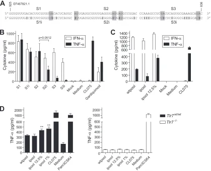
[image:7.585.65.520.68.364.2]A-to-I editing in a proportion of viral RNA potentiates TLR7 sensing.Having shown that (i) purified viral RNA with greater inosine content (Fig. 4) and (ii) RNA from infected cells with greater viral hyperediting (Fig. 5) were more immunostimulatory, we next sought to validate the direct contribution of A-to-I editing on synthetic RNAs encompassing a 90-nt hyperedited portion of PR8 RNA previously analyzed by sequencing (Fig. 6A) (reference 31and data not shown). We wished to exclude possible self RNA cleavage products that could have contaminated our viral RNA preparations (36). Unexpectedly, comparison of the immuno-stimulatory profiles of each individual 30-nt segment with (S1i, S2i, and S3i) or without (S1, S2, and S3) inosine editing showed no increase in cytokine production with inosine editing for any of the fragments (Fig. 6B). Noteworthy, there appeared to be a degree of correlation between the frequency of TLR8-specific AU motifs (Fig. 6A, shaded in gray) (17) that were converted to IU motifs and
the negative impact of A-to-I editing on TNF-␣production. As such, S2i, which lost three AU motifs, displayed a near-significant (P⫽0.0512) decrease in the induction of TNF-␣compared to S2 (Fig. 6B). Nonetheless, we speculated that formation of complex intermolecular secondary structures could be at play in the effect of inosine on TLR7/8 sensing of viral RNA seen inFig. 4and5, and therefore we studied pools of 30-nt ssRNAs with or without ino-sine editing (Fig. 6C). While no increase in cytokine production was seen using pure pools of inosine-modified RNA (condition “ipool”), the addition of a small quantity of inosine-modified RNAs (12.5%) to the pool of native RNAs resulted in an⬃2-fold increase in TNF-␣production and a small increase in IFN-␣levels in human PBMCs (Fig. 6C, compare “ipool 12.5%” with “wtpool” conditions). Significant potentiation of cytokine production with as low as 1% of edited transcripts was confirmed in immortalized BMMs fromTlr7wtmice, but not fromTlr7⫺/⫺mice, for TNF-␣
0 50 100 150 200 250 0 20 40 60 80 100 200 300 IF N
-α (
pg/
m
l) TNF
-α ( pg/ m l) P R8+CIP
ΔNS1+CIPCells
onl y Mo ck CL07 5 ns * TNF-α IFN-α R el a ti ve hy per -edi te d P R8 RNA / Gapdh PR 8 ΔNS1 0.0 0.5 1.0 1.5 2.0 2.5 P R8 ΔNS1 Cells onl y
Mock CL07
5 0 500 1000 1500 0 50 100 150 200 600 750 900 IF N
-α (
pg/ m l) TN F -α ( pg/ m l) TNF-α IFN-α
D
siADARsiNC5 0.0 0.2 0.4 0.6 0.8 1.0 1.2 Re la tiv e A d ar-1 e x p re s s io n Tlr7 -/-Tlr7wt/wt nsΔNS1+
CIPΔNS1
Cells onlyCL075 Pam3CSK4 0 50 100 150 200 250 2000 2500 3000 3500 TN (p g /ml ) *** ** ΔNS 1+CI P ΔNS1 Cells only CL075 Pam3CSK4 0 200 400 600 2000 3000 4000 TN (p g /ml ) *
E
siADARsiNC5 siADAR+CIPsiNC5+CIP MockR-848 0 100 200 300 400 800 1000 1200 TN (p g /ml ) siADARsiNC5 siADAR+CIPsiNC5+CIP MockR-848 0 200 400 600 800 IFN ( pg/ m l)F
* ns *** ***FIG 5RNA from influenza virus-infected cells activates Tlr7, independent of 5=-triphosphate. (A) Total RNA purified from LA-4 cells infected with two strains of PR8 influenza virus (PR8-⌬NS1, shown as “⌬NS1,” lacks theNS1gene, which interacts with Adar-1 and decreases A-to-I editing [31]) was transfected in human PBMCs (1.3g/ml), and cytokine production was measured after overnight incubation. The “Cells only” condition refers to RNA from noninfected LA-4 cells. (B) The same RNA as described for panel A was treated with CIP (⫹CIP) (see Materials and Methods), prior to transfection into human PBMCs (1.6g/ml) and analysis of cytokine production after overnight incubation. Data shown in panels A and B are averaged from a minimum of two independent experiments in biological triplicate. Unpaired two-tailedttests comparing PR8 and PR8-⌬NS1 conditions are shown in panel B. (C) The same RNA from infected cells as described for panel A was reverse transcribed and analyzed by qPCR for levels of hyperedited PR8 RNA relative to the levels of mouseGapdhmRNA. RT-qPCR data shown are averaged from two independent experiments. (D) Total RNA from PR8-⌬NS1-infected LA-4 cells with (⫹CIP) or without CIP was transfected inTlr7wt/wtandTlr7⫺/⫺mouse BMMs (2.6g/ml). Cytokine production was measured by specific ELISA following overnight incubation. Results shown are averaged from a minimum of two independent experiments in biological triplicate. Unpaired two-tailedttests using “Cells only” as a reference are shown. (E) Total RNA from PR8-⌬NS1-infected LA-4 cells previously transfected with siADAR or the nontargeting control siNC5 was analyzed by RT-qPCR forAdar-1 mRNA levels (see Materials and Methods). The data shown are averaged from technical triplicates, relative to the levels of mouseGapdhmRNA. (F) The RNA described in panel E was treated with CIP (⫹CIP) or left untreated, prior to transfection into human PBMCs (1.6g/ml for nontreated RNA, 3.2g/ml for CIP-treated RNA) and analysis of cytokine production after overnight incubation. The data are averaged from two blood donors in biological triplicate. Unpaired two-tailedttests are shown. SEM is shown in panels A, B, D, E, and F.
on November 7, 2019 by guest
http://jvi.asm.org/
[image:8.585.68.522.66.386.2](Fig. 6D) and interleukin-6 (IL-6) production (data not shown). These results establish that small proportions of A-to-I hype-redited RNAs can facilitate sensing of viral RNA by Tlr7.
DISCUSSION
ADAR-1L-mediated A-to-I editing is a critical component of the antiviral response against several viruses, including measles and influenza A viruses (5). A recent report analyzing the A-to-I edit-ing content of influenza A viral RNA from infected chicken fibro-blasts suggested that A-to-I editing is frequent during viral repli-cation, potentially modifying up to 1% of viral RNA (31). In our analyses of two strains of influenza A virus, we show that as much as 0.8% of viral RNA can be edited following standard propaga-tion of virus in hens’ eggs (as carried out for the producpropaga-tion of influenza vaccine). In accord with the work of Suspene et al., this suggests that A-to-I editing is indeed a frequent event during viral replication (31). We also confirm that both genomic and the rep-lication template of influenza A virus can be edited (31). We found that an important proportion of RNAs with at least three A-to-G
mutations had in fact at least six A-to-G mutations, indicative of their hyperediting. However, our observations suggest that hype-redited viral RNAs represent only a fraction of all A-to-I edited RNAs. While such mutations would directly impact the affected clones and their ability to produce effective viruses, it is however most likely that such direct effects on⬍1% of the clones do not account for much of the overall antiviral properties of ADAR-1L-mediated A-to-I editing. Rather, inosine incorporation in struc-tured viral RNAs could facilitate recognition of nonself RNA (11, 14,31). Under this rationale, a small proportion of edited viral RNA could elicit a strong antiviral effect through the downstream induction/amplification of type I IFN signaling. This is supported by two independent reports directly implicating a role for inosine residues in the activation of TLR3 (11,12).
In the current study, we set out to investigate the impact of inosine incorporation into ssRNAs on TLR7/8 sensing. We pro-vide several lines of epro-vidence supporting the direct effect of inosine incorporation into ssRNA on the potentiation of TLR7/8 sensing. First, we observed that inosine addition to short synthetic
immu-S1
5’GGUGUUUGACACUUCGUGUUACACUCAUGC AUUGAUGCGUUUGAGGUGAUGAUGCCGGAC CCAAAGCCUCUACUCAGUGCGAAAGCAUAC 3’ 5’GGUGUUUGICICUUCGUGUUICICUCAUGC IUUGAUGCGUUUGIGGUGIUGIUGCCGGIC CCIIIGCCUCUICUCIGUGCGIIIGCAUIC 3’
S2 S3
S1i S2i S3i
925 EF467821.1 836
B
S1 S1i S2 S2i S3 S3i Moc
k
Med ium
CL07 5
Gar diqui
mod
0 200 400 600 800 1000
C
y
tok
ine (
pg/
m
l) p=0.0512
wtpo
ol
ipoo
l
ipool
12.
5% Mo
ck
Medi
um
CL075
0 100 200 300 400 600 800 1000 1200 1400
C
y
tok
ine (
pg/
m
l)
C
wtpo ol
ipoo l ipoo
l 12. 5%
ipoo l 1%
Me dium Pam
3CSK4
0 100 200 300 400 500 600 1600 2000
** **
TN
F
-α
(p
g
/m
l)
CL07 5
Tlr7
-/-wtpo ol
ipool ipoo
l 12 .5%
ipoo l 1%
CL07 5 Mediu
m Pam
3CSK4
0 100 200 300 400 500 600 1600 2000
TN
F
-α
(p
g
/m
l) Tlr7
wt/wt
D
TNF-α IFN-α
TNF-α IFN-α
FIG 6Inosine incorporation in a proportion of viral RNA potentiates Tlr7 sensing. (A) Sequence details of the synthetic small RNAs used, covering the region spanning residues 836 to 925 of GenBank sequenceEF467821.1(antisense strand). Ribo-inosines are shown in bold, and AU motifs mutated to IU motifs are shaded in gray. (B) The indicated ssRNAs were transfected with DOTAP to a final 100 nM in human PBMCs and incubated overnight. Cytokine production was measured by specific ELISA. Data shown are averaged from three independent experiments in three blood donors, in biological triplicate. Unpairedttest is shown. (C, D) Indicated pools of pure ssRNAs (“wtpool” consists of S1, S2, and S3, while “ipool” consists of S1i, S2i, and S3i, in a 1:1:1 ratio) or a mixture of both pools (12.5% and 1% ipool conditions) were transfected with DOTAP to a final concentration of 100 nM in human PBMCs (C) or 200 nM inTlr7wt/wtand
Tlr7⫺/⫺mouse BMMs (D). Cytokine production was measured by specific ELISA following overnight incubation. Results shown are representative of two independent experiments in biological triplicate in two blood donors (C) and averaged from three independent experiments in biological triplicate (D). (D) Unpaired two-tailedttests using “wtpool” as a reference are shown. SEM is shown in panels B, C, and D.
on November 7, 2019 by guest
http://jvi.asm.org/
[image:9.585.83.502.67.407.2]nostimulatory ssRNA molecules could result in increased produc-tion of IFN-␣or TNF-␣by human PBMCs, in a sequence-specific manner (Fig. 1,2, and3). Given that TLR7 and TLR8 recognize different RNA motifs (17), the sequence-specific potentiation of IFN-␣or TNF-␣by inosines is indicative of the specific recruit-ment of TLR7/8. Critically, ablation of IFN-␣ production of B-406-AS-i with a TLR7 antagonist in human PBMCs, together with the lack of an effect of B-406-AS-i in mouseTlr7⫺/⫺pDCs, directly implicates TLR7 in the detection of inosine-modified ssRNA (Fig. 3). In addition, the finding that the inosine-rich ssIA promoted specific induction of TNF-␣in both human PBMCs and PMA-activated THP-1 monocytic cells but not in primary mouse BMMs supports the notion that RNA sensing by human TLR8 can also be facilitated by inosine incorporation (Fig. 1). This is in accord with the observation that the double-stranded form of ssIA, dsIA, did not elicit an increase in TNF-␣production, given that TLR8 sensing is strongly repressed by annealing of comple-mentary oligonucleotides (18).
Second, we demonstrate that a nonimmunostimulatory ino-sine-rich ssRNA, ss41-L-I (lacking uridine residues), cannot elicit an immune response following DOTAP-mediated delivery in hu-man PBMCs. We and others have previously demonstrated that TLR7/8 sensing is strongly reliant on the presence of uridine res-idues (19, 37), further implicating TLR7/8 in the detection of inosine-modified immunostimulatory RNAs (Fig. 2). Third, it has previously been speculated that TLR7/8 sensing of ssRNAs relates to their tendency to form inter- and intramolecular uridine-rich secondary structures (37). In agreement with this, we have previ-ously observed that the position of uridine modifications within the secondary structure of an ssRNA has an important effect on immunostimulation (22). Given the ability of inosine to form wobble base pairing with adenosine, cytidine, and uridine, inosine incorporation is expected to directly impact on the ability of an ssRNA to form secondary structures. In accord with a role for inosine in the modulation of TLR7/8 sensing through structural variations, we demonstrate here that the position of inosine incor-poration is critical to the effect on TLR7/8 sensing (Fig. 1). In addition, we establish a direct correlation between the inosine-mediated modulation of inter- and intramolecular ssRNA struc-tures and activation of TLR7 with B-406-AS-i (Fig. 3). Nonethe-less, the effect of inosine on immune stimulation is not limited to its structural activity and was not reproduced by its DNA analog deoxy-inosine (ssdIA), while also favoring structural variations (Fig. 2). This inhibition of immunostimulatory activity by the DNA analog of inosine is similar to that seen with deoxy-uridine residues, which repress sensing of ssRNAs when replacing uridine residues (37).
TLR7/8 can be activated by ssRNA and dsRNA, although the immunostimulatory role of dsRNA is greater when the affinity for the dsRNA duplex is weaker, particularly for TLR8 (18,19). In the context of viral infection, we therefore hypothesized that ADAR-1L-mediated A-to-I editing of structured RNAs could facilitate TLR7/8 sensing by destabilizing dsRNA structures. However, im-munostimulatory analyses of individual ssRNAs mimicking a known structured hyperedited region of influenza A virus indi-cated that A-to-I editing did not enhance TLR7/8 sensing when present in 100% of the molecules (Fig. 6). In fact, an⬃50% de-crease in human TNF-␣was even visible with one of the sequences (S2i) and when the sequences were pooled, indicating an inhibi-tory effect of A-to-I editing on TLR8 sensing. While
counterintui-tive in light of our other results with ssRNAs where inosine incor-poration was not restricted to adenosine residues (Fig. 1,2, and3), we propose that this relates to the loss of TLR8-specific AU-rich motifs (changed to IU motifs) (17). In line with this concept, S2i ssRNA lost three AU motifs following A-to-I editing and displayed a marked decrease in TNF-␣, while IFN-␣levels were unaffected when transfected in human PBMCs. Further involving TLR8 in this event, pools of inosine-modified ssRNAs did not promote such a decrease in TNF-␣production in mouse macrophages, in which Tlr8 does not contribute to ssRNA sensing (13). It is also possible that changes in secondary structure less favorable to the detection of these ssRNAs are also at play.
Nonetheless, under the rationale that A-to-I editing affected only small proportions of viral RNAs, we postulated that the ob-served structural effects of inosine on ssIA and B-406-AS-i ssRNA could increase the formation of complex intermolecular struc-tures in A-to-I edited RNA, even if present in a small proportion of sequences. Accordingly, we demonstrate here that physiological levels (between 1 and 12.5%) of inosine-modified ssRNA mole-cules can potentiate RNA sensing and result in increased TNF-␣ production by human and mouse macrophages. We propose that this potentiation is mostly TLR8 dependent in human PBMCs, as revealed by the preferential increase in TNF-␣production. As such, A-to-I editing exhibits a dual activity, which can both po-tentiate and antagonize TLR8 sensing, the latter diminishing with a biologically relevant low concentration of inosine-modified RNAs. Noteworthy, we have previously shown that human TLR7 was necessary for ssRNA sensing in human monocytic cells (22), and it is thus most likely that TLR7 is also at play in the macro-phage sensing of inosine-modified RNA. We directly establish a role for mouse Tlr7 in the effect of inosine-modified ssRNA mol-ecules, through the use ofTlr7⫺/⫺macrophages (Fig. 6). This role for Tlr7 in the increased detection of A-to-I edited viral RNA is consistent with recent reports demonstrating a critical role for mouse Tlr7 in the immune response to lymphocytic choriomen-ingitis virus (LCMV) (15, 16), which is subject to ADAR-1L hyperediting during replication (10).
Finally, in support of a direct role for A-to-I editing in the sensing of viral RNA by TLR7/8, we show a correlation between the proportion of hyperedited influenza virus RNA and the im-munostimulatory activity of this viral RNA in human PBMCs. As with our observations on pools of inosine-modified RNAs, TNF-␣ was predominantly affected by the overall proportion of hype-redited RNA, suggesting preferential modulation of TLR8 activity. Critically, we also demonstrate that phagocytosis of total RNA from influenza virus-infected cells resulted in TNF-␣production by human and mouse macrophages, independently of 5= -triphos-phate residues and in an Adar-1-dependent manner. Although the direct antiviral activity of 5=-triphosphate-dependent IFN-␣ pro-duction was clearly predominant in these experiments, these re-sults confirm a contributing role for human TLR8 and mouse Tlr7 to the sensing of RNA from infected epithelial cells. Because mod-ified cellular RNA is essentially nonimmunostimulatory, it re-mains to be determined whether inosine residues directly compete with inhibitory moieties such as 2=-O-methyl bases, to result in TLR7/8 sensing. Altogether, our results support a role for TLR7/8 in the production of TNF-␣ by macrophages in response to phagocytosed infected epithelial cells. In addition to its direct pro-inflammatory activity, TNF-␣was previously shown to enhance influenza A virus-induced chemokine expression by epithelial
on November 7, 2019 by guest
http://jvi.asm.org/
cells (38). This suggests a possible feedback loop between epithe-lial cells and macrophages, increasing immune cell recruitment to the infected locus.
Collectively, our data establish a novel role for TLR7/8 in the detection of A-to-I edited viral RNA. We propose that Adar-1-mediated inosine incorporation into viral RNA acts to facilitate recognition of nonself RNA by TLR7/8, allowing infected cells to be identified when cleared by phagocytes, such as macrophages. It is unlikely that inosine-modified viral RNA contributes directly to the antiviral response against infected epithelial cells, as they lack TLR7/8 (13). We speculate that A-to-I editing of structured viral RNA helps to counterbalance the repressive effect of self-RNA from phagocytosed infected cells, thus helping to distinguish be-tween self and nonself RNA, together with other modifications such as 5=-triphosphate (1). Along with reports on TLR3 sensing (11,12), the implications of our work may be far-reaching, as our results suggest a novel paradigm whereby A-to-I editing of viral RNA acts to facilitate detection of nonself RNA.
ACKNOWLEDGMENTS
This work was supported by funding from the Australian National Health and Medical Research Council (1022144 to M.P.G. and 1006590 to B.R.G.W), a Monash University Faculty of Medicine, Nursing and Health Sciences Strategic Grant (2012), and the Victorian Government’s Opera-tional Infrastructure Support Program.
We are grateful to Vivien Vasic, Trevor Wilson, Ross Chapman, and Jodee Gould (Monash Health Translation Precinct Medical Genomics Facility—Australian Cancer Research Foundation Centre for Cancer Genomic Medicine) for their help with Ion Torrent sequencing, Scott Rose (Integrated DNA Technologies Inc.) for his help in the production of the RNA oligonucleotides, and Kun Xiao (La Trobe University) for his help with mouse work.
Mark A. Behlke and Ashley M. Jacobi are employed by Integrated DNA Technologies Inc. (IDT), which offers oligonucleotides for sale sim-ilar to some of the compounds described in the manuscript. IDT, how-ever, is not a publicly traded company, and these authors do not own any shares or hold equity in IDT.
REFERENCES
1.Hornung V, Ellegast J, Kim S, Brzozka K, Jung A, Kato H, Poeck H, Akira S, Conzelmann KK, Schlee M, Endres S, Hartmann G. 2006. 5=-Triphosphate RNA is the ligand for RIG-I. Science314:994 –997.http: //dx.doi.org/10.1126/science.1132505.
2.Kariko K, Buckstein M, Ni H, Weissman D.2005. Suppression of RNA recognition by Toll-like receptors: the impact of nucleoside modification and the evolutionary origin of RNA. Immunity23:165–175.http://dx.doi .org/10.1016/j.immuni.2005.06.008.
3.Robbins M, Judge A, Liang L, McClintock K, Yaworski E, MacLachlan I.2007. 2=-O-methyl-modified RNAs act as TLR7 antagonists. Mol. Ther.
15:1663–1669.http://dx.doi.org/10.1038/sj.mt.6300240.
4.Zust R, Cervantes-Barragan L, Habjan M, Maier R, Neuman BW, Ziebuhr J, Szretter KJ, Baker SC, Barchet W, Diamond MS, Siddell SG, Ludewig B, Thiel V.2011. Ribose 2=-O-methylation provides a molecular signature for the distinction of self and nonself mRNA dependent on the RNA sensor Mda5. Nat. Immunol.12:137–143.http://dx.doi.org/10.1038 /nrm3071,http://dx.doi.org/10.1038/ni.1979.
5.Ward SV, George CX, Welch MJ, Liou LY, Hahm B, Lewicki H, de la Torre JC, Samuel CE, Oldstone MB.2011. RNA editing enzyme adeno-sine deaminase is a restriction factor for controlling measles virus replica-tion that also is required for embryogenesis. Proc. Natl. Acad. Sci. U. S. A.
108:331–336.http://dx.doi.org/10.1073/pnas.1017241108.
6.Higuchi M, Maas S, Single FN, Hartner J, Rozov A, Burnashev N, Feldmeyer D, Sprengel R, Seeburg PH. 2000. Point mutation in an AMPA receptor gene rescues lethality in mice deficient in the RNA-editing enzyme ADAR2. Nature406:78 – 81.http://dx.doi.org/10.1038/35017558. 7.Peng Z, Cheng Y, Tan BC, Kang L, Tian Z, Zhu Y, Zhang W, Liang Y,
Hu X, Tan X, Guo J, Dong Z, Bao L, Wang J.2012. Comprehensive analysis of RNA-Seq data reveals extensive RNA editing in a human tran-scriptome. Nat. Biotechnol. 30:253–260. http://dx.doi.org/10.1038/nbt .2122.
8.Nishikura K, Yoo C, Kim U, Murray JM, Estes PA, Cash FE, Liebhaber SA.1991. Substrate specificity of the dsRNA unwinding/modifying activ-ity. EMBO J.10:3523–3532.
9.George CX, Gan Z, Liu Y, Samuel CE. 2011. Adenosine deaminases acting on RNA, RNA editing, and interferon action. J. Interferon Cytokine Res.31:99 –117.http://dx.doi.org/10.1089/jir.2010.0097.
10. Zahn RC, Schelp I, Utermohlen O, von Laer D.2007. A-to-G hyper-mutation in the genome of lymphocytic choriomeningitis virus. J. Virol.
81:457– 464.http://dx.doi.org/10.1128/JVI.00067-06.
11. Liao JY, Thakur SA, Zalinger ZB, Gerrish KE, Imani F.2011. Inosine-containing RNA is a novel innate immune recognition element and re-duces RSV infection. PLoS One 6:e26463. http://dx.doi.org/10.1371 /journal.pone.0026463.
12. Marshall-Clarke S, Downes JE, Haga IR, Bowie AG, Borrow P, Pennock JL, Grencis RK, Rothwell P.2007. Polyinosinic acid is a ligand for toll-like receptor 3. J. Biol. Chem.282:24759 –24766.http://dx.doi.org/10.1074 /jbc.M700188200.
13. Sarvestani ST, Williams BR, Gantier MP.2012. Human toll-like receptor 8 can be cool too: implications for foreign RNA sensing. J. Interferon Cytokine Res.32:350 –361.http://dx.doi.org/10.1089/jir.2012.0014. 14. Gantier MP, Williams BR.2011. Making sense of viral RNA sensing. Mol.
Ther.19:1578 –1581.http://dx.doi.org/10.1038/mt.2011.168.
15. Macal M, Lewis GM, Kunz S, Flavell R, Harker JA, Zuniga EI.2012. Plasmacytoid dendritic cells are productively infected and activated through TLR-7 early after arenavirus infection. Cell Host Microbe11:
617– 630.http://dx.doi.org/10.1016/j.chom.2012.04.017.
16. Walsh KB, Teijaro JR, Zuniga EI, Welch MJ, Fremgen DM, Blackburn SD, von Tiehl KF, Wherry EJ, Flavell RA, Oldstone MB.2012. Toll-like receptor 7 is required for effective adaptive immune responses that pre-vent persistent virus infection. Cell Host Microbe11:643– 653.http://dx .doi.org/10.1016/j.chom.2012.04.016.
17. Forsbach A, Nemorin JG, Montino C, Muller C, Samulowitz U, Vicari AP, Jurk M, Mutwiri GK, Krieg AM, Lipford GB, Vollmer J. 2008. Identification of RNA sequence motifs stimulating sequence-specific TLR8-dependent immune responses. J. Immunol.180:3729 –3738.http: //www.jimmunol.org/content/180/6/3729.
18. Ablasser A, Poeck H, Anz D, Berger M, Schlee M, Kim S, Bourquin C, Goutagny N, Jiang Z, Fitzgerald KA, Rothenfusser S, Endres S, Hart-mann G, Hornung V.2009. Selection of molecular structure and delivery of RNA oligonucleotides to activate TLR7 versus TLR8 and to induce high amounts of IL-12p70 in primary human monocytes. J. Immunol.182:
6824 – 6833.http://dx.doi.org/10.4049/jimmunol.0803001.
19. Gantier MP, Tong S, Behlke MA, Irving AT, Lappas M, Nilsson UW, Latz E, McMillan NA, Williams BR.2010. Rational design of immuno-stimulatory siRNAs. Mol. Ther.18:785–795.http://dx.doi.org/10.1038 /mt.2010.4.
20. Gantier MP, Williams BRG.2010. Monitoring innate immune recruit-ment by siRNAs in mammalian cells. Methods Mol. Biol.623:21–33.http: //dx.doi.org/10.1007/978-1-60761-588-0_2.
21. Gantier MP.2013. Strategies for designing and validating immunostimu-latory siRNAs. Methods Mol. Biol.942:179 –191.http://dx.doi.org/10 .1007/978-1-62703-119-6_10.
22. Gantier MP, Tong S, Behlke MA, Xu D, Phipps S, Foster PS, Williams BR.2008. TLR7 is involved in sequence-specific sensing of single-stranded RNAs in human macrophages. J. Immunol.180:2117–2124.http://www .jimmunol.org/content/180/4/2117.
23. Sweet MJ, Leung BP, Kang D, Sogaard M, Schulz K, Trajkovic V, Campbell CC, Xu D, Liew FY.2001. A novel pathway regulating lipopo-lysaccharide-induced shock by ST2/T1 via inhibition of Toll-like receptor 4 expression. J. Immunol. 166:6633– 6639. http://www.jimmunol.org /content/166/11/6633.full.pdf.
24. Lund JM, Alexopoulou L, Sato A, Karow M, Adams NC, Gale NW, Iwasaki A, Flavell RA.2004. Recognition of single-stranded RNA viruses by Toll-like receptor 7. Proc. Natl. Acad. Sci. U. S. A.101:5598 –5603. http://dx.doi.org/10.1073/pnas.0400937101.
25. Vremec D, Pooley J, Hochrein H, Wu L, Shortman K.2000. CD4 and CD8 expression by dendritic cell subtypes in mouse thymus and spleen. J. Immunol.164:2978 –2986.http://www.jimmunol.org/content/164/6/297 8.full.pdf.