Stomatitis Virus Replication by Regulating Cell Survival and Cellular
Gene Expression
Phat X. Dinh,a,bAnshuman Das,a,bRodrigo Franco,aAsit K. Pattnaika,b School of Veterinary Medicine and Biomedical Sciencesa
and Nebraska Center for Virology,b
University of Nebraska—Lincoln, Lincoln, Nebraska, USA
The heterogeneous nuclear ribonucleoprotein K (hnRNP K) is a member of the family of hnRNPs and was recently shown in a genome-wide small interfering RNA (siRNA) screen to support vesicular stomatitis virus (VSV) growth. To decipher the role of hnRNP K in VSV infection, we conducted studies which suggest that the protein is required for VSV spreading. Virus binding to cells, entry, and nucleocapsid uncoating steps were not adversely affected in the absence of hnRNP K, whereas viral genome tran-scription and replication were reduced slightly. These results indicate that hnRNP K is likely involved in virus assembly and/or release from infected cells. Further studies showed that hnRNP K suppresses apoptosis of virus-infected cells, resulting in in-creased cell survival during VSV infection. The inin-creased survival of the infected cells was found to be due to the suppression of
proapoptotic proteins such as Bcl-XSand Bik in a cell-type-dependent manner. Additionally, depletion of hnRNP K resulted in
not only significantly increased levels of T-cell-restricted intracellular antigen 1 (TIA1) but also switching of the expression of the two isoforms of the protein (TIA1a and TIA1b), both of which inhibited VSV replication. hnRNP K was also found to sup-port expression of several cellular proteins known to be required for VSV infection. Overall, our studies demonstrate hnRNP K to be a multifunctional protein that supports VSV infection via its role(s) in suppressing apoptosis of infected cells, inhibiting the expression of antiviral proteins, and maintaining the expression of proteins required for the virus.
T
he K homology (KH) domain-containing subfamily ofheter-ogeneous nuclear ribonucleoproteins (hnRNPs) has five members, namely, the hnRNP K (the prototypic member of the subfamily), the poly(C) binding protein 1 (PCBP1, also known as hnRNP E1), PCBP2 (hnRNP E2), PCBP3, and PCBP4. All mem-bers of this subfamily carry three KH domains, which are
respon-sible for binding to C- or U-rich regions in RNA and/or DNA (1).
These proteins, and in particular, hnRNP K, are known to partic-ipate in various cellular processes such as chromatin organization, mRNA translation, regulation of transcription and splicing, RNA
shuttling, mRNA and/or protein stability, and cell survival (2,3).
hnRNP K interacts with numerous cellular partners, including
oncogenic proteins such as tyrosine kinases (Lck and c-Src) (4)
and serine/threonine kinases (extracellular signal-regulated
ki-nase/mitogen-activated protein kinase [ERK/MAPK]) (5), and
plays critical roles in cell growth, tissue development and
differ-entiation including red blood cell differdiffer-entiation (6), ovary
devel-opment (7), and neuronal development (8). The observation that
hnRNP K is highly expressed in multiple cancerous tissues (9–12)
suggests its possible roles in cancer development and tumorigen-esis. On the other hand, its sequestration, deficiency, or
degrada-tion marks the initial step for apoptotic progression (13–15).
hnRNP K has also been demonstrated to play key roles in many
viral infections. While interacting with the 5=untranslated region
(UTR), it supports replication of enterovirus 71 (16,17); its
inter-action with the hepatitis B virus (HBV) genome leads to increased
viral DNA synthesis (18,19). Dengue virus and herpes simplex
virus 1 (HSV-1) also have been shown to require the functions of
hnRNP K in progeny virus production (20,21). hnRNP K not only
serves as a splicing factor for Tat/Rev exon 3 of HIV-1 (22) but also
interacts with viral components of Sindbis virus, chikungunya virus, hepatitis C virus, African swine fever virus, human
cyto-megalovirus (CMV), and Epstein-Barr virus (23–28) to support
virus growth.
Vesicular stomatitis virus (VSV) is an enveloped,
nonseg-mented, negative-stranded RNA virus in theRhabdoviridaefamily
and replicates exclusively in the cytoplasm of infected cells. Re-cently, we demonstrated that PCBP2 and PCBP1 (PCBP1/2), two members of the KH-domain-containing subfamily of hnRNPs, inhibit VSV growth by negatively regulating viral gene expression
(29). Although the mechanism by which the PCBPs inhibit viral
gene expression and virus growth is unknown at this time, further studies have revealed that the infected cells induce formation of stress granule (SG)-like structures that contain not only PCBP2 but also other cellular RNA-binding proteins such as the T-cell-restricted intracellular antigen 1 (TIA1) and TIA1-related (TIAR)
proteins, which have been shown to inhibit VSV replication (30).
hnRNP K resides predominantly in the nucleus (31); however,
studies have shown that in VSV-infected cells, it is translocated
into the cytoplasm (32). The reason(s) for this altered subcellular
localization in infected cells is unclear, but it is possible that hn-RNP K might be directly or indirectly involved in VSV replication and growth. This contention is further strengthened by the iden-tification of hnRNP K as one of the host factors required for VSV infection in a genome-wide small interfering RNA (siRNA) screen
(33). Since both PCBP2 and hnRNP K proteins are in the same
subfamily with similar domain organizations and functions, it is
Received9 May 2013Accepted30 June 2013
Published ahead of print10 July 2013
Address correspondence to Asit K. Pattnaik, [email protected].
Copyright © 2013, American Society for Microbiology. All Rights Reserved.
doi:10.1128/JVI.01257-13
on November 7, 2019 by guest
http://jvi.asm.org/
surprising to observe opposite effects of these two proteins on VSV infection. In this communication, we conducted studies to examine the role of hnRNP K in supporting VSV infection. Our studies show that hnRNP K supports virus growth and infection by enhancing the cell viability of infected cells. Further studies reveal that the enhanced cell viability is due to increased levels of
several antiapoptotic factors such as Bcl-XL, Bcl-2, and Bag1
and/or decreased levels of proapoptotic factors such as Bcl-XSand
Bik in a cell-type-dependent manner. In addition, our studies re-veal that hnRNP K modulates expression of certain cellular pro-teins that have been found to be required for VSV replication, while also downregulating expression of certain cellular proteins previously shown to inhibit virus replication. The key findings reported here suggest that hnRNP K supports VSV growth by prolonging survival of the virus-infected cells and modulating ex-pression of cellular factors influencing virus growth.
MATERIALS AND METHODS
Cell culture and reagents.Monolayer cultures of HeLa (ATCC CCL2), HEK293, Huh7, and MCF-7 cells were maintained in Dulbecco’s modi-fied Eagle’s medium (DMEM) supplemented with 8% fetal bovine serum (FBS) and the antibiotics penicillin (100 units/ml), kanamycin (20 units/ ml), and streptomycin (20 units/ml) (PKS). Baby hamster kidney (BHK-21) cells were maintained in minimal essential medium (MEM) supple-mented with 5% fetal bovine serum (FBS) and the antibiotics (PKS). Immortalized murine embryonic fibroblasts (MEFs) from wild-type (wt) and TIA1-knockout mice (34) were obtained from P. Anderson (Harvard University) and were maintained as described previously (30).
Viruses and VSV RNP or nucleocapsid (NC) preparation.Stocks of wt VSV, VSV expressing enhanced green fluorescent protein (eGFP) and eGFP fused to the VSV phosphoprotein (VSV-eGFP and VSV-PeGFP, respectively) were prepared as described earlier (35,36). VSV⌬G (VSV lacking the G protein) virus encoding PeGFP and VSV ribonucleocapsids (RNPs) were prepared as previously described (29). VSV-PLuc was con-structed by replacing the eGFP coding sequence in the VSV-PeGFP back-bone (35) with the coding sequence of the luciferase (Luc) protein of Renilla reniformis. First, the coding sequence ofRenillaluciferase was amplified from plasmid pRL-TK (Promega) by primers containing an NotI site. After digestion with NotI, the fragment was inserted into a VSV-PeGFP plasmid (35), which was digested with NotI to remove the eGFP sequences. This resulted in the insertion of the entire coding se-quences of luciferase in frame following amino acid 196 of the P protein. Subsequently, VSV-PLuc was recovered according to a protocol described previously (36).
Virus titration and infection.Virus titers were determined by plaque assay on BHK-21 and HeLa cells. Virus infection in each experiment was performed at a multiplicity of infection (MOI) of 1 or 0.01 PFU per cell, except where indicated otherwise.
Antibodies.Anti-M (23H12) monoclonal antibodies were kindly pro-vided by D. Lyles. Antibodies for actin (mouse monoclonal; sc-47778), PCBP1 (goat polyclonal; 16504), PCBP2 (mouse monoclonal; sc-101136), hnRNP K (mouse monoclonal; sc-28380), TIA1 (goat clonal; sc-1751), GS28 (rabbit polyclonal; sc-15270), NF90 (rabbit poly-clonal; sc-22530), procaspase 3 (mouse monopoly-clonal; sc-166589), alanine deaminase-like (ADAL) (rabbit polyclonal; sc-138086), FLICE-inhibitory protein (FLIP) (mouse monoclonal; sc-5276), Bad (mouse monoclonal; sc-8044), Bak (rabbit polyclonal; sc-832), and Noxa (rabbit polyclonal; sc-30209) were purchased from Santa Cruz Biotechnology, Inc. Anti-hemagglutinin (HA) monoclonal antibody HA-7 (H3663), goat anti-rab-bit immunoglobulin (IgG)-horseradish peroxidase (A6154), goat anti-mouse IgG-horseradish peroxidase (A4416), and rabbit anti-goat IgG-horseradish peroxidase (A4174) were obtained from Sigma-Aldrich. Antibody for cleaved caspase 3 (p17) (rabbit polyclonal; 9661) was pur-chased from Cell Signaling.
Plasmid constructs. The plasmid encoding the full-length TIA1a (variant 2, NM_022173.2) has been described before (30). TIA1b (variant 1, NM_022037.2) and hnRNP K (variant 1, NM_002140.3) were ampli-fied from total RNA of HeLa cells using the specific primers shown in
Table 1. The PCR products were digested with KpnI and EcoRI and cloned into pcDHA as described previously (29). These plasmids were named pcDHA-TIA1b and pcDHA-hnRNP K. These constructs express the in-serted gene with the HA epitope at the N terminus of the protein. Expres-sion of the proteins can be driven by either a CMV promoter or T7 pro-moter located upstream of the coding sequences.
siRNA-mediated silencing.For depletion of PCBP1, PCBP2, TIA1, and hnRNP K in HeLa, HEK293, Huh7, MCF-7, or BHK-21 cells, siRNAs (pool of four different siRNAs) targeting PCBP1, PCBP2, TIA1 (catalog numbers M-012243-01-0005, M-012002-01-0005, and L-013042-00-0005, respectively; Dharmacon), and hnRNP K (catalog numbers J-011692-05, -06, -07, and -08, respectively, for duplexes 1, 2, 3, and 4; Dharmacon) were transfected at a final concentration of 10 to 20 nM (except where indicated otherwise), according to the protocol of reverse transfection, with Lipofectamine RNAiMax (Invitrogen) as recom-mended by the manufacturer. At 24 h posttransfection (hpt), the trans-fection mix was replaced with DMEM containing 10% FBS and PKS, and the cells were incubated further for 40 to 48 h to allow knockdown of the gene. A nontargeting (NT) siRNA (catalog number 1027281; Qiagen) which does not target any of the known mammalian genes was used as a control siRNA.
Plasmid DNA and RNP (or NC) transfection.Transfection of plas-mids was performed using Lipofectamine 2000 (Invitrogen) as per the manufacturer’s instructions. At 4 hpt, the transfection mix was replaced with complete growth medium and incubated for 40 to 44 h before being used for assays. Transfection of viral RNPs was also performed using the same procedure as used for plasmid transfection and as described previ-ously (29).
WB.Western blotting (WB) and quantification of the protein bands were performed as described before (29). Concentrations of antibodies used were as follows: M, 1:1,000 to 1:3,000; actin, 1:3,000 to 1:5,000; pro-caspase 3 and PCBP2, 1:3,000; hnRNP K, 1:15,000; HA, 1:10,000; PCBP1, TIA1, FLIP, Bad, Bak, and Noxa, 1:250 to 1:500 each; GS28, NF90, ADAL, and cleaved caspase 3 (p17), 1:1,000 each. Secondary antibodies including goat anti-rabbit immunoglobulin (IgG)-horseradish peroxidase, goat an-ti-mouse IgG-horseradish peroxidase, and rabbit anti-goat IgG-horserad-ish peroxidase were used at dilutions of 1:500 to 1:10,000.
qRT-PCR.The primers, probes, and the methods used for quantifica-tion of VSV P mRNA and antigenome RNA by quantitative real-time PCR (qRT-PCR) were as described previously (33).
Semiquantitative RT-PCR.Total RNA from cells was extracted with TRIzol (Invitrogen). First-strand cDNA was synthesized using total RNA and oligo(dT) primer (Roche), followed by PCR amplification with spe-cific primers (Table 1) to determine the mRNA levels of anti- and proapo-ptotic proteins. For an internal control, oligo(dT)-derived cDNA was used to amplify ribosomal protein L32 (L32) mRNA using specific prim-ers as described before (37). PCR cycling conditions used were 20 s at 94°C, 30 s at 54°C, and 30 s at 72°C for 28 to 40 cycles.
Cell viability assay and VSV entry assay based on luciferase activity.
A total of 2⫻104HeLa cells/well were reverse transfected with 10 nM NT
or hnRNP K siRNA in opaque-walled, round-bottom, black 96-well plates using Lipofectamine RNAiMax (Invitrogen). For cell viability assays, at 60 hpt, cells were washed with 1⫻phosphate-buffered saline (PBS) and then mock infected or infected with VSV-eGFP at an MOI of 0.1 in a volume of 40l/well. After 1 h of incubation at 37°C, 60l of complete medium was added to each well, and the incubation continued for time points indi-cated in the figures. Cell viability was determined by directly adding 100l of CellTiter-Glo Reagent (Promega) to each well as per the manufacturer’s instructions. Luciferase activity was recorded in a Veritas Microplate Lu-minometer (Turner Biosystems). For a VSV entry assay based on lucifer-ase activity, at 60 hpt, the cells were infected with VSV-PLuc at an MOI of
on November 7, 2019 by guest
http://jvi.asm.org/
300 for 1 h on ice in a volume of 40l/well, washed, and incubated at 37°C. At the time points indicated in the figures, luciferase assays were performed by using a dual-luciferase assay kit (Promega).
Statistical analysis. Statistical analyses were performed using the MIXED procedure of SAS (SAS Institute, Inc., Cary, NC). The statistical model included “treatment” as a fixed effect. Data are presented as means⫾ standard errors of means. APvalue of⬍0.05 was considered statistically significant.
RESULTS
hnRNP K is required for VSV infection. In a previous
high-throughput siRNA screening assay (33), we identified hnRNP K as
a cellular factor required for VSV infection. To understand further the role of hnRNP K in VSV infection, we examined how deple-tion of hnRNP K affects virus growth in cultured HeLa cells. Treating the cells with a pool of siRNAs specific for hnRNP K resulted in a greater than 90% reduction in the levels of hnRNP K compared to those seen in cells treated with a nontargeting (NT)
siRNA or in mock-treated cells (Fig. 1A). Infection of hnRNP
K-depleted cells with VSV resulted in a 5- to 6-fold reduction in viral protein expression, as examined by monitoring the levels of the viral M protein, compared to those in NT siRNA-transfected
cells (Fig. 1AandB). Evaluation of viral growth kinetics revealed
that the virus growth was significantly attenuated in cells depleted of hnRNP K at all time points examined. At a low multiplicity of infection (MOI of 0.01), the virus yield at 24 h postinfection (hpi) was reduced over 15-fold in hnRNP K-depleted cells compared to
the yield from control NT siRNA-treated cells (Fig. 1C). A similar
difference in growth kinetics and virus yield was also observed with the use of a higher MOI (1.0), and the overall virus yield was diminished by about 5-fold (data not shown). The requirement of
hnRNP K for VSV infection was also observed in MCF-7, Huh-7,
and HEK293 (data not shown) cells as well as in MEFs (seeFig. 4).
While depletion of hnRNP K resulted in significant inhibition of VSV growth, ectopic expression of HA-tagged hnRNP K in cells yielded reproducibly a 2-fold increase in viral protein expression,
as indicated by the M protein levels (Fig. 1D), and a similar 2-fold
increase in overall virus growth was observed (data not shown). Taken together, the results from depletion and overexpression studies suggest that hnRNP K is required for VSV growth and infection.
The requirement for hnRNP K is primarily at the level of
virus propagation.To determine the step(s) at which hnRNP K is
required for VSV infection, we next examined the relative levels of viral mRNA and full-length antigenomic RNAs synthesized in in-fected cells depleted of hnRNP K. In VSV infection at a low MOI (0.01), we observed that the levels of M mRNAs and antigenomic RNAs in hnRNP K-depleted cells were significantly decreased, representing approximately 20% of those in NT siRNA-treated
cells (Fig. 2A). This was consistently seen in infected cells at early
(6 h) (data not shown) and late times (12 h) (Fig. 2A)
postinfec-tion. However, at a high MOI (1.0), the reduction in viral mRNA and antigenomic RNA levels in hnRNP K-depleted cells was less pronounced. The levels of these RNAs were about 70% of those
seen in NT siRNA-treated cells (Fig. 2B). The observations that
[image:3.585.39.523.76.355.2]viral RNA synthesis was less affected by hnRNP K depletion at a high MOI (a condition in which most of the cells in the culture were infected) but that it was significantly reduced at a low MOI (a condition in which majority of the cells in the culture were unin-fected at early times of infection but could potentially be inunin-fected with recently produced virions) suggest that although hnRNP K
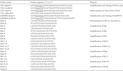
TABLE 1Primers used in this study
Primer name Primer sequence (5=to 3=)a Purpose
TIA1-KpnI-F ATATGGTACCATGGAGGACGAGATGCCCAAG Amplification and cloning of TIA1a and TIA1b
TIA1-EcoRI-R ATATGAATTCTCACTGGGTTTCATACCCTGCC
TIA1-KpnI-F ATATGGTACCATGGAGGACGAGATGCCCAAG Differentiation of TIA1a from TIA1b
TIA1-spliced-R GATCACCAACAAAGACATGGAAATG
hnRNPK-KpnI-F ATATGGTACCATGGAAACTGAACAGCCAG Amplification and cloning of hnRNP K hnRNPK-EcoRI-R ATATGAATTCTTAGAAAAACTTTCCAGAATACTG
BclX-F CATATCAGAGCTTTGAACAG Differentiation of Bcl-XLfrom Bcl-XS
BclX-R TCATTTCCGACTGAAGAGTG
Bax-F CAGCTCTGAGCAGATCATG Amplification of Bax
Bax-R CACAGGGCCTTGAGCAC
Bik-F TCTCCAGAGACATCTTGATG Amplification of Bik
Bik-R GTTCGCAGGACACCCAG
Bid-F TGGACCGTAGCATCCCTC Amplification of Bid
Bid-R CAGTCCATCCCATTTCTGG
Mcl-1-F CGGGAGGGCGACTTTTG Amplification of Mcl-1
Mcl-1-R GAACTCCACAAACCCATCC
Bcl2-A1-F CAGTGCGTCCTACAGATAC Amplification of Bcl2-A1
Bcl2-A1-R GCCTTATCCATTCTCCTGTG
Bag1-F CAGCAATGAGAAGCACGAC Amplification of Bag1
Bag1-R TGCAGAGAGCTTCAGCTTG
Bcl-2-F CGATAACCGGGAGATAGTG Amplification of Bcl-2
Bcl-2-R CACCGTGGCAAAGCGTC
Bcl-W-F CTGGTGGCAGACTTTGTAG Amplification of Bcl-W
Bcl-W-R CATCCACTCCTGCACTTG
XIAP-F TGGCACGAGCAGGGTTTC Amplification of XIAP
XIAP-R CTCGGGTATATGGTGTCTG
aUnderlined sequences indicate restriction sites.
on November 7, 2019 by guest
http://jvi.asm.org/
may play a role in viral gene expression to some extent, it likely plays a more prominent role(s) at another step (or other steps) in the virus replication cycle.
To further elucidate the role of hnRNP K in the VSV life cycle, we investigated the requirement of hnRNP K at VSV entry/un-coating and assembly/budding steps. Transfection of VSV nucleo-capsids (NCs) directly into cells bypasses the viral entry and
un-coating steps, resulting in expression of the viral genes (29,33).
Thus, we examined the effect of depletion of hnRNP K on viral gene expression in cells transfected with the viral NCs. Results show that NC transfection led to only a slight decrease (approxi-mately 30%) in the levels of the viral M protein in cells treated with siRNA for hnRNP K compared to levels in NT siRNA-transfected
cells (Fig. 2C, lanes 3 and 4). In contrast, an 8- to 10-fold reduction
in M protein levels was observed in cells infected at a low MOI
(0.01) with VSV (Fig. 2C, lanes 1 and 2). Infection of cells with
VSV⌬G virus (a virus that lacks the G gene and cannot produce
infectious particles, thereby limiting the infection with this virus, which is incompetent in budding, to a single cycle only) also led to
a slight decrease (approximately 30%) in the levels of the viral M protein in hnRNP K siRNA-treated cells compared to the control
NT siRNA-treated cells (Fig. 2C, lanes 5 and 6). Furthermore,
using a high MOI (1.0) of VSV, we observed viral gene expression at 4 hpi (a time at which viral gene expression is readily detectable
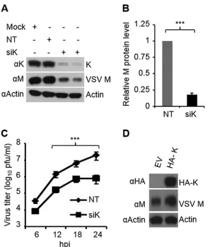
FIG 1hnRNP K is required for VSV infection. (A) Suppression of VSV rep-lication in cells depleted of hnRNP K. HeLa cells were transfected with 10 nM siRNA for NT (lane 2) or 10 nM and 20 nM hnRNP K (siK) (lanes 3 and 4, respectively) for 60 h. Cells were then infected with VSV at an MOI of 0.01 for 12 h. Equal amounts of cell lysates were analyzed by WB with anti-hnRNP K and anti-M antibodies. Actin served as the loading control. (B) Quantification of M protein levels (arbitrary units) from three independent experiments as described in panel A with error bars representing the standard errors of means. The level of M expression in the NT siRNA-treated sample was set at 1. ***, P⬍0.001. (C) Multicycle growth of VSV in cells depleted of hnRNP K. siRNA (10 nM) treatment and virus infections were performed as described in panel A; virus titers in supernatants collected at indicated time points were deter-mined by plaque assay and are expressed as log10PFU/ml. Statistical
signifi-cance was determined for virus titers at 12, 18, and 24 hpi. ***,P⬍0.001. (D) VSV infection is enhanced in hnRNP K-overexpressing cells. HeLa cells trans-fected with 1.5g of empty vector (EV) or an HA-hnRNP K (HA-K)-encoding plasmid for 48 h were infected with VSV at an MOI of 0.01 for 12 h, and cell lysates were analyzed by WB using anti-M or anti-HA antibodies. Actin served as the loading control.␣, anti; siK, siRNA targeting hnRNP K.
FIG 2hnRNP K is required for VSV propagation. (A and B) VSV mRNA and antigenomic RNA levels in cells depleted of hnRNP K and infected with the virus at a low (0.01) or high (1.0) MOI (panels A and B, respectively) were determined by qRT-PCR. Experimental conditions were as described in the legend ofFig. 1A. Error bars represent the standard errors of means from three independent experiments. ***,P⬍0.001; *,P⬍0.05. (C) hnRNP K is not required for early stages of the VSV life cycle. HeLa cells were transfected with 10 nM siRNA for NT or targeting hnRNP K for 60 h. Cells were then treated as follows: infected with VSV at an MOI of 0.01 for 12 h (lanes 1 and 2), super-transfected for 6 h with viral NC prepared from VSV (lanes 3 and 4), infected with VSV⌬G at an MOI of 0.5 for 8 h (lanes 5 and 6), or infected with VSV at an MOI of 1 for 4 h (lane 7 and 8). Cell lysates corresponding to equal amounts of total proteins were analyzed by WB with hnRNP K and M anti-bodies. Actin served as the loading control. (D) HeLa cells were transfected with siRNA as described in the legend ofFig. 1A. At 60 hpt, cells were infected with VSV-PLuc at an MOI of 300. After 1 h of virus adsorption on ice, cells were washed with PBS, and cell extracts at various times postincubation were analyzed for luciferase activity. The luciferase activity in NT siRNA-treated cells at various times postinfection was set at 1. Data show relative luciferase activity and are expressed as the averages of three independent experiments, with error bars representing the standard errors of means. NS, nonsignificant. (E) HeLa cells were transfected with siRNA as described in the legend ofFig. 1A. At 60 hpt, cells were infected with VSV-PeGFP at an MOI of 0.05 or VSV⌬G at an MOI of 0.5 for 12 h. The percentages of infected cells were determined by counting the number of cells expressing green fluorescence from PeGFP. Data show relative percent infection after normalizing the value of cells treated with siRNA targeting hnRNP K to that of NT siRNA-treated cells, which is set at 100, and are expressed as average of three independent experiments with error bars representing the standards error of means. ***, P⬍0.001; NS, nonsignificant.
on November 7, 2019 by guest
http://jvi.asm.org/
[image:4.585.61.268.64.310.2] [image:4.585.318.522.66.375.2]but virus assembly/budding is minimal) to be reduced by only
about 30% in cells treated with an siRNA for hnRNP K (Fig. 2C,
lanes 7 and 8) compared to that in NT siRNA-transfected cells. These results show that bypassing entry/uncoating steps (NC
transfection), using assembly/budding-incompetent (VSV⌬G)
virus, or using high MOIs did not adversely affect overall viral gene expression. On the other hand, a significant reduction in viral gene expression under conditions of VSV infection at a low MOI suggests that the supportive roles of hnRNP K in VSV infec-tion are manifested when the infecinfec-tion undergoes multiple cycles, indicating that this cellular protein may be involved in virus entry/ uncoating or assembly/budding steps.
To determine if virus entry and uncoating steps are affected by depletion of hnRNP K, we used a recombinant virus (VSV-PLuc) which encodes a P-luciferase (P-Luc) fusion protein, in which the entire coding region of luciferase is fused in frame following amino acid 196 of the P protein. The VSV-PLuc virus is similar to
VSV-PeGFP described previously (35) and incorporates P-Luc
within virus particles. Luciferase activity can be readily detected in extracellular virions and in infected cells (data not shown). Cells transfected with NT or hnRNP K siRNA were infected with VSV-PLuc at an MOI of 300 to allow readily detectable levels of lucif-erase. Following virus adsorption on ice, cells were washed, and luciferase activity was determined in extracts of cells at 0, 0.5, 1, and 1.5 h postincubation at 37°C to enable virus entry and
uncoat-ing. Results (Fig. 2D) show that luciferase activity in these cells was
not significantly affected in the presence or absence of hnRNP K, indicating that this protein does not play any discernible role in
virus entry and uncoating steps. Furthermore, using a low MOI (0.01), a multicycle virus growth condition, we observed that the percentage of cells infected with wt VSV increased with time in the presence of hnRNP K, while in cells depleted of hnRNP K, the relative percentage of infected cells remained low, reaching up to
about 15% of that of NT siRNA-treated cells at 16 hpi (Fig. 2E).
On the other hand, we found no differences in the percent infec-tion when these cells were infected with the budding-incompetent VSV⌬G (Fig. 2E). Taken together, the results suggest that hnRNP K is required for virus egress in infected cells and that its depletion may negatively affect assembly and/or release of virus required for further rounds of infection.
hnRNP K alters the expression of TIA1, a known negative
regulator of VSV replication.Since hnRNP K is known to
influ-ence expression of many cellular genes, we wondered whether its depletion alters expression of cellular genes that have been shown to be involved in VSV replication. Previous studies from our lab-oratory showed that PCBP2 and PCBP1 as well as TIA1 inhibit
VSV replication (29,30). Thus, we examined the levels of these
proteins in cells depleted of hnRNP K by siRNA transfection.
Fig-ure 3Ashows that knockdown of hnRNP K had no measurable effect on the levels of PCBP1/2. Expression levels of another cel-lular protein, NF90, which is known to inhibit replication of
sev-eral other viruses such as HIV-1 (38), Ebola virus (39), and
influ-enza virus (40), was also not affected by hnRNP K depletion (Fig.
3A). Interestingly, under this condition, we observed a
reproduc-ibly greater than 2-fold increase in the total amount of TIA1
pro-tein (Fig. 3BandC). Strikingly, hnRNP K depletion resulted in a
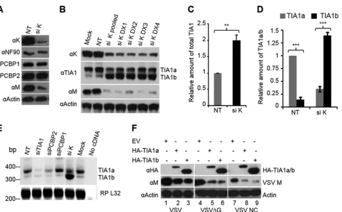
FIG 3hnRNP K regulates the expression of the two isoforms of TIA1 but not PCBP1/2. (A) Depletion of hnRNP K does not affect the expression of PCBP1/2. HeLa cells were transfected with NT or hnRNP K siRNA at a concentration of 10 nM, and at 60 hpt, cells were infected with VSV at an MOI of 0.01 for 12 h. Cell lysates were used for WB to detect the indicated proteins. (B to D) hnRNP K depletion results in increased levels of total TIA1 and the TIA1b isoform. HeLa cells were mock transfected or transfected with NT or hnRNP K siRNA as a pool of four duplexes (pooled) or as individual duplexes (si DX1 to si DX4) at a concentration of 10 nM, and at 60 hpt, the cells were infected with VSV at an MOI of 0.01 for 12 h. Cell lysates were used for WB to detect the indicated proteins (B). Quantification of total TIA1 protein levels (arbitrary units) from three independent experiments is shown in panel C while the levels of TIA1a and TIA1b isoforms are shown in panel D. The level of TIA1a expression in the NT siRNA-treated sample was set at 1. ***,P⬍0.001; **,P⬍0.01. (E) Switching of TIA1 isoform expression is regulated by hnRNP K protein at the mRNA level. The experiment was done as described in panel B. Total RNA was subjected to semiquantitative RT-PCR to detect the two isoforms of TIA1. Ribosomal protein (RP) L32 mRNA served as an internal control. (F) Overexpression of TIA1a and TIA1b inhibits VSV gene expression. HeLa cells transfected with 1.5g of empty vector (EV) or HA-TIA1a- or HA-TIA1b-encoding plasmids for 48 h were infected with VSV at an MOI of 0.1 for 12 h (lanes 1 to 3) or with VSV⌬G at an MOI of 0.5 for 8 h (lanes 4 to 6) or supertransfected for 6 h with viral NC prepared from VSV (lanes 7 to 9), and cell lysates were analyzed by WB using anti-M or anti-HA antibodies. Actin served as the loading control. siPCBP1, siRNA targeting PCBP1; siPCBP2, siRNA targeting PCBP2; siTIA1, siRNA targeting TIA1.
on November 7, 2019 by guest
http://jvi.asm.org/
[image:5.585.128.461.63.269.2]dramatic shift in the ratio of the two isoforms (TIA1a and TIA1b)
of the protein (Fig. 3BandD). In mock-treated or NT
siRNA-treated cells, the amount of TIA1b was approximately 12% of TIA1a, whereas in hnRNP K-depleted cells, TIA1b was detected at
significantly higher levels (about 3-fold) than TIA1a (Fig. 3B).
These results were observed when individual siRNAs specific for
hnRNP K or pooled siRNAs were used (Fig. 3B), indicating that
the effect of hnRNP K protein on altered expression of TIA1 iso-forms was not due to off-target effects. The altered expression of
the two isoforms was detected at the mRNA level (Fig. 3E),
indi-cating that hnRNP K regulates the expression of these two iso-forms at the transcription and/or splicing level. Similar results were also obtained in HEK293 cells (data not shown) and wt MEFs (seeFig. 4D).
Since depletion of hnRNP K led to increased levels of the TIA1b isoform and inhibition of virus replication, we examined whether TIA1b inhibits VSV replication, as was demonstrated for
the TIA1a isoform previously (30). In VSV-infected cells
overex-pressing HA-tagged TIA1b, the viral M protein levels were
re-duced (Fig. 3F). The reduction in M protein levels was observed in
TIA1b-expressing cells infected with VSV (Fig. 3F, lanes 1 to 3) or
VSV⌬G (lanes 4 to 6) or in NC-transfected cells (lanes 7 to 9),
suggesting that TIA1b inhibits VSV replication. Thus, it appears that hnRNP K depletion results in increased accumulation of TIA1b, which is also responsible for inhibition of VSV replication.
hnRNP K is required for survival of VSV-infected cells.
hn-RNP K plays essential roles in cell survival. Its cleavage by different granzymes initiates cell death processes, and its depletion primes the cells to undergo programmed cell death under various
apo-ptotic stimuli (9,13,41,42). While performing our studies, we
observed that VSV-infected cells depleted of hnRNP K exhibited enhanced cytopathic effects and cell death compared to the in-fected cells not depleted of the protein. Therefore, we examined if hnRNP K plays any role in cell survival during VSV infection. Depletion of hnRNP K did not affect the viability of mock-in-fected cells significantly; however, the viability of VSV-inmock-in-fected
cells was significantly reduced with time postinfection (Fig. 4A).
The results show that viability of VSV-infected cells depleted of hnRNP K was reduced to approximately 63% and 25% at 12 and 18 hpi, respectively, whereas the viability of NT siRNA-treated cells infected with VSV was reduced only to 92% and 60%,
respec-tively, at the corresponding times postinfection (Fig. 4A).
Consis-tent with the increase in cell death, the amount of cleaved caspase 3 (p17) was found to be 7-fold higher in cells lacking hnRNP K at
12 hpi (Fig. 4BandC).
Since TIA1 is a proapoptotic protein (43,44), we wondered if
the upregulation of TIA1 in hnRNP-depleted cells is responsible for the enhanced apoptosis observed in these cells infected with VSV. To this end, we examined the apoptosis occurring in wt and
TIA1⫺/⫺MEFs (34) when hnRNP K is silenced by siRNA and
FIG 4hnRNP K is required for survival of VSV-infected cells. (A) Knockdown of hnRNP K reduces cell survival during VSV infection. HeLa cells were transfected with NT or hnRNP K siRNA at concentration of 10 nM for 60 h. Cells were mock infected or infected with VSV at an MOI of 0.01. At 6, 12, and 18 hpi, cell survival was determined by a CellTiter-Glo Kit. The percentage of cell viability in NT siRNA-transfected and mock-infected culture at the beginning of infection was set at 100%. Data from three independent experiments are shown with error bars representing the standard errors of means. **,P⬍0.01; NS, nonsignificant. (B) Caspase 3 activation is more pronounced in infected cells lacking hnRNP K. The experiment was conducted as described in panel A. Infected cells were lysed and analyzed at the indicated time points to detect procaspase 3, activated caspase 3 (p17), hnRNP K, and VSV M protein using specific antibodies. Actin served as a loading control. (C) Quantification of p17 protein levels (arbitrary units) from three independent experiments as described in panel B. The level of activated caspase 3 (p17) in cells treated with siRNA targeting hnRNP K at 18 hpi was set arbitrarily at 1, and the relative levels of p17 in other samples were then determined. Error bars represent the standard errors of means from three independent experiments. ***,⬍0.001; **,P⬍0.01. (D) wt MEF or TIA1⫺/⫺MEF
cells were transfected with NT or hnRNP K siRNA at a concentration of 20 nM for 60 h. Cells were mock infected (mock) or infected with VSV at an MOI of 1. At 4 and 8 hpi, cells were lysed and subjected to WB with the indicated antibodies. (E) Quantification of p17 levels (arbitrary units) from three independent experiments, with error bars representing the standard errors of means. The level of p17 in wt MEFs treated with siRNA targeting hnRNP K was set arbitrarily at 1, and the relative levels of p17 in other samples were then determined. ***,P⬍0.001.
on November 7, 2019 by guest
http://jvi.asm.org/
[image:6.585.125.461.67.298.2]subsequently infected with VSV. Results (Fig. 4DandE) show that activation of caspase 3 (as measured by the levels of p17) occurred
to similar extents in VSV-infected wt and TIA1⫺/⫺MEFs depleted
of hnRNP K, indicating that VSV-induced cell death in hnRNP K-depleted cells is independent of TIA1.
VSV-mediated cell death in hnRNP K-depleted cells is asso-ciated with increased levels of proapoptotic proteins and
de-creased levels of antiapoptotic proteins.VSV infection
upregu-lates the expression of proapoptotic proteins such as Noxa and Bak that are involved in virus-mediated apoptosis in cell culture (45–47). On the other hand, hnRNP K is known to support the expression of antiapoptotic proteins such as the caspase inhibitor
protein XIAP (X-linked inhibitor of apoptosis protein) (12),
FLICE-inhibitory protein (FLIP) (9), and Bcl-XLbut suppresses
the expression of proapoptotic Bcl-XS(42). Therefore, we
exam-ined if depletion of hnRNP K affects the expression of anti- and/or proapoptotic proteins that may result in altered survival of VSV-infected cells. Our results show that the levels of Noxa, Bad, and FLIP were unchanged while the expression of Bak decreased in both HeLa and HEK293 cells depleted of hnRNP K and infected
with VSV (Fig. 5A). Through semiquantitative RT-PCR, we found
that the mRNA levels of proapoptotic factors Bax and Bid were not altered significantly in the absence of hnRNP K, whereas the level of another proapoptotic factor, Bik, was upregulated over 4-fold (Fig. 5B). The mRNA levels of antiapoptotic factors Bag1 and Bcl-2A1 but not Bcl-W or XIAP under similar experimental con-ditions were slightly reduced, whereas the level of Bcl-2 was
down-regulated greater than 3-fold (Fig. 5C).
A previous study reported that depletion of hnRNP K by siRNA treatment led to a significant decrease in the level of mRNA
for the antiapoptotic protein Bcl-XLand an increase in the
pro-apoptotic Bcl-XSmRNAs in PC-3 cells as well as several other cell
lines, but no significant differences were observed in HeLa cells
(42), which was suggested to be due to less efficient depletion of
the protein in this cell line. Therefore, it was of interest to examine
the mRNA levels of Bcl-XLand Bcl-XSin HeLa cells depleted of
greater than 90% of hnRNP K using the pool of four siRNAs
described in our studies above (Fig. 1A). Results from such an
experiment show (Fig. 5B) that the mRNA levels of Bcl-XLand
Bcl-XSremained unchanged in VSV-infected HeLa cells depleted
of hnRNP K compared to levels in the infected cells not depleted of the protein. In contrast to the results in HeLa cells, we observed a
significant decrease of Bcl-XL and a concomitant increase in
Bcl-XSmRNA levels in HEK293 cells under similar experimental
conditions (Fig. 5D). Interestingly, the level of Bik mRNA was also
reduced significantly (Fig. 5D). These results indicate that the
VSV-mediated apoptosis in hnRNP K-depleted cells is associated with alterations in the balance of /antiapoptotic Bcl-2
pro-teins (decrease of antiapoptotic propro-teins such as Bcl-XL, Bag1, and
Bcl-2 and an increase of proapoptotic factors including Bcl-XSand
Bik) though the identity of these proteins might differ in different cell lines.
hnRNP K maintains expression of cellular proteins required
for VSV infection.Since hnRNP K is involved in regulating
ex-pression of many cellular genes (1), it is possible that depletion of
hnRNP K might influence levels of cellular proteins that are known to be required for VSV replication, leading to inhibition of virus replication. Recently, we reported through a genome-wide siRNA screen the requirement of several host factors for VSV
rep-lication at various stages of the virus infection cycle (33). Among
these, alanine deaminase-like (ADAL) protein was shown to be required for the VSV assembly/budding step, whereas GBF1, ARF1, and the proteins of the coatomer I (COPI) complex were
required for VSV genome transcription and replication (33,48).
Therefore, we examined if hnRNP K silencing affects the expres-sion of some of these cellular factors. Results show that hnRNP K
FIG 5VSV-mediated cell death in hnRNP K-depleted cells is associated with increased levels of proapoptotic proteins Bcl-XSand Bik. (A) HeLa or HEK293 cells
transfected with siRNAs for 60 h were infected with VSV at an MOI of 1 for 6 h. The levels of proapoptotic proteins Bak, Bad, Noxa, and antiapoptotic factor FLIP were detected by WB using specific antibodies. (B and C) mRNA levels of proapoptotic proteins (B) or antiapoptotic proteins (C) in hnRNP K-depleted HeLa cells infected with VSV. siRNA treatment and virus infections were performed as described in panel A. Total RNA was converted to cDNA using oligo(dT) primers followed by PCR with specific primers. The mRNA level of cellular ribosomal protein L32 was used as an internal control. A DNA ladder (bp) is shown on the right. Values at the top or bottom of the lanes represent relative levels of mRNAs, using levels in the NT lane as 1. The Bcl-XSlevel is relative to the Bcl-XL
level in the NT lane. The asterisk indicates the DNA product of an undocumented isoform of Bag1 or a nonspecific product. (D) mRNA levels of antiapoptotic or proapoptotic proteins in hnRNP K-depleted HEK293 cells infected with VSV. siRNA treatment, virus infection, and mRNA detection were as described above.
on November 7, 2019 by guest
http://jvi.asm.org/
[image:7.585.114.473.65.259.2]depletion by siRNA did not alter the levels of COPI proteins such
as COPB1 and COPZ or GBF1 (Fig. 6A). Furthermore, under the
same conditions, the level of GS28, a putative protein of thecis
-Golgi compartment that is involved in endoplasmic reticulum
(ER)-Golgi transport (49) and apoptosis (50,51), was not affected
either (Fig. 6A). On the other hand, depletion of hnRNP K
re-sulted in a 3- to 4-fold reduction in the levels of ARF1 and ADAL
proteins (Fig. 6BandC). These results suggest that hnRNP K
regulates expression of certain cellular proteins required for VSV replication.
DISCUSSION
hnRNP K is a multifunctional protein in the subfamily of hnRNPs which carry three KH domains and is involved in gene expression and mRNA metabolism. Previously, our genome-wide screening study identified hnRNP K as a factor required for VSV infection
(33). In this communication, we conducted further studies to
de-cipher the role of hnRNP K in supporting VSV infection. Our results reveal that hnRNP K primarily supports VSV infection at the level of virus propagation, but it is also involved in viral
repli-cation and transcription to some extent (Fig. 1and2). Further
studies showed that hnRNP K is required for suppressing apopto-sis induction and promoting survival of infected cells, which in turn supports VSV growth. We found that hnRNP K helps main-tain the low levels of expression of proapoptotic proteins such as
BiK and Bcl-XSand the high levels of expression of antiapoptotic
proteins such as Bcl-XL, Bag1, and Bcl-2. In addition to the role of
hnRNP K in cell death suppression, our study found that hnRNP K supports expression of several cellular factors required for VSV replication and downregulates expression of some of the cellular factors known to inhibit VSV replication. Thus, the studies pre-sented here reveal critical roles of hnRNP K in VSV infection through multiple mechanisms. The supportive role of hnRNP K in VSV infection is observed in all cell lines examined in this study,
whereas its role in suppression of apoptosis is mediated by a bal-ance between several pro- and antiapoptotic factors and is cell type dependent.
hnRNP K is reported to play diverse roles in many virus infec-tions. Its interactions with viral components and/or cellular fac-tors support the replication of viruses, including enterovirus 71, Sindbis virus, dengue virus, chikungunya virus, hepatitis C virus, HBV, HIV-1, HSV-1, African swine fever virus, human
cytomeg-alovirus, and Epstein-Barr virus (16,18–28). Interestingly, in
hu-man cytomegalovirus-infected cells, hnRNP K is essential for
vi-rus replication as it promotes the viability of infected cells (28).
Although our studies reveal such a role for hnRNP K in VSV replication, it may also be involved in virus assembly and/or re-lease. So, how then does hnRNP K facilitate VSV assembly and/or release from infected cells? The answer to this critical question requires further investigation, including examination of interac-tions of viral proteins with hnRNP K. It is possible that hnRNP K may directly or indirectly interact with the viral components to mediate virus assembly or egress. Since hnRNP K is known to be
involved in cellular gene expression and signal transduction (3), it
is also possible that it modulates expression of certain cellular
proteins that are important for VSV egress, such as ADAL (Fig. 6B
andC), which was shown before to be required for VSV assembly
and budding (33). Alternatively, hnRNP K may recruit factors
necessary for virus egress through its K protein-interacting (KI) domain.
Although interaction of VSV proteins with hnRNP K is being examined currently, one required role for hnRNP K in VSV infec-tion is found to be at the level of maintaining the viability of infected cells. VSV-infected cells exhibited significantly reduced cytopathic effects and did not undergo apoptosis as fast in the presence of hnRNP K as in cells lacking this protein. Thus, hnRNP K suppresses VSV-induced apoptosis. Examination of the levels of
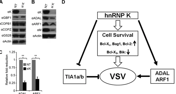
FIG 6hnRNP K regulates expression of cellular proteins required for VSV infection. (A and B) HeLa cells transfected with siRNA for 60 h were infected with VSV at an MOI of 0.01 for 12 h. The levels of the proteins were detected by WB using specific antibodies. (C) Quantification of ADAL and ARF1 protein levels (arbitrary units) from three independent experiments, with error bars showing the standard errors of means. The level of ADAL or ARF1 in the NT lane was arbitrarily set at 1. **,P⬍0.01. (D) Involvement of hnRNP K in regulating expression of cellular proteins that affect VSV infection. hnRNP K inhibits TIA1a/b expression, which is known to inhibit VSV replication. Increased levels of antiapoptotic proteins such as Bcl-XL, Bag1, and Bcl-2 and/or reduction in the levels
of apoptotic proteins Bcl-XSand Bik promotes cell survival for increased VSV replication, while maintenance of factors such as ADAL and ARF1 is required for
VSV replication.
on November 7, 2019 by guest
http://jvi.asm.org/
[image:8.585.114.475.68.262.2]several factors involved in apoptosis suggested that increased apoptosis of VSV-infected cells lacking hnRNP K does not involve Noxa and Bak, two proapoptotic proteins previously shown to
mediate VSV-induced cell death (45–47). Rather, significantly
in-creased levels of Bik or Bcl-XSappear to regulate apoptosis in
hnRNP K-depleted cells by regulating the balance of the pro-/
antiapoptotic Bcl-2 proteins. In HeLa cells, the levels of Bcl-XS
remained unchanged in the presence or absence of hnRNP K, but Bik was upregulated over 4-fold, which presumably was
responsi-ble for increased cell death. In contrast, in HEK293 cells, Bcl-XS
levels were increased significantly in the absence of hnRNP K
along with decreased levels of Bcl-XLwhile the level of Bik was
significantly reduced, suggesting that in this cell type, the decrease
in the Bcl-XL/Bcl-XSratio may primarily regulate apoptosis of
VSV-infected cells. Thus, it appears that hnRNP K regulates the balance between pro- and antiapoptotic factors in a cell-type-de-pendent manner so as to allow infected cells to survive longer for increased virus growth. It should be noted, however, that consid-ering the multiple roles played by hnRNP K in many cellular pro-cesses, it is possible that suppression of apoptosis may not be solely responsible for increased growth of VSV in infected cells. Further studies would have to be conducted to delineate additional roles of hnRNP K, if any, in supporting VSV growth.
hnRNP K is known to function closely with p53, a well-known mediator of apoptosis, at multiple levels to coregulate cell fate. Together with p53, hnRNP K binds to promoters to upregulate a
number of genes encoding DNA damage response factors (52),
and in combination with large intergenic noncoding RNA p21 (lincRNA-p21), it regulates global gene repression downstream of
a p53-mediated response (53). Evidence also suggests that hnRNP
K acts as a cell survival enhancer independent of p53 by maintain-ing a high level of antiapoptotic proteins, includmaintain-ing FLIP and
XIAP (9,12). Studies presented here reveal that the role of hnRNP
K in survival of VSV-infected cells is independent of its interaction with p53. This interpretation is consistent with the observations that VSV-induced apoptosis in the absence of hnRNP K occurs with similar kinetics in both p53-inactive cells (HeLa and HEK293) and wild-type p53-active cells such as MCF-7 and MEF cells. Additionally, apoptosis induced by other factors such as cy-cloheximide treatment or other virus infections (lymphocytic choriomeningitis virus [LCMV] and human parainfluenza virus type 3 [HPIV3]) was also enhanced in hnRNP K-depleted cells (data not shown). These observations highlight the essential role of hnRNP K independent of p53 in protecting cells from apoptosis triggered by different stimuli.
VSV is being used as an oncolytic agent for various types of
tumors (55–58). One of the hallmark features of most cancer cells
is the defective type I interferon response and signaling (59),
which allows viruses such as VSV to grow rapidly and kill the cells.
hnRNP K is upregulated in many types of cancerous cells (10,11,
60) and is considered a prognostic marker and a target for tumor
therapy (11,61). The results showing that hnRNP K supports VSV
propagation further add to the understanding of why tumors pro-vide an appropriate microenvironment for VSV growth.
Our studies also reveal a novel role of hnRNP K in suppressing not only the expression of the TIA1b isoform (a differentially spliced isoform of TIA1a) but also the overall levels of TIA1. The function of these two isoforms in cells is poorly understood, ex-cept that TIA1b displays greater splicing-stimulatory activity than
TIA1a (62). Previously, we along with others demonstrated that
TIA1 inhibited VSV replication (30,63) although the
contribu-tion of the TIA1b isoform was not investigated. Our present re-sults demonstrate that TIA1b is inhibitory to VSV replication. Since expression of TIA1 is mediated by many cellular factors
including miRNAs (64,65), it would be of interest to examine how
hnRNP K regulates or is regulated by these factors, which may provide further understanding of the role of cellular factors in-volved in VSV replication.
The results presented here do not exclude the involvement of hnRNP K in supporting VSV assembly and budding. Although not much is known about the direct role(s) of hnRNP K in lipid metabolism, in protein trafficking, in the growth of plasma mem-brane, or in secretory pathways, which are directly involved in virus assembly and budding, hnRNP K regulates the expression of 15-lipoxygenase, a key enzyme in the metabolism of
phospholip-ids and internal membrane (66). Studies of the hnRNP K
interac-tome revealed more than 100 interacting partners and determined that hnRNP K protein is present in the nucleus, cytoplasm,
mito-chondria, and the vicinity of the plasma membrane (31). Thus, it
is possible that hnRNP K exerts its positive impact on VSV growth through one or more of its interacting partners.
In summary, we demonstrate here a role(s) of hnRNP K in VSV
infection (Fig. 6D). The mechanisms by which hnRNP K
pro-motes VSV infection involve the following: (i) promoting cell
sur-vival via suppression of proapoptotic proteins (Bik and Bcl-XS)
and upregulation of antiapoptotic proteins (Bcl-XL, Bag1, and
Bcl-2); (ii) suppression of expression of VSV-inhibitory protein TIA1; and (iii) upregulation of cellular factors favoring VSV in-fection such as ARF1 and ADAL. Our data also provide a better understanding of why tumor cells provide a more conducive en-vironment for VSV replication. Overall, our studies presented here suggest a multifunctional role of hnRNP K in VSV infection and prompt us to undertake additional research to uncover its involvement in virus-host interactions.
ACKNOWLEDGMENT
We thank Z. H. Gill for excellent assistance in the laboratory.
REFERENCES
1.Choi HS, Hwang CK, Song KY, Law PY, Wei LN, Loh HH. 2009. Poly(C)-binding proteins as transcriptional regulators of gene expression. Biochem. Biophys. Res. Commun.380:431– 436.
2.Ostareck-Lederer A, Ostareck DH.2012. Precision mechanics with mul-tifunctional tools: how hnRNP K and hnRNPs E1/E2 contribute to post-transcriptional control of gene expression in hematopoiesis. Curr. Protein Pept. Sci.13:391– 400.
3.Bomsztyk K, Denisenko O, Ostrowski J.2004. hnRNP K: one protein multiple processes. Bioessays26:629 – 638.
4.Ostareck-Lederer A, Ostareck DH, Rucknagel KP, Schierhorn A, Moritz B, Huttelmaier S, Flach N, Handoko L, Wahle E.2006. Asymmetric arginine dimethylation of heterogeneous nuclear ribonucleoprotein K by protein-arginine methyltransferase 1 inhibits its interaction with c-Src. J. Biol. Chem.281:11115–11125.
5.Habelhah H, Shah K, Huang L, Ostareck-Lederer A, Burlingame AL, Shokat KM, Hentze MW, Ronai Z.2001. ERK phosphorylation drives cytoplasmic accumulation of hnRNP-K and inhibition of mRNA transla-tion. Nat. Cell Biol.3:325–330.
6.Naarmann IS, Harnisch C, Flach N, Kremmer E, Kuhn H, Ostareck DH, Ostareck-Lederer A.2008. mRNA silencing in human erythroid cell maturation: heterogeneous nuclear ribonucleoprotein K controls the ex-pression of its regulator c-Src. J. Biol. Chem.283:18461–18472. 7.Wang N, Zhang P, Guo X, Zhou Z, Sha J. 2011. Hnrnpk, a protein
differentially expressed in immature rat ovarian development, is required for normal primordial follicle assembly and development. Endocrinology 152:1024 –1035.
on November 7, 2019 by guest
http://jvi.asm.org/
8.Laursen LS, Chan CW, ffrench-Constant C.2011. Translation of myelin basic protein mRNA in oligodendrocytes is regulated by integrin activa-tion and hnRNP-K. J. Cell Biol.192:797– 811.
9.Chen LC, Chung IC, Hsueh C, Tsang NM, Chi LM, Liang Y, Chen CC, Wang LJ, Chang YS.2010. The antiapoptotic protein, FLIP, is regulated by heterogeneous nuclear ribonucleoprotein K and correlates with poor overall survival of nasopharyngeal carcinoma patients. Cell Death Diff. 17:1463–1473.
10. Tang FM, Li WM, Chen Y, Wang DM, Han J. 2008. Expression of hnRNP K in lung adenocarcinoma cells. Sichuan Da Xue Xue Bao Yi Xue Ban39:823– 826.
11. Wu CS, Chang KP, Chen LC, Chen CC, Liang Y, Hseuh C, Chang YS. 2012. Heterogeneous ribonucleoprotein K and thymidine phosphorylase are independent prognostic and therapeutic markers for oral squamous cell carcinoma. Oral Oncol.48:516 –522.
12. Xiao Z, Ko HL, Goh EH, Wang B, Ren EC.2013. hnRNP K suppresses apoptosis independent of p53 status by maintaining high levels of endog-enous caspase inhibitors. Carcinogenesis [Epub ahead of print.] doi:10 .1093/carcin/bgt085.
13. van Domselaar R, Quadir R, van der Made AM, Broekhuizen R, Bovenschen N.2012. All human granzymes target hnRNP K that is essen-tial for tumor cell viability. J. Biol. Chem.287:22854 –22864.
14. White M, Xia G, Gao R, Wakamiya M, Sarkar PS, McFarland K, Ashizawa T.2012. Transgenic mice with SCA10 pentanucleotide repeats show motor phenotype and susceptibility to seizure: a toxic RNA gain-of-function model. J. Neurosci. Res.90:706 –714.
15. Gao FH, Wu YL, Zhao M, Liu CX, Wang LS, Chen GQ.2009. Protein kinase C-delta mediates down-regulation of heterogeneous nuclear ribo-nucleoprotein K protein: involvement in apoptosis induction. Exp. Cell Res.315:3250 –3258.
16. Lin JY, Li ML, Huang PN, Chien KY, Horng JT, Shih SR. 2008. Heterogeneous nuclear ribonuclear protein K interacts with the enterovi-rus 71 5=untranslated region and participates in virus replication. J. Gen. Virol.89:2540 –2549.
17. Shih SR, Stollar V, Li ML.2011. Host factors in enterovirus 71 replica-tion. J. Virol.85:9658 –9666.
18. Zhang W, Zhang X, Tian C, Wang T, Sarkis PT, Fang Y, Zheng S, Yu XF, Xu R.2008. Cytidine deaminase APOBEC3B interacts with hetero-geneous nuclear ribonucleoprotein K and suppresses hepatitis B virus ex-pression. Cell Microbiol.10:112–121.
19. Ng LF, Chan M, Chan SH, Cheng PC, Leung EH, Chen WN, Ren EC. 2005. Host heterogeneous ribonucleoprotein K (hnRNP K) as a potential target to suppress hepatitis B virus replication. PLoS Med.2:e163. doi:10 .1371/journal.pmed.0020163.
20. Schmidt T, Striebinger H, Haas J, Bailer SM.2010. The heterogeneous nuclear ribonucleoprotein K is important for herpes simplex virus-1 propagation. FEBS Lett.584:4361– 4365.
21. Kanlaya R, Pattanakitsakul SN, Sinchaikul S, Chen ST, Thongboonkerd V.2010. Vimentin interacts with heterogeneous nuclear ribonucleopro-teins and dengue nonstructural protein 1 and is important for viral repli-cation and release. Mol. Biosyst.6:795– 806.
22. Marchand V, Santerre M, Aigueperse C, Fouillen L, Saliou JM, Van Dorsselaer A, Sanglier-Cianferani S, Branlant C, Motorin Y. 2011. Identification of protein partners of the human immunodeficiency virus 1 tat/rev exon 3 leads to the discovery of a new HIV-1 splicing regulator, protein hnRNP K. RNA Biol.8:325–342.
23. Bourai M, Lucas-Hourani M, Gad HH, Drosten C, Jacob Y, Tafforeau L, Cassonnet P, Jones LM, Judith D, Couderc T, Lecuit M, Andre P, Kummerer BM, Lotteau V, Despres P, Tangy F, Vidalain PO.2012. Mapping of chikungunya virus interactions with host proteins identified nsP2 as a highly connected viral component. J. Virol.86:3121–3134. 24. Burnham AJ, Gong L, Hardy RW.2007. Heterogeneous nuclear
ribonu-clear protein K interacts with Sindbis virus nonstructural proteins and viral subgenomic mRNA. Virology367:212–221.
25. Hsieh TY, Matsumoto M, Chou HC, Schneider R, Hwang SB, Lee AS, Lai MM.1998. Hepatitis C virus core protein interacts with heteroge-neous nuclear ribonucleoprotein K. J. Biol. Chem.273:17651–17659. 26. Hernaez B, Escribano JM, Alonso C.2008. African swine fever virus
protein p30 interaction with heterogeneous nuclear ribonucleoprotein K (hnRNP-K) during infection. FEBS Lett.582:3275–3280.
27. Gross H, Hennard C, Masouris I, Cassel C, Barth S, Stober-Grasser U, Mamiani A, Moritz B, Ostareck D, Ostareck-Lederer A, Neuenkirchen N, Fischer U, Deng W, Leonhardt H, Noessner E, Kremmer E, Grasser
FA.2012. Binding of the heterogeneous ribonucleoprotein K (hnRNP K) to the Epstein-Barr virus nuclear antigen 2 (EBNA2) enhances viral LMP2A expression. PLoS One 7:e42106. doi:10.1371/journal.pone .0042106.
28. van Domselaar R, de Poot SA, Remmerswaal EB, Lai KW, ten Berge IJ, Bovenschen N.2013. Granzyme M targets host cell hnRNP K that is essential for human cytomegalovirus replication. Cell Death Differ.20: 419 – 429.
29. Dinh PX, Beura LK, Panda D, Das A, Pattnaik AK.2011. Antagonistic effects of cellular poly(C) binding proteins on vesicular stomatitis virus gene expression. J. Virol.85:9459 –9471.
30. Dinh PX, Beura LK, Das PB, Panda D, Das A, Pattnaik AK. 2013. Induction of stress granule-like structures in vesicular stomatitis virus-infected cells. J. Virol.87:372–383.
31. Mikula M, Dzwonek A, Karczmarski J, Rubel T, Dadlez M, Wyrwicz LS, Bomsztyk K, Ostrowski J.2006. Landscape of the hnRNP K protein-protein interactome. Proteomics6:2395–2406.
32. Pettit Kneller EL, Connor JH, Lyles DS.2009. hnRNPs Relocalize to the cytoplasm following infection with vesicular stomatitis virus. J. Virol.83: 770 –780.
33. Panda D, Das A, Dinh PX, Subramaniam S, Nayak D, Barrows NJ, Pearson JL, Thompson J, Kelly DL, Ladunga I, Pattnaik AK.2011. RNAi screening reveals requirement for host cell secretory pathway in infection by diverse families of negative-strand RNA viruses. Proc. Natl. Acad. Sci. U. S. A.108:19036 –19041.
34. Piecyk M, Wax S, Beck AR, Kedersha N, Gupta M, Maritim B, Chen S, Gueydan C, Kruys V, Streuli M, Anderson P.2000. TIA-1 is a transla-tional silencer that selectively regulates the expression of TNF-alpha. EMBO J.19:4154 – 4163.
35. Das SC, Nayak D, Zhou Y, Pattnaik AK.2006. Visualization of intracel-lular transport of vesicular stomatitis virus nucleocapsids in living cells. J. Virol.80:6368 – 6377.
36. Das SC, Pattnaik AK.2004. Phosphorylation of vesicular stomatitis virus phosphoprotein P is indispensable for virus growth. J. Virol.78:6420 – 6430.
37. Beura LK, Sarkar SN, Kwon B, Subramaniam S, Jones C, Pattnaik AK, Osorio FA.2010. Porcine reproductive and respiratory syndrome virus nonstructural protein 1modulates host innate immune response by an-tagonizing IRF3 activation. J. Virol.84:1574 –1584.
38. Agbottah ET, Traviss C, McArdle J, Karki S, St Laurent GC, 3rd, Kumar A.2007. Nuclear Factor 90(NF90) targeted to TAR RNA inhibits tran-scriptional activation of HIV-1. Retrovirology 4:41. doi:10.1186/1742 -4690-4-41.
39. Shabman RS, Leung DW, Johnson J, Glennon N, Gulcicek EE, Stone KL, Leung L, Hensley L, Amarasinghe GK, Basler CF.2011. DRBP76 associates with Ebola virus VP35 and suppresses viral polymerase func-tion. J. Infect. Dis.204(Suppl 3):S911–S918.
40. Wang P, Song W, Mok BW, Zhao P, Qin K, Lai A, Smith GJ, Zhang J, Lin T, Guan Y, Chen H.2009. Nuclear factor 90 negatively regulates influenza virus replication by interacting with viral nucleoprotein. J. Virol. 83:7850 –7861.
41. White MC, Gao R, Xu W, Mandal SM, Lim JG, Hazra TK, Wakamiya M, Edwards SF, Raskin S, Teive HA, Zoghbi HY, Sarkar PS, Ashizawa T.2010. Inactivation of hnRNP K by expanded intronic AUUCU repeat induces apoptosis via translocation of PKC␦to mitochondria in spinocer-ebellar ataxia 10. PLoS Genet. 6:e1000984. doi:10.1371/journal.pgen .1000984.
42. Revil T, Pelletier J, Toutant J, Cloutier A, Chabot B.2009. Heteroge-neous nuclear ribonucleoprotein K represses the production of pro-apoptotic Bcl-xS splice isoform. J. Biol. Chem.284:21458 –21467. 43. Tian Q, Streuli M, Saito H, Schlossman SF, Anderson P. 1991. A
polyadenylate binding protein localized to the granules of cytolytic lym-phocytes induces DNA fragmentation in target cells. Cell67:629 – 639. 44. Tian Q, Taupin J, Elledge S, Robertson M, Anderson P.1995.
Fas-activated serine/threonine kinase (FAST) phosphorylates TIA-1 during Fas-mediated apoptosis. J. Exp. Med.182:865– 874.
45. Samuel S, Tumilasci VF, Oliere S, Nguyen TL, Shamy A, Bell J, Hiscott J.2010. VSV oncolysis in combination with the BCL-2 inhibitor obatoclax overcomes apoptosis resistance in chronic lymphocytic leukemia. Mol. Ther.18:2094 –2103.
46. Lallemand C, Blanchard B, Palmieri M, Lebon P, May E, Tovey MG. 2007. Single-stranded RNA viruses inactivate the transcriptional activity
on November 7, 2019 by guest
http://jvi.asm.org/
of p53 but induce NOXA-dependent apoptosis via post-translational modifications of IRF-1, IRF-3 and CREB. Oncogene26:328 –338. 47. Pearce AF, Lyles DS.2009. Vesicular stomatitis virus induces apoptosis
primarily through Bak rather than Bax by inactivating Mcl-1 and Bcl-XL. J. Virol.83:9102–9112.
48. Cureton DK, Burdeinick-Kerr R, Whelan SP.2012. Genetic inactivation of COPI coatomer separately inhibits vesicular stomatitis virus entry and gene expression. J. Virol.86:655– 666.
49. Subramaniam VN, Peter F, Philp R, Wong SH, Hong W.1996. GS28, a 28-kilodalton Golgi SNARE that participates in ER-Golgi transport. Sci-ence272:1161–1163.
50. Lee HO, Byun YJ, Cho KO, Kim SY, Lee SB, Kim HS, Kwon OJ, Jeong SW.2011. GS28 protects neuronal cell death induced by hydrogen perox-ide under glutathione-depleted condition. Korean J. Physiol. Pharmacol. 15:149 –156.
51. Sun NK, Huang SL, Chien KY, Chao CC.2012. Golgi-SNARE GS28 potentiates cisplatin-induced apoptosis by forming GS28-MDM2-p53 complexes and by preventing the ubiquitination and degradation of p53. Biochem. J.444:303–314.
52. Haley B, Paunesku T, Protic M, Woloschak GE. 2009. Response of heterogeneous ribonuclear proteins (hnRNP) to ionising radiation and their involvement in DNA damage repair. Int. J. Rad. Biol.85:643– 655. 53. Huarte M, Guttman M, Feldser D, Garber M, Koziol MJ,
Kenzelmann-Broz D, Khalil AM, Zuk O, Amit I, Rabani M, Attardi LD, Regev A, Lander ES, Jacks T, Rinn JL.2010. A large intergenic noncoding RNA induced by p53 mediates global gene repression in the p53 response. Cell 142:409 – 419.
54. Reference deleted.
55. Huang TG, Ebert O, Shinozaki K, Garcia-Sastre A, Woo SL. 2003. Oncolysis of hepatic metastasis of colorectal cancer by recombinant vesic-ular stomatitis virus in immune-competent mice. Mol. Ther.8:434 – 440. 56. Ebert O, Shinozaki K, Huang TG, Savontaus MJ, Garcia-Sastre A, Woo SL.2003. Oncolytic vesicular stomatitis virus for treatment of orthotopic hepatocellular carcinoma in immune-competent rats. Cancer Res.63: 3605–3611.
57. Chang G, Xu S, Watanabe M, Jayakar HR, Whitt MA, Gingrich JR. 2010. Enhanced oncolytic activity of vesicular stomatitis virus encoding SV5-F protein against prostate cancer. J. Urol.183:1611–1618. 58. Wollmann G, Rogulin V, Simon I, Rose JK, van den Pol AN.2010.
Some attenuated variants of vesicular stomatitis virus show enhanced on-colytic activity against human glioblastoma cells relative to normal brain cells. J. Virol.84:1563–1573.
59. Critchley-Thorne RJ, Simons DL, Yan N, Miyahira AK, Dirbas FM, Johnson DL, Swetter SM, Carlson RW, Fisher GA, Koong A, Holmes S, Lee PP.2009. Impaired interferon signaling is a common immune defect in human cancer. Proc. Natl. Acad. Sci. U. S. A.106:9010 –9015. 60. Chen LC, Liu HP, Li HP, Hsueh C, Yu JS, Liang CL, Chang YS.2009.
Thymidine phosphorylase mRNA stability and protein levels are increased through ERK-mediated cytoplasmic accumulation of hnRNP K in naso-pharyngeal carcinoma cells. Oncogene28:1904 –1915.
61. Benelli R, Monteghirfo S, Balbi C, Barboro P, Ferrari N.2009. Novel antivascular efficacy of metronomic docetaxel therapy in prostate cancer: hnRNP K as a player. Int. J. Cancer124:2989 –2996.
62. Izquierdo JM, Valcarcel J.2007. Two isoforms of the T-cell intracellular antigen 1 (TIA-1) splicing factor display distinct splicing regulation activ-ities. Control of TIA-1 isoform ratio by TIA-1-related protein. J. Biol. Chem.282:19410 –19417.
63. Li W, Li Y, Kedersha N, Anderson P, Emara M, Swiderek KM, Moreno GT, Brinton MA.2002. Cell proteins TIA-1 and TIAR interact with the 3=
stem-loop of the West Nile virus complementary minus-strand RNA and facilitate virus replication. J. Virol.76:11989 –12000.
64. Bomben R, Dal-Bo M, Benedetti D, Capello D, Forconi F, Marconi D, Bertoni F, Maffei R, Laurenti L, Rossi D, Del Principe MI, Luciano F, Sozzi E, Cattarossi I, Zucchetto A, Rossi FM, Bulian P, Zucca E, Nicoloso MS, Degan M, Marasca R, Efremov DG, Del Poeta G, Gaidano G, Gattei V.2010. Expression of mutated IGHV3-23 genes in chronic lymphocytic leukemia identifies a disease subset with peculiar clinical and biological features. Clin. Cancer Res.16:620 – 628.
65. Calin GA, Cimmino A, Fabbri M, Ferracin M, Wojcik SE, Shimizu M, Taccioli C, Zanesi N, Garzon R, Aqeilan RI, Alder H, Volinia S, Rassenti L, Liu X, Liu CG, Kipps TJ, Negrini M, Croce CM. 2008. MiR-15a and miR-16-1 cluster functions in human leukemia. Proc. Natl. Acad. Sci. U. S. A.105:5166 –5171.
66. Ostareck DH, Ostareck-Lederer A, Wilm M, Thiele BJ, Mann M, Hentze MW.1997. mRNA silencing in erythroid differentiation: hnRNP K and hnRNP E1 regulate 15-lipoxygenase translation from the 3=end. Cell89:597– 606.