Regions Important for Episome Segregation, Replication, and
Persistence
Erika De León Vázquez, Vincent J. Carey, Kenneth M. Kaye
Departments of Medicine, Brigham and Women’s Hospital and Harvard Medical School, Boston, Massachusetts, USA
Kaposi’s sarcoma-associated herpesvirus (KSHV) latency-associated nuclear antigen (LANA) is a 1,162-amino-acid protein that
mediates the maintenance of episomal viral genomes in latently infected cells. The two central components of episome
persis-tence are DNA replication with each cell division and the segregation of DNA to progeny nuclei. LANA self-associates to bind
KSHV terminal-repeat (TR) DNA and to mediate its replication. LANA also simultaneously binds to TR DNA and mitotic
chro-mosomes to mediate the segregation of episomes to daughter nuclei. The N-terminal region of LANA binds histones H2A and
H2B to attach to mitotic chromosomes, while the C-terminal region binds TR DNA and also associates with chromosomes. Both
the N- and C-terminal regions of LANA are essential for episome persistence. We recently showed that deletion of all internal
LANA sequences results in highly deficient episome maintenance. Here we assess independent internal LANA regions for effects
on episome persistence. We generated a panel of LANA mutants that included deletions in the large internal repeat region and in
the unique internal sequence. All mutants contained the essential N- and C-terminal regions, and as expected, all maintained the
ability to associate with mitotic chromosomes in a wild-type fashion and to bind TR DNA, as assessed by electrophoretic
mobil-ity shift assays (EMSA). Deletion of the internal regions did not reduce the half-life of LANA. Notably, deletions within either the
repeat elements or the unique sequence resulted in deficiencies in DNA replication. However, only the unique internal sequence
exerted effects on the ability of LANA to retain green fluorescent protein (GFP) expression from TR-containing episomes
defi-cient in DNA replication, consistent with a role in episome segregation; this region did not independently associate with mitotic
chromosomes. All mutants were deficient in episome persistence, and the deficiencies ranged from minor to severe. Mutants
deficient in DNA replication that contained deletions within the unique internal sequence had the most-severe deficits. These
data suggest that internal LANA regions exert critical roles in LANA-mediated DNA replication, segregation, and episome
per-sistence, likely through interactions with key host cell factors.
K
aposi’s sarcoma-associated herpesvirus (KSHV), also known
as human herpesvirus 8 (HHV-8), is a gamma-2-herpesvirus.
KSHV is tightly associated with Kaposi’s sarcoma, primary
effu-sion lymphoma (PEL), and multicentric Castleman’s disease (
1–
4
). In tumor cells, KSHV infection is predominantly latent, and
only a small subset of viral genes are expressed. Cells latently
in-fected with KSHV have multiple copies of the viral genome
main-tained as extrachromosomal, covalently closed circular (ccc) DNA
(episomes) (
1
,
5
). Latency-associated nuclear antigen (LANA) is
necessary and sufficient for episome persistence in the absence of
other viral genes (
6
,
7
).
LANA is an 1,162-amino-acid protein that contains a
proline-rich region, a central acidic repeat region, and a putative leucine
zipper (see
Fig. 1
). For episomes to persist, DNA must replicate
with each cell cycle and must then segregate to daughter nuclei
during mitosis. LANA fulfills both of these functions. Both the
N-and C-terminal regions of LANA are necessary for episome
per-sistence. The C-terminal region binds directly to a specific
se-quence in the KSHV terminal-repeat (TR) elements, and this
binding is required for LANA-mediated replication and episome
persistence (
6
,
8–15
). The KSHV genome contains approximately
40 TR copies (
16
,
17
), and each TR has two adjacent LANA
bind-ing sites (
11
,
12
,
18
). LANA also segregates KSHV DNA to
daugh-ter nuclei by tethering episomes to mitotic chromosomes during
mitosis. The association of LANA with chromosomes (
7
,
19–21
) is
necessary for episome persistence (
22
). LANA has two
indepen-dent chromosome binding regions, located in its N- and
C-termi-nal regions (
19
,
20
,
22–26
) (see
Fig. 1
). The N-terminal region is
the dominant chromosome association region and binds to
mi-totic chromosomes by interacting directly with histones H2A and
H2B. This interaction is important for DNA replication and is
essential for episome maintenance (
12
,
22
,
27
).
Although the N- and C-terminal regions of LANA are essential
for episome persistence, we showed recently that they are not
suf-ficient for efsuf-ficient episome maintenance. Fusion of the N-and
C-terminal regions of LANA resulted in mutants that retained the
ability to associate with chromosomes and could also bind and
replicate TR DNA; however, these LANA deletion mutants were
highly deficient in episome maintenance (
28
). This work now
in-vestigates the importance of individual internal regions of LANA
for episome persistence. DNA replication was reduced by multiple
different deletions spanning the internal sequence. Retention of
green fluorescent protein (GFP) expression from
replication-de-ficient TR-containing episomes localized to an N-terminal
inter-nal region, consistent with a role for this sequence in episome
segregation. Mutants in which both DNA replication and the
Received7 May 2013Accepted29 August 2013 Published ahead of print4 September 2013
Address correspondence to Kenneth M. Kaye, [email protected]. Copyright © 2013, American Society for Microbiology. All Rights Reserved. doi:10.1128/JVI.01243-13
on November 7, 2019 by guest
http://jvi.asm.org/
unique N-terminal region were disrupted had the most-severe
episome persistence defects.
MATERIALS AND METHODS
Cell lines.BJAB and Loukes cells were maintained in RPMI medium containing 10% bovine growth serum (BGS) (HyClone) or 10% FetalPlex
(Gemini) and 15g/ml gentamicin. Cells stably expressing LANA were
grown in a medium containing hygromycin B at 200 U/ml (Calbiochem). KSHV-infected BCBL-1 cells were maintained in RPMI medium
contain-ing 20% BGS or 20% FetalPlex and 15g/ml gentamicin.
Plasmids.All T7 epitope-tagged LANA constructs have the first 3 LANA amino acids deleted and were cloned into the pSG5 oligonucleotide
(28). pT7 and pT7LANA have been described previously (28). To generate
the pSG5 oligonucleotide T7LANA⌬33-273, pT7LANA was digested with
BamHI, and the large LANA BamHI fragment was cloned into the pSG5 oligonucleotide, which was then termed pSG5 oligonucleotide LANA 273. To generate the N-terminal portion for pSG5 oligonucleotide
T7LANA⌬33-273, T7LANA 4-32 was amplified using primers NotI T7
tag-F (ATAAGAATGCGGCCGCCCACCATGGCATCGATGACAGGT GGC) and LANA273N-R (TTGATATCCTCTTTCCGGAGACCTGTTT CG). Primer LANA273N-R adds an extra nucleotide to correct the open
reading frame (ORF) and an EcoRV site at the 5=end. After amplification,
T7LANA 4-32 was digested and was ligated into the NotI/EcoRV sites from pSG5 oligonucleotide LANA 273, resulting in pSG5 oligonucleotide
T7LANA⌬33-273, which has amino acids 33 to 273 deleted, encodes
amino acids glycine, tyrosine, and glutamine, and includes an EcoRV site in place of the deleted sequence. To generate pSG5 oligonucleotide
T7LANA⌬465-929, the SmaI/PstI fragment from pSG5 oligonucleotide
T7LANA (28) (LANA 4-463) was cloned into pBluescript KS(⫺) (pBS), and
the resulting construct was termed pBS LANA 4-464 (amino acid 464 is re-constituted after cloning into pBS). The C-terminal LANA portion of
T7LANA⌬465-929 was amplified with primers LANA930Pst-F (TCCACTG
CAGCAGGAGACGGTGGAAGAGCC) and LANA1162EcoXbaHind-R (TTAAGCTTTCTAGAGATATCTTATGTCATTTCCTGTGGAG
AGTCCC). LANA930Pst-F adds a PstI site at the 5= end, and
LANA1162EcoXbaHind-R adds EcoRV, XbaI, and HindIII sites at the 3= end, of the amplification product. After amplification, the C-terminal region of LANA was cloned into the PstI/HindIII site of pBS LANA 4-464,
and the resulting construct was termed pBS LANA⌬465-929. The SmaI/
HindIII fragment from pBS LANA⌬465-929 was then cloned into the
pT7 vector, and the resulting construct was termed pT7 LANA⌬465-929.
pT7 LANA⌬465-929 was digested with KpnI and EcoRV, and the LANA
fragment was used to replace the KpnI/EcoRV LANA fragment from
pSG5 oligonucleotide T7LANA⌬33-929 (28) to generate pSG5
oligonu-cleotide T7LANA⌬465-929, which encodes amino acids Leu and Gln and
has a PstI restriction site in place of amino acids 465 to 929. To generate
pSG5 oligonucleotide T7LANA⌬33-495, the PstI/BamHI fragment from
pT7LANA was cloned into pBS, and the resulting construct was termed pBS LANA 496-1162. The N-terminal region of LANA was amplified from
pT7LANA using primers NotI T7 tag-F and LANA495N-R (5=TTGATA
TCTCTTTCCGGAGACCTGTTTCG 3=). LANA495N-R adds an EcoRV
site at the 3=end of the amplified PCR fragment. The amplified fragment
was then cloned into the XhoI/EcoRV site of pBS LANA 496-1162, and the
resulting construct was termed pBS LANA⌬33-495. pBS LANA⌬33-495
was then digested with HindIII and NruI, and the LANA fragment was used to replace the HindIII/NruI fragment from a pSG5 oligonucleotide
T7LANA N-C fusion mutant, described previously (28), to generate pSG5
oligonucleotide T7LANA⌬33-495, which encodes Asp, Ile, Glu, and Phe
and includes EcoRV and EcoRI sites in place of amino acids 33 to 495. To
generate pSG5 oligonucleotide T7LANA⌬332-929, the N-terminal
portion (T7LANA 4-331) was amplified from pSG5 oligonucleotide
T7LANA⌬465-929 using primers NotI T7 tag-F and LANA331Pst-R (5=
GGGGCTGCAGATCATCCTTATTGTCATTGTC 3=). LANA331Pst-R
adds a PstI site to the 3=end of the amplified product. The PCR product
was then digested with PstI (a second PstI site is located upstream of the
LANA ORF and 3=to the T7 epitope) and was used to replace the PstI
fragment from pSG5 oligonucleotide T7LANA⌬465-929 to generate
pSG5 oligonucleotide T7LANA⌬332-929, which encodes amino acids
Leu and Gln and has a PstI restriction site in place of amino acids 332 to
929. pSG5 oligonucleotide T7LANA⌬264-929 was generated by PCR.
T7LANA 4-263 was amplified from pT7LANA using primers NotI T7
tag-F and LANA263Xho-R (5=CTCGAGTGCTGCGGGAGATGTAGGC
GGTTGGCGTGGCGG 3=). LANA 930-1162 was then amplified with
primers LANA1162 EcoRV-R (5=TGATATCTTATGTCATTTCCTGTG
GAGAGTCCC 3=) and LANA930Xho-F (5=GCAGCACTCGAGCAGGA
GACGGTGGAAGAGCC 3=). The products of the N- and C-terminal LANA amplifications were then combined and amplified with primers NotI T7 tag-F and LANA1162 EcoRV-R. The final PCR product,
T7LANA⌬264-929, was then cloned into the NotI/EcoRV sites of the
pSG5 oligonucleotide to generate pSG5 oligonucleotide T7LANA⌬
264-929, which encodes amino acids Leu and Glu and has an XhoI site in place of amino acids 264 to 929. pSG5 oligonucleotide T7LANA and pSG5
oligonucleotide T7LANA⌬465-497 have been described previously (28).
pEGFP LANA 1-32 has been described previously (22). To generate
pEGFP LANA 1-331, LANA 1-331 was amplified by PCR from pEGFP
LANA (22) using forward primer GFP LANA 1-331wt Fwd (CCGGAA
TTCCGCAATGGCGCCCCCGGGAATGCGCCTGAGGTCGGGA) and reverse primer GFP LANA 331stopKpn Rev (CGGGGTACCCTATTAAT CATCCTTATTGTCATTGTC). To generate pEGFP LANA 1-331 GMR,
LANA 1-331 GMR was amplified from pEGFP LANA GMR (22) using
primer GFP LANA 1-331 GMR Fwd (CCGGAATTCCGCAATGGCGCC CCCGGCGGCCGCGCTGAGG) and primer GFP LANA 331stopKpn Rev. LANA 33-331 was amplified from pEGFP LANA using primer GFP LANA 33-331 Fwd (CCGGAATTCCGCATGTGACCTTGGCGATGACC TACATCTA) and primer GFP LANA 331stopKpn Rev. After amplifica-tion, PCR fragments were digested with EcoRI and KpnI and were ligated
into the EcoRI/KpnI sites of pEGFP-NLS (29). All constructs were
con-firmed by sequencing. p8TR contains eight copies of the KSHV terminal-repeat unit (TR) cloned into pRep9 (Invitrogen), which was first modified
by deleting the sequence between ClaI and KpnI (7,22). p8TR-gB is a
modified version of p8TR that contains a small target sequence cloned into the HindIII site and is used to measure p8TR replication by real-time
PCR (30). p2TR-GFP and p2TR-⌬RE-GFP were kindly provided by Rolf
Renne (31). Each contains two copies of the TR and also a GFP expression
cassette. p2TR-⌬RE-GFP lacks the 32-bp replication element (RE), which
is adjacent to the two LANA binding sites present in each TR (32); this RE
is required for LANA-mediated replication but not for the binding of LANA to the TR. Therefore, the plasmid can be used to investigate the ability of LANA to segregate TR-containing DNA in the absence of repli-cation.
Generation of BJAB cells stably expressing LANA proteins.Ten
mil-lion BJAB cells were transfected in 400l of RPMI medium at 200 V and
960F in a 0.4-cm-gap cuvette with a Bio-Rad electroporator (7). pSG5
oligonucleotide plasmids (70g) encoding T7LANA or the T7LANA
de-letion mutants were cotransfected with a plasmid carrying the
hygromy-cin resistance gene downstream of a simian virus 40 promoter (10g)
into BJAB cells. After 48 h, cells were seeded at 1,000/well into 96-well plates and were selected for hygromycin B resistance (200 U hygromycin B/ml; Calbiochem). Clones resistant to hygromycin B were screened for LANA expression using Western blotting and immunofluorescence.
Fluorescence microscopy.For metaphase spreads of cells stably
ex-pressing LANA or LANA mutants, 0.5⫻106cells/ml were incubated
overnight in 1g/ml of colcemid (Calbiochem). Colcemid-treated cells
were swollen in hypotonic buffer (1% sodium citrate, 10 mM CaCl2, 10
mM MgCl2) for 5 min, spread onto slides by a Cytospin system (Thermo
Scientific Shandon), and fixed for 10 min in 4% paraformaldehyde (Poly-sciences) in phosphate-buffered saline. To detect LANA or the LANA mutants, cell spreads were incubated with an anti-T7 tag monoclonal antibody (1:1,000; Novagen), which detects a single epitope in T7 LANA, or an anti-LANA monoclonal antibody (1:500; Advanced
on November 7, 2019 by guest
http://jvi.asm.org/
gies), which detects a multicopy epitope located in the LANA repeat re-gion. For the secondary antibody, an Alexa Fluor 488-conjugated anti-mouse antibody or an Alexa Fluor 488-conjugated anti-rat antibody (1: 2,000; Molecular Probes) was used. Cells were counterstained with
propidium iodide (1g/ml; Molecular Probes), and Aqua-Poly/Mount
(Polysciences) was applied to coverslips. Microscopy was performed with a Zeiss Axioskop microscope, PCM 2000 hardware, and C-Imaging soft-ware (Compix, Inc.).
For GFP fusion proteins, 5⫻106BJAB cells were transfected with 5g
of NLS, pEGFP LANA 1-32, NLS LANA 1-331, pEGFP-NLS LANA 1-331 GMR, or pEGFP-pEGFP-NLS LANA 33-331. Twenty-four
hours posttransfection, 2⫻106cells were seeded in 6-well plates at 0.5⫻
106/ml and were treated with colcemid at 1.5g/ml for 8 h. Cells were
then swollen in hypotonic buffer, spread onto slides, fixed, and counter-stained with propidium iodide as described above.
Half-life determination.Due to intrinsic cycloheximide resistance of
these BJAB cells, Loukes cells were used to assess the half-life (t1/2) of
LANA. Ten million Loukes cells were transfected with 7g of the vector
and LANA or LANA deletion mutants, using Amaxa Nucleofector
pro-gram X-005, in 150l solution V. Cells were seeded in 6 ml of medium in
T25 flasks and were kept in log phase. Two days after transfection, 1⫻106
cells were seeded in 6-well plates at 0.5⫻106/ml and were treated with 100
g/ml cycloheximide (Calbiochem). Cells were collected at serial time
points up to 15 h, because longer incubation resulted in cell death. As a control, BCBL-1 cells were similarly incubated with cycloheximide. LANA was detected with an affinity-purified antibody against the C-ter-minal region of LANA. As a control for effective arrest of protein synthesis by cycloheximide, c-Myc expression was detected with antibody 9E10
(Santa Cruz Biotechnology). Blots were stripped, and␣-tubulin was
de-tected as a loading control.
DNA replication assay.To assess LANA-mediated DNA replication,
cells were transfected as described previously (30). Briefly, 10 million cells
were transfected with 5g of p8TR-gb by using Amaxa Nucleofactor
program O-17 and solution V. After transfection, cells were seeded in 6-well plates in 5 ml of medium. Twenty-four hours after transfection,
cells were transferred to 25-cm2flasks, and⬃5⫻106cells were harvested
for the isolation of low-molecular-weight DNA using the Hirt method
(33). The remaining cells were kept in log-phase growth by resuspending
cells daily at 0.4 million/ml. Seventy-two hours after transfection, low-molecular-weight DNA was again harvested from cells. For Hirt DNA extraction, cells were lysed in 1 ml lysis buffer (0.6% SDS, 10 mM EDTA,
10 mM Tris-HCl [pH 7.5], 50g/ml RNase A) per 5 million cells and were
incubated at 37°C for 2 h. NaCl was then added to a final concentration of 1 M NaCl, and cells were incubated overnight at 4°C. After a 30-min
centrifugation at 11,000⫻gand 4°C, DNA was extracted once with
phe-nol-chloroform (1:1) and twice with chloroform–isoamyl alcohol (24:1) and was ethanol precipitated, and the DNA pellet was washed with 70% ethanol, air dried, and resuspended in buffer (10 mM Tris [pH 8], 0.1 mM EDTA).
For real-time PCR, primers gB-F (5= TCAAGACCACCTCCTCC
ATGA 3=) and gB-R (5=CGGCCCAACATATCGTTGAC 3=) and a
fluo-rescent probe (5=6-carboxyfluorescein [FAM]-TCGCCCGGCTGATCG
CAGTT-6-carboxytetramethylrhodamine [TAMRA] 3=) were used as
described previously (30). For the PCR, 300 nM primers gB-F and gB-R,
250 nM fluorescent probe, and TaqMan Universal master mix (Applied
Biosystems) were combined in a final volume of 25l. PCRs were
per-formed using an ABI 7300 real-time PCR system (Applied Biosystems) (1 cycle at 50°C for 2 min; 1 cycle at 95°C for 10 min; 40 cycles of 95°C for 30 s
and 60°C for 1 min). To detect total p8TR-gB, 2g of Hirt DNA was
digested with 30 U of EcoRV (New England Biolabs [NEB]) in a 50-l
reaction mixture overnight at 37°C, followed by heat inactivation of EcoRV at 80°C for 30 min. PCR amplification was performed in triplicate
using 3.75l of the EcoRV-digested product as the template. Amplified
DNA was quantified using a standard curve from a p8TR-gB plasmid
dilution series. To detect replicated p8TR-gB, 6g of Hirt DNA was
digested with 60 U DpnI in NEB buffer 2 in a 50-l reaction mixture
overnight at 37°C, followed by exonuclease III (ExoIII) (NEB) treatment (120 U) for 30 min at 37°C to reduce the background due to incompletely
DpnI digested DNA (30,34). After heat inactivation of ExoIII at 70°C for
30 min, the volumes of the digested products were increased to 60l by
using NEB buffer 2, and DNA was digested with 80 U of EcoRV at 37°C for at least 6 h. EcoRV was then heat inactivated at 80°C for 30 min. PCR
amplification was performed in triplicate with 4.5l of each digested
product as the template.
To normalize the replication of each test sample, the total amount of p8TR-gB present at 24 h posttransfection was divided by the amount detected in BJAB control cells and was termed the ratio for normalization. The amount of replicated DNA detected in the sample at 3 days posttrans-fection was divided by the corresponding ratio for normalization to ob-tain the normalized replication of the particular test sample. To calculate the fold replication of each sample compared to that of BJAB control cells, the normalized replication of the sample was divided by the normalized replication of BJAB control cells.
EMSA.For electrophoretic mobility shift assays (EMSA), LANA and
LANA mutants were translatedin vitrousing a TNT Quick Coupled
re-ticulocyte lysate system (Promega). Similar amounts ofin vitro-translated
proteins, as assessed by Western blotting, were incubated in DNA binding buffer [20 mM Tris (pH 7.5), 10% glycerol, 50 mM KCl, 0.1 mM
dithio-threitol, 10 mM MgCl2, 1 mM EDTA, 20g/ml of poly(dI-dC)] with
50,000 cpm of32P-labeled oligonucleotide TR-13 for 30 min at room
temperature. TR-13 contains a 20-bp LANA binding sequence (6). Bound
complexes were resolved on a 5% nondenaturing polyacrylamide gel, and signals were detected by autoradiography.
Selection of G418-resistant cells and Gardella gel analysis.BJAB cells alone and BJAB cells stably expressing T7LANA or T7LANA deletion
mutants were cotransfected with 4g of GFP and 30g of p8TR using a
Bio-Rad electroporator as described above. GFP transfection efficiencies were assessed at 24 h posttransfection by determining the percentage of fluorescent cells by microscopy or by fluorescence-activated cell sorting (FACS) and were similar for different transfections. Seventy-two hours posttransfection, LANA or LANA mutant protein expression was assayed by Western blotting, and cells were seeded in 96-well plates at 1,000/well
in a medium containing G418 (600g/ml) (Gibco or Gemini).
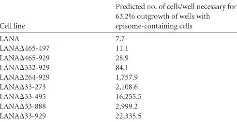
Alterna-tively, for limiting-dilution assays, cells were seeded in 96-well plates at 1,000/well, 100/well, 10/well, or 1/well in a medium containing G418. GraphPad Prism was used to perform nonlinear regression analyses with LANA limiting-dilution outgrowth values to compare episome mainte-nance efficiencies. Gardella analyses were performed on G418-resistant
clones byin situlysis of cells in gel-loading wells with protease (Sigma) and
sodium dodecyl sulfate (35), followed by electrophoresis in
Tris-borate-EDTA. DNA was transferred to a nylon membrane, and KSHV DNA was
detected using a32P-labeled TR probe. Signals were detected by
autora-diography.
GFP retention assay.Ten million BJAB cells alone or
LANA-express-ing BJAB cells in log-phase growth were transfected with 5⫻1010copies
(0.08 pmol) of plasmid p2TR-GFP (407 ng DNA) or p2TR-⌬RE-GFP
(467 ng DNA), using Amaxa Nucleofactor program O-17, in 150l of
solution V. After transfection, cells were seeded in 5 ml medium in a six-well plate. Eighteen to 20 h posttransfection, cells were sorted for GFP-positive cells (BD FACSAria sorter) and were suspended in RPMI
medium containing hygromycin B and Normocin (50g/ml; Invivogen)
at a concentration of 150,000 cells/ml (day zero). The cell concentration was measured daily by FACS for the first 5 days after sorting, and the percentages of GFP-positive cells were monitored daily and were plotted
over periods of 14 days for p2TR-GFP and 7 days for p2TR-⌬RE-GFP.
After reaching a concentration of⬃1⫻106/ml, cells were kept in
log-phase growth by reducing the concentration to⬃0.2⫻105/ml. Cell
con-centrations were measured by FACS using CountBright Absolute Count-ing Beads (Invitrogen).
Although all LANA mutant cell lines grew similarly, some small
on November 7, 2019 by guest
http://jvi.asm.org/
ferences in growth occurred after sorting. To compare the efficiencies of plasmid segregation of the different LANA mutants while accounting for differences in rates of cell growth, the percentages of GFP-positive cells were compared at a mutant-specific, plasmid-specific time point
(de-notedt*sp, where s is the mutant [strain] and p is the plasmid) at which the
cell concentration reachede1.5(4.48) times the concentration measured at
day 1 after cell seeding. The factore1.5was used because this was well
within the approximately linear part of the growth pattern for all trans-fected cells. To estimate the date posttransfection at which cells reached
e1.5times the concentration at day 1, growth curve models were fitted to
replicate data for each of the transfections. Repeated observations of GFP retention were made on replicates for each mutant, and quadratic logistic regression was used to model the relationship between the proportion of cells expressing GFP and the time that had elapsed from the initial trans-fection for each replicate transtrans-fection. For each mutant-plasmid
combi-nation, the estimated proportion of cells expressing GFP att*spfor
trans-fected cells was obtained using replicate-specific logistic regression parameter estimates. The distributions of these growth-corrected GFP expression rates are summarized in boxplots. Nonparametric tests of the null hypothesis that different LANA mutants have common medians were performed for 8 families of comparisons. For cell lines transfected
with p2TR-GFP, group A, composed of LANA, LANA⌬465-929, and
LANA⌬332-929, was compared to group B, composed of LANA⌬33-273,
LANA⌬33-495, LANA⌬33-888, LANA⌬33-929, and LANA⌬33-949;
group A was also compared to LANA⌬264-929. In addition, LANA⌬
264-929 was compared to group B and, individually, to LANA⌬33-495. The
same sets of comparisons were made for cell lines transfected with
p2TR-⌬RE-GFP. Holm’s procedure (36) for adjusting inferences for multiple
comparisons was used to calculatePvalues for the comparisons described
above (seeFig. 6). All computations for the GFP retention analyses were
performed using R, version 2.15.2.
RESULTS
LANA internal-deletion mutants associate with mitotic
chro-mosomes.
To persist stably in proliferating cells, KSHV episomes
must replicate with each cell division and must segregate
effi-ciently to daughter cell nuclei. The N- and C-terminal portions of
LANA are essential for these processes. In addition, internal
LANA sequences are critical for function, since deletion of the
internal sequences by fusion of the N- and C-terminal portions of
LANA results in highly deficient episome maintenance (
28
). To
investigate the roles of distinct LANA internal regions in episome
persistence, we generated a panel of LANA deletion mutants
termed
LANA
⌬
465-497,
LANA
⌬
33-495,
LANA
⌬
33-273,
LANA
⌬
465-929, LANA
⌬
332-929, and LANA
⌬
264-929 (
Fig. 1
).
Although all mutants contain the N- and C-terminal
chromo-some association regions, we wanted to confirm that each
associ-ates with mitotic chromosomes, since this function is essential for
DNA tethering and episome persistence. BJAB cells alone or
BJAB cells stably expressing LANA, LANA
⌬
465-497, LANA
⌬
33-495, LANA
⌬
33-273, LANA
⌬
465-929, LANA
⌬
332-929, or
LANA
⌬
264-929 (
Fig. 1
) were arrested in metaphase by overnight
treatment with colcemid, and the subcellular localization of
LANA was analyzed by confocal microscopy. LANA (green) was
detected using an antibody against LANA or against the T7 tag;
mitotic chromosomes were stained with propidium iodide (red).
As expected, no LANA staining was detected in BJAB cells (
Fig. 2a
and
f
). In agreement with previous results, LANA associated with
mitotic chromosomes (yellow, resulting from the colocalization
of green and red), with concentrations near telomeres and
centro-meres (
Fig. 2b
and
g
; arrows indicate peritelomeric staining, and
arrowheads
indicate
pericentromeric
staining)
(
28
,
37
).
LANA
⌬
465-497 (
Fig. 2c
and
h
), LANA
⌬
33-273 (
Fig. 2d
and
i
),
LANA
⌬
33-495 (
Fig. 2e
and
j
), LANA
⌬
465-929 (
Fig. 2k
),
LANA
⌬
332-929 (
Fig. 2l
), and LANA
⌬
264-929 (
Fig. 2m
)
associ-ated with mitotic chromosomes and also displayed preferential
localization near the centromeres and telomeres, like LANA. Since
the anti-LANA antibody detects the central repeat elements that
are deleted in LANA
⌬
465-929, LANA
⌬
332-929, and LANA
⌬
264-929, these mutants were detected only with an antibody to the
N-terminal T7 epitope tag. These data demonstrate that deletion
of the internal domains does not affect the ability of LANA to
interact with mitotic chromosomes.
LANA internal-deletion mutants bind TR DNA.
Since LANA
DNA binding is mediated through the C-terminal portion of
Episome Positive
Chr om.
Assoc . DNA
Binding DNAReplic.
5 13 996 1139
TR DNA binding Self-association Chrom. assoc
19/20 (95%)
+++
+
+
DEP Q
∆465-929
20/22 (91%)
++
+
NT P∆332-929
5 60 270 330 430 750 770 840 931 1162
13/33 (39%)
+
+
+
∆264-929 P∆33-929
+
+
+
3/44 (7%)*∆33-949
+
+
+
0/28 (0%)*22/23 (96%)
++++
+
+
∆465-497 P DE Q LZ EQE
14/44 (32%)
++
+
+
∆33-273 DE Q LZ EQE
34/34 (100%)
++++
+
+
LANA P DE Q LZ EQE
3/32 (9%)
+
+
+
∆33-495 Q LZ EQE
∆33-888
+
+
+
9/36 (25%)*Chrom. H2A/H2B
binding
33 331
Segregation
DNA Segreg
.
+++
+++
++
+
+
NT+
+++
+
+
Fold Deficiency
1.0
1.4
2904.0 389.9 2113.5 274.2 228.6 10.9 3.8
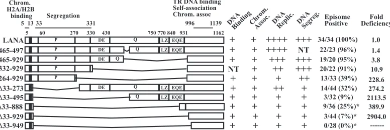
---FIG 1Schematic diagram of KSHV LANA and LANA deletion mutants. Indicated are the proline-rich region (P), the aspartate and glutamate (DE), glutamine (Q), and glutamate and glutamine (EQE) regions, and the putative leucine zipper (LZ). The DE, Q, EQE, and LZ regions all contain repeat elements. The shaded
area represents region I of the N-terminal nuclear localization signal (NLS) within amino acids 24 to 30 (20,69). The C-terminal portion of LANA can also
localize to nuclei, but an NLS has not been precisely mapped. Amino acids 5 to 13 mediate chromosome association through interaction with histones H2A/H2B. Amino acids 996 to 1139 contain TR DNA binding, self-association, and chromosome association functions. The capabilities for TR DNA binding, mitotic
chromosome association, DNA replication, DNA segregation (as suggested by retention of p2TR-⌬RE-GFP), and episome persistence for each of the constructs
are shown on the right. For episome persistence, fractions indicate the number of G418-resistant cell lines with episomes divided by the total number of G418-resistant cell lines assayed by Gardella analysis; percentages are given in parentheses. Fold deficiencies in episome maintenance ability were determined by
dividing the value for each mutant inTable 1by that for WT LANA. NT, not tested. LANA deletion mutants marked with asterisks, and their abilities to bind TR
DNA, associate with chromosomes, and maintain p8TR episomes, have been described previously (28), but their TR DNA replication and segregation capabilities
were further investigated here.
on November 7, 2019 by guest
http://jvi.asm.org/
[image:4.585.100.488.70.200.2]LANA, which is present in all mutants, we expected all LANA
deletion proteins to bind TR DNA. However, since LANA TR
DNA binding is essential for episome persistence (
14
), we wanted
to confirm DNA binding, and we assessed this by electrophoretic
mobility shift assays (EMSA). LANA or each deletion mutant was
translated
in vitro
and was incubated with a radiolabeled probe
containing the LANA TR binding site. Incubation of rabbit
reticu-locyte lysate (RRL) with TR DNA in the absence of LANA did not
shift the TR probe (
Fig. 3
, lanes 1 and 7). In agreement with
pre-vious results (
6
,
23
,
28
,
38
), incubation of wild-type (WT) LANA
with the TR probe resulted in two major complexes (
Fig. 3
, lanes 2
and 8, top two arrows). LANA
⌬
465-497 (
Fig. 3
, lane 3) and
LANA
⌬
33-273 (
Fig. 3
, lane 4) also generated two complexes.
LANA
⌬
33-495 (
Fig. 3
, lane 5) bound TR DNA and formed several
detectable complexes. LANA
⌬
465-929 bound TR DNA, but as a
single complex (
Fig. 3
, lane 6). Previous experiments had
indi-cated that LANA amino acids 900 to 929 are responsible for
gen-erating multiple EMSA complexes (
28
). LANA
⌬
264-929 (
Fig. 3
,
lane 9) also bound TR DNA in one predominant complex.
Therefore, all LANA deletion mutants retained the ability to bind
TR DNA.
Deletion of the internal repeat elements does not reduce the
half-life of LANA.
We investigated whether deletion of internal
regions reduces the half-life of LANA, since previous work
sug-gested that the repeat elements enhance the stability of LANA
(
39
). We assessed the half-life of LANA in KSHV-infected
BCBL-1 cells or after LANA expression in uninfected Loukes
cells. Treatment with cycloheximide and assessment by
immu-noblotting at serial time points demonstrated a
t
1/2of
⬃
9 to 12 h
in both BCBL-1 and Loukes cells (
Fig. 4A
). LANA
⌬
33-273,
LANA
⌬
33-495, LANA
⌬
465-929, LANA
⌬
332-929, LANA
⌬
264-929, LANA
⌬
33-888, LANA
⌬
33-929, and LANA
⌬
33-949 each had
a half-life similar to that of LANA (
Fig. 4A
). As a control for
effective arrest of protein synthesis by cycloheximide, we also
de-tected c-Myc, which had a
t
1/2of
⬃
1 to 3 h in the presence of
LANA expression and a
t
1/2of
⬃
0.5 to 1 h in the absence of LANA
(
Fig. 4B
). LANA is known to enhance the stability of c-Myc (
40
,
41
). Therefore, deletion of the internal LANA regions, including
the repeat elements, did not reduce the half-life of LANA.
The LANA deletion mutants are deficient in TR DNA
repli-cation.
LANA-mediated DNA replication is essential for KSHV
episome persistence. The C-terminal portion of LANA binds
di-rectly to KSHV TR DNA to mediate replication (
11–15
,
42
), and
the N-terminal chromosome binding region is also critical for
replication (
12
,
15
,
42
). In addition, we showed previously that
deletion of all internal LANA sequences reduced the efficiency of
DNA replication (
28
). Therefore, we wanted to assess the
contri-butions of specific internal LANA regions to DNA replication.
To assay for TR DNA replication, BJAB cells alone and BJAB
cells stably expressing LANA, LANA
⌬
465-497, LANA
⌬
33-273,
LANA
⌬
33-495, LANA
⌬
465-929, LANA
⌬
332-929, LANA
⌬
264-929, LANA
⌬
33-929, LANA
⌬
33-949, or LANA
⌬
33-888 (
Fig. 1
)
were transfected with p8TR-gB. LANA
⌬
33-929, LANA
⌬
33-949,
and LANA
⌬
33-888 are LANA deletion mutants lacking all
inter-nal sequences that were previously shown to have reduced TR
DNA replication by Southern blot assays for DpnI-resistant DNA
(
28
) and were included for comparison. The transfected plasmid
p8TR-gB is Dam-methylated DNA, purified from Dam
methy-lase-positive bacteria, and contains 8 copies of the KSHV TR
ele-ments. Dam methylation renders the DNA susceptible to DpnI
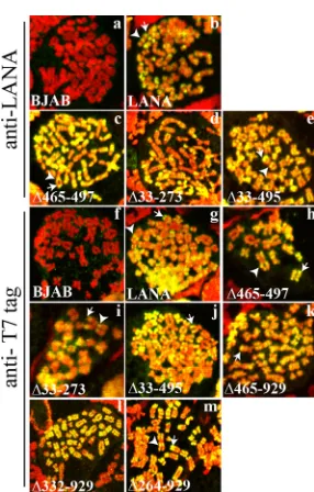
FIG 2LANA and LANA deletion mutants associate with mitoticchromo-somes, with preferential localization to areas near centromeres and telomeres. BJAB cells alone and BJAB cells stably expressing different LANA mutants were arrested in metaphase with colcemid and were analyzed for LANA localization by confocal microscopy. LANA (green) was detected with an antibody against LANA or against the T7 epitope tag. Chromosomes were counterstained with propidium iodide (red). The overlay of green and red results in yellow. White arrowheads and arrows indicate pericentromeric and peritelomeric localiza-tion of LANA, respectively. Brightness and contrast were similarly adjusted for the panels, and individual panels were uniformly adjusted using Adobe
Pho-toshop. Magnification,⫻630.
RRL LANA
∆465-497 ∆33-273 ∆33-495 ∆465-929
RRL LANA ∆264-929
1 2 3 4 5 6 7 8 9
free probe
O
FIG 3Deletion of internal regions of LANA does not abolish TR DNA binding as assessed by EMSA. A radiolabeled TR DNA probe was incubated with rabbit reticulocyte lysate (RRL) (lanes 1 and 7) or similar amounts (as detected by
Western blotting) ofin vitro-translated LANA (lanes 2 and 8), LANA⌬465-497
(lane 3), LANA⌬33-273 (lane 4), LANA⌬33-495 (lane 5), LANA⌬465-929
(lane 6), or LANA⌬264-929 (lane 9). After incubation, complexes were
re-solved on a nondenaturing polyacrylamide gel. Brightness and contrast were uniformly adjusted with Adobe Photoshop. All panels are from the same gel. Arrows indicate LANA-TR or mutated LANA-TR complexes. O, gel origin.
on November 7, 2019 by guest
http://jvi.asm.org/
[image:5.585.91.234.61.285.2] [image:5.585.362.480.66.264.2]digestion. Since mammalian cells lack Dam methylase, DNA that
undergoes replication after transfection is resistant to DpnI
diges-tion.
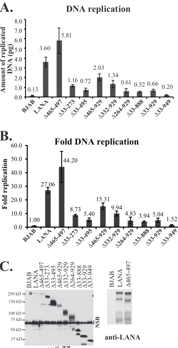
Figure 5
shows the normalized DNA replication (
Fig. 5A
) and
fold replication (
Fig. 5B
) of LANA and LANA mutants over that
for BJAB control cells. LANA efficiently mediated DNA
replica-tion at 27.06-fold over the control level, and LANA
⌬
465-497,
which contains only a 33-amino-acid deletion within the
glu-tamine-rich repeat region, also efficiently replicated DNA at
44.20-fold over the control level. In agreement with previous
re-sults using Southern blot detection of DpnI-resistant DNA (
28
)
that showed reductions in replication, deletion of all internal
se-quences in LANA
⌬
33-888, LANA
⌬
33-929, and LANA
⌬
33-949
resulted in dramatically reduced DNA replication, at only 3.94-,
5.04-, and 1.52-fold over the control level, ranging from 5.6% to
18.6% of the WT level.
Deletion of smaller internal regions resulted in different levels
of reduction in DNA replication. LANA
⌬
465-929, in which most
of the glutamine-rich internal repeat regions and all of the
pre-dicted leucine zipper are deleted, had 15.31-fold replication over
that of the control, a level modestly reduced at 56.7% of WT
rep-lication. LANA
⌬
332-929, which lacks all internal repeat elements,
had 9.94-fold replication over the control level, equivalent to
36.7% of the WT level. LANA
⌬
264-929, which lacks only 68
ad-ditional amino acids relative to LANA
⌬
332-929, replicated DNA
at only 4.83-fold over the control level, or 17.8% of the WT level,
suggesting that these additional amino acids are important for
replication. Deletion of unique sequence, including the
proline-rich region, in LANA
⌬
33-273 resulted in 8.73-fold replication, or
32.3% of WT LANA replication. Notably, LANA
⌬
33-495, which
lacks the unique internal sequence (LANA amino acids 33 to 331)
and some of the repeat elements, showed substantially reduced
replication at 5.40-fold over the control level, or 20.0% of the WT
level. The reductions in replication were not due to reduced
pro-tein expression, since all mutants were expressed at levels at least
equivalent to that of WT LANA (
Fig. 5B
). Therefore, deletions
within either the unique internal sequence of LANA or the repeat
regions resulted in reduced DNA replication.
The unique internal sequence of LANA is critical for the
re-tention of GFP expression from replication-deficient TR
epi-somes.
We wanted to assess the episome segregation of the LANA
mutants, since segregation of KSHV DNA to daughter nuclei
fol-lowing mitosis is essential for episome persistence; otherwise,
DNA is degraded. The N- and C-terminal portions of LANA,
which mediate chromosome attachment and KSHV DNA binding
to tether episomes to mitotic chromosomes, are necessary for this
process. However, the role of the internal LANA sequence is
un-known. Each KSHV TR contains two adjacent LANA binding sites
and an adjacent 32-bp replication element that is required for
LANA-mediated replication (
32
). We used GFP expression from
FIG 4Deletion of internal LANA regions does not reduce the half-life of LANA. Cells were treated with cycloheximide to arrest protein synthesis, and LANAlevels were assessed by immunoblotting. (A) Results for BCBL-1 cells (⬃1.5⫻105cells loaded per lane) and for Louckes cells (⬃3⫻105cells loaded per lane)
transfected with LANA, LANA⌬33-273, LANA⌬33-495, LANA⌬465-929, LANA⌬332-929, LANA⌬264-929, LANA⌬33-888, LANA⌬33-929, or LANA⌬33-949
are shown at time zero and at serial time points after cycloheximide arrest. Untransfected cells (not transf.) show a comigrating cross-reactive band with
LANA⌬264-929. LANA was detected with affinity-purified antibody directed against the C-terminal region of LANA. Tubulin immunoblots are shown below
each panel. (B) c-Myc was detected in the same cells as those used for panel A at time zero and at serial time points in order to assess the evidence for effective cycloheximide arrest. Results for untransfected cells are also shown. Tubulin blots are shown below each panel.
on November 7, 2019 by guest
http://jvi.asm.org/
[image:6.585.36.541.70.358.2]TR-containing DNA to monitor for the presence of episomes. To
investigate the abilities of the LANA deletion mutants to retain
GFP expression from TR DNA, BJAB cells alone or stably
express-ing LANA, LANA
⌬
465-929, LANA
⌬
332-929, LANA
⌬
264-929,
LANA
⌬
33-273, LANA
⌬
33-495, LANA
⌬
33-888, LANA
⌬
33-929,
[image:7.585.74.252.68.415.2]or LANA
⌬
33-949 were transfected with equimolar amounts of
p2TR-GFP or p2TR-
⌬
RE-GFP (kindly provided by Rolf Renne)
(
31
). p2TR-GFP contains two TR elements and a GFP expression
cassette. p2TR-
⌬
RE-GFP is identical to p2TR-GFP except that it
lacks the replication element (RE). Since p2TR-
⌬
RE-GFP retains
LANA binding sites, this plasmid can be used to assess the ability
of LANA to segregate TR episomes in the absence of replication.
Eighteen to 20 h after transfection with p2TR-GFP or p2TR-
⌬
RE-GFP, cells were sorted to collect GFP-positive cells and were
seeded at 1.5
⫻
10
5cells/ml. GFP expression and cell
concentra-tions were assessed daily.
Figure 6A
is a boxplot showing the percentages of GFP-positive
cells after transfection with p2TR-GFP, which is capable of both
LANA-mediated DNA replication and segregation. In the absence
of selection, TR DNA is expected to be lost from proliferating cells,
and LANA reduces the rate of this loss (
30
,
31
,
43
). To account for
slightly different growth rates after the sorting process, the
per-centages of GFP-positive cells were compared after a constant
amount of cell proliferation (e
1.5, or
⬃
4.48-fold) for each cell line,
which generally occurred
⬃
3 to 4 days postsorting and was well
within the approximate linear growth range for all cell lines. In the
absence of LANA, GFP expression was rapidly lost from nearly all
BJAB cells (
Fig. 6A
). In contrast,
⬎
80% of LANA-expressing cells
(median value) retained GFP. The abilities of LANA
⌬
33-273,
LANA
⌬
33-495, LANA
⌬
33-888, LANA
⌬
33-929, and LANA
⌬
33-949 to maintain p2TR-GFP were all substantially reduced from
that of LANA; for each of these mutants, fewer than 20% of cells
(median value) were GFP positive. Notably, LANA
⌬
465-929
maintained p2TR-GFP at nearly WT levels. For both LANA
⌬
332-929 and LANA
⌬
264-929, the ability to maintain p2TR-GFP was
reduced to intermediate levels, with about 45% and 30% of cells
expressing GFP. Holm’s procedure was used to adjust the
P
values
used to measure the statistical significance of tests comparing the
groups; the adjusted
P
values may be individually compared to a
significance threshold of 5%. Comparison of group A, composed
of LANA, LANA
⌬
465-929, and LANA
⌬
332-929, with group B,
which included LANA
⌬
33-273, LANA
⌬
33-495, LANA
⌬
33-888,
LANA
⌬
33-929, and LANA
⌬
33-949, yielded a
P
value of 2.68
⫻
10
⫺16; comparison of group A to LANA
⌬
264-929 yielded a
P
value of 1.57
⫻
10
⫺5.
P
values for the comparison of LANA
⌬
264-929 to group B and individually to LANA
⌬
33-495 were 2.24
⫻
10
⫺7and 1.17
⫻
10
⫺3, respectively (
Fig. 6A
).
In contrast to p2TR-GFP, transfection with p2TR-
⌬
RE-GFP
assesses the ability of LANA to retain TR DNA in the absence of
replication, since loss of the RE abolishes the ability of LANA to
replicate DNA. Therefore, GFP retention is expected to directly
reflect LANA-mediated segregation. As expected, nearly all BJAB
cells, which lack LANA, rapidly lost GFP expression (
Fig. 6B
). In
contrast, about 50% of cells expressing LANA expressed GFP.
No-tably, both LANA
⌬
465-929 and LANA
⌬
332-929 retained
p2TR-⌬
RE-GFP at wild-type levels. In contrast, as with p2TR-GFP,
LANA
⌬
33-273, LANA
⌬
33-495, LANA
⌬
33-888, LANA
⌬
33-929,
and LANA
⌬
33-949 were all highly deficient in the ability to retain
GFP expression (
Fig. 6B
), whereas LANA
⌬
264-929 had an
inter-mediate phenotype. Holm’s procedure was again used to adjust
P
values. Comparison of group A (LANA, LANA
⌬
465-929, and
LANA
⌬
332-929) with group B (LANA
⌬
33-273, LANA
⌬
33-495,
LANA
⌬
33-888, LANA
⌬
33-929, and LANA
⌬
33-949) resulted in a
P
value of 6.32
⫻
10
⫺16; comparison of group A with LANA
⌬
264-929 yielded a
P
value of 2.69
⫻
10
⫺5; comparison of LANA
⌬
264-1.00 27.06
44.20
8.73 5.40
15.31 9.94
4.83 3.94 5.041.52 0.0
10.0 20.0 30.0 40.0 50.0 60.0
Fold replication
Fold DNA replication
BJAB
∆33-495 ∆33-273 ∆465-497 LANA
∆332-929 ∆465-929 ∆33-88
8
∆264-929 ∆33-92 9
∆33-94 9
A.
B.
250 kD
150 kD
37 kD 50 kD 75 kD 100 kD
BJAB LANA ∆465-497 ∆33-273 ∆33-495 ∆465-929 ∆332-929 ∆264-929 ∆33-888 ∆33-929 ∆33-949
anti-T7
anti-LANA *
*
BJAB LANA ∆465-497
NSB
0.0 1.0 2.0 3.0 4.0 5.0 6.0 7.0 8.0
0.13 3.60
0.72 1.16 5.81
0.61 1.34
0.52 2.03
0.660.20
Amount of replicated
DNA (pg)
BJAB
∆264-929
∆33-495∆465-929∆332-929 ∆33-888∆33-929∆33-949 ∆33-27
3
∆465-497 LANA
DNA replication
C.
FIG 5Deletion of internal LANA regions results in decreased DNA replica-tion. (A and B) BJAB cells alone or BJAB cells stably expressing LANA or different LANA deletion mutants were transfected with p8TR-gB, which con-tains 8 copies of the TR. Hirt DNA was isolated at 24 h posttransfection for the assessment of transfection efficiencies and again at 72 h posttransfection for the assessment of replicated DNA. The amounts of p8TR-gB present in the Hirt DNA samples were quantified by real-time PCR. To normalize for trans-fection efficiencies, the amount of p8TR-gB detected in the samples at 24 h posttransfection was divided by the amount detected in BJAB control cells, and that result was termed the ratio of normalization. These values were close to 1. To normalize the amount of replicated p8TR-gB, the amount of replicated p8TR DNA detected at 72 h was divided by the corresponding ratio of normal-ization for each mutant. To calculate the fold p8TR-gB replication relative to the negative control (BJAB cells), the normalized replication of the samples was divided by the normalized replication detected for the BJAB control cells. The results represent averages for 4 experiments. (C) Western blotting of BJAB cells alone or BJAB cells expressing LANA or LANA mutants at the time of
DNA replication. (Left) Anti-T7 epitope blot. Approximately 3.5⫻105cells
were loaded per lane. NSB, nonspecific band. Asterisks indicate WT LANA and
full-length LANA⌬33-888 bands. (Right) Anti-LANA blot of LANA and
LANA⌬465-497. Approximately 1.5⫻105cells were loaded per lane.
Bright-ness and contrast within each panel were uniformly adjusted with Adobe Pho-toshop. Deletions of the central repeat regions result in increased LANA
ex-pression (28,39).
on November 7, 2019 by guest
http://jvi.asm.org/
929 with group B yielded a
P
value of 5.60
⫻
10
⫺6; and
compari-son of LANA
⌬
264-929 with LANA
⌬
33-495 yielded a
P
value of
1.75
⫻
10
⫺2(
Fig. 6B
). The reductions in LANA-mediated GFP
retention were not due to reduced protein expression, because all
mutants were expressed at levels higher than that of WT LANA
(
Fig. 6C
). It remains possible that increased expression of
LANA
⌬
465-929 or LANA
⌬
332-929 may be masking a defect for
these mutants, although this is unlikely, since the expression of
LANA
⌬
465-929 was only modestly higher than that of WT LANA.
These results indicate that the unique N-terminal LANA internal
sequence within amino acids 33 to 331, which are deleted in group
B, is critical for the retention of p2TR-
⌬
RE-GFP, consistent with
an important role in episome segregation, while the central repeat
elements (deleted in LANA
⌬
332-929 and LANA
⌬
465-929) are
dispensable for this function.
LANA amino acids 33 to 331 do not independently associate
with mitotic chromosomes.
Since LANA residues 33 to 331 were
implicated as critical for episome segregation, we assessed whether
this region could independently associate with mitotic
chromo-somes. GFP LANA 1-32, GFP LANA 1-331, and GFP LANA
33-331 each contain the designated LANA residues fused
down-stream of GFP. GFP LANA 1-32 contains LANA residues 5 to 13,
which bind histones H2A/H2B on the nucleosome surface to
me-diate chromosome association. As expected, GFP (green) did not
associate with mitotic chromosomes (red), while GFP LANA 1-32
(green) painted chromosomes (the overlay of green and red
gen-erates yellow) (
Fig. 6D
). GFP 1-331 also associated tightly with
chromosomes. GFP LANA 1-331
5GMR
7, which contains alanine
substitutions at the indicated residues that abolish interactions
with histone H2A/H2B and with chromosomes (
22
,
27
), did not
associate with chromosomes. GFP LANA 33-331 (green), which
lacks the histone-interacting LANA residues 5 to 13, also did not
associate with chromosomes (
Fig. 6D
). Therefore, LANA residues
33 to 331 do not independently associate with mitotic
chromo-somes.
Both the unique internal sequence and repeat elements have
roles in episome persistence.
We investigated the abilities of the
internal LANA deletion mutants to mediate episome persistence.
BJAB cells alone or expressing either LANA, LANA
⌬
465-497,
LANA
⌬
33-495, LANA
⌬
33-273, LANA
⌬
465-929, LANA
⌬
332-929, or LANA
⌬
264-929 were transfected with a plasmid
contain-ing 8 copies of the KSHV terminal repeats (p8TR), seeded at 1,000
cells/well into microtiter plates, and selected for G418 resistance
(which is encoded on the plasmid vector). After 7 to 8 days of
G418 selection,
⬎
94% of LANA and LANA
⌬
465-497 wells had
robust, G418-resistant outgrowth that was macroscopically visible
and could be expanded to 24-well plates. LANA
⌬
465-929 also had
G418-resistant growth in
⬎
94% of the wells, but the cells could
not be expanded to 24-well plates until
⬃
10 to 11 days of selection.
LANA
⌬
332-929 had somewhat less-robust G418-resistant
out-A.
B.
BJ
AB
∆
33−273
∆
33−495
∆
33−888
∆
33−929
∆
33−949
∆
264−929 ∆332−929 ∆465−929 LANA 0
20 40 60 80 100
2TR - GFP
% GFP positiv
e
p=1.57 x 10⁻⁵
p=2.68 x 10⁻¹⁶
p=1.17 x 10⁻³
p=2.24 x 10⁻⁷
BJ
AB
∆
33−273 ∆33−495 ∆33−888 ∆33−929 ∆33−949
∆
264−929
∆
332−929
∆
465−929 LANA 0
20 40 60 80 100
2TR∆RE - GFP
% GFP positiv
e
p=5.60 x 10⁻⁶
p=6.32 x 10⁻¹⁶
p=2.69 x 10⁻⁵
p=1.75 x 10⁻²
C.
BJAB LANA ∆33-273 ∆33-495 ∆465-929 ∆332-929 ∆264-929 ∆33-888 ∆33-929 ∆33-949 BJAB LANA
*
250 kD 150 kD
50 kD 37 kD 50 kD 75 kD 100 kD
Top: anti-T7 Bottom: anti-tubulin
anti-LANA
D.
GFP NLS
GFP LANA 1-331 GMR
GFP LANA 33-331 no GFP
GFP LANA 1-331 GFP
LANA 1-32
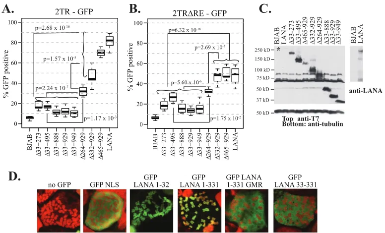
FIG 6The unique internal LANA sequence is important for the retention of GFP expression from episomal TR-containing DNA. (A) Ten million BJAB cells
alone or BJAB cells stably expressing LANA or the different LANA deletion mutants were transfected with 5⫻1010copies (5,000 copies per cell) of p2TR-GFP.
Eighteen to 20 h posttransfection, cells were sorted for GFP expression and were seeded at similar densities. GFP expression was monitored daily by FACS for 14
days. To account for some small differences in cell growth after sorting, GFP expression was compared at a time point when cells had reached a concentratione1.5
times that at day 1 of seeding.Pvalues for comparisons between the indicated groups are shown. (B) Cell lines were assessed as for panel A but were initially
transfected with p2TR-⌬RE-GFP, which is identical to p2TR-GFP except for deletion of the RE, abolishing LANA-mediated DNA replication. GFP expression
was monitored for 7 days. The results in panels A and B are each from six experiments. (C) Western blotting of BJAB cells alone or BJAB cells expressing LANA or LANA mutants at the time of DNA segregation. (Left) (Top) Anti-T7 epitope blot; (bottom) anti-tubulin blot. (Right) Anti-LANA blot. Brightness and
contrast were uniformly adjusted within each panel with Adobe Photoshop. Approximately 3.5⫻105cells were loaded per lane for the anti-T7 and anti-tubulin
blots, and⬃1.0⫻105cells were loaded per lane for the anti-LANA blot. (D) GFP NLS, GFP LANA 1-32, GFP LANA 1-331, GFP LANA 1-331 GMR, and GFP
LANA 33-331 were each expressed in BJAB cells. Cells were arrested in metaphase with colcemid. GFP is green, and chromosomes were counterstained with propidium iodide (red). The overlay of green and red generates yellow. Brightness and contrast in individual panels were uniformly adjusted using Adobe
Photoshop. Magnification,⫻630.
on November 7, 2019 by guest
http://jvi.asm.org/
[image:8.585.99.487.66.301.2]growth, in 90% of wells (average value), and cells could be
ex-panded at
⬃
11 to 12 days of selection. Like the negative-control
BJAB cells, LANA
⌬
33-495, LANA
⌬
33-273, and LANA
⌬
264-929
had G418-resistant outgrowth in fewer than 90% of wells and
could not be expanded until
⬃
12 to 14 days under G418 selection.
The lower G418-resistant outgrowth of BJAB cells than of
LANA-expressing cells is due to the need for integration of p8TR DNA,
which is a rare event compared with episome persistence. These
results were not due to lower transfection efficiencies as assessed
by GFP cotransfection. Therefore, these G418-resistant
out-growth results were consistent with the notion that LANA
⌬
465-929 has wild-type or only slightly reduced episome maintenance
efficiency, while that of LANA
⌬
332-929 may be modestly
reduced. In contrast, LANA
⌬
33-495, LANA
⌬
33-273, and
LANA
⌬
264-929 appeared either not to mediate episome
persis-tence or to do so with greatly reduced efficiency.
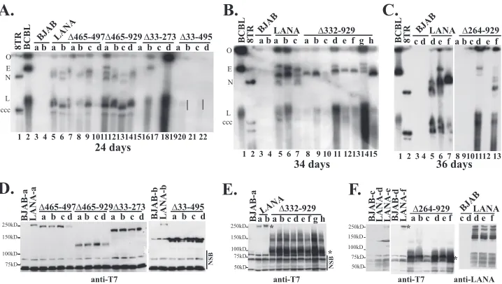
To detect the presence of p8TR episomes, Gardella gel analyses
were performed on G418-resistant cell lines. In Gardella gels, live
cells are loaded into the wells of an agarose gel and are lysed
in situ
in the presence of SDS and protease. During electrophoresis, large
chromosomal DNA remains at the gel origin, while
extrachromo-somal DNA (as large as several hundred kilobases) migrates into
the gel. DNA is then detected by Southern blotting by using a
radiolabeled probe. Analysis of BCBL-1 cells, a KSHV-infected
primary effusion lymphoma (PEL) cell line, results in a slowly
migrating band representing the viral episome and more quickly
migrating linear DNA that is due to lytic replication of the virus
(
Fig. 7A
, lane 2, and B and C, lanes 1). As expected, G418-resistant
BJAB cells lacking LANA did not have episomal DNA (
Fig. 7A
through C, lanes 3 and 4). In contrast, and in agreement with
previous results, BJAB cells expressing LANA (
Fig. 7A
, lanes 5 and
6, and B and C, lanes 5 to 7) or LANA
⌬
465-497 (
Fig. 7A
, lanes 7 to
10) all contained episomal DNA (
28
). As observed previously (
6
,
22
,
28
), much of the episomal DNA migrated more slowly than the
covalently closed circular (ccc) p8TR plasmid DNA. This slower
migration is due to duplication of the TR sequence and
multim-erization of input plasmids (
6
,
28
). Like LANA and LANA
⌬
465-497, LANA
⌬
465-929 (
Fig. 7A
, lanes 11 to 14) and LANA
⌬
332-929 (
Fig. 7B
, lanes 8 to 15) also efficiently maintained TR episomes
in all lanes. From a total of 11 experiments, LANA had episomes in
34/34 (100%) G418-resistant clones; from 4 experiments,
LANA
⌬
465-497 had episomes in 22/23 (96%); from 5
experi-ments, LANA
⌬
465-929 had episomes in 19/20 (95%); and from 3
experiments, LANA
⌬
332-929 had episomes in 20/22 (91%) (
Fig.
1
). Therefore, deletion of most of the glutamine-rich internal
re-peat regions and the predicted leucine zipper in LANA
⌬
465-929,
or deletion of all of the internal repeat sequence in LANA
⌬
332-929, had only had modest effects on the ability of LANA to
main-tain episomes in these assays.
In contrast, LANA
⌬
264-929, LANA
⌬
33-273, and LANA
⌬
33-495 were highly compromised for episome persistence. Gardella
analysis showed that LANA
⌬
33-273 (
Fig. 7A
, lanes 15 to 18) had
A.
D.
anti-T7
NSB
250kD
150kD
100kD
75kD
∆465-497∆465-929∆33-273 ∆33-495
BJAB-a LANA-aa b c d a b c d a b c d BJAB-b LANA-b a b c d
F.
E.
a b a b c d e f g h
BJAB-a LANA
∆332-929
250kD
150kD
100kD 75kD
50kD *
*
NSB
anti-T7
B.
C.
BCBL 8TR
BJABLANA ∆264-929 c d d e f a b c d e f
1 2 3 4 5 6 7 8 9101112 13
36 days
1 2 3 4 5 6 7 8 9 10 11 12131415
34 days
BCBL 8TR BJA
B
LANA ∆332-929
a b a b c a b c d e f g h O
ccc L E
N
BJABLANA
c d d e f
anti-LANA 250kD
150kD
100kD
75kD 50kD
BJAB-c LANA-d LANA-e
d-B
A
J
B LANA-f a b c d e f
∆264-929
anti-T7
* *
BCBL
8TR a b a b a b c d a b c d a b c d a b c d O
E
ccc
1 2 3 4 5 6 7 8 9 1011121314151617 181920 21 22
L
24 days
BJA B
LANA∆465-497∆465-929 ∆33-273 ∆33-495
N
FIG 7LANA mutants have differing levels of episome maintenance deficiencies as assessed by Gardella gel analyses. BJAB cells alone, or BJAB cells stably expressing LANA or LANA deletion mutants, were transfected with p8TR. Seventy-two hours posttransfection, cells were seeded in 96-well plates and were selected for G418 resistance. Gardella cell analysis was performed to detect p8TR episomes from G418-resistant cell lines expanded from the microtiter plates.
Approximately 1⫻106cells were loaded per lane in Gardella gels. (A) Gardella gel containing a naked p8TR plasmid (lane 1), BCBL-1 cells (a KSHV-infected
primary effusion lymphoma cell line) (lane 2), p8TR-transfected, G418-resistant BJAB cells (lanes 3 and 4), or BJAB cells stably expressing LANA (lanes 5 and-6),
LANA⌬465-497 (lanes 7 to 10), LANA⌬465-929 (lanes 11 to 14), LANA⌬33-273 (lanes 15 to 18), or LANA⌬33-495 (lanes 19 to 22). Vertical lines (lanes 20 and
22) indicate a faint episomal signal. The gel origin (O), BCBL-1 episomal (E) and linear (L) forms (linear due to lytic replication), and p8TR covalently closed circular (ccc) DNA and nicked (N) DNA are indicated. Gardella gel analysis was performed after 24 days of G418 selection. (B) Gardella gel with BCBL-1 cells (lane 1), naked p8TR plasmid (lane 2), p8TR-transfected, G418-resistant BJAB cells (lanes 3 and 4), or BJAB cells stably expressing LANA (lanes 5 to 7) or
LANA⌬332-929 (lanes 8 to 15). Gardella gel analysis was performed after 34 days of G418 selection. (C) Gardella gel with BCBL-1 cells (lane 1), naked p8TR
plasmid (lane 2), p8TR-transfected, G418-resistant BJAB cells (lanes 3 and 4), or BJAB cells stably expressing LANA (lanes 5 to 7) or LANA⌬264-929 (lanes 8 to
13). Gardella gel analysis was performed after 36 days of selection. (D to F) Immunoblot analyses for LANA or LANA mutants expressed in G418-resistant cell lines used in Gardella gel analyses. Panels D to F correspond to panels A to C, respectively. LANA was detected using an anti-T7 monoclonal antibody or an
anti-LANA antibody. Approximately 3.5⫻105cells were loaded per lane for anti-T7 blots and⬃1.5⫻105cells per lane for the anti-LANA blot. Asterisks indicate
LANA⌬332-929 or LANA bands. Brightness and contrast in individual panels were uniformly adjusted using Adobe Photoshop. Lowercase letters indicate
individually selected G418-resistant cell lines. NSB, nonspecific bands.
on November 7, 2019 by guest
http://jvi.asm.org/
[image:9.585.114.473.67.270.2]episomes in only two of four lanes, LANA
⌬
264-929 (
Fig. 7C
, lanes
8 to 13) had episomes in only two of the six lanes, and LANA
⌬
33-495 (
Fig. 7A
, lanes 19 to 22) had only very faint signals in two of
four lanes. Overall, from 7 experiments, LANA
⌬
264-929 had
epi-somes in 13/33 G418-resistant cell lines (39%); from 8
experi-ments, LANA
⌬
33-273 had episomes in 14/44 (32%); and from 4
experiments, LANA
⌬
33-495 had episomes in 3/32 (9%) (
Fig. 1
).
All mutants were expressed at levels at least as high as that of
LANA (
Fig. 7D
to
F
), so deficits were not due to reduced protein
expression. Protein expression levels were highest for LANA
⌬
264-929 and LANA
⌬
332-929 (
Fig. 7E
and
F
). This is due to the
dele-tion of the internal repeat regions, which exert inhibitory effects
on LANA expression (
39
). Therefore, the lower levels of episome
persistence for LANA
⌬
264-929, LANA
⌬
33-273, and LANA
⌬
33-495 were not due to decreased protein expression. These results
show that LANA mutants with deletions that included the unique
internal sequence (LANA amino acids 33 to 331) had the largest
episome maintenance deficits.
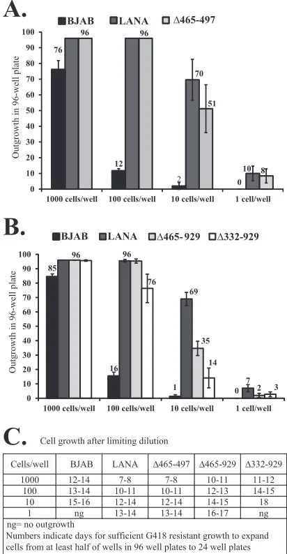
Assessment of the episome maintenance deficiencies of
LANA⌬332-929 and LANA⌬465-929 in limiting-dilution
as-says.
Although both LANA
⌬
465-929 and LANA
⌬
332-929
medi-ated long-term episome maintenance of TR DNA and maintained
episomes in 95% and 91% of G418-resistant cell lines (
Fig. 1
),
respectively, the G418-resistant outgrowth of both was delayed by
several days relative to that of LANA after plating at 1,000 cells/
well, as described above. In contrast, LANA
⌬
465-497 did not
ex-hibit delayed outgrowth. These differences may be due to fewer
G418-resistant LANA
⌬
465-929 and LANA
⌬
332-929
episome-containing cells in each well with outgrowth, resulting in delayed
G418-resistant outgrowth. To further investigate these
differ-ences, we performed limiting-dilution assays with
p8TR-trans-fected cells.
First, we assessed LANA
⌬
465-497 as a control. BJAB cells alone
or BJAB cells stably expressing LANA or LANA
⌬
465-497 were
transfected with p8TR, seeded in 96-well plates at 1,000, 100, 10,
and 1 cell/well, and selected for G418 resistance (
Fig. 8A
). Cells
expressing LANA and LANA
⌬
465-497 had 100% well outgrowth
when seeded at 1,000 cells/well and 100 cells/well. When seeded at
10 cells/well or 1 cell/well, LANA had G418-resistant outgrowth in
70 and 10 wells on average, respectively, while LANA
⌬
465-497
had outgrowth in 51 and 8 wells, respectively. In contrast, BJAB
cells lacking LANA had much lower G418-resistant outgrowth,
which occurred in 76, 12, 2, and 0 wells after seeding at 1,000, 100,
10, and 1 cell/well, respectively. Similarly, the number of days
necessary for cells to expand sufficiently for transfer from 96-well
to 24-well plates was the same for LANA and LANA
⌬
465-497 but
was significantly higher for BJAB cells (
Fig. 8C
). Therefore, LANA
and LANA
⌬
465-497 showed similar abilities to mediate
G418-resistant outgrowth after limiting dilution, while BJAB cells,
which lack LANA and rely on the integration of p8TR for G418
resistance, were much less efficient.
We then performed limiting-dilution experiments after
trans-fection of BJAB cells alone or BJAB cells expressing LANA,
LANA
⌬
465-929, or LANA
⌬
332-929. In results similar to those
for the previous experiment, after transfection with p8TR, LANA
showed G418-resistant outgrowth in 96, 96, 69, and 7 wells on
average after seeding at 1,000, 100, 10, and 1 cell/well (
Fig. 8B
).
LANA
⌬
465-929 showed slightly reduced G418-resistant
out-growth, in 96, 96, 35, and 2 cells/well after seeding at 1,000, 100,
10, and 1 cell/well. The outgrowth of LANA
⌬
332-929 was further
reduced, occurring in 96, 76, 14, and 3 cells/well after seeding at
1,000, 100, 10, and 1 cell/well (
Fig. 8B
). The reduced outgrowth of
LANA
⌬
465-929 and LANA
⌬
332-929 relative to that of LANA was
also reflected in the number of days necessary for cells to expand
sufficiently for transfer from 96-well plates to 24-well plates (
Fig.
8C
). The reductions in G418-resistant outgrowth for LANA
⌬
465-929 and LANA
⌬
332-929 were not due to differences in
transfec-tion efficiencies as assessed by GFP cotransfectransfec-tion. Therefore,
LANA
⌬
465-929 was modestly less efficient than LANA, and
85
16
1 0
96
69
7 96
35
2 76
14
3 0
10 20 30 40 50 60 70 80 90 100
1000 cells/well 100 cells/well 10 cells/well 1 cell/well
Outgrowth in 96-well plate
BJAB LANA ∆465- 929 ∆332-929
Cell growth after limiting dilution
Cells/well BJAB LANA ∆465-497 ∆465-929 ∆332-929
1000 12-14 7-8 7-8 10-11 11-12 100 13-14 10-11 10-11 12-13 14-15
10 15-16 12-14 12-14 14-15 18 1 ng 13-14 13-14 16-17 ng ng= no outgrowth
Numbers indicate days for sufficient G418 resistant growth to expand cells from at least half of wells in 96 well plates to 24 well plates
76
12
2 0
70
10
96 96
51
8
0 10 20 30 40 50 60 70 80 90 100
1000 cells/well 100 cells/well 10 cells/well 1 cell/well
Outgrowth in 96-well plate
BJAB LANA ∆465-497
A.
B.
C.
FIG 8LANA⌬465-929 and LANA⌬332-929 exhibit deficiencies in limiting-dilution outgrowth after TR DNA transfection and G418 selection. (A) BJAB
cells alone and BJAB cells stably expressing LANA or LANA⌬465-497 were
transfected with plasmid p8TR and were seeded in 96-well plates at different cell concentrations