Virus
p36 Replicase-Associated Protein Interacts with the Host Cell
ESCRT-I Component Vps23
Lynn G. L. Richardson,a*Eric A. Clendening,aHyukho Sheen,bSatinder K. Gidda,aK. Andrew White,bRobert T. Mullena
Department of Molecular and Cellular Biology, University of Guelph, Guelph, Ontario, Canadaa
; Department of Biology, York University, Toronto, Ontario, Canadab
ABSTRACT
Like most positive-strand RNA viruses, infection by plant tombusviruses results in extensive rearrangement of specific host cell
organelle membranes that serve as the sites of viral replication. The tombusvirusTomato bushy stunt virus(TBSV) replicates
within spherules derived from the peroxisomal boundary membrane, a process that involves the coordinated action of various viral and cellular factors, including constituents of the endosomal sorting complex required for transport (ESCRT). ESCRT is comprised of a series of protein subcomplexes (i.e., ESCRT-0 -I, -II, and -III) that normally participate in late endosome biogene-sis and some of which are also hijacked by certain enveloped retroviruses (e.g., HIV) for viral budding from the plasma
mem-brane. Here we show that the replication ofCarnation Italian ringspot virus(CIRV), a tombusvirus that replicates at
mitochon-drial membranes also relies on ESCRT. In plant cells, CIRV recruits the ESCRT-I protein, Vps23, to mitochondria through an interaction that involves a unique region in the N terminus of the p36 replicase-associated protein that is not conserved in TBSV or other peroxisome-targeted tombusviruses. The interaction between p36 and Vps23 also involves the Vps23 C-terminal steadi-ness box domain and not its N-terminal ubiquitin E2 variant domain, which in the case of TBSV (and enveloped retroviruses) mediates the interaction with ESCRT. Overall, these results provide evidence that CIRV uses a unique N-terminal sequence for the recruitment of Vps23 that is distinct from those used by TBSV and certain mammalian viruses for ESCRT recruitment. Char-acterization of this novel interaction with Vps23 contributes to our understanding of how CIRV may have evolved to exploit key differences in the plant ESCRT machinery.
IMPORTANCE
Positive-strand RNA viruses replicate their genomes in association with specific host cell membranes. To accomplish this, cellu-lar components responsible for membrane biogenesis and modeling are appropriated by viral proteins and redirected to assem-ble membrane-bound viral replicase complexes. The diverse pathways leading to the formation of these replication structures are poorly understood. We have determined that the cellular ESCRT system that is normally responsible for mediating late
en-dosome biogenesis is also involved in the replication of the tombusvirusCarnation Italian ringspot virus(CIRV) at
mitochon-dria. Notably, CIRV recruits ESCRT to the mitochondrial outer membrane via an interaction between a unique motif in the viral protein p36 and the ESCRT component Vps23. Our findings provide new insights into tombusvirus replication and the virus-induced remodeling of plant intracellular membranes, as well as normal ESCRT assembly in plants.
T
ombusviruses are positive-strand RNA [(⫹)RNA] viruses thatinfect a wide range of plant species and replicate at host cell membranes derived specifically from either peroxisomes (e.g.,
Tomato bushy stunt virus[TBSV]) or mitochondria (e.g., Carna-tion Italian ringspot virus[CIRV]) (1). Upon infection and de-pending on the tombusvirus, the peroxisomal or mitochondrial (outer) membranes progressively proliferate and invaginate, re-sulting in the formation of hundreds of spherules that serve to concentrate viral and host cell factors required for synthesis of the viral RNA genome and to protect nascent viral RNAs from
degra-dation by host cell defenses (2,3). Concomitant with these
mor-phological changes, the modified organelles also form large ap-pendages and coalesce, yielding aggregated structures that no longer resemble the organelles from which they were derived
(1,4).
The morphological transformation of peroxisomes or mito-chondria in tombusvirus-infected cells involves two viral replica-tion proteins: an auxiliary viral RNA-binding protein and an RNA-dependent RNA polymerase, referred to as p33 and p92,
respectively, in TBSV, or p36 and p95, respectively, in CIRV (5).
Both sets of replicase proteins are essential for viral genome
rep-lication (6, 7) and are encoded by overlapping open reading
frames (ORFs), and p92 and p95 are products of translational read-through of an amber stop codon in p33 and p36, respectively
(8,9). Consequently, the N-terminal portion of p92/p95 is
iden-tical to p33/p36. Both sets of replicase proteins are also integral membrane proteins, each possessing two transmembrane do-mains (TMDs), as well as unique targeting signals that mediate
Received27 December 2013Accepted18 March 2014
Published ahead of print26 March 2014
Editor:A. Simon
Address correspondence to Robert T. Mullen, [email protected].
* Present address: Lynn G. L. Richardson, Department of Biochemistry and Molecular Biology, University of Massachusetts, Amherst, Massachusetts, USA.
Copyright © 2014, American Society for Microbiology. All Rights Reserved.
doi:10.1128/JVI.03840-13
on November 7, 2019 by guest
http://jvi.asm.org/
their specific sorting to either peroxisomes or mitochondria (4,
10,11) and thus dictate the intracellular site for viral replication.
Numerous host cell factors involved in tombusvirus replica-tion have been identified as part of several large-scale genomic and
proteomic studies performed with TBSV and Saccharomyces
cerevisiaeas a model host (12). Among these factors are several components of endosomal sorting complex required for transport
(ESCRT). ESCRT is a network of⬃20 soluble proteins that, in
noninfected cells, are sequentially recruited from the cytosol and assembled into several multiprotein subcomplexes (ESCRT-0, -I, -II, and -III) at the late endosomal surface, where they participate in sorting of ubiquitinated membrane-bound cargo proteins into intraluminal vesicles derived from the endosomal boundary membrane during multivesicular body (MVB) biogenesis. Ac-cording to models based primarily on studies with yeasts and
mammalian cells (13), ESCRT assembly begins with ESCRT-0
rec-ognizing ubiquitinated cargo in the endosomal membrane and recruiting ESCRT-I to the endosomal surface. ESCRT-I also partic-ipates in ubiquitinated cargo sorting and recruits ESCRT-II, which subsequently recruits ESCRT-III, which forms polymeric filaments that drive membrane vesiculation. Thereafter, the AAA-ATPase Vps4 (vacuolar protein sorting 4) catalyzes the disassembly of ESCRT-III, a process that is coupled with membrane fission and results in the re-cycling of ESCRT(III) subunits to the cytosol, while the mature MVB fuses with the lysosome/vacuole, where its contents are degraded. Interestingly, while the ESCRT machinery and its interaction net-work are relatively well conserved and have similar cellular functions
in plants compared to yeasts and mammals (14,15), some key
differ-ences exist, which are not well understood. For example, homologs of
mammalian and yeast ESCRT-0 do not exist in plants (16), and
in-stead, recent work has identified the TOL family of proteins to be involved in early recognition of ubiquitinated cargo and their sorting
to MVBs (17). However, the relationship between cargo recognition
by TOL proteins and subsequent ESCRT recruitment and assembly has yet to be investigated.
In addition to MVB biogenesis, certain ESCRT components
also participate in other important cellular processes involving membrane deformation, including scission of the midbody
dur-ing cytokinesis or budddur-ing of enveloped retroviruses (18,19). In
cells infected with human immunodeficiency virus (HIV), for ex-ample, ESCRT is initially redirected or “hijacked” to the plasma membrane via interactions between peptide late-domain motifs in the HIV structural protein, Gag p6, and the ESCRT-I protein
Tsg101 and/or the ESCRT accessory protein Alix (20). Among the
most common and best characterized of these late-domain motifs is a proline-rich sequence (i.e., P[S/T]AP), which interacts with the ubiquitin E2 variant (UEV) domain of Tsg101 via a binding mechanism that mimics the interaction of certain ESCRT-0
pro-teins with Tsg101 (21,22).
Analogous to retroviral budding, the proposed model for the role of ESCRT during TBSV infection is that ESCRT is recruited to the surface of the peroxisome to facilitate invagination of the per-oxisomal membrane, as well as to concentrate and assemble the
viral replicase complexes within nascent spherules (23). Evidence
in support of this model includes the identification of several ESCRT proteins in a genome-wide screen for factors involved in
TBSV replication in yeast (24) and the inhibition of TBSV
repli-case activity in yeast and plant cells upon overexpression of
mu-tant versions of various ESCRT proteins (23). In addition,
coex-pression of the TBSV replicase protein p33 and the yeast ortholog of mammalian Tsg101, Vps23p, in yeast cells results in the
relo-calization of Vps23p to peroxisomes (23). TBSV p33 also interacts
with yeast Vps23p in a manner that depends on the N-terminal UEV domain of Vps23p and two (mono)ubiquitinated lysine res-idues in p33, as well as a peptide sequence in p33 (i.e., PSVP) that
resembles the PSAP late-domain motif of HIV Gag p6 (25).
Given the ultrastructural similarities of the modified peroxi-somal and mitochondrial membranes in TBSV- and CIRV-in-fected cells, respectively, as well as the ability of chimeric versions of these two viruses to produce the corresponding
organelle-spe-cific membrane rearrangements (5, 11, 26), it seems likely that
CIRV relies on ESCRT in a manner similar to TBSV. However,
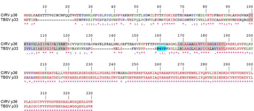
FIG 1Alignment of the deduced amino acid sequences of CIRV p36 and TBSV p33. Sequences were obtained from GenBank (accession numbersCAA59477.2 andNP_062898.1) and aligned using ClustalW. Identical and similar amino acids in each protein are colored red and green or blue, respectively, and indicated also with asterisks and colons or periods, respectively. The numbers represent specific amino acid residues in full-length p36 (330 residues). Putative TMDs (shown on a gray background) were determined using TOPCONS and visual inspection, and the late-domain-like motif identified in the intervening loop sequence of p33 (23) but absent in p36 is shown on a bright blue background.
Richardson et al.
on November 7, 2019 by guest
http://jvi.asm.org/
[image:2.585.75.509.59.249.2]CIRV p36 does not possess a late-domain-like motif resembling
that identified in TBSV p33 (Fig. 1) (23) (and in mammalian
en-veloped retroviruses [20]), implying that CIRV uses a distinct
mechanism to recruit ESCRT to mitochondrial membranes. Con-sistent with this premise, in this study, we show that CIRV p36 binds to and recruits Vps23 to mitochondria in plant cells via a unique N-terminal sequence in p36 that is not present in TBSV p33. We show also that, in contrast to TBSV p33 (and mammalian enveloped retroviruses), the interaction between p36 and Vps23 does not require the UEV domain of Vps23 but, instead, requires the C-terminal steadiness box domain (StBox) of Vps23, which in yeasts and mammals is important for the assembly of ESCRT-I (27–30).
Collectively, our results highlight a unique mechanism for ESCRT recruitment by CIRV that utilizes a Vps23-interacting motif that appears to be distinct from other viruses that co-opt ESCRT, such as TBSV and HIV. This novel mechanism may be reflective of the absence of an ESCRT-0 complex, which raises new mechanistic questions about general features of ESCRT assembly in plants.
MATERIALS AND METHODS
Recombinant DNA procedures and reagents.Molecular biology re-agents were purchased either from New England BioLabs, Promega, PerkinElmer Life Sciences Inc., Stratagene, or Invitrogen. Custom oligo-nucleotides were synthesized by Sigma-Aldrich Ltd. All DNA constructs were verified using automated sequencing performed at the University of Guelph Genomics Facility. PCR-based mutagenesis was carried out using the QuikChange site-directed mutagenesis kit (Stratagene).
Plasmid construction.The majority of the plasmids used in this study have been described previously, including the following binary plasmids used inAgrobacteriuminfiltration and rub inoculation ofNicotiana ben-thamianaleaves: pHST20/TBSV and pHST20/CIRV, encoding the entire TBSV and CIRV genomes, respectively (8,31); pTRV1 and pTRV2, en-coding RNA1 and RNA2 of the bipartiteTobacco rattle virus(TRV) ge-nome (32), which were obtained from theArabidopsisBiological Resource Center (ABRC); pMO4-AtSKD1[E232Q], encoding the green fluorescent protein (GFP) fused to the N terminus of a mutant version (i.e., a glutamic acid at position 232 replaced with glutamine) ofArabidopsis thalianaVps4 (also referred to as SKD1 [33]), which was kindly provided by M. Otegui (University of Wisconsin); and pRCS2, serving as an “empty” binary vec-tor control (34). All binary plasmids, as well as all other plant expression plasmids based on pRTL2, pSAT, and pUC18 vectors (see below), contain the cauliflower mosaic virus 35S promoter (CaMV 35S).
Plant expression plasmids used in transient transformations of Nico-tiana tabacumBright Yellow-2 (BY-2) cells include the following plasmids that have been described previously (11,35): pRTL2/p36, encoding CIRV p36; pRTL2/Myc-p36, encoding p36 with an N-terminal Myc epitope tag; pRTL2/p95, encoding CIRV p95, in which the p36 amber stop codon was mutated to a tyrosine codon; pRTL2/Rep, encoding the overlapping CIRV open reading frame 1 (ORF1) and ORF2 that encode both p36 and p95; p361-90-CAT and p3690-190-CAT, encoding the N-terminal 90 or 90 to 190 amino acid residues of p36 fused to the N terminus of chloramphenicol acetyltransferase (CAT), respectively; pRTL2/Myc-Vps23, pRTL2/GFP-Vps23, pUC18/Vps23-GFP, and pRTL2/HA-pRTL2/GFP-Vps23, encoding either the Myc or hemagglutinin (HA) epitope tag or the green fluorescent protein fused to the N or C terminus of isoform A ofArabidopsisVps23 (referred to as Vps23 in this study); pRLT2/Myc-Vps28 and pRTL2/Myc-Vps25, encoding N-terminal Myc-tagged Arabidopsis Vps28 isoform A and ArabidopsisVps25, respectively. Other previously described plant expres-sion vectors include the following plasmids: pRTL2/RFP, encoding the red fluorescent protein (RFP) (36); Syp21 and pUC18/GFP-Syp52, which encode GFP fused to the N termini of theArabidopsis mem-brane-bound Qa-SNAREs (solubleN-ethylmaleimide-sensitive factor
at-tachment protein receptors) Syp21 (Syntaxin of plants 21) and Syp52 (37) proteins (kindly provided by M. Sato [Kyoto University]); pSAT2/Cherry-PTS1, encoding the monomeric cherry fluorescent protein fused to the C-terminal 10 amino acid residues of pumpkin hydroxypyruvate reduc-tase, including its type 1 peroxisomal targeting signal (38); pRTL2/TIC40-RFP, encoding theArabidopsis40-kDa component of the translocon at the inner membrane of chloroplasts fused to the N terminus of RFP (39); and pSAT4A/AtPAP26-mCherry, encoding theArabidopsispurple acid phos-phatase isoform 26 fused to the N terminus of the monomeric cherry fluorescent protein (40). Yeast two-hybrid expression vectors, pGADT7 and pGBKT7 (Clontech) encodingArabidopsisVps23, Vps25, Vps37, and Vps28 were also previously described (35).
Plasmids used for bimolecular fluorescence complementation (BiFC) as-says were based on pSAT4/nEYFP and pSAT4/cEFYP, which encode the N-terminal and C-N-terminal halves of the enhanced yellow fluorescent protein (nEYFP and cEYFP), respectively (kindly provided by S. Gelvin [Purdue Uni-versity]) (41). Briefly, pSAT4/p36-cYFP, p3691-330-cYFP, and pSAT4/nYFP-Vps23 were constructed by PCR amplifying the p36 or pSAT4/nYFP-Vps23 open reading frames (ORFs), or portions thereof, and cloned into pSAT4/nEFYP or pSAT4/cEFYP. Complete details on the oligonucleotide primers used for gen-erating these and other plasmids are available upon request.
Plant expression vectors encoding modified (truncated) versions of p36 (i.e., p3691-330, p3668-330, and p3623-330), Vps23 (GFP-UEV, Myc-CC⫹StBox [CC stands for coiled-coil, and StBox stands for steadiness box], Myc-CC, Myc-StBox, and Myc-UEV⫹CC) were generated by PCR amplification of the sequence corresponding to the indicated proteins or protein domains and ligated into either pRTL2/MCS (containing a mul-tiple cloning site [MCS]) (42), pRTL2/Myc-MCS (encoding an initiation methionine, followed by the Myc epitope tag and then an MCS), pRTL2/ GFP-MCS (encoding GFP followed by an MCS) (14), or pUC18/NheI-GFP (encoding pUC18/NheI-GFP with a 5=unique NheI restriction site) (43). Similarly, yeast two-hybrid expression vectors encoding p36 or p361-90or modified (truncated) versions of Vps23 (UEV, CC⫹StBox, CC, StBox, and UEV⫹CC) were generated by PCR amplification and subcloned into pGBKT7 (encoding the GAL4 DNA-binding domain [BD] and a Myc epitope tag, followed by an MCS) or pGADT7 (encoding the GAL4 acti-vation domain [AD] and an HA epitope tag, followed by an MCS), respec-tively.
For construction of pRTL2/Myc-NtVps23, the 3=sequence ofVPS23 fromNicotiana tabacumwas obtained by 3=rapid amplification of cDNA ends (RACE)-PCR using cDNA synthesized fromN. tabacumsuspension cell mRNA, and a gene-specific forward primer based on the availableN. tabacum VPS23expressed sequence tag (EST) (NCBI accession number EB680173). Subsequently, full-lengthNtVPS23was amplified from BY-2 cDNA using gene-specific primers and ligated into pRTL2/Myc-MCS (via pCR2.1TOPO serving as a shuttle vector), yielding pRTL2/Myc-NtVps23. pRTL2/Myc-NtVps23 was then used as the template DNA for construct-ing pRTL2-Myc/NtUEV, which includes the entire UEV domain ofN. tabacumVps23.
pRTL2/p36⌬7-22 was constructed using PCR-based site-directed mu-tagenesis, whereby sequences encoding amino acid residues 7 to 22 in the p36 ORF were deleted using the appropriate forward and reverse muta-genic primers and pRTL2/p36 as the template DNA. pRTL2/p331-74 -p3691-330and pRTL2/p361-28-p3313-74-p3691-330were constructed in the following manner. First, sequences encoding the N-terminal 74 amino acid residues of p33 were amplified from pRTL2/p33 (4), and the resulting PCR products were ligated into pRTL2/p3691-330, yielding pRTL2/ p331-74-p3691-330. Next, sequences encoding the N-terminal 12 amino acid residues of p33 were replaced (via successive site-directed mutagen-esis reactions and pRTL2/p331-74-p3691-330 as the initial template DNA) with sequences encoding the N-terminal 28 residues of p36, yielding pRTL2/p361-28-p3313-74-p3691-330. pRTL2/Myc-p361-90-TraB and pRTL2/Myc-p361-28-TraB, encoding an N-terminal Myc epitope tag fused to the N-terminal 90 or 28 amino acids of p36 followed by the C-terminal 34 or 75 amino acid residues ofArabidopsisTraB (At1g05270),
on November 7, 2019 by guest
http://jvi.asm.org/
respectively, were constructed by first introducing (using PCR-based site-directed mutagenesis and pRTL2/Myc-TraB as the template DNA) an NheI site immediately upstream of either codon 338 or codon 297 (of 371) in the TraB ORF. The modified plasmids were then digested with NheI (removing sequences coding for residues 1 to 337 or 1 to 296 of TraB and the N-terminal Myc epitope tag) followed by ligation with NheI-digested PCR products encoding the N-terminal 90 or 28 residues of p36, along with an appended Myc epitope tag from pRTL2/Myc-p36 (see above). pRTL2/Myc-TraB was constructed by ligating the full-length TraB ORF (cDNA provided by the ABRC) into pRTL2/Myc-MCS.
Agrobacteriuminfiltration and rub inoculation ofN. benthamiana and RNA gel blot analysis.N. benthamianaplants were grown in cham-bers at 21°C with a 16-h/8-h light/dark cycle. The leaves of approximately 3-week-old plants were infiltrated or, for experiments involving TRV, triple infiltrated with cultures ofAgrobacterium tumefaciens(LBA4404) carrying the appropriate binary vectors. Procedures involving Agrobacte-riumhave been described previously (4). Rub inoculations were per-formed 2 days after infiltration with 10g of plasmid DNA encoding full-length infectious TBSV or CIRV cDNA diluted in 30l of RNA inoc-ulation buffer (11,44). Approximately 4 to 6 days after inoculation or, for experiments involving TRV, 2 or 4 days after infiltration, the leaves were flash frozen in liquid nitrogen followed by total RNA extraction (45). Aliquots of isolated RNA were separated in nondenaturing 1.2% (wt/vol) agarose gels, and viral RNAs were detected by electrophoretic transfer to nylon (Hybond-N; Amersham Biosciences) followed by incubation with 32P-end-labeled oligonucleotide probes complementary to the CIRV and TBSV genome or TRV RNA1 genome (45,46). Complete details of oligo-nucleotides used for RNA gel blot analysis are available upon request. Labeled RNAs were visualized using a phosphorimager. Results presented are representative of at least two separate experiments.
Biolistic bombardment and fluorescence microscopy of BY-2 cells. N. tabacumBright Yellow-2 suspension cell cultures were maintained and prepared for biolistic bombardment with a Biolistic PDS-1000/He parti-cle delivery system (Bio-Rad Laboratories) as described previously (47). Bombarded cells were incubated for⬃4 to 8 h to allow for expression and sorting of the introduced gene product(s). Cells were fixed in 4% (wt/vol) formaldehyde, followed by permeabilization with 0.01% (wt/vol) pec-tolyase Y-23 (Kyowa Chemical Products) and either 0.3% (vol/vol) Triton X-100, which permeabilizes the plasma membrane and all organellar membranes, or 25g/ml digitonin, which permeabilizes only the plasma membrane. Details on the differential detergent permeabilization of BY-2 cells have been previously described (48).
Primary and dye-conjugated secondary antibodies used for immuno-fluorescence staining of cells and their sources were as follows: rabbit anti-Myc IgGs and mouse anti-hemagglutinin (anti-HA) IgGs (Bethyl Laboratories); mouse anti-Myc antibodies in hybridoma medium (Princeton University, Monoclonal Antibody Facility); mouse anti-␣ -tu-bulin (Sigma-Aldrich Ltd.); mouse anti-CAT antibodies in hybridoma medium (provided by S. Subramani); mouse anti-maize-ATPase anti-bodies in hybridoma medium (49); rabbit anti-cytochromecoxidase sub-unit II (CoxII) IgGs (50); rabbit anti-p36 IgGs raised against a synthetic peptide corresponding to an amino acid sequence either in the C terminus of p36 (residues 218 to 237 [4] or the intervening loop [residues 147 to 160]) of p36 (Cedarlane Laboratories); goat mouse and goat anti-rabbit Alexa Fluor 488 IgGs (Molecular Probes); and goat anti-anti-rabbit rhodamine red-X IgGs (Jackson ImmunoResearch Laboratories).
Microscopic images of cells were acquired using an Axioscope 2 MOT epifluorescence microscope (Carl Zeiss Inc.) or a Leica DM RBE micro-scope. Figure compositions and merged images were generated using Openlab (Improvision) or Northern Eclipse (Empix Imaging Inc.) soft-ware and Adobe Photoshop CS (Adobe Systems). Images presented in all figures are representative of the results obtained from analyzingⱖ25 in-dependently transformed cells from at least three separate experiments. Colocalization of proteins was quantified using the ImageJ plugin “Co-localization Finder” and methods as described previously (51).
Pear-son’s correlation coefficientrvalues of⫺1.0 to 1.0 are considered to be equivalent to all of the pixels from the regions of interest within the indi-vidual red and green channels of the images being 100% noncolocalized to 100% colocalized, respectively.
BiFC assays.Bimolecular fluorescence complementation assays were performed as previously described (11). BY-2 cells were transformed via biolistic bombardment with plasmid DNA encoding RFP serving as a transformation control and an internal reference for assessing any cell-to-cell variability in RFP/YFP fluorescence values due to differences in pro-tein expression, together with nYFP-Vps23, and p36-cYFP or p3691-330 -cYFP. Transformed cells were visualized (via epifluorescence microscopy) based on RFP fluorescence, and both RFP and reconstituted YFP fluores-cence intensities were collected with identical image acquisition settings (e.g., gain, offset, and exposure). Acquisition settings, amounts of plasmid DNA bombarded, and postbombardment cell incubation times employed in BiFC assays were chosen based on preliminary optimization experi-ments aimed at minimizing the possibility of nonspecific interactions. Likewise, p3691-330-cYFP, rather than empty cYFP vector, was chosen as a potential negative control based on guidelines for assessing membrane-bound protein interactions using the BiFC assay (52). The mean intensity of RFP and YFP fluorescence in transformed cells was calculated by de-fining the boundary of each cell followed by quantification of the mean pixel intensity using ImageJ software. The raw data for at least 25 cells were then expressed as a mean YFP-to-RFP ratio and a Student two-tailedttest assuming unequal variance between samples was used to determine sta-tistical significance. Results shown are representative of three indepen-dent experiments.
Yeast two-hybrid analysis.Yeast two-hybrid assays were carried out as described previously (53) with some modifications (35). Yeast cells (PJ69-4A) containing pGADT7 (activation domain fusions) and pGBKT7 (DNA-binding domain fusions) plasmids were cultured in synthetic dex-trose medium (2% [wt/vol] dexdex-trose, 0.67% [wt/vol] yeast nitrogen base without amino acids, 2 g/liter synthetic mix of amino acid supplements [SD-Leu,Trp; Bufferad]), diluted in a 1:5 dilution series, and then replica plated on agar plates containing SD-Leu,Trp or SD-Leu,Trp,His,Ade. Re-sults of growth assays presented in figures are representative of the reRe-sults obtained from analyzing three isolated yeast colonies from at least two separate cotransformations. In addition, all fusion proteins described in this study were confirmed to be properly expressed based on Western blot analysis of protein lysates obtained from yeast (co)transformed with two-hybrid plasmids, as described elsewhere (35).
In vitrocoimmunoprecipitations.Myc-tagged versions of p361-90, p36, and Vps28 were synthesizedin vitrousing the TNT T7 coupled re-ticulocyte lysate system (Promega), with the corresponding pGBKT7-based plasmids serving as the template DNA. S-epitope- tagged Vps23 (S-Vps23) was expressed inEscherichia coliBL21 Codon Plus (Strat-agene). Cultures were grown to an optical density at 600 nm (OD600) of
⬃0.6 to 0.8, and protein expression was induced with 1 mM isopropyl- -D-thiogalactopyranoside (IPTG) (Sigma-Aldrich Ltd.) at 30°C for 3 h. For experiments involving “mock” lysate,E. colicells were transformed with empty vector (pET29a). Total soluble proteins were isolated in extraction buffer using a French press, and lysate was cleared by centrifugation as described elsewhere (54). Total soluble protein from cleared S-Vps23-containing or mock lysates were then separated using SDS-PAGE and stained with Coomassie blue R250 to ensure approximately equal total protein. Coimmunoprecipitations were carried out as described else-where (54), and proteins were resolved by SDS-PAGE, subjected to West-ern blotting using rabbit Myc or mouse S-tag (Novagen) anti-bodies, and detected using chemiluminescence.
RESULTS
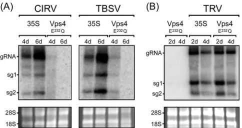
CIRV replication inN. benthamianais disrupted by a mutant of
Vps4.To begin to test whether ESCRT plays a role in CIRV
repli-cation, a dominant-negative version of theArabidopsisESCRT
protein Vps4 (Vps4E232Q) was expressed in CIRV-infectedN.
ben-Richardson et al.
on November 7, 2019 by guest
http://jvi.asm.org/
thamianaleaves. Vps4 is an AAA-ATPase, and Vps4E232Qcontains a Glu-to-Gln mutation in the ATPase domain that blocks ATP
hydrolysis and causes defects in endosomal protein sorting (33,
55), presumably by preventing disassembly of the endogenous
ESCRT-III machinery at the late endosomal surface, as it does in
yeast (56). Leaves ofN. benthamiana, which is a host of
tombus-viruses, including CIRV (57), were infiltrated withAgrobacterium
harboring a plasmid expressing Vps4E232Q(33) (agro-infiltrated).
The same leaves were then rub inoculated 2 days later with a CaMV 35S promoter-containing plasmid encoding the full-length
infectious CIRV cDNA (11), and 4 or 6 days thereafter, viral
genomic and subgenomic RNAs were analyzed by Northern blot-ting.
As shown inFig. 2A, compared to CIRV-infectedN.
benthami-analeaves that were agro-infiltrated with an empty vector (35S),
expression of Vps4E232Q dramatically reduced the amounts of
CIRV genomic RNA and both subgenomic mRNAs (sg1 and sg2 mRNA). Consistent with the results reported previously on the
inhibition of TBSV replication inN. benthamianaby
overexpres-sion of another dominant-negative, ATPase-deficient veroverexpres-sion of
ArabidopsisVps4, i.e., Vps4K178A(23), we found that
overexpres-sion of Vps4E232Q also inhibited the accumulation of TBSV
genomic and subgenomic mRNAs (Fig. 2A). Overexpression of
Vps4E232Qdid not, however, inhibit the replication of theTobacco rattle virus(TRV) inN. benthamianaleaves (Fig. 2B), which is a
(⫹)RNA tobravirus (46) and does not rely on ESCRT for its
rep-lication (23). This confirms that the inhibition of CIRV (and
TBSV) replication by Vps4E232Qis not simply a consequence of an
indirect, inhibitory effect(s) due to the overexpression of this ESCRT mutant in plant cells. As expected, no viral RNAs were
detectable in mock-infected N. benthamiana leaves expressing
Vps4E232Q(results are presented for TRV only [Fig. 2B]).
CIRV p36 recruits Vps23 to mitochondria in plant cells. Given the well-documented role of Tsg101 (the mammalian ho-molog of Vps23) in HIV budding from the plasma membrane in
mammalian cells (21,22) and that yeast Vps23p is redirected to
peroxisomes in yeast cells coexpressing TBSV p33 (23), we tested
whether, in an analogous manner, CIRV causes the relocalization
of Vps23 to mitochondria in plant cells. Toward this end,N.
taba-cumBY-2 suspension-cultured cells—which serve as a
well-char-acterized system for studying protein targeting in plant cells (57),
including viral proteins (58, 59)—were transiently
(co)trans-formed (via biolistic bombardment) with plasmids encoding full-length CIRV and/or an N-terminal Myc epitope-tagged version of
ArabidopsisVps23 (Myc-Vps23) and then processed for immu-noepifluorescence microscopy.
As shown inFig. 3Aand consistent with previously published
results (11), both the p36 and p95 replicase proteins in
CIRV-transformed BY-2 cells localized to endogenous
-ATPase-con-taining mitochondria, which as a consequence of the expression of the viral proteins, were conspicuously altered (i.e., aggregated) in terms of their intracellular distribution. For comparison
pur-poses, refer to the normal appearance of the-ATPase-containing
mitochondria that are distributed throughout the cytosol in a
nontransformed BY-2 cell (Fig. 3A). Also consistent with
previ-ously published results (35), Myc-Vps23 localized in BY-2 cells to
the cytosol and late endosomes, shown by its partial colocalization
with GFP-Syp21 (Fig. 3A), which is a well-known late endosomal
marker protein (38). In contrast, coexpression of CIRV and
Myc-Vps23 in the same cell resulted in relocalization of Myc-Myc-Vps23 to
CIRV-induced aggregated mitochondria (Fig. 3B), which was
confirmed as such based on immunostaining of both Myc-Vps23 and endogenous mitochondrial CoxII in the same batch of
CIRV-and Myc-Vps23-cotransformed cells (Fig. 3B). These results
sup-port the premise that, similar to HIV and TBSV, CIRV causes a change in the intracellular localization of Vps23.
We determined next whether the replicase proteins p36 and/or p95 expressed outside of the context of full-length CIRV were capable of recruiting Vps23 to mitochondria in plant cells. As
shown inFig. 3C, coexpression of Myc-Vps23 with either or both
replicase proteins resulted in the relocalization of Myc-Vps23 to
aggregated mitochondria (Fig. 3C), similar to when Myc-Vps23
was coexpressed with full-length CIRV (Fig. 3B). Colocalization
between p36 and Myc-Vps23 is also shown inFig. 3Dusing
con-focal microscopy, and quantification of colocalization in images obtained from medial (midcell) optical sections using the mean
Pearson’s correlation coefficient,r, confirmed a high degree of
fluorescence signal overlap for both sets of proteins (see the legend toFig. 3Dforrvalues). In contrast, confocal microscopy and quan-tification of colocalization confirmed that Myc-Vps23 was not asso-ciated with mitochondria in BY-2 cells in the absence of coexpressed
p36, as expected (seeFig. 3Dand the legend forrvalue).
We also performed a number of additional control experi-ments to confirm that the recruitment of Vps23 to mitochondria by p36 was not dependent on the appended peptide sequence or
fluorescent reporter protein (Fig. 3E) or the heterologous
expres-sion ofArabidopsisVps23 in tobacco cells (Fig. 3F) and that it was
specific for Vps23, since at least two other ESCRT proteins, Vps28
(ESCRT-I) and Vps25 (ESCRT-II), were not recruited by p36 (Fig.
3G). Moreover, we demonstrated that the appearance (i.e.,
distri-bution) of organelles other than mitochondria was unaffected in
p36 (co)expressing cells (Fig. 3H), indicating that colocalization
of Vps23 and p36 at mitochondria is not simply due to a general aggregation of organelles.
Taken together, the results presented inFig. 3 indicate that
FIG 2Inhibition of CIRV and TBSV replication in N. benthamianaby Vps4E232Q. (A) RNA blot analysis ofN. benthamianaleaves infiltrated with
Agrobacteriumharboring either an empty vector (35S) or a vector encoding ArabidopsisVps4E232Q, followed by rub inoculation with full-length infectious
CIRV or TBSV cDNA. The relative positions of the CIRV and TBSV genomic (gRNA) and the two subgenomic mRNAs (sg1 and sg2) are shown to the left of the gel. The numbers of days after infiltration or inoculation are indicated above the lanes. (B) RNA blot analysis ofN. benthamianaleaves agro-infil-trated with either Vps4E232Qalone, empty vector (35S), and two other vectors
(TRV1 and TRV2) encoding the full-length infectious TRV genome (TRV), or Vps4E232Q, TRV1, and TRV2. In panels A and B, the 28S and 18S rRNAs in the
corresponding ethidium bromide-stained agarose gels are shown as a loading control.
on November 7, 2019 by guest
http://jvi.asm.org/
[image:5.585.43.285.66.194.2]FIG 3Relocalization of Vps23 from the cytosol and late endosomes to mitochondria in BY-2 cells coexpressing CIRV p95 and/or p36. (A to C) Representative epifluorescence micrographs of BY-2 cells (co)transformed (as indicated by panel labels) with either full-length CIRV, Rep (both p95 and p36 together), p95, p36, Myc-Vps23, or GFP-Syp21, or mock transformed with empty plasmid DNA and then immunostained for the endogenous mitochondrial protein-ATPase (top right-hand photo in panel A). All cells shown inFig. 3were formaldehyde fixed and processed for immunofluorescence microscopy as described in Materials and Methods. Note that p95 and p36 were immunodetected using primary antibodies raised against a synthetic peptide that corresponds to an amino acid sequence in both proteins. p95 is produced by the translational read-through of the p36 amber stop codon (8). Selected transformed cells were also immunostained (as indicated) for endogenous mitochondrial-ATPase or CoxII. Also shown are the corresponding merge and differential interference contrast (DIC) images. The yellow color in the merged images indicates colocalization, and the white arrowheads in panel A indicate obvious examples of Myc-Vp23 and GFP-Syp21 colocalization at late endosomes. Bar⫽10m. (D) Representative confocal micrographs of BY-2 cells cotransformed with either p36 and Myc-Vps23 (top and middle rows), or GFP-Syp21 and Myc-Vps23 (bottom row), which were immunostained for Myc-Vps23 and endogenous CoxII (GFP fluorescence is not shown). All images are medial (midcell) optical sections of cells, and boxes represent the portions of the cells shown at higher magnification in the panels on the right. The Pearson’s correlation coefficientrvalues were as follows:r⫽0.81 for p36 and Myc-Vps23,r⫽0.71 for Myc-Vps23 and CoxII, andr⫽0.21 for Myc-Vps23 and CoxII. (E to H) Representative epifluorescence micrographs of BY-2 cells cotransformed (as indicated) with either p36 and either N-terminal HA epitope- or GFP-tagged Vps23 (E); p36 and a Myc-taggedN. tabacumVps23 (F), p36 and Myc-tagged Vps28 or Vps25 (G), or p36 and various organelle marker fusion proteins, including Cherry-PTS1 (peroxisome), GFP-Syp52 (early endosome/trans-Golgi network), GFP-Syp21 (late endosome), TIC40-RFP (plastid), PAP26-Cherry (lytic vacuole) (H). Bar⫽10m.
on November 7, 2019 by guest
http://jvi.asm.org/
[image:6.585.76.511.36.564.2]CIRV specifically recruits the ESCRT-I component Vps23 to mi-tochondria in plant cells and that the p36 replicase protein is the minimal viral component necessary for this recruitment.
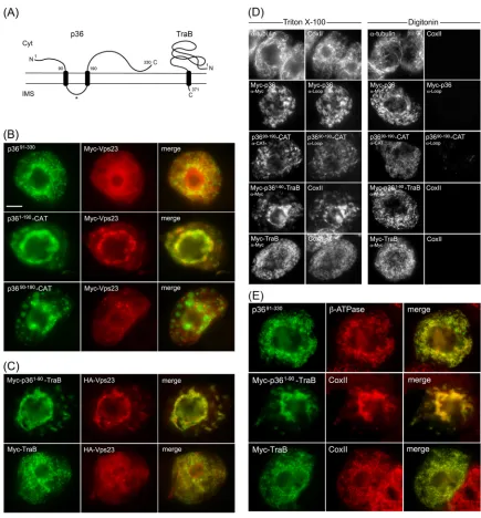
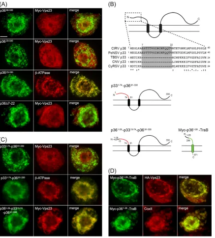
The cytosol-facing N-terminal region of p36 recruits Vps23
to mitochondria.On the basis of previously published models
(11,26) and as illustrated inFig. 4A,p36 is an integral outer
mi-tochondrial membrane protein that is orientated with both the N-and C-terminal regions facing the cytosol. p36 also possesses two TMDs and an intervening loop region that contains the mito-chondrial targeting signal and is predicted to face the
intermem-brane space (11). Keeping in mind this predicted topology, we
began to explore regions within p36 that could be responsible for recruitment of Vps23 to mitochondria by examining mutant ver-sions of p36 lacking the cytosol-facing N- and/or C-terminal por-tions of the protein, since these should be available to interact with cytosolic Vps23.
As shown inFig. 4B, Myc-Vps23 localized to the cytosol and
not to mitochondria when coexpressed with p3691-330, a mutant of
p36 in which the protein’s cytosol-facing, N-terminal 90 amino
acid residues were deleted (see the legend toFig. 4Bfor thervalue
of coexpressed Myc-Vps23 and p3691-330, based on confocal
mi-croscopy). In contrast, p361-190-CAT, which is a previously
char-acterized p36 mutant wherein the cytosol-facing C-terminal re-gion of the protein (i.e., residues 191 to 330) was replaced with the
passenger protein CAT (11), retained the ability to recruit
Myc-Vps23 to mitochondria (Fig. 4B). Taken together, these data
indi-cate that the N terminus, but not the C terminus, of p36 is neces-sary for the recruitment of Vps23 to mitochondria.
Consistent with this conclusion, Myc-Vps23 was not recruited
to mitochondria when coexpressed with p3690-190-CAT (Fig. 4B),
which lacks the N-terminal 90 residues of p36, and also has its
cytosol-facing C terminus replaced with CAT (11). These results
further indicate that the intervening loop region of p36 alone is not capable of recruiting Vps23 to mitochondria, as expected
from the protein’s predicted topology (Fig. 4A), wherein the
in-tervening loop faces the mitochondrial intermembrane space and, thus, is inaccessible to interact with Vps23 in the cytosol. Confir-mation that the intervening loop in both full-length p36 and p3690-190-CAT is oriented toward the mitochondrial
intermem-brane space was obtained using BY-2 cell differential detergent
permeabilization assays (Fig. 4D). For instance, applied
antibod-ies specific for a peptide sequence within the intervening loop of
p36 were able to immunodetect the protein (or p3690-190-CAT) in
cells only when both the plasma membrane and mitochondrial membranes (and all other organelle membranes) were permeab-ilized with Triton X-100, but not when only the plasma membrane was permeabilized with digitonin. We also confirmed that all of the p36 mutants mentioned above were properly targeted to tochondria based on their colocalization with an endogenous
mi-tochondrial marker protein (results are presented for p3691-330
only [Fig. 4E]).
To further demonstrate the importance of the N-terminal re-gion (i.e., residues 1 to 90) of p36 in the recruitment of Vps23 to mitochondria, we replaced the N terminus of a mitochondrial outer membrane protein (TraB) with that of p36 and tested the fusion protein’s ability to recruit coexpressed Vps23 to
mitochon-dria. TraB is a “tail”-anchored membrane protein (Fig. 4A) (60),
consisting of (i) an N-terminal region that faces the cytosol and represents the majority of the protein, (ii) a single TMD located near its C terminus, and (iii) a short C-terminal tail region
orien-tated toward the intermembrane space that, along with the TMD, constitutes the protein’s mitochondrial outer membrane
target-ing information (11). As shown inFig. 4C, Myc-p361-90-TraB,
consisting of a Myc epitope tag fused to the N-terminal 90 amino acids of p36 and the C-terminal TMD and tail of TraB (i.e., resi-dues 348 to 371), colocalized with coexpressed HA epitope-tagged
Vps23 (HA-Vps23; refer also toFig. 3E) at mitochondria in BY-2
cells; mitochondrial recruitment was confirmed by colocalization
of Myc-p361-90-TraB and endogenous CoxII in the same batch of
Myc-p361-90-TraB- and HA-Vps23-cotransformed cells (Fig. 4E).
In contrast, HA-Vps23 did not colocalize with full-length
Myc-TraB at mitochondria (Fig. 4CandE) (refer also to theFig. 4C
legend for thervalue for coexpressed Myc-TraB and HA-Vps23,
based on confocal microscopy), a result that was not due to an
aberrant topology of Myc-TraB, which like Myc-p361-90-TraB is
orientated with its N terminus facing the cytosol (Fig. 4D).
Overall, the data presented inFig. 4indicate that the
cytosol-facing N-terminal portion of p36, which represents the first 90 amino acids of the protein, is both necessary and sufficient for recruitment of Vps23 to mitochondria in plant cells.
The N-terminal region of p36 interacts directly with Vps23. We investigated next whether the N-terminal region of p36 inter-acts directly with Vps23 by employing several different ap-proaches. For example, yeast two-hybrid assays revealed that
co-expression of p361-90 and Vps23 resulted in significant yeast
growth on high selection media (Fig. 5A), indicating that the two
fusion proteins interact. Similarly, coexpression of Vps23 and ESCRT-I protein Vps28 resulted in growth on high selection me-dia, which has been reported previously using the yeast
two-hy-brid assay (36,61,62). As a negative control and consistent with
ourin vivodata (Fig. 3G), no interaction was observed between
p361-90and the ESCRT-II component Vps25. Likewise,
coexpres-sion of p361-90, Vps23, Vps28, or Vps25 with the corresponding
empty vectors yielded no yeast growth on high selection media (Fig. 5A).
The interaction between p36 and Vps23 was confirmed usingin
vitrocoimmunoprecipitation. Briefly, recombinant S-epitope-tagged
Vps23 (S-Vps23) from clearedE. colicell lysate was immobilized on
S-protein agarose and then incubated with in vitro-translated,
Myc-tagged versions of either full-length p36 (Myc-p36), the
N-terminal 90 amino acids of p36 (Myc-p361-90), or Vps28
(Myc-Vps28); Myc-Vps28 served as a positive control based on its pre-viously published interaction with Vps23 via
coimmuno-precipitation (35,63). For a negative control, the same threein
vitro-translated proteins were incubated with S-protein agarose
preincubated with cleared cell lysate prepared fromE. coli
trans-formed with empty vector, but containing an approximately equal
amount of total protein (Fig. 5B, right panel). As shown also in
Fig. 5B, both Myc-p36 and Myc-p361-90, similar to Myc-Vps28,
were coimmunoprecipitated with immobilized S-Vps23, but not by the corresponding empty vector negative controls (marked by
solid arrowheads inFig. 5B, left panel), indicating that both
full-length p36 and the N terminus of p36 interact directly with Vps23. We further demonstrated the interaction between p36 and
Vps23 using thein vivoBiFC assay. In this experiment, p36 or p36
lacking its N terminus (p3691-330) fused to the C-terminal half of
YFP (p36-cYFP and p3691-330-cYFP, respectively) were each
coex-pressed with Vps23 fused to the N-terminal half of YFP (nYFP-Vps23) in BY-2 cells. In addition, cells were transformed with cytosolic RFP, which was used to identify transformed cells and
on November 7, 2019 by guest
http://jvi.asm.org/
FIG 4The cytosol-facing N-terminal region of p36 is both necessary and sufficient for relocalizing Vps23 to mitochondria in BY-2 cells. (A) Topology models of p36 and TraB in the mitochondrial outer membrane based on previously published results (11,26,50) and those presented in panel D. The numbers show the numbers of specific amino acid residues in full-length p36 (330 residues) and TraB (371 residues), including those in the names of the modified versions of p36 and p36-CAT (and p36-TraB) shown in panels B to E. The asterisk represents the relative position of the peptide sequence in the intervening loop of p36 used to generate the antiloop antibodies used in panel D. The N and C termini of the proteins are shown. Cyt, cytosol; IMS, intermembrane space. (B and C) Representative epifluorescence micrographs of BY-2 cells cotransformed with proteins as indicated by panel labels. Cells were formaldehyde fixed and processed for immunofluorescence epimicroscopy as described in the legend toFig. 3. The Pearson’s correlation coefficientrvalues based on confocal microscopy of cells coexpressing p3691-330and Myc-Vps23 or Myc-TraB and HA-Vps23 werer⫽0.31 andr⫽0.25, respectively. Bar⫽10m. (D) Representative epifluorescence
micrographs of BY-2 cells either nontransformed (top row) or cotransformed with the indicated proteins, fixed with formaldehyde, and then permeabilized with either Triton X-100 (which permeabilizes both the plasma membrane and all organellar membranes) or digitonin (which permeabilizes only the plasma membrane) (16). The cells were then processed for immunoepifluorescence microscopy using primary antibodies raised against endogenous cytosolic␣-tubulin, endogenous mitochondrial matrix protein CoxII (anti-CoxII [␣-CoxII]), the Myc epitope tag (anti-Myc [␣-Myc]), an amino acid sequence in the intervening loop of p36 (antiloop [␣-Loop]), or the bacterial passenger protein CAT (anti-CAT [␣-CAT]), as indicated by panel labels. Note that the presence of immuno-staining in digitonin-permeabilized cells indicates that endogenous␣-tubulin and the expressed protein’s appended Myc epitope tag(s) or CAT moiety are exposed to the cytosol. Conversely, endogenous CoxII or the intervening loop sequence of p36 is not immunodetectable in the same corresponding digitonin-permeabilized cells. (E) Representative epifluorescence micrographs of BY-2 cells transformed with p3691-330, Myc-p361-90-TraB, or Myc-TraB and
immuno-stained for endogenous mitochondrial-ATPase or CoxII. Note that the mitochondria in the cells expressing p361-90-TraB, but not Myc-TraB, were aggregated,
similar to the mitochondrial aggregation observed in cells expressing full-length p36 (Fig. 3). Richardson et al.
on November 7, 2019 by guest
http://jvi.asm.org/
[image:8.585.73.508.62.530.2]also served as an internal normalization for reconstituted YFP fluorescence, minimizing differences due to cell-to-cell variability
in expression levels. p3691-330, which does not recruit Vps23 to
mitochondria in plant cells (Fig. 4B), served as a negative control.
As shown inFig. 5C, BY-2 cells cotransformed with p36-cYFP and
nYFP-Vps23 displayed significantly more YFP/RFP fluorescence
than cells cotransformed with p3691-330-cYFP and nYFP-Vps23.
While we cannot rule out the possibility that the relatively small amount of YFP/RFP fluorescence still observed upon
coexpres-sion of p3691-330-cYFP and nYFP-Vps23 may be a result of weaker
or, as discussed below, “secondary” interactions between the C terminus of p36 and Vps23, these results indicate that, just as in
yeast two-hybrid andin vitro coimmunoprecipitation assays (Fig.
5AandB), p36 and Vps23 interact in plant cells in a manner that
is dependent upon the N terminus of p36.
A unique polypeptide sequence near the N terminus of p36 is both necessary and sufficient for recruiting Vps23 to
mitochon-dria.Since the N-terminal 90-amino-acid sequence of p36
con-tains no sequence motifs that match the peptide late-domain mo-tifs (e.g., P[S/T]AP) responsible for the recruitment of ESCRT by
enveloped retroviral proteins (20) or by TBSV p33 (23,25), we
carried out a mutational analysis of this region to define any po-tentially novel Vps23 recruitment motifs. The first mutants
exam-ined were those in which either the N-terminal 67 (p3668-330) or 22
(p3623-330) amino acid residues were deleted from the protein. As
shown inFig. 6A, both p3668-330and p3623-330, which were
prop-erly targeted to mitochondria (results are presented for p3623-330
only [Fig. 6A), did not recruit Myc-Vps23. Instead, Myc-Vps23
localized in these cells primarily to the cytosol and/or late
endo-somes, just as it does when it is expressed on its own (Fig. 3A) or
coexpressed with the p36 mutant lacking the entire
90-amino-acid-long N-terminal region (p3691-330) (Fig. 4B). These data
sug-gest that at least the first 22 amino acids of the p36 N terminus are essential for recruiting Vps23 to mitochondria.
Comparison of the N-terminal sequences of p36 with its
coun-terparts from various other tombusviruses (Fig. 6B) revealed that
only p36 and the replicase protein ofPelargonium necrotic spot
virus(PeNSV), which like CIRV, replicates at host cell membranes
derived specifically from mitochondria (64), possess a unique and
identical stretch of 16 amino acids (residues 7 to 22) that is not present in the replicase proteins of tombusviruses that replicate
at peroxisome-derived membranes (i.e., TBSV p33,Cucumber
necrosis virus [CNV] p33, Cymbidium ringspot virus [CyRSV] p33). Notably, this unique sequence in CIRV p36 is due to the initiation codon of the p36 open reading frame being positioned further upstream than the corresponding codon in TBSV or
CyRSV (5,8) and while it contains a negative determinant for
satellite RNA replication (65), it is not suspected to play a
signifi-cant role in virus symptom development (66). Nonetheless, we
FIG 5The N-terminal region of p36 interacts with Vps23. (A) p361-90
inter-acts with Vps23 in the yeast two-hybrid assay. Yeast strains were cotrans-formed with the indicated pairs of binding domain (BD) and GAL4-activiating domain (AD) fusion proteins or the corresponding empty BD or AD control plasmids, denoted by a hyphen in the BD or AD column. Serial (1:5) dilutions of cells were spotted onto agar plates containing either low-stringency media (SD-Leu,Trp) or high-stringency media (SD-Leu, Trp,His,Ade), where cell growth is dependent on two-hybrid protein interac-tions. (B) In vitro coimmunoprecipitation of p36 or p361-90 and Vps23.
Whole-cell lysates ofE. colitransformed with S-Vps23 or the corresponding empty vector were incubated with S-protein agarose andin vitro-translated (IVT) full-length Myc-p36, Myc-p361-90, or Myc-Vps28, and washed. Eluted
proteins (and 10% of the total IVT reaction mixture) were subjected to SDS-PAGE and protein blotting and then probed with either anti-Myc or anti-S-tag antibodies. The solid (black) arrowheads indicate the relative positions of spe-cific immunodetected proteins; the open (white) arrowheads indicate nonspe-cific, immunodetected proteins. Note that the immunodetected proteins indi-cated by the solid arrowheads are not present in the coimmunoprecipitation reactions with lysates ofE. colicontaining empty vector. Shown to the right is a Coomassie blue-stained gel of total soluble protein from lysates containing S-Vps23 or empty vector prior to incubation with S-protein agarose, confirm-ing equivalent input. The numbers to the left of the gels are the molecular masses (in kilodaltons) of protein standards. (C) Interaction of p36 and Vps23 in the BiFC assay. Representative epifluorescence micrographs of BY-2 cells
triple transformed (as indicated) with RFP, nYFP-Vps23, and either p36-cYFP or p3691-330-cYFP. Relative RFP and YFP fluorescence values of transformed
cells, which are delineated by the solid lines in the panels on the right were quantified using ImageJ (refer to Materials and Methods for details). Note the relatively low YFP fluorescence in the representative cell coexpressing the neg-ative control, p3691-330-cYFP. At least 25 transformed cells were analyzed from
at least three independent experiments, and the mean YFP-to-RFP ratios (plus standard deviation [SD]) are plotted in the bar graph on the right. The two asterisks denote a statistically significant difference between the two samples (Pⱕ0.001).
on November 7, 2019 by guest
http://jvi.asm.org/
[image:9.585.43.284.75.501.2]chose to focus on whether this unique region in p36 (and PeNSV p33) is involved in the recruitment of Vps23 based on the inability
of p3623-330to recruit Vps23 to mitochondria. As shown inFig.
6A, Myc-Vps23 did not localize to mitochondria when
coex-pressed with a p36 mutant lacking residues 7 through 22 (p36⌬
7-22), supporting the premise that this region in p36 is specifically involved in Vps23 recruitment. As a complementary approach, we also replaced the N-terminal 90 residues of p36 with the
equiva-FIG 6A unique 16-amino-acid-long sequence at the N terminus of p36 is both necessary and sufficient for relocalizing Vps23 to mitochondria in BY-2 cells. (A) Representative epifluorescence micrographs of BY-2 cells cotransformed with proteins as indicated by panel labels and immunostained (as indicated) for endogenous mitochondrial-ATPase. Numbers in the name of the construct denote the specific amino acid residues derived from full-length p36 (330 residues) or specific residues deleted from p36. Bar⫽10m. (B) Amino acid sequence alignment of the N termini of various tombusvirus p36 and p33 proteins and cartoon illustrations of p33-36 and p36-TraB hybrid proteins. Sequences were obtained from GenBank and aligned using ClustalW. Identical and similar amino acids in each protein are indicated with asterisks and colons or periods, respectively. The unique amino acid sequence present in CIRV p36 and PeNSV p33 (residues 7 to 22) but absent in TBSV, CNV, and CyRSV p33 are shaded gray. Cartoons depict the structure and topology of p33–p36 and p36-TraB hybrid proteins in the mitochondrial outer membrane. Lines representing amino acid sequences from p33, p36, and TraB are colored red, black, and green, respectively. Numbers represent specific amino acid residues derived from either full-length p33 (296 residues), p36 (330 residues), or TraB (371 residues) and correspond to the numbers in the names of the p33–p36 and p36-TraB hybrid proteins described in panels C and D. (C and D) Representative epifluorescence micrographs of BY-2 cells cotransformed with proteins as indicated by panel labels and immunostained (as indicated) for endogenous mitochondrial-ATPase. Numbers in the name of the construct denote the specific amino acid residues derived from full-length p36 or p33 and are as illustrated in panel B.
Richardson et al.
on November 7, 2019 by guest
http://jvi.asm.org/
[image:10.585.78.515.65.549.2]lent N-terminal soluble region of TBSV p33 (residues 1 to 74),
which is similarly orientated toward the cytosol (4,26), but does
not contain the unique sequence found within the N terminus of
p36 (Fig. 6B). Moreover, this N-terminal region of p33 is not
considered on its own to be sufficient for interacting with Vps23
(23). As shown inFig. 6C, the resulting fusion protein, p331-74
-p3691-330, did not colocalize with Myc-Vps23 at mitochondria
(re-fer to Fig. 6C, which shows colocalization between p331-74
-p3691-330and endogenous-ATPase for evidence that this fusion
protein was properly targeted to mitochondria). In contrast, when
the first 28 residues of p36 were reintroduced into p331-74
-p3691-330, yielding p361-28-p3313-74-p3691-330 (Fig. 6B), the
re-cruitment of Myc-Vps23 to mitochondria was restored (Fig. 6C).
We also showed that the N-terminal 28 amino acid residues of p36 were capable of recruiting Vps23 to mitochondria when this region was fused to the N terminus of TraB. That is, similar to
Myc-p361-90-TraB (Fig. 4C), Myc-p361-28-TraB, which as
illus-trated inFig. 6B, consists of the N-terminal 28 residues of p36
fused to a portion of the cytosol-facing domain of TraB (in order to preserve the overall length of the soluble N-terminal region), as well as its C terminus, also recruits HA-Vps23 to mitochondria in
coexpressing cells (Fig. 6D).
Taken together, the data presented inFig. 6indicate that the
unique N-terminal 16-amino-acid-long sequence present in CIRV p36, but absent in the replicase proteins from tombusvi-ruses that rely on peroxisomal membranes for their replication, contains a novel Vps23 recruitment motif.
Recruitment of Vps23 to mitochondria by p36 requires its
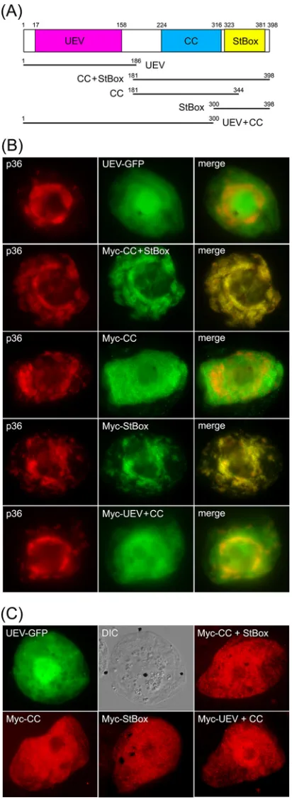
StBox domain rather than the UEV domain.As illustrated inFig.
7A,ArabidopsisVps23, similar to Vps23 in all species examined, consists of three unique structural/functional domains, including (i) an N-terminal UEV domain, (ii) a central coiled-coil (CC) region, which is involved in ESCRT-I protein-protein interac-tions, and (iii) a C-terminal steadiness box (StBox), which is also involved in protein interactions with other ESCRT-I components,
as well as regulating Vps23 protein turnover (63). Given the
im-portance of the UEV domain in facilitating the interaction
be-tween Vps23 and p33 during recruitment to peroxisomes (23) and
the interactions between Tsg101 and HIV Gag p6 at the plasma
membrane (20) and between certain ESCRT-0 proteins and
Vps23/Tsg101 during MVB biogenesis in yeasts and mammals
(13), we postulated that the UEV domain might also be involved
in the recruitment of Vps23 to mitochondria by p36.
To investigate this possibility, the UEV domain of Vps23 fused to green fluorescent protein (UEV-GFP) was coexpressed with
full-length p36 in BY-2 cells. As shown inFig. 7B, UEV-GFP was
not localized to mitochondria in cells coexpressing p36, but in-stead localized throughout the cytosol, just as it does when ex-pressed on its own, as do all other Vps23 mutants described in this
study [see below]) (Fig. 7C). The lack of recruitment of the UEV
domain by p36 was somewhat unexpected and led us to investigate next whether the CC and/or StBox domains play a role in its
re-cruitment to mitochondria by p36. As shown inFig. 7B,aVps23
mutant consisting of both the CC and StBox domains fused to an
FIG 7The C-terminal StBox of Vps23 is necessary and sufficient for its recruit-ment to mitochondria by p36 in BY-2 cells. (A) Schematic diagram illustrating the domain organization ofArabidopsisVps23 based on Spitzer et al. (63). Numbers denote the specific amino acid residues derived from full-length Vps23 (398 residues) that delineate the protein’s UEV, CC, and StBox domains, which are colored red, blue, and yellow, respectively, and which are depicted below as lines representing the various Vps23 truncation mutants described in panels B and C. Numbers denote the specific amino acid residues at the N and C termini of each Vps23 mutant. (B and C) Representative epifluorescence micrographs of BY-2 cells (co)transformed with proteins as indicated by panel labels.
Names of mutants represent the specific domain(s) derived from Vps23, as illustrated in panel A. The corresponding merged images (B) and the corre-sponding DIC images of the UEV-GFP-transformed BY-2 cells (C) are also shown.
on November 7, 2019 by guest
http://jvi.asm.org/
[image:11.585.61.267.68.631.2]N-terminal Myc epitope tag (i.e., Myc-CC⫹StBox; refer to
sche-matic diagram inFig. 7A), colocalized with coexpressed p36 at
mitochondria in BY-2 cells, reinforcing the notion that the re-cruitment of Vps23 by p36 is mediated by a region(s) other than
the UEV domain. As shown also inFig. 7B,aVps23 mutant
con-sisting of the CC domain alone (Myc-CC) localized to the cytosol in cells coexpressing p36, while the StBox domain (Myc-StBox) alone was recruited to p36-containing mitochondria. These re-sults suggest that Vps23 recruitment is mediated by the StBox domain. Indeed, the Vps23 mutant consisting of the UEV and CC
domains (Myc-UEV⫹CC) did not colocalize with coexpressed
p36 but instead remained in the cytosol (Fig. 7B), just as it does
when coexpressed without full-length p36 (Fig. 7C). Together,
these results indicate that the C-terminal StBox domain is critical for the recruitment of Vps23 to mitochondria by p36 and that the UEV domain either does not play a role or plays a minor role that is undetectable using this method.
Consistent with this conclusion, yeast two-hybrid assays
per-formed with p361-90and the equivalent set of Vps23 mutants as
those described above revealed that only those proteins
contain-ing the StBox (i.e., CC⫹StBox and StBox) conferred yeast growth
on high selection media (Fig. 8A). A notable exception was the
UEV⫹CC mutant, which when coexpressed with p361-90, also
re-sulted in growth on high selection media, albeit relatively less than
that observed for yeast coexpressing CC⫹StBox, StBox, or
full-length Vps23 (Fig. 8A). However, as mentioned above, the
equiv-alent UEV-CC mutant is not recruited to mitochondria by
full-length p36 in plant cells (Fig. 7B), suggesting that if the CC
domain does indeed participate in the recruitment of Vps23 by p36, it does so to a lesser extent than the StBox domain does.
While we confirmed that UEV⫹CC (or any of the other Vps23
mutants) does not autoactivate the yeast two-hybrid reporter
genes (Fig. 8B), we cannot rule out the possibility that the
ob-served p361-90/UEV⫹CC interaction may be the result of a
“bridge” by an endogenous yeast ESCRT component(s). More-over, and as discussed in more detail below, we also confirmed that all of the Vps23 mutants (and full-length Vps23) interacted in
yeast two-hybrid assays with theArabidopsisESCRT-I proteins
Vps28 and Vps37 (Fig. 8CandD) in ways that are consistent with
how these domains are involved in the corresponding ESCRT
pro-tein-protein interactions in yeasts and mammals (27–30). For
in-stance, like their yeast and mammalian counterparts, the
interac-tion ofArabidopsisVps28 and Vps23 is mediated by the Vps23
StBox domain (Fig. 8C), while the interaction of Arabidopsis
Vps37 and Vps23 is mediated by the Vps23 CC domain, although the Vps23 UEV domain also appears to be involved in
Vps23-Vps37 binding (Fig. 8D).
DISCUSSION
Understanding how viruses appropriate host cell organelles in or-der to carry out their replication is not only an important step in the development of strategies to prevent infection, it often pro-vides novel insights into the molecular mechanisms underlying organelle biogenesis and dynamics in uninfected cells. For
exam-ple, recent studies examining how theBunyavirus Tomato spotted
wilt virusbuds from the endoplasmic reticulum (ER) in plant cells
or howTurnip mosaic virusinfection leads to the fusion of
ER-derived vesicles with chloroplasts has shed new light on the
for-mation of ER exit sites (67) and the role(s) of the previously
uncharacterized SNARE protein Syp71 (68). Likewise, the
charac-terization of the late-domain motifs in certain retroviral structural proteins and their interaction with the host cell ESCRT-I protein
Tsg101 (69) was a key step in the subsequent discovery that similar
peptide motifs exist in certain ESCRT-0 proteins. As such, it is now known that, in uninfected cells, the sequential recruitment of the ESCRT subcomplexes (i.e., ESCRT-0, -I, -II, and -III) during MVB biogenesis begins with ESCRT-0 binding to ESCRT-I and that retroviral proteins such as the HIV Gag p6 mimic ESCRT-0 in order to hijack ESCRT-I (Tsg101) to the sites of viral budding at
the plasma membrane (20).
FIG 8The N-terminal region of p36, like Vps28, interacts with the C-terminal StBox of Vps23 in the yeast two-hybrid assay. (A to D) Yeast strains were cotrans-formed with the indicated pairs of GAL4-binding domain (BD) and GAL4-acti-vating domain (AD) fusion proteins or the corresponding empty BD or AD con-trol plasmids (indicated by a hyphen in the BD or AD column). Serial (1:5) dilutions of cells were spotted onto agar plates containing either low-stringency medium (SD-Leu,Trp) or high-stringency medium (SD-Leu,Trp,His,Ade), where cell growth is dependent on two-hybrid protein interactions. Note that neither full-length Vps23, nor any of the truncation mutants of Vps23, Vps28, and Vps37 autoactivated the two-hybrid reporter gene system on their own.
Richardson et al.