Complementary Effects of Interleukin-15 and Alpha Interferon Induce
Immunity in Hepatitis B Virus Transgenic Mice
Marianna Di Scala,a,bItziar Otano,cIrene Gil-Fariña,aLucia Vanrell,a*Mirja Hommel,aCristina Olagüe,a,bAfrica Vales,a,b Miguel Galarraga,dLaura Guembe,b,eCarlos Ortiz de Solorzano,b,dIndrajit Ghosh,fMala K. Maini,cJesús Prieto,a,b,g,h Gloria González-Aseguinolazaa,b
Gene Therapy and Regulation of Gene Expression Program, Center for Applied Medical Research (CIMA), Pamplona, Spaina
; Instituto de Investigación Sanitaria de Navarra (IdisNA), Pamplona, Spainb
; Division of Infection and Immunityc
and Centre for Sexual Health and HIV Research,f
UCL, London, United Kingdom; Imaging Unit and Cancer Imaging Laboratory, CIMA, Pamplona, Spaind
; Department of Morphology, CIMA, Pamplona, Spaine
; University Clinic of Navarra, Pamplona, Spaing
; CIBERehd, University of Navarra, Pamplona, Spainh
ABSTRACT
In chronic hepatitis B (CHB), failure to control hepatitis B virus (HBV) is associated with T cell dysfunction. HBV transgenic
mice mirror many features of the human disease, including T cell unresponsiveness, and thus represent an appropriate model in
which to test novel therapeutic strategies. To date, the tolerant state of CD8
ⴙT cells in these animals could be altered only by
strong immunogens or by immunization with HBV antigen-pulsed dendritic cells; however, the effectors induced were unable to
suppress viral gene expression or replication. Because of the known stimulatory properties of alpha interferon (IFN-
␣
) and
in-terleukin-15 (IL-15), this study explored the therapeutic potential of liver-directed gene transfer of these cytokines in a murine
model of CHB using adeno-associated virus (AAV) delivery. This combination not only resulted in a reduction in the viral load
in the liver and the induction of an antibody response but also gave rise to functional and specific CD8
ⴙimmunity.
Further-more, when splenic and intrahepatic lymphocytes from IFN-
␣
- and IL-15-treated animals were transferred to new HBV carriers,
partial antiviral immunity was achieved. In contrast to previous observations made using either cytokine alone, markedly
atten-uated PD-L1 induction in hepatic tissue was observed upon coadministration. An initial study with CHB patient samples also
gave promising results. Hence, we demonstrated synergy between two stimulating cytokines, IL-15 and IFN-
␣
, which, given
to-gether, constitute a potent approach to significantly enhance the CD8
ⴙT cell response in a state of immune hyporesponsiveness.
Such an approach may be useful for treating chronic viral infections and neoplastic conditions.
IMPORTANCE
With 350 million people affected worldwide and 600,000 annual deaths due to HBV-induced liver cirrhosis and/or
hepatocellu-lar carcinoma, chronic hepatitis B (CHB) is a major health problem. However, current treatment options are costly and not very
effective and/or need to be administered for life. The unprecedented efficacy of the strategy described in our paper may offer an
alternative and is relevant for a broad spectrum of readers because of its clear translational importance to other chronic viral
infections in which a hyporesponsive antigen-specific T cell repertoire prevents clearance of the pathogen.
W
orldwide, 350 million people suffer from chronic hepatitis B
(CHB), and approximately 600,000 people die annually
be-cause of hepatitis B virus (HBV)-induced liver cirrhosis and/or
hep-atocellular carcinoma (
1
). The host immune response to HBV
anti-gens is a critical factor determining the outcome of infection. While
patients with self-limited, acute HBV develop strong, multispecific T
cell responses to viral antigens, these responses are weak and narrowly
focused in chronic HBV carriers (
2
,
3
). In these patients,
HBV-spe-cific CD4
⫹and CD8
⫹T cells display an exhausted phenotype
char-acterized by failure to proliferate and failure to produce gamma
in-terferon (IFN-
␥
), tumor necrosis factor alpha (TNF-
␣
), and
interleukin-2 (IL-2) after stimulation with viral antigens (
4
,
5
).
A cytokine that has received much attention for the treatment
of chronic hepatitis B and C infections is IFN-
␣
. As a recombinant
protein, it has been demonstrated to be effective in a proportion of
patients (
6
,
7
); however, patients with high viral loads and normal
serum transaminase levels seem particularly resistant to IFN-
␣
therapy (
8
). While IFN-
␣
was shown to have a direct degrading
effect on viral DNA (
9
) and to induce the expansion and activation
of NK cells (
10
), it did not effectively support the expansion
and/or survival of CD8
⫹T cells from patients with CHB (
8
).
In-terestingly, IFN-
␣
can facilitate the response of CD8
⫹T cells to
IL-15 stimulation by inducing the expression of IL-15 receptor
subunit alpha (IL-15R
␣
) (
11
). Moreover, there is evidence
indi-cating that long-lasting persistence of IFN-
␣
-primed CD8
⫹T cells
is favored by their enhanced responsiveness to IL-15 (
12
).
IL-15 is a powerful stimulatory cytokine that plays a key role in
lymphocyte function and homeostasis. It is involved in various
activation, proliferation, and differentiation processes of CD8
⫹T
Received25 May 2016Accepted12 July 2016
Accepted manuscript posted online20 July 2016
CitationDi Scala M, Otano I, Gil-Fariña I, Vanrell L, Hommel M, Olagüe C, Vales A, Galarraga M, Guembe L, Ortiz de Solorzano C, Ghosh I, Maini MK, Prieto J, González-Aseguinolaza G. 2016. Complementary effects of interleukin-15 and alpha interferon induce immunity in hepatitis B virus transgenic mice. J Virol 90:8563–8574.doi:10.1128/JVI.01030-16.
Editor:G. McFadden, University of Florida
Address correspondence to Gloria González-Aseguinolaza, [email protected].
*Present address: Lucia Vanrell, Cátedra de Inmunología, Facultad de Química, Instituto de Higiene, Universidad de la República, Montevideo, Uruguay.
Copyright © 2016 Di Scala et al. This is an open-access article distributed under the terms of theCreative Commons Attribution 4.0 International license.
crossmark
on November 7, 2019 by guest
http://jvi.asm.org/
cells (
13
), NK cells (
14
), and CD4
⫹T cells (
15
,
16
). IL-15 has been
reported to be capable of rescuing tolerant CD8
⫹T cells for use in
adoptive immunotherapy of established tumors (
17
), and in
com-bination with retinoic acid, it abrogated tolerance to dietary
anti-gens (
18
). Importantly, hepatic overexpression of IL-15 has
re-cently been implicated in inducing an anti-HBV response,
possibly by mediating IFN-

induction (
19
).
This study explored the therapeutic potential of liver-directed
gene transfer of IFN-
␣
and IL-15, alone or in combination, in a
murine model of chronic HBV (
20
) by use of adeno-associated
virus (AAV) delivery. Despite their limitations, HBV transgenic
(HBVTg) mice are widely used for elucidating immune responses
in CHB and evaluating therapeutic strategies for CHB (
21
). To
date, it has been shown that strong immunogens or immunization
with HBV antigen-pulsed dendritic cells was able to alter the
tol-erant state of CD8
⫹T cells in these animals; however, the effectors
induced were unable to suppress viral gene expression or
replica-tion (
22
,
23
). The results presented here show that combining
IL-15 with IFN-
␣
resulted in a functional and specific CD8
⫹re-sponse, which was reflected by a decrease in the level of HBV core
antigen (HBcAg) in the liver, and could confer partial immunity
upon splenocyte transfer to HBV-infected recipients.
MATERIALS AND METHODS
Animals and manipulations.HBVTg mice were kindly provided by Fran-cis V. Chisari (20). Mice were bred and maintained under pathogen-free conditions at the animal facility of the University of Navarra. For experi-ments, they were matched for age (6 to 10 weeks), sex (male), and levels of HBV DNA and HBV surface antigen (HBsAg) in serum. Age-matched C57BL/6 wild-type (WT) males were purchased from Harlan Laborato-ries (Barcelona, Spain). The experimental design was approved by the Ethical Committee for Animal Testing of the University of Navarra.
For all procedures, animals were anesthetized by intraperitoneal injec-tion of a mixture of xylazine (Rompun 2%; Bayer) and ketamine (Imal-gene 500; Merial) (1:9, vol/vol).
HBVTg mice were injected intravenously (i.v.) with AAV at a dose of 1.5⫻1012viral genomes (vg)/kg, except for the control group, which received 3⫻1012vg/kg.
Blood was collected by bleeding from the retro-orbital plexus, and serum samples were obtained by centrifugation of total blood.
For adoptive transfer experiments, C57BL/6 mice were injected with a recombinant AAV vector (5⫻1012vg/kg) carrying the HBV 1.3 genome
as described previously (28) in order to establish an alternative model of chronic HBV infection. Donor cells, consisting of 106intrahepatic
lym-phocytes (IHL) and 107splenocytes, were injected i.v. into the
retro-or-bital plexus in a final volume of 125l.
Viral construction, production, and purification.The recombinant AAV8 (rAAV8) vector with WT AAV2 inverted terminal repeats (ITRs) was produced as described previously (24,25). The expression cassette in the AAV–IL-15 vector consists of the murine IL-15 gene (GenBank acces-sion numberDQ083237.1) under the regulation of a liver-specific pro-moter (25). The expression cassette in the AAV–IFN-␣vector has been described previously and contains of the murine IFN-␣1 gene under the regulation of the elongation factor 1␣promoter (24). A luciferase-encod-ing AAV (AAV-LUC) served as a control (24).
DNA and RNA analysis.Total DNA and RNA were isolated from livers as described previously (25). Real-time PCR-based quantification of HBcAg and glyceraldehyde-3-phosphate dehydrogenase (GADPH) DNA and RNA was performed using SYBR green master mix (Applied Biosys-tems, Foster City, CA). The following primers were used: HBcAg sense (5=-TTCGCACTCCTCCAGCTTAT-3=) and antisense (5=-GGCGAGGG AGTTCTTCTTCTA-3=), GAPDH sense (5=-TGCACCACCAACTGCTT A-3=) and antisense (5=-CAGAAGACTGTGGATGGCCCCTC-3=), and
luciferase sense (5=-TCGAGGAGCCTTCAGGATT-3=) and antisense (5= -TTTTGGCGAAGAAGGAGAAT-3=).
HBV DNA was isolated from 20-l serum samples using the High Pure viral nucleic acid kit (Roche).
For Southern blot analysis of HBV replication intermediates, frozen liver and kidney tissues were mechanically pulverized, and total DNA was isolated and was digested with HindIII as described previously (20). Before electro-phoresis, all DNA samples were digested with RNase A (Roche) at 10 mg/ml and 37°C overnight (o/n). Nylon filters (Hybond-N⫹; Amersham) were hy-bridized with a32P-radiolabeled HBV-specific DNA probe.
Determination of serum ALT levels.Levels of alanine aminotransfer-ase (ALT) in blood were measured using commercial kits (Sigma Chem-icals, St. Louis, MO) and a Hitachi automatic analyzer (Boehringer Mann-heim, Indianapolis, IN).
ELISAs for IL-15, IFN-␣, and HBsAg levels in serum.The concentration of IL-15 in serum was determined using the Mouse IL-15/IL-15R Complex ELISA (enzyme-linked immunosorbent assay) Ready-SET-Go! set (eBiosci-ence), and the serum IFN-␣concentration was determined using the Ver-iKine Mouse Interferon Alpha ELISA kit (PBL Assay Science). HBsAg levels were also determined by ELISA (Bioelisa HBsAg 3.0; Biokit).
Liver histology, IHC, and TUNEL staining.Liver sections were fixed in 4% paraformaldehyde (Panreac), embedded in paraffin, sectioned (thickness, 5m), and stained with hematoxylin and eosin (H&E). Im-munohistochemistry (IHC) for HBcAg (B0586; Dako, Glostrup, Den-mark) and PD-L1 (ab58810; Abcam) was performed using the EnVision system (Dako) according to the manufacturer’s recommendations.In situ
detection of cells with DNA double-strand breaks was performed by the terminal deoxynucleotidyltransferase-mediated dUTP-biotin nick end la-beling (TUNEL) staining method by use of anIn SituCell Death Detection kit (Roche) according to instructions. Images were acquired digitally with an Axio Imager. MI microscope (Zeiss, Germany) using an in-house MetaMorph macro (Molecular Devices, USA). All images were stored in uncompressed 24-bit color TIFF format. Images were automatically ana-lyzed using a plugin developed for Fiji, ImageJ, at NIH.
Isolation of IHL.Mice were perfused via the portal vein with 10 ml of Ca2⫹- and Mg2⫹-free phosphate buffer solution preheated to 37°C. After perfusion, the vena cava was cut and the liver extracted. After incubation in 10 ml of phosphate-buffered saline (PBS) containing 1,000 U of type II collagenase (Gibco) for 20 min, the liver homogenate was passed through a 70-m nylon cell strainer, and the cell suspension was centrifuged at 1,200 rpm for 10 min. The cell pellet was resuspended in 40% Percoll and was centrifuged at 1,800 rpm for 10 min. Red blood cells were removed using a lysis buffer (Gibco); the remaining cells were washed and were resuspended in RPMI 1640 medium.
In vitroanalysis of lymphocyte proliferation.Intrahepatic lympho-cytes obtained from HBV transgenic mice treated with LUC, IFN-␣, IL-15, or IL-15 plus IFN-␣were isolated, labeled with carboxyfluorescein succinimidyl ester (CFSE), and cultured for 3 days in the presence or absence of 10g/ml of HBcAg peptide (epitope C93-100). The prolifera-tion of CD8⫹T cells was analyzed by flow cytometry.
Antibody and pentamer staining.Cells were labeled with the fol-lowing antibodies according to the manufacturer’s recommendations: allophycocyanin (APC)-conjugated CD19 (CD19-APC) (clone PeCa1; ImmunoTools), CD4-APC/Cy7 (clone GK1.5), CD44-APC/Cy7 (clone IM7), CD8a-Pacific Blue (clone 53.6.7), NK1.1-APC (clone PK136), PD-1 conjugated with fluorescein isothiocyanate (FITC) or brilliant violet 421 (clone 29F.1A12) (all from BioLegend), and CD8a-FITC (clone 1D3; eBioscience).
For pentamer staining. 2⫻106isolated cells were incubated for 10
min at room temperature (RT) with major histocompatibility complex (MHC) class I peptide pentamers (H-2Kb/ILSPFLPLL for HBsAg [epitope S208 –216], H-2Kb/MGLKFRQL for HBcAg [epitope C93–100], or
H-2Kb/SIINFEKL for ovalbumin [OVA] [epitope OVA257–264];
Pro-Immune). Cells were then washed and were stained with the respective surface antibodies according to the manufacturer’s recommendations.
Di Scala et al.
on November 7, 2019 by guest
http://jvi.asm.org/
Data were acquired with an 8-color BD FACSCanto II system equipped with three lasers (488 nm, 633 nm, and 405 nm) or with a 4-color FACSCalibur system equipped with two lasers (argon [488 nm] and diode [635 nm]). Fluorescence-activated cell sorter (FACS) data were analyzed with FlowJo or DIVA software.
In vivokilling assay (IVK).Splenocytes isolated from HBVTg or WT mice were pulsed with HBV peptide C93–100or S208 –216, or with an
irrel-evant peptide (ovalbumin; epitope OVA257–264) (all from Think
Pep-tides), for 30 min at 37°C. Cells were then labeled with 5M (for HBV peptides) or 0.5M (for the control peptide) CFSE (Invitrogen), and 5⫻ 106 cells of each population were transferred intravenously to mice.
Twenty-four hours later, animals were sacrificed, liver and spleen lym-phocytes isolated, and CFSE staining analyzed by FACS.
Depletion of lymphocyte subpopulations and PD-L1 blockade.CD8 T cell depletion studies were performed by intraperitoneal adminis-tration of 100g of an anti-mouse CD8 antibody (clone H35-17.2). Doses were scheduled starting 3 days before vector injection and at days 0, 3, 7, and 14 after virus injection. Depletion levels were deter-mined to be⬎98%. Irrelevant mouse immunoglobulin isotypes were used as controls. NK cell depletion studies were performed by intra-peritoneal administration of 200g of anti-mouse NK1.1 antibodies (clone PK136). Doses were scheduled daily starting 2 days before virus injection and at days 1, 3, 7, and 14 after virus injection. Depletion levels were determined to be⬎98%. Irrelevant mouse immunoglobu-lin isotypes were used as controls.
For in vivo PD1–PD-L1 blockade, rat anti–mouse PDL-1 (clone 10F.9G2; catalog no. BE0101; BioXCell) or a rat IgG2b isotype-matched control antibody (LTF-2; catalog no. BE0090; BioXCell) was injected in-traperitoneally 1 day before AAV–IL-15 or AAV-LUC injection and at days 0, 3, 6, and 9 after injection.
Patients and isolation of peripheral blood mononuclear cells (PBMCs).
PBMCs were isolated by Ficoll gradient centrifugation from heparinized blood samples taken from a total of 21 patients with CHB. Ethical approval was obtained through UK NHS REC 11/LO/0421, and each patient gave writ-ten informed consent. All patients were negative for anti-HCV and anti-HIV antibodies and were treatment naïve. Patients had a median age of 40 years (range, 27 to 60 years), and 2/21 were positive for HBV envelope antigen (HBeAg). The median HBV DNA level was 1,400 international units (IU)/ml (range, below the limit of quantification [BLQ] to 2⫻108IU/ml), and the
median ALT level was 31 IU/liter (range, 18 to 474 IU/liter).
HBV peptide stimulation of human PBMCs.Patient PBMCs were stimulated with 1M overlapping long peptides spanning the whole HBV core protein (genotype D [ayw]; JPT Peptide Technologies) in the presence or absence of IFN-␣(103IU/ml), IL-15 (10 ng/ml; R&D),
or both. Cells were cultured in complete RPMI medium supplemented with 20 IU/ml IL-2 for 10 days at 37°C. IL-2 and medium were re-freshed on day 4 of culture. On day 9, PBMCs were restimulated with 1M HBV peptide in the presence of brefeldin A (1g/ml; Sigma) for 16 h. For intracellular cytokine staining, cells were fixed and perme-abilized using a Cytofix/Cytoperm kit (BD Biosciences) for 15 min at 4°C and were then stained with phycoerythrin (PE)-conjugated anti-granzyme B (anti-GrB; Biolegend) and anti-IFN-␥BV421 (BD) for 30 min at 4°C. Samples were analyzed using a FACS LSRII flow cytometer (BD Biosciences).
Statistical analysis.Data are presented as means⫾standard errors of the means (SEM) and were analyzed for significance by the Studentttest. Differences among groups were analyzed by one-way analysis of variance (ANOVA) followed by a Bonferroni multiple-comparison test using GraphPad Prism software, version 5.0. (Significance is indicated by aster-isks in the figures as follows: *,P⬍0.05; **,P⬍0.01; ***,P⬍0.001; ****,
P⬍0.0001.)
For analysis of the reduction of viral replication and the decrease in the number of HBV core-positive nuclei induced by AAV–IFN-␣ com-bined with AAV–IL-15, the fold inhibition (FI) for each treatment was calculated as the ratio of the level of viral RNA obtained from the livers of mice treated with AAV-LUC to the level of viral RNA obtained from the livers of mice treated with AAV–IFN-␣(FIIFN-␣), AAV-IL-15 (FIIL-15), or
both (FIcombination). The synergy index (S) for the combination of
AAV–IFN-␣and AAV–IL-15 was calculated as described previously (47) from the fold inhibition by use of the equationS⫽FIcombination⫺
(FIIFN-␣⫹FIIL-15)/(FIIFN-␣⫹FIIL-15). The critical value ofSis zero and indicates an additive effect, while a positive value forS shows synergism.
RESULTS
Antiviral effect of IL-15–IFN-
␣
combination treatment in
HBVTg mice.
The immunomodulatory potential of IL-15 and
IFN-
␣
in a chronic HBV setting was investigated using an HBVTg
mouse model (
20
). HBVTg mice and C57BL/6 WT mice received
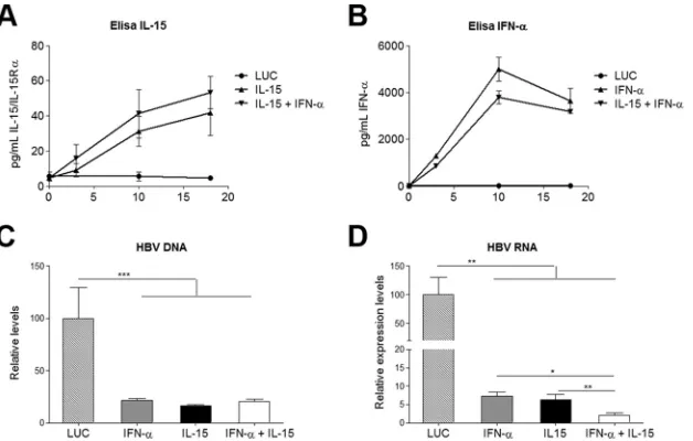
FIG 1Cytokine expression and reduced HBV nucleic acids. (A and B) Concentrations of the IL-15–IL-15R␣complex (A) and IFN-␣(B) in serum after vector injection. Results are representative of 3 independent experiments and represent means⫾SEM for triplicates. (C and D) Ten days after vector injection, animals were sacrificed and livers assessed for the presence of HBV DNA (C) and HBV RNA (D). Data were normalized to GADPH levels, and the means⫾SEM for 5 to 6 animals/group are shown. *,P⬍0.05; **,P⬍0.01; ***,P⬍0.001.
Induction of HBV Immunity by IFN-␣–IL-15 Combination
on November 7, 2019 by guest
http://jvi.asm.org/
[image:3.585.138.452.66.266.2]a single intravenous (i.v.) injection of AAV–IFN-
␣
, AAV–IL-15, a
combination of both, or an equivalent dose of a control vector
expressing the luciferase reporter gene (LUC). Serum cytokine
levels were measured at different time points after vector injection
(
Fig. 1A
and
B
).
To evaluate the direct antiviral effect elicited by the different
treatments, total HBV-specific DNA and RNA in the liver were
analyzed by quantitative real-time PCR. IFN-
␣
, IL-15, and the
combination treatment exerted similar inhibitory effects on HBV
replication (
Fig. 1C
). However, transcriptional inhibition was
more pronounced in animals that had received the combination
therapy (
Fig. 1D
) with a significant synergy index (S
⫽
3,476
⫾
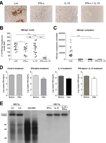
FIG 2Synergistic antiviral effect of combined IL-15 and IFN-␣treatment. (A) HBcAg protein expression in the liver, determined by IHC. A representative image is shown for each treatment group. (B and C) Fields were analyzed automatically to determine the number of hepatocytes positive for HBcAg in the nucleus (B) or the area of HBcAg staining in the cytoplasm (C). Each symbol represents the mean value for 10 different sections per mouse. Means⫾SEM for each group (n⫽
16) are also shown. (D) HBV genome copy numbers 10 days after vector injection. Graphs show means (n⫽5)⫾SEM. *,P⬍0.05; ***,P⬍0.001; ****,P⬍
0.0001; ns, nonsignificant. (E) HBV replicative intermediates in the liver by Southern blot analysis 10 days after vector injection. RC, relaxed-circular DNA; DS, double-stranded DNA; SS, single-stranded DNA; Luc, HBVTg mice treated with AAV-Luc; AAV-HBV, C57BL/6 WT mice receiving AAV-HBV; IFN-␣, HBVTg mouse treated with AAV-IFN-␣; IL-15, HBVTg mouse treated with AAV-IL15; IFN-␣ ⫹IL-15, HBVTg mouse treated with AAV-IFN-␣ ⫹AAV-IL-15. Results from representative animals from each treatment group are shown.
Di Scala et al.
on November 7, 2019 by guest
http://jvi.asm.org/
[image:4.585.114.474.65.548.2]2,390). The robustness of the antiviral effect of the combined
ther-apy was confirmed by immunohistochemical analysis of HBcAg
expression (
Fig. 2A
), as well as by a quantitative method,
deter-mining the number of core-positive nuclei per field (
Fig. 2B
) and
the area of HBcAg cytoplasmic staining per field (
Fig. 2C
). IFN-
␣
or IL-15 alone reduced the presence of core protein in the
cyto-plasm of hepatocytes but had little effect on the expression of core
protein in the nuclei. In contrast, when combined, IFN-
␣
and
IL-15 were able to eliminate nuclear core protein expression,
showing strong synergy (S
⫽
4,673
⫾
1,853).
The direct antiviral effects of the different treatments were also
analyzed by measuring the viral load in serum and hepatic HBV
DNA replicative intermediates by Southern blotting. In all
treat-ment groups, viral loads in the serum were significantly reduced
and HBV DNA replicate intermediates in the liver were
undetect-able (
Fig. 2D
and
E
).
As an important aspect of the immune response against HBV,
the presence of antibodies against HBsAg was measured by ELISA
20 days after treatment. Animals that had received IFN-
␣
alone or
the combination of IFN-
␣
and IL-15 developed anti-HBsAg, and
antibody levels were higher in the group receiving the
combina-tion treatment (data not shown).
The IL-15–IFN-
␣
combination treatment induces
immune-mediated liver damage in HBVTg mice.
Liver histology showed
no signs of inflammation at day 10 posttreatment in HBVTg mice
treated with IFN-
␣
or the control, while marked portal and
peri-portal infiltrates were observed in the IL-15 and IL-15–IFN-
␣
groups (data not shown), and this pattern remained unchanged
on day 20 (data not shown). However, on day 20, the livers of the
combination group presented with extensive areas of
hepatocel-lular necrosis surrounded by dense lymphocytic infiltrates (
Fig.
3A
). TUNEL staining showed large areas of positive nuclei in
HBVTg mice treated with IL-15 plus IFN-
␣
, while staining was
negative for all other groups (
Fig. 3B
and data not shown). In
parallel, marked hypertransaminasemia was observed in these
an-imals (
Fig. 3C
). Importantly, neither periportal infiltrates nor
liver damage was observed in WT mice subjected to the
combina-tion therapy (
Fig. 3A
and
B
; also data not shown).
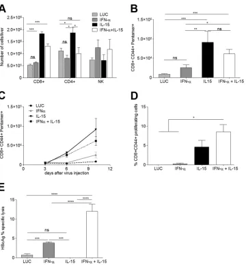
AAV–IL-15 enhances HBV-specific T cell responses in
HBVTg mice.
To explore the nature of the liver infiltrate further,
its cellular composition was analyzed in more detail. IL-15 alone
or in combination with IFN-
␣
induced an increase in total
lym-phocyte numbers (data not shown). While IL-15 induced a
marked rise in the number of CD8
⫹T cells and a moderate
eleva-tion of CD4
⫹T cells (
Fig. 4A
), only the number of CD8
⫹cells, not
that of CD4
⫹T cells, was also significantly increased in the
com-bination treatment group. Another cell type that has been
impli-cated in virus control and clearance (reviewed in reference
27
), the
NK cell, was found in greatly elevated numbers in response to
IFN-
␣
; however, IL-15 seemed to partially counteract this
expan-sion in the combination therapy (
Fig. 4A
).
Since CD8
⫹T cells are one of the key players in clearing acute
HBV infection, the number of HBV-specific CD8
⫹T cells in the
liver was determined using H-2Kb pentamers specific for HBcAg
epitope C
93–100. Both IL-15- and IFN-
␣
–IL-15-treated mice
showed significantly more intrahepatic CD8
⫹CD44
⫹pentamer-positive T cells than the IFN-
␣
and control groups (
Fig. 4B
and data not shown). Similar results were obtained by using
pentamers specific for HBsAg (data not shown); however, no
OVA-specific T cells were detected in the different treatment
FIG 3The IL-15–IFN-␣combination treatment induces immune-mediated liver damage in HBVTg mice but not in WT mice. (A) Representative images of necroinflammatory lesions in HBVTg (top) and C57BL/6 (bottom) mice on day 20 after vector administration. (B) TUNEL-positive nuclei (right) and 4=,6-diamidino-2-phenylindole (DAPI) staining (left) of the same mice. (C) Serum ALT levels in HBVTg mice at days 10 and 20 after vector administration. Results are expressed as means⫾SEM for 5 to 6 animals/group (***,P⬍0.001).
Induction of HBV Immunity by IFN-␣–IL-15 Combination
on November 7, 2019 by guest
http://jvi.asm.org/
[image:5.585.136.450.68.332.2]groups (data not shown). To further characterize the response,
the numbers of HBV-specific CD8
⫹lymphocytes in the liver were
analyzed at different time points after treatment. While in IFN-
␣
-treated mice HBcAg-specific CD8
⫹T cells were detected in very
low numbers at day 3 after treatment and increased only
moder-ately thereafter, the number of pentamer-positive cells in the livers
of animals treated with IL-15 or the combination increased
signif-icantly over time. The increase was highest in the IL-15 treatment
group (
Fig. 4C
). Only a very low pentamer-positive population
could be detected in control animals, and this population is likely
to represent the tolerant HBcAg-specific population. The
re-sponse to HBsAg reflected the rere-sponse to HBcAg in all groups
(data not shown). Furthermore, while CD8
⫹lymphocyte
expan-sion was observed in the livers of C57BL/6 mice treated with IL-15
or IL-15–IFN-
␣
, no HBV- or OVA-specific CD8 T cells were
de-tected (data not shown). When isolated intrahepatic lymphocytes
(IHL) were stimulated
ex vivo
on day 10, a robust proliferative
response to C
93–100(MGLKFRQL) could be observed in
IL-15-and IL-15–IFN-
␣
-treated animals, and this response was more
intense with the combination treatment (
Fig. 4D
).
To test if HBV-specific CD8 T cells were able to exert effector
functions, an
in vivo
killing assay (IVK) was performed 10 days
after the administration of the different treatments. Splenocytes
isolated from HBVTg or WT mice were loaded with epitope
C
93–100or with an irrelevant peptide (OVA
257–264), labeled with 5
M or 0.5
M CFSE, respectively, and transferred i.v. to the
groups of mice treated as before. Killing was analyzed 24 h after
transfer. No specific lysis was detected in animals treated with the
control or IL-15 alone, while in mice that had received IFN-
␣
or
IFN-
␣
plus IL-15, specific killing of HBV peptide-loaded cells
could be observed (
Fig. 4E
). The percentage of lysis was
signifi-cantly higher with the combination treatment than with IFN-
␣
alone. Similar results were obtained in an IVK assay using HBsAg
epitope S
208 –215(ILSPFLPL) (data not shown). No HBV-specific
CD8
⫹T cells or cytotoxic activity was seen in WT mice (data not
shown).
FIG 4IL-15-induced expansion of HBcAg-specific CD8⫹T cells requires combination with IFN-␣to render them functional CTLs. The mononuclear liver infiltrate was isolated and analyzed on day 10 after vector administration. (A and B) The absolute numbers of CD8⫹, CD4⫹, and NK cells per liver (A) and the number of HBcAg-specific CD8⫹T cells per liver (B) are shown (n⫽7). (C) The increase in the number of HBcAg-specific CD8⫹T cells was analyzed over time (n⫽3/time point). (D) Proliferative response of HBcAg-specific CD8⫹T cells upon peptide stimulation.In vitrostimulation was performed in triplicate with cells obtained from 3 mice per group. (E) Proportion of lysed HBcAg-loaded target cellsin vivo(n⫽5). Results are representative of 3 independent experiments. All graphs show means⫾SEM. *,P⬍0.05; **,P⬍0.01; ***,P⬍0.001; ****,P⬍0.001; ns, nonsignificant.
Di Scala et al.
on November 7, 2019 by guest
http://jvi.asm.org/
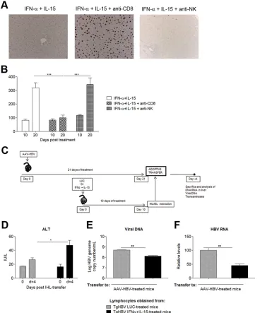
[image:6.585.115.475.62.449.2]CD8
ⴙT cells play an essential role in the antiviral response to
HBV.
To investigate the importance of CD8
⫹T cells and NK cells
in mediating the antiviral response elicited by the combination
therapy, HBVTg mice treated with IL-15 plus IFN-
␣
were divided
into three groups. Group 1 was depleted of CD8
⫹T cells; group 2
was depleted of NK cells; and group 3 received an isotype control
antibody. The expression of HBcAg in the liver, which appeared to
be a sensitive indicator of the intensity of the antiviral response,
was assessed by IHC. As shown in
Fig. 5A
, depletion of CD8
⫹lymphocytes completely abrogated the antiviral effect of the
com-bination therapy, while NK cell depletion had little impact on the
antiviral response. Moreover, while the combined treatment
re-sulted in an elevation of serum transaminases, this alteration was
absent in the treated transgenic mice subjected to depletion of
CD8
⫹T cells (
Fig. 5B
). These findings indicate that CD8
⫹lym-phocytes are the key players in the antiviral response and that the
disappearance of virus from the liver is not solely caused by a
direct antiviral effect of the cytokines.
Importantly, transfer of splenic and intrahepatic lymphocytes
from HBVTg mice treated with IL-15 plus IFN-
␣
, but not from those
receiving the LUC control, to C57BL/6 mice injected with an AAV
vector carrying the HBV 1.3 genome—another model of chronic
HBV hepatitis (
28
)—induced moderate hypertransaminasemia as
well as a reduction in HBV viremia (
Fig. 5C
to
E
). Additionally,
sig-nificantly lower levels of hepatic HBV RNA could be observed at day
4 after transfer (
Fig. 5F
), while DNA levels remained unchanged (data
FIG 5Combined IL-15–IFN-␣treatment confers partial protective CD8 immunity. (A) HBVTg mice treated with AAV–IL-15 plus AAV–IFN-␣were depleted of CD8⫹or NK cells. Hepatic HBcAg expression was analyzed 10 days later. Representative images from each group are shown. (B) Serum ALT levels (IU/liter) in HBVTg mice depleted of CD8⫹or NK cells at day 10 after vector administration. (C) Scheme of the adoptive transfer protocol. (D) Serum ALT levels at day 0 and day 4 (d⫹4) post-IHL transfer. (E and F) HBV genome copy numbers (E) and HBV RNA copies (F) on day 4 after IHL transfer. Results are normalized to GADPH levels. All data are presented as mean (n, 3 to 4)⫾SEM. *,P⬍0.05; **,P⬍0.01; ***,P⬍0.001.
Induction of HBV Immunity by IFN-␣–IL-15 Combination
on November 7, 2019 by guest
http://jvi.asm.org/
[image:7.585.111.479.67.510.2]not shown). In contrast, LUC DNA and RNA expression did not
change in control mice infected with AAV-LUC prior to lymphocyte
transfer (data not shown). Taken together, these data suggest that the
antiviral effector functions of lymphocytes rescued from tolerance by
IL-15–IFN-
␣
treatment are not a bystander effect and can be
trans-ferred to chronically HBV infected mice.
IL-15 upregulates PD-1 and PD-L1 in the inflammatory
infil-trate of the liver, an effect counteracted by IFN-
␣
.
PD-1 and PD-L1
constitute a key control point of the CD8
⫹T cell response and have
been thought to negatively regulate viral responses (
5
). Since both
PD-1 and PD-L1, the receptor and its ligand, can be upregulated by
IFN-
␣
and IL-15 (
26
,
29–31
), their expression was analyzed on
hepa-tocytes and IHL. The proportion of intrahepatic PD-1
⫹CD8
⫹cells
was increased in all treatment groups, especially in the presence of
IL-15 (
Fig. 6A
). Surprisingly coinfection with IFN-
␣
seemed to offset
this effect to a significant degree. On NK cells, PD-1 upregulation was
observed only in the context of IL-15, and again, coadministration of
IFN-
␣
seemed to dampen this effect (
Fig. 6B
). When PD-L1
expres-sion on the surfaces of hepatocytes (
Fig. 6C
) and relative RNA levels
in liver extracts (
Fig. 6D
) were analyzed, they were also found to be
increased in IL-15-treated mice. However, as in lymphocytes and NK
cells, combination treatment with IFN-
␣
reduced IL-15-induced
PD-L1 expression. For intrahepatic dendritic cells, the same
observa-tion was made (data not shown).
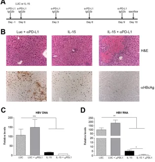
PD-L1 blockade enhances the rescue effect of IL-15.
Since the
PD-1–PD-L1 interaction has been implicated in the induction and
maintenance of peripheral tolerance (
32
,
33
) and exhaustion
(re-viewed in reference
34
), the effect of PD-L1 expression on the
effector functions of IHL expanded in IL-15-treated HBVTg mice
was investigated. An anti-PD-L1 blocking antibody was
adminis-tered intraperitoneally before and after injection of AAV–IL-15 or
a control (
Fig. 7A
). IHC performed on day 10 revealed large areas
of necrosis (
Fig. 7B
, top) and a simultaneous rise in ALT levels
(data not shown), as well as an almost-complete disappearance of
nuclear HBV core protein from the livers of animals treated with
IL-15 in combination with anti-PD-L1 (
Fig. 7B
, bottom). Analysis
of HBV DNA and RNA levels in the liver by quantitative PCR
showed that IL-15 alone or in combination with anti-PD-L1
sig-nificantly reduced both HBV DNA (
Fig. 7C
) and RNA (
Fig. 7D
)
levels; the decrease in HBV RNA levels was significantly more
pronounced in the last group. Importantly, IL-15 plus PD-L1
blockade did not cause any tissue damage in wild-type mice.
Un-expectedly, PD-L1 blockade in mice that had received IFN-
␣
–
IL-15 treatment did not augment the infiltration of CD8 cells or
their response over that with combination treatment alone. Only a
very subtle effect, if any, on the reduction in the number of
HBcAg
⫹cells could be observed (data not shown).
Treatment with IL-15 plus IFN-
␣
can restore effector
func-tion and expand the HBV-specific CD8 populafunc-tion in some CHB
patients.
To test if our findings would be transferable to
chroni-cally infected patients, a first
in vitro
study with PBMC samples
from a small cohort of patients with CHB was performed. Upon
stimulation in IL-2-supplemented medium in the presence or
ab-sence of an HBV peptide and the cytokines indicated in
Fig. 8
, the
addition of IL-15 resulted in unspecific bulk expansion of the live
CD8 T cell population in culture, while IFN-
␣
reduced the
popu-lation (
Fig. 8A
). Interestingly, as in the mouse study presented in
this paper, IL-15 partially compensated for this effect. In the
pres-ence of both cytokines, the overall proportion of HBV-specific
granzyme B-producing CD8 T cells (
Fig. 8B
), as well as the
num-ber of patients responding (
Fig. 8C
), was significantly increased
relative to that with either cytokine alone, especially in the
pres-ence of an HBV peptide. The percentage of IFN-
␥
-producing CD8
T cells upon HBV peptide stimulation was also increased by either
IFN-
␣
or IL-15 alone or by their combination in approximately
half of the 21 patients with CHB tested (
Fig. 8D
). Because of the
global CD8 expansion induced by the combination, the absolute
increase in HBV-specific CD8 T cells can be expected to be even
FIG 6IL-15-induced expression of PD-1 and PD-L1 on liver infiltrates is reduced by IFN-␣. (A and B) Ten days after vector administration, PD-1 expression on intrahepatic CD8⫹(A) and NK⫹(B) cells was analyzed by flow cytometry. (C) Mean fluorescence intensity (MFI) of PD-L1 expressed on the surfaces of purified hepatocytes. (D) Relative expression of PD-1 in RNA extracted from liver. Results are expressed as means (n⫽6)⫾SEM. *,P⬍0.05; **,P⬍0.01; ***,
P⬍0.001; ****,P⬍0.0001.
Di Scala et al.
on November 7, 2019 by guest
http://jvi.asm.org/
[image:8.585.138.454.65.287.2]more significant. Further studies will require HLA/peptide
dex-tramers to dissect antigen-specific and bystander boosting in a
larger cohort of patients with CHB.
DISCUSSION
The hallmark of CHB is immune dysfunction, which results in
persistent infection (
3
,
4
). Because of their stimulatory properties,
cytokines such as IFN-
␣
and IL-15 are considered good candidates
to aid the functional recovery of HBV-specific lymphocytes (
17
,
35
), and recent work has also highlighted the potential of IFN-
␣
to
stimulate cell-intrinsic antiviral mechanisms in HBV-infected
hepatocytes (
9
). However, while IFN-
␣
therapy could strongly
drive the expansion and activation of NK cells, it did not reactivate
HBV-specific T cells in CHB patients (
7
,
8
). To investigate
whether it would be possible to increase the antiviral potential of
IFN-
␣
and to more efficiently activate the adaptive arm of the
immune response by combining it with the stimulatory cytokine
IL-15, an HBVTg mouse model was used (
20
). These mice present
with dysfunctional, unresponsive HBV-specific CD8 T cells and
have been widely used to test immunostimulatory therapies aimed
at clearing HBV infection (
22
,
23
,
36
,
37
).
The presence of cytoplasmic HBcAg is an indicator of active
HBV replication, whereas nuclear HBcAg is highly stable,
persist-ing in the absence of HBV replication (
38
,
39
). In accordance with
previous findings (
9
), significant decreases in hepatic HBV DNA
and HBV RNA levels were obtained upon infection with
AAV-expressed IL-15 or IFN-
␣
alone, indicating an attenuation of HBV
transcriptional activity and an abrogation of viral replication.
However, a high proportion of hepatocyte nuclei remained
HBcAg positive. In stark contrast, coinfection with both cytokines
resulted in the complete disappearance of HBV core protein from
hepatocytes, likely reflecting the successful initiation of an
anti-HBV immune response capable of controlling viral replication.
Hence, the efficacy of the combined therapy appeared to depend
on the synergistic activities of the two cytokines. In terms of
cel-lular immunity, in line with existing literature, IL-15 triggered a
robust expansion of the HBV-specific CD8 T cell population (
17
,
35
), while IFN-
␣
induced differentiation into effector cytotoxic T
lymphocytes (CTL) without major cellular expansion (
40
). When
combined, HBV-specific CD8 T lymphocytes not only infiltrated
the liver in greater numbers but were also able to exert enhanced
cytotoxic activity, as reflected in increased serum ALT levels as
well as liver pathology. Importantly, no immune response against
an irrelevant H-2Kb peptide was detected, and no HBV- or
OVA-specific CD8 T cell responses were present in the livers of C57BL/6
wild-type mice receiving the same treatment. In contrast, IHL
FIG 7The combination of IL-15 therapy with PD-L1 blockade induces immune-mediated liver damage in HBVTg mice and a strong antiviral effect. (A) Treatment scheme. (B) Representative images of liver sections stained with H&E (upper panels) or stained for core antigen (lower panels). (C and D) HBV (C) and HBV RNA (D) relative levels on day 10 after vector administration. Results are normalized to GADPH levels. Data are presented as means (n⫽5)⫾SEM. *,P⬍0.05; **,P⬍0.01; ***,P⬍0.001.
Induction of HBV Immunity by IFN-␣–IL-15 Combination
on November 7, 2019 by guest
http://jvi.asm.org/
[image:9.585.135.450.70.400.2]from HBVTg mice that had received IL-15 only, although
numer-ous, were not able to kill target cells
in vivo
or induce
hepatocel-lular necrosis. These data support the notion that liver damage
was at least partially mediated by the complementary effects of
IFN-
␣
and IL-15 on specific CD8 T cells and was not only a
by-stander effect. The successful recovery of HBV-specific CD8
⫹ef-fector cells with the combination treatment and their importance
for viral clearance were further corroborated upon their systemic
elimination by repeated anti-CD8 administration: the previously
observed antiviral effect of the cytokine coadministration
disap-peared, and HBc protein remained detectable in hepatocyte
nu-clei. Therefore, the viral clearance observed in the liver was not
due solely to the direct, antiviral effects of IFN-
␣
and/or IL-15.
Although in recent years, a potential antiviral role in chronic
HBV infection has been attributed to NK cells (
27
,
41
), the
ther-apeutic approach presented here does not seem to support this
notion. Both alone and in combination with IL-15, IFN-
␣
resulted
in elevated NK cell numbers; however, in contrast to the results
obtained for CD8
⫹cells, depletion of NK cells did not result in
significant recovery of HBcAg
⫹hepatocytes. This finding is in line
with the observations of Yin et al. (
19
), who reported an
NK-independent effect of IL-15 on HBV.
FIG 8Combination treatment with IL-15 and IFN-␣can restore partial CD8 effector function in some patients with chronic hepatitis B. PBMCs from patients with CHB were cultured in IL-2-supplemented medium in the presence/absence of HBV peptide and the indicated cytokines. (A) Cumulative data showing the percentage of CD8 T cells after 10 days of culture. Numbers of live CD8 T cells were measured by using a live/dead staining kit to exclude dead cells. (B) Cumulative data showing the percentage of granzyme B-positive CD8⫹cells in the presence of IFN-␣, IL-15, or both in the absence or presence of HBV peptide stimulation. (C and D) Individual responses of CHB patients, expressed as the percentage of CD8⫹cells producing granzyme B (C) or IFN-␥(D). Data represent means⫾SEM. *,P⬍0.05; **,P⬍0.01; ***,P⬍0.001. Viral loads (VL), expressed as log10IU per milliliter, and ALT levels, expressed as IU per liter, are given
below the graphs. n.a., not available; BLQ, below the limit of quantification.
Di Scala et al.
on November 7, 2019 by guest
http://jvi.asm.org/
[image:10.585.112.475.59.513.2]The exact role of PD-1–PD-L1 interaction in the induction and
maintenance of T cell tolerance, as well as in exhaustion, is still being
disputed and appears rather complex. However, it could be shown
that PD-1 blockade can restore T cell function either directly, by using
blocking antibodies (
42
,
43
), or indirectly, by blockade of IFN-
␣
(
26
,
31
). In contrast to the latter reports, in our hands, the high increase in
the level of PD-1 expressed on intrahepatic CD8
⫹- and NK cells from
HBVTg mice treated with AAV–IL-15 alone was significantly
re-duced in mice that had received the IL-15–IFN-
␣
combination
ther-apy. Thus, our data point to a role of IFN-
␣
in prohibiting
IL-15-induced PD-L1 upregulation, an effect that would be crucial for
licensing effector T lymphocytes to attack target cells. It was reported
previously that PD-1 does not directly mediate apoptosis but that its
expression levels correlate to the susceptibility of the cell to apoptosis
(
29
). In this study, pretreatment with common
␥
– chain cytokines,
IL-15 included, induced PD-1 upregulation but did not interfere with
expansion, enhancement of some effector functions, or survival. This
treatment did, however, inhibit the exertion of certain effector
func-tions. The work presented here points to the synergy of IL-15 and
IFN-
␣
in overcoming these obstacles. How this may happen in detail
requires more exploration. One possibility is the upregulation of
other stimulating cytokines/receptors on either the T cells themselves
or another cell type, such as dendritic cells. Our approach is
corrob-orated by a recently published study that investigated the roles of Bim,
PD-1, and Socs-1 in the onset and degree of acute liver damage in a T
cell receptor (TCR) transgenic model. The authors concluded that
strategies aimed at overcoming the block in effector function would
be more efficient at clearing chronic hepatitis than those purely
aim-ing at prolongaim-ing survival (
44
).
The functional block could also be overcome by blocking the
PD-1–PD-L1 interaction and resulted in extensive areas of
hepa-tocellular necrosis and complete clearance of viral antigens from
the livers of IL-15-treated mice, indicating that this therapy is
equally effective in restoring the anti-HBV immune response.
Although the use of vector-delivered cytokines—and IL-15 in
particular— comes with many potential problems in a clinical
set-ting, our work demonstrated that the synergistic potential of the
ap-propriate combination of proliferation- and effector-inducing
cyto-kines can result in the recovery of dysfunctional T cell populations.
This work provides new insight into how to tune existing IFN-
␣
treat-ment of CHB and also offers a therapeutic approach for chronic viral
infections and neoplastic conditions. As shown, preliminary results
from human studies indicate that this approach may be successful,
although the addition of IL-15 appears to promote global as well as
HBV-specific T cell expansion, as recently described for HIV patients
(
45
). Such bystander T cell responses may increase the risk of hepatic
flares, in line with the necroinflammatory responses observed in our
mouse model, but could also contribute to a heterologous antiviral
response (
46
). Further in depth studies will have to be done to explore
the synergistic effects of IFN-
␣
and other proliferation-promoting
common
␥
-chain cytokines and their potential efficacy for CHB
pa-tients.
ACKNOWLEDGMENTS
We thank Francis V. Chisari for kindly providing the HBVTg mice, Tomas Aragon and Rafael Aldabe for discussions, and Elena Ciordia, Alberto Espinal, and CIFA staff for animal care and vivarium management.
We declare no conflict of interest.
FUNDING INFORMATION
This work was supported by UTE project CIMA, Fundación Barrié de la Maza y Condesa de Fenosa, Fundación Fuentes Dutor, CIBERehd Insti-tuto de Salud Carlos III, SAF2009-08524 and SAF2012-39578 (G.G.-A.) from the Spanish Department of Science. M.D.S. was funded by a fellow-ship from Fondo de Investigaciones Sanitarias, (FI09/00647) and I.G.-F. and L.V. received FPI grants (BES-2010-038245 and BES-2007-17050, respectively). I.O. is funded by an EASL fellowship and M.K.M. by a Well-come Trust Senior Investigator Award. C.O.D.S. received MINECO grant DPI2015-64221-C2-2. The funders had no role in study design, data col-lection and interpretation, or the decision to submit the work for publi-cation.
REFERENCES
1.Trepo C, Chan HL, Lok A.2014. Hepatitis B virus infection. Lancet 384:2053–2063.http://dx.doi.org/10.1016/S0140-6736(14)60220-8. 2.Guidotti LG, Chisari FV.2006. Immunobiology and pathogenesis of viral
hepatitis. Annu Rev Pathol1:23– 61.http://dx.doi.org/10.1146/annurev .pathol.1.110304.100230.
3.Rehermann B, Nascimbeni M.2005. Immunology of hepatitis B virus and hepatitis C virus infection. Nat Rev Immunol5:215–229.http://dx .doi.org/10.1038/nri1573.
4.Boni C, Fisicaro P, Valdatta C, Amadei B, Di Vincenzo P, Giuberti T, Laccabue D, Zerbini A, Cavalli A, Missale G, Bertoletti A, Ferrari C. 2007. Characterization of hepatitis B virus (HBV)-specific T-cell dysfunc-tion in chronic HBV infecdysfunc-tion. J Virol81:4215– 4225.http://dx.doi.org/10 .1128/JVI.02844-06.
5.Maier H, Isogawa M, Freeman GJ, Chisari FV. 2007. PD-1:PD-L1 interactions contribute to the functional suppression of virus-specific CD8⫹T lymphocytes in the liver. J Immunol178:2714 –2720.http://dx .doi.org/10.4049/jimmunol.178.5.2714.
6.Degasperi E, Vigano M, Aghemo A, Lampertico P, Colombo M.2013. PegIFN-␣2a for the treatment of chronic hepatitis B and C: a 10-year history. Expert Rev Anti Infect Ther 11:459 – 474.http://dx.doi.org/10 .1586/eri.13.37.
7.Micco L, Peppa D, Loggi E, Schurich A, Jefferson L, Cursaro C, Panno AM, Bernardi M, Brander C, Bihl F, Andreone P, Maini MK.2013. Differential boosting of innate and adaptive antiviral responses during pegylated-interferon-alpha therapy of chronic hepatitis B. J Hepatol58: 225–233.http://dx.doi.org/10.1016/j.jhep.2012.09.029.
8.Penna A, Laccabue D, Libri I, Giuberti T, Schivazappa S, Alfieri A, Mori C, Canetti D, Lampertico P, Vigano M, Colombo M, Loggi E, Missale G, Ferrari C.2012. Peginterferon-alpha does not improve early peripheral blood HBV-specific T-cell responses in HBeAg-negative chronic hepatitis. J Hepatol 56:1239 –1246.http://dx.doi.org/10.1016/j.jhep.2011.12.032.
9.Lucifora J, Xia Y, Reisinger F, Zhang K, Stadler D, Cheng X, Sprinzl MF, Koppensteiner H, Makowska Z, Volz T, Remouchamps C, Chou WM, Thasler WE, Huser N, Durantel D, Liang TJ, Munk C, Heim MH, Browning JL, Dejardin E, Dandri M, Schindler M, Heikenwalder M, Protzer U.2014. Specific and nonhepatotoxic degradation of nuclear hepatitis B virus cccDNA. Science343:1221–1228.http://dx.doi.org/10 .1126/science.1243462.
10. Markova AA, Mihm U, Schlaphoff V, Lunemann S, Filmann N, Bremer B, Berg T, Sarrazin C, Zeuzem S, Manns MP, Cornberg M, Herrmann E, Wedemeyer H.2014. PEG-IFN alpha but not ribavirin alters NK cell phenotype and function in patients with chronic hepatitis C. PLoS One 9:e94512.http://dx.doi.org/10.1371/journal.pone.0094512.
11. Jayaraman A, Jackson DJ, Message SD, Pearson RM, Aniscenko J, Caramori G, Mallia P, Papi A, Shamji B, Edwards M, Westwick J, Hansel T, Stanciu LA, Johnston SL, Bartlett NW.2014. IL-15 complexes induce NK- and T-cell responses independent of type I IFN signaling during rhinovirus infection. Mucosal Immunol7:1151–1164.http://dx .doi.org/10.1038/mi.2014.2.
12. Hervas-Stubbs S, Mancheno U, Riezu-Boj JI, Larraga A, Ochoa MC, Alignani D, Alfaro C, Morales-Kastresana A, Gonzalez I, Larrea E, Pircher H, Le Bon A, Lopez-Picazo JM, Martin-Algarra S, Prieto J, Melero I.2012. CD8 T cell priming in the presence of IFN-␣renders CTLs with improved responsiveness to homeostatic cytokines and recall anti-gens: important traits for adoptive T cell therapy. J Immunol189:3299 – 3310.http://dx.doi.org/10.4049/jimmunol.1102495.
13. Schluns KS, Williams K, Ma A, Zheng XX, Lefrancois L. 2002.
Induction of HBV Immunity by IFN-␣–IL-15 Combination
on November 7, 2019 by guest
http://jvi.asm.org/
Requirement for IL-15 in the generation of primary and memory an-tigen-specific CD8 T cells. J Immunol168:4827– 4831.http://dx.doi .org/10.4049/jimmunol.168.10.4827.
14. Huntington ND.2014. The unconventional expression of IL-15 and its role in NK cell homeostasis. Immunol Cell Biol92:210 –213.http://dx.doi .org/10.1038/icb.2014.1.
15. Van Belle TL, Dooms H, Boonefaes T, Wei XQ, Leclercq G, Grooten J. 2012. IL-15 augments TCR-induced CD4⫹T cell expansion in vitro by inhibiting the suppressive function of CD25 high CD4⫹T cells. PLoS One 7:e45299.http://dx.doi.org/10.1371/journal.pone.0045299.
16. Chen XL, Bobbala D, Cepero Donates Y, Mayhue M, Ilangumaran S, Ramanathan S.2014. IL-15trans-presentation regulates homeostasis of CD4⫹T lymphocytes. Cell Mol Immunol11:387–397.http://dx.doi.org /10.1038/cmi.2014.13.
17. Teague RM, Sather BD, Sacks JA, Huang MZ, Dossett ML, Morimoto J, Tan X, Sutton SE, Cooke MP, Ohlen C, Greenberg PD. 2006. Interleukin-15 rescues tolerant CD8⫹T cells for use in adoptive immu-notherapy of established tumors. Nat Med12:335–341.http://dx.doi.org /10.1038/nm1359.
18. DePaolo RW, Abadie V, Tang F, Fehlner-Peach H, Hall JA, Wang W, Marietta EV, Kasarda DD, Waldmann TA, Murray JA, Semrad C, Kupfer SS, Belkaid Y, Guandalini S, Jabri B.2011. Co-adjuvant effects of retinoic acid and IL-15 induce inflammatory immunity to dietary anti-gens. Nature471:220 –224.http://dx.doi.org/10.1038/nature09849. 19. Yin W, Xu L, Sun R, Wei H, Tian Z.2012. Interleukin-15 suppresses
hepatitis B virus replication via IFN-production in a C57BL/6 mouse model. Liver Int32:1306 –1314.http://dx.doi.org/10.1111/j.1478-3231.2012 .02773.x.
20. Guidotti LG, Matzke B, Schaller H, Chisari FV.1995. High-level hepatitis B virus replication in transgenic mice. J Virol69:6158 – 6169.
21. Inuzuka T, Takahashi K, Chiba T, Marusawa H.2014. Mouse models of hepatitis B virus infection comprising host-virus immunologic interactions. Pathogens3:377–389.http://dx.doi.org/10.3390/pathogens3020377. 22. Shimizu Y, Guidotti LG, Fowler P, Chisari FV. 1998. Dendritic cell
immunization breaks cytotoxic T lymphocyte tolerance in hepatitis B vi-rus transgenic mice. J Immunol161:4520 – 4529.
23. Buchmann P, Dembek C, Kuklick L, Jager C, Tedjokusumo R, von Freyend MJ, Drebber U, Janowicz Z, Melber K, Protzer U.2013. A novel therapeutic hepatitis B vaccine induces cellular and humoral immune re-sponses and breaks tolerance in hepatitis B virus (HBV) transgenic mice. Vaccine31:1197–1203.http://dx.doi.org/10.1016/j.vaccine.2012.12.074. 24. Di Scala M, Gil-Farina I, Vanrell L, Sanchez-Bayona R, Alignani D,
Olague C, Vales A, Berraondo P, Prieto J, Gonzalez-Aseguinolaza G. 2015. Chronic exposure to IFN␣drives medullar lymphopoiesis towards T-cell differentiation in mice. Haematologica100:1014 –1022.http://dx .doi.org/10.3324/haematol.2014.
25. Gil-Farina I, Di Scala M, Vanrell L, Olague C, Vales A, High KA, Prieto J, Mingozzi F, Gonzalez-Aseguinolaza G. 2013. IL12-mediated liver inflammation reduces the formation of AAV transcriptionally active forms but has no effect over preexisting AAV transgene expression. PLoS One8:e67748.http://dx.doi.org/10.1371/journal.pone.0067748. 26. Teijaro JR, Ng C, Lee AM, Sullivan BM, Sheehan KC, Welch M,
Schreiber RD, de la Torre JC, Oldstone MB.2013. Persistent LCMV infection is controlled by blockade of type I interferon signaling. Science 340:207–211.http://dx.doi.org/10.1126/science.1235214.
27. Rehermann B.2015. Natural killer cells in viral hepatitis. Cell Mol Gastroenterol Hepatol1:578–588.http://dx.doi.org/10.1016/j.jcmgh.2015.09.004.
28. Huang YH, Fang CC, Tsuneyama K, Chou HY, Pan WY, Shih YM, Wu PY, Chen Y, Leung PS, Gershwin ME, Tao MH.2011. A murine model of hepatitis B-associated hepatocellular carcinoma generated by adeno-associated virus-mediated gene delivery. Int J Oncol39:1511–1519.http: //dx.doi.org/10.3892/ijo.2011.1145.
29. Kinter AL, Godbout EJ, McNally JP, Sereti I, Roby GA, O’Shea MA, Fauci AS.2008. The common gamma-chain cytokines IL-2, IL-7, IL-15, and IL-21 induce the expression of programmed death-1 and its ligands. J Immunol 181:6738 – 6746.http://dx.doi.org/10.4049/jimmunol.181.10.6738. 30. Terawaki S, Chikuma S, Shibayama S, Hayashi T, Yoshida T, Okazaki
T, Honjo T.2011. IFN-␣directly promotes programmed cell death-1 transcription and limits the duration of T cell-mediated immunity. J Im-munol186:2772–2779.http://dx.doi.org/10.4049/jimmunol.1003208. 31. Wilson EB, Yamada DH, Elsaesser H, Herskovitz J, Deng J, Cheng G,
Aronow BJ, Karp CL, Brooks DG.2013. Blockade of chronic type I
interferon signaling to control persistent LCMV infection. Science340: 202–207.http://dx.doi.org/10.1126/science.1235208.
32. Francisco LM, Sage PT, Sharpe AH.2010. The PD-1 pathway in toler-ance and autoimmunity. Immunol Rev236:219 –242.http://dx.doi.org/10 .1111/j.1600-065X.2010.00923.x.
33. Keir ME, Butte MJ, Freeman GJ, Sharpe AH.2008. PD-1 and its ligands in tolerance and immunity. Annu Rev Immunol26:677–704.http://dx.doi .org/10.1146/annurev.immunol.26.021607.090331.
34. Wherry EJ, Kurachi M.2015. Molecular and cellular insights into T cell exhaustion. Nat Rev Immunol 15:486 – 499. http://dx.doi.org/10.1038 /nri3862.
35. Miyagawa F, Tagaya Y, Kim BS, Patel HJ, Ishida K, Ohteki T, Wald-mann TA, Katz SI.2008. IL-15 serves as a costimulator in determining the activity of autoreactive CD8 T cells in an experimental mouse model of graft-versus-host-like disease. J Immunol181:1109 –1119.http://dx.doi .org/10.4049/jimmunol.181.2.1109.
36. Berraondo P, Di Scala M, Korolowicz K, Thampi LM, Otano I, Suarez L, Fioravanti J, Aranda F, Ardaiz N, Yang J, Kallakury BV, Tucker RD, Vasquez M, Menne S, Prieto J, Gonzalez-Aseguinolaza G.2015. Liver-directed gene therapy of chronic hepadnavirus infection using interferon alpha tethered to apolipoprotein A-I. J Hepatol63:329 –336.http://dx.doi .org/10.1016/j.jhep.2015.02.048.
37. Backes S, Jager C, Dembek CJ, Kosinska AD, Bauer T, Stephan AS, Dislers A, Mutwiri G, Busch DH, Babiuk LA, Gasteiger G, Protzer U. 2016. Protein-prime/modified vaccinia virus Ankara vector-boost vacci-nation overcomes tolerance in high-antigenemic HBV-transgenic mice. Vaccine34:923–932.http://dx.doi.org/10.1016/j.vaccine.2015.12.060. 38. Burrell CJ, Gowans EJ, Marmion BP.1985. High levels of cytoplasmic
hepatitis B core antigen as reliable marker of HBV DNA replication. Lan-ceti:454 – 455.
39. Gowans EJ, Burrell CJ, Jilbert AR, Marmion BP.1985. Cytoplasmic (but not nuclear) hepatitis B virus (HBV) core antigen reflects HBV DNA syn-thesis at the level of the infected hepatocyte. Intervirology24:220 –225.
http://dx.doi.org/10.1159/000149646.
40. Marshall HD, Prince AL, Berg LJ, Welsh RM.2010. IFN-␣and self-MHC divert CD8 T cells into a distinct differentiation pathway character-ized by rapid acquisition of effector functions. J Immunol185:1419 –1428.
http://dx.doi.org/10.4049/jimmunol.1001140.
41. Stelma F, de Niet A, Tempelmans Plat-Sinnige MJ, Jansen L, Takken-berg RB, Reesink HW, Kootstra NA, van Leeuwen EM.2015. Natural killer cell characteristics in patients with chronic hepatitis B virus (HBV) infection are associated with HBV surface antigen clearance after combi-nation treatment with pegylated interferon alfa-2a and adefovir. J Infect Dis212:1042–1051.http://dx.doi.org/10.1093/infdis/jiv180.
42. Bengsch B, Martin B, Thimme R.2014. Restoration of HBV-specific CD8⫹T cell function by PD-1 blockade in inactive carrier patients is linked to T cell differentiation. J Hepatol61:1212–1219.http://dx.doi.org /10.1016/j.jhep.2014.07.005.
43. Tzeng HT, Tsai HF, Liao HJ, Lin YJ, Chen L, Chen PJ, Hsu PN.2012. PD-1 blockage reverses immune dysfunction and hepatitis B viral persis-tence in a mouse animal model. PLoS One7:e39179.http://dx.doi.org/10 .1371/journal.pone.0039179.
44. Vo M, Holz LE, Wong YC, English K, Benseler V, McGuffog C, Azuma M, McCaughan GW, Bowen DG, Bertolino P.2016. Effector T cell function rather than survival determines extent and duration of hepatitis in mice. J Hepatol64:1327–1133.http://dx.doi.org/10.1016/j.jhep.2016.01.040. 45. Younes SA, Freeman ML, Mudd JC, Shive CL, Reynaldi A, Panigrahi
S, Estes JD, Deleage C, Lucero C, Anderson J, Schacker TW, Dav-enport MD, McCune JM, Hunt PW, Lee SA, Serrano-Villar S, Debernardo RL, Jacobson JM, Canaday DH, Sekaly RP, Rodriguez B, Sieg SF, Lederman MM. 2016. IL-15 promotes activation and expansion of CD8⫹T cells in HIV-1 infection. J Clin Invest126:2745– 2756.http://dx.doi.org/10.1172/JCI85996.
46. Sandalova E, Laccabue D, Boni C, Tan AT, Fink K, Ooi EE, Chua R, Shafaeddin Schreve B, Ferrari C, Bertoletti A.2010. Contribution of her-pesvirus specific CD8 T cells to anti-viral T cell response in humans. PLoS Pathog6:e1001051.http://dx.doi.org/10.1371/journal.ppat.1001051. 47. Abad X, Razquin N, Abad A, Fortes P.2010. Combination of RNA
inter-ference and U1 inhibition leads to increased inhibition of gene expression. Nucleic Acids Res38:e136.http://dx.doi.org/10.1093/nar /gkq299.
Di Scala et al.
on November 7, 2019 by guest
http://jvi.asm.org/