Localization Signals in the Adeno-Associated Virus Serotype 2
Assembly-Activating Protein
Lauriel F. Earley,a,bYasuhiro Kawano,b,cKei Adachi,bXiao-Xin Sun,bMu-Shui Dai,bHiroyuki Nakaib
Departments of Molecular Microbiology and Immunologya
and Molecular and Medical Genetics,b
Oregon Health and Science University School of Medicine, Portland, Oregon, USA; Takara Bio Inc., Otsu, Shiga, Japanc
ABSTRACT
Assembly-activating protein (AAP) of adeno-associated virus serotype 2 (AAV2) is a nucleolar-localizing protein that plays a critical role in transporting the viral capsid VP3 protein to the nucleolus for assembly. Here, we identify and characterize AAV2 AAP (AAP2) nuclear (NLS) and nucleolar (NoLS) localization signals near the carboxy-terminal region of AAP2 (amino acid
po-sitions 144 to 184) (AAP2144 –184). This region contains five basic-amino-acid-rich (BR) clusters, KSKRSRR (AAP2BR1), RRR
(AAP2BR2), RFR (AAP2BR3), RSTSSR (AAP2BR4), and RRIK (AAP2BR5), from the amino terminus to the carboxy terminus. We created 30 AAP2BR mutants by arginine/lysine-to-alanine mutagenesis or deletion of AAP2BRs and 8 and 1 green
fluores-cent protein (GFP)-AAP2BR and-galactosidase–AAP2BR fusion proteins, respectively, and analyzed their intracellular
local-ization in HeLa cells by immunofluorescence microscopy. The results showed that AAP2144 –184has redundant multipartite NLSs
and that any combinations of 4 AAP2BRs, but not 3 or less, can constitute a functional NLS-NoLS; AAP2BR1 and AAP2BR2 play the most influential role for nuclear localization, but either one of the two AAP2BRs is dispensable if all 4 of the other AAP2BRs are present, resulting in 3 different, overlapping NLS motifs; and the NoLS is shared redundantly among the five AAP2BRs and functions in a context-dependent manner. AAP2BR mutations not only resulted in aberrant intracellular localization, but also attenuated AAP2 protein expression to various degrees, and both of these abnormalities have a significant negative impact on capsid production. Thus, this study reveals the organization of the intermingling NLSs and NoLSs in AAP2 and provides insights into their functional roles in capsid assembly.
IMPORTANCE
Adeno-associated virus (AAV) has become a popular and successful vector forin vivogene therapy; however, its biology has yet
to be fully understood. In this regard, the recent discovery of the assembly-activating protein (AAP), a nonstructural, nucleolar-localizing AAV protein essential for viral capsid assembly, has provided us a new opportunity to better understand the funda-mental processes required for virion formation. Here, we identify clusters of basic amino acids in the carboxy terminus of AAP from AAV serotype 2 (AAV2) that act as nuclear and nucleolar localization signals. We also demonstrate their importance in maintaining AAP expression levels and efficient production of viral capsids. Insights into the functions of AAP can elucidate the requirements and process for AAV capsid assembly, which may lead to improved vector production for use in gene therapy. This study also contributes to the growing body of work on nuclear and nucleolar localization signals.
A
deno-associated virus (AAV) is a small, single-stranded DNA virus from the parvovirus family that has become a successful vector for gene delivery. The recent achievements in the field of AAV vector research have called attention to the incompletely understood life cycle of the virus. The AAV genome comprises two genes,repandcap, which encode the nonstructural Rep proteins and the structural VP proteins, respectively. The AAV virion is composed of 60 subunits comprising the three VP proteins, VP1, VP2, and VP3, encoded by a single open reading frame (ORF) in thecapgene. Recently, a nonstructural viral protein encoded by an alternative ORF within thecapgene was identified and termed assembly-activating protein (AAP) for its indispensable role in capsid formation (1–3). The AAP (AAP2) from AAV serotype 2 (AAV2) is a nucleolar-localizing protein that binds to VP proteins through interacting domains in the amino (N) terminus of AAP2 (1), transports the VP proteins to the nucleolus, and promotes capsid assembly (3). Therefore, AAP2 is expected to have both a nuclear localization signal (NLS) and a nucleolar localization sig-nal (NoLS) within its protein sequence. However, suchorganelle-targeting sequences in AAP2 remain to be identified and charac-terized.
The most common mechanism for targeting a protein to the nucleus is by an NLS that is recognized by one of the nuclear import proteins, termed importins, which are part of the large family of transport proteins known as karyopherins (4). Classical NLSs can be either monopartite, such as the PKKKRKV sequence
Received28 October 2014Accepted22 December 2014
Accepted manuscript posted online31 December 2014
CitationEarley LF, Kawano Y, Adachi K, Sun X-X, Dai M-S, Nakai H. 2015. Identification and characterization of nuclear and nucleolar localization signals in the adeno-associated virus serotype 2 assembly-activating protein.
J Virol 89:3038 –3048.doi:10.1128/JVI.03125-14.
Editor:M. J. Imperiale
Address correspondence to Hiroyuki Nakai, [email protected]. Copyright © 2015, American Society for Microbiology. All Rights Reserved.
doi:10.1128/JVI.03125-14
on November 7, 2019 by guest
http://jvi.asm.org/
in simian virus 40 (SV40) large T antigen (5), or bipartite, such as the KRPAATKKAGQAKKKK sequence in nucleophosmin (6,7). These classical NLSs are bound by the adaptor protein impor-tin-␣, which is then bound by importin-, forming a heterotri-meric complex consisting of the two importin proteins and the cargo protein. Importin-mediates nuclear entry of the hetero-trimer through the nuclear pores by its increasing affinity for nucleoporins along the inside of the nuclear pore complex (8). If the cargo protein also contains an NoLS, it can then be targeted to the nucleolus through charge-based interactions (9) or interac-tions with nucleolar proteins (10,11), although the specific re-quirements defining nucleolar localization are not as well under-stood as those for nuclear import.
As AAP2 is able to localize to the nucleolus (3), we hypothe-sized that it would contain both an NLS and an NoLS responsible for this intracellular localization and that these signals would be critical to its function in capsid assembly. Because a protein region rich in basic amino acid residues is a hallmark of NLSs and NoLSs, we tested our hypothesis on the carboxy (C)-terminal region of AAP2, amino acid positions 144 to 184 (AAP2144 –184), where there are five basic-amino-acid-rich (BR) clusters. By fusing green flu-orescent protein (GFP) or the-galactosidase protein with an AAP2 protein segment of interest and by creating a series of argi-nine/lysine-to-alanine mutations or deletions in AAP2144 –184, we were able to identify NLSs and NoLSs and elucidate their redun-dant and overlapping nature. Mutations in this NLS- or NoLS-containing region resulted in not only aberrant intracellular local-ization, but also substantial reduction in AAP2 expression and capsid production, showing the multifaceted functional impor-tance of the NLS-NoLS in AAP2.
MATERIALS AND METHODS
Plasmid construction.pCMV-FLAG-cmAAP2 is a plasmid expressing a codon-modified (cm) version of the wild-type AAP2 with an N-terminal FLAG tag under the control of the human cytomegalovirus (CMV) im-mediate-early gene enhancer/promoter (12). This FLAG-tagged AAP2 is translated from the ATG start codon. The codon-modified AAP2 ORF was utilized to maximize expression in human cells and to prevent recom-bination between the AAV2-RepVP3 viral genome (12) and AAP2 plas-mid DNA through the homologous sequence during AAV production in human embryonic kidney (HEK 293) cells. pCMV-FLAG-cmAAP2 mu-tant plasmids were created by site-directed mutagenesis. pCMV-GFP is an enhanced GFP (eGFP)-expressing plasmid under the control of the same CMV enhancer/promoter as that in pCMV-FLAG-cmAAP2 and was used to construct plasmids expressing GFP fused with the AAP2144 –184peptide at its C terminus. pAAV2-RepVP3 is a plasmid that expresses all the Rep proteins and the VP3 protein from AAV2 but does not express VP1, VP2, or AAP2 (12). An adenovirus helper plasmid, pHelper, was purchased from Agilent. pCMV-AAV2VP3 is a plasmid expressing the AAV2 VP3 protein under the same CMV enhancer/promoter (12). pAAV-CMV-lacZ is a plasmid containing an alcohol dehydrogenase (Adh)-lacZ fusion transgene under the control of the CMV enhancer/promoter (13). This plasmid expresses cytosolic-galactosidase in cells transfected with the plasmid. To construct the pAAV-CMV-lacZ-AAP2144 –184plasmid ex-pressing-galactosidase fused with the AAP2144 –184peptide, the peptide-coding nucleotide sequence was introduced at the N terminus between the start and second codons of theAdh-lacZgene ORF in the plasmid pAAV-CMV-lacZ. pCMV-FLAG-cmAAP2-GFP is a plasmid expressing the wild-type full-length AAP2 fused with a FLAG tag and GFP at the N terminus and C terminus, respectively. This protein configuration allows simultaneous detection of the wild-type AAP2 in cells using three differ-ent approaches: anti-FLAG antibody immunostaining, direct detection of GFP fluorescence, and anti-GFP antibody immunostaining.
Cells.HEK 293 cells (AAV293) were purchased from Stratagene. The HeLa human cervical cancer cell line was obtained from the American Type Culture Collection (ATCC). HEK 293 cells and HeLa cells were grown in Dulbecco’s modified Eagle’s medium (DMEM) (Lonza, Basel, Switzerland) supplemented with 10% fetal bovine serum (FBS),L
-glu-tamine, and penicillin-streptomycin.
Immunofluorescence microscopy and data analysis.HeLa cells were seeded on coverslips in 12-well plates and transfected with plasmid DNA using polyethyleneimine (PEI). Forty-eight hours after transfection, the cells were fixed with 4% paraformaldehyde at room temperature, perme-abilized with 0.2% Tween 20, blocked with 8% bovine serum albumin (BSA), and stained with mouse monoclonal anti-FLAG M2 antibody (F1804; Sigma-Aldrich, St. Louis, MO) and rabbit polyclonal anti-nucle-ostemin antibody (AB5689 [Millipore, Billerica, MA] or sc-67012 [Santa Cruz Biotechnology, Dallas, TX]), followed by DAPI (4= ,6-diamidino-2-phenylindole), Alexa Fluor 488-AffiniPure goat anti-mouse IgG antibody (115-545-166; Jackson ImmunoResearch, West Grove, PA), and Cy3-Af-finiPure goat anti-rabbit IgG antibody (111-165-144; Jackson Im-munoResearch). For imaging of AAP2 and VP proteins together, the cells were stained with rat monoclonal anti-DYKDDDDK (FLAG) antibody (NBP1-06712; Novus Biological, Littleton, CO), mouse monoclonal anti-AAV VP1/VP2/VP3 antibody (B1) (03-61058; American Research Prod-ucts, Inc., Waltham, MA), and rabbit polyclonal nucleostemin anti-body (sc-67012; Santa Cruz Biotechnology), followed by DAPI, Alexa Fluor 488-AffiniPure goat anti-mouse IgG antibody (115-545-166; Jack-son ImmunoResearch), Cy3-AffiniPure donkey anti-rat IgG antibody (712-165-153; Jackson ImmunoResearch), and Alexa Fluor 647-Affini-Pure goat anti-rabbit IgG antibody (111-605-144; Jackson Immuno-Research). For a subcellular-localization analysis of-galactosidase, the cells were treated in the same manner as for the double immunostaining of AAP2 and nucleostemin described above, except that mouse monoclo-nal anti--galactosidase antibody (B0271; Sigma-Aldrich) was used in place of mouse monoclonal anti-FLAG M2 antibody. For antigen retrieval by protease treatment, transfected HeLa cells were fixed with 4% parafor-maldehyde at 48 h posttransfection, permeabilized with 0.2% Tween 20, incubated with trypsin (2.5g/ml) at 37°C for 10 min, treated with 1 mM phenylmethylsulfonyl fluoride (PMSF) at room temperature for 1 min, and immunostained using mouse monoclonal anti-eGFP antibody (F56-6A1.2.3; Thermo Scientific, Waltham, MA) and rat monoclonal anti-DY KDDDDK (FLAG) antibody (NBP1-06712; Novus Biological, Littleton, CO), followed by Alexa Fluor 647-AffiniPure donkey anti-mouse IgG antibody (A-31571; Invitrogen, Grand Island, NY) and Cy3-AffiniPure donkey anti-rat IgG antibody (712-165-153; Jackson ImmunoResearch). The cells were imaged on a Zeiss LMS 710 laser scanning confocal micro-scope. In this study, we defined NLS (⫹) proteins as those that showed exclusive nuclear accumulation, NoLS (⫹) proteins as those that showed obvious nucleolar enrichment, and nucleolar exclusion (Ex) proteins as those that showed clear exclusion from the nucleolus.
The functional role of each AAP2BR in nuclear and nucleolar localiza-tion was evaluated in the following manner. The following mutants har-boring a pair of AAP2BR mutations showed impaired nuclear trafficking: AAP2mt12, -mt17, -mt21, -mt22, and -mt27. In these 5 mutants, when correction of one AAP2BR mutation back to the wild-type amino acid sequence resulted in an NLS (⫹) phenotype, we interpreted it as showing that the AAP2BR that was corrected has an NLS role. Likewise, we focused on the nucleolar-excluded mutants AAP2mt17, -mt22, and -mt27 har-boring a pair of AAP2BR mutations to assess the NoLS role of each AAP2BR. In these 3 mutants, when correction of one AAP2BR mutation back to the wild-type amino acid sequence resulted in an NoLS (⫹) phe-notype, we interpreted it as showing that the AAP2BR that was corrected has an NoLS role. The NLS roles of a given pair of AAP2BRs were also assessed statistically as described below.
X-Gal staining.HEK 293 cells were seeded on a 6-well plate and trans-fected with 2 g of either pAAV-CMV-lacZ or
pAAV-CMV-lacZ-AAP2144 –184 plasmid DNA. Twenty-four hours after transfection, the
on November 7, 2019 by guest
http://jvi.asm.org/
cells were fixed with 2% formaldehyde-0.2% glutaraldehyde in phos-phate-buffered saline (PBS) for 5 min, washed with PBS, and stained with 5 mM FeK4(CN)6–5 mM FeK3(CN)6–2 mM MgCl2 –5-bromo-4-chloro-3-indolyl--D-galactopyranoside (X-Gal) (1 mg/ml) in PBS at
37°C for 1 h.
AAV production and an ELISA specific for intact AAV2 particles. AAV2 VP3-only viral particles were produced by transcomplementation, where VP3 and AAP2 (the wild type or mutant) were expressed in HEK 293 cells from two separate plasmids (12). In brief, HEK 293 cells were transfected with pAAV2-RepVP3, pCMV-FLAG-cmAAP2 (the wild type or mutant), and pHelper using PEI in 6-cm dishes. Forty-eight hours after transfection, the cells were washed in PBS and resuspended in 100l of HEPES-buffered saline (pH 7.4). The cells were lysed by three freeze-thaw cycles using a dry-ice– ethanol bath, and the supernatants were collected. The resulting cell lysates containing viral particles were then subjected to an A20 antibody-based intact AAV2 capsid-specific enzyme-linked im-munosorbent assay (ELISA) using the AAV2 Titration ELISA Kit (Progen, Heidelberg, Germany) according to the manufacturer’s instructions.
Western blot analysis.HEK 293 cells were transfected with either the wild-type or mutant pCMV-FLAG-cmAAP2 plasmid DNA. Forty-eight hours posttransfection, the HEK 293 cells were lysed in radioimmunopre-cipitation assay (RIPA) buffer containing protease inhibitors (Complete Mini; Roche, Indianapolis, IN). Protein concentrations in the cell lysates were determined with a DC Protein Assay Kit (Bio-Rad, Hercules, CA). The same amount of total cell lysates (60 or 80g per lane) was separated on an 8% SDS-PAGE gel, transferred onto a polyvinylidene difluoride (PVDF) membrane, and reacted with mouse monoclonal anti-FLAG M2 antibody and monoclonal mouse anti-␣-tubulin antibody (sc-32293; Santa Cruz Biotechnology), followed by goat polyclonal anti-mouse IgG antibody (sc-2055; Santa Cruz Biotechnology) conjugated to horseradish peroxidase. The signals on the blots were visualized and quantified using the FluorChem M system (ProteinSimple, Santa Clara, CA). The data were collected from a biologically duplicated set of experiments.
Statistical analyses.Differences in AAV particle production yields were statistically assessed by the two-tailed Welchttest. An unconditional exact test was used to statistically evaluate the association between the presence or absence of a given combination of 2 intact AAP2BRs and the presence or absence of an NLS using a 2-by-2 contingency table. For the purpose of this statistical analysis, the NLS (⫹) and NLS (⫺) mutants among AAP2mt10 to -mt27 were defined as those that showed exclusively nuclear accumulation and those that did not belong to the NLS (⫹) cat-egory, respectively. There were 9 NLS (⫹) and 9 NLS (⫺) AAP2BR mu-tants. The null hypothesis is that there is no association between AAV2BRs and NLSs. Since we analyzed only 18 combinations out of all 25 possible combinations of AAP2BR mutations that leave at least 2 intact AAP2BRs (10 combinations of 2 intact BRs with 3 mutated BRs, 10 combinations of 3 intact BRs with 2 mutated BRs, and 5 combinations of 4 intact BRs with 1 mutated BR), a range ofPvalues are given in which all the possible outcomes from the 7 AAP2BR mutants that were not assessed are taken into account. ThePvalues that would be obtained if all 25 AAP2BR mu-tants were analyzed are expressed asP25. ThePvalues obtained from the 18 AAP2BR mutants are expressed asP18.
RESULTS
The C terminus of AAP2 contains both an NLS and an NoLS. AAP2 has an amino acid stretch rich in basic amino acid residues, AAP2144 –184, near the C terminus (Fig. 1). This region harbors a KRSR sequence that matches the classical monopartite NLS motif, K(K/R)X(K/R) (14), and a putative NoLS, RRIK (15, 16), and shares features characteristic of NoLSs, including a high propor-tion of basic amino acids and proximity to the C terminus (17). It is also possible that these two basic amino acid clusters might serve as a bipartite NLS with a long linker sequence (14,18,19). There-fore, we hypothesized that AAP2144 –184contains both an NLS and an NoLS. To test this hypothesis, we transfected HeLa cells with
the pCMV-GFP-AAP2144 –184plasmid expressing the GFP fused with the AAP2144 –184peptide at its C terminus or with pCMV-GFP, the control parental GFP plasmid devoid of the AAP2144 –184 peptide. Forty-eight hours posttransfection, the control GFP was observed diffusely throughout the cytoplasm and nucleoplasm, while the GFP-AAP2144 –184fusion protein was localized predom-inantly to the nucleolus (Fig. 2AandB). Since GFP can diffuse into the nucleus without an NLS due to its small size (27 kDa), we also investigated whether the AAP2144 –184peptide could target a large cytoplasmic protein,-galactosidase (⬃120 kDa), to the nucleo-lus. Both immunofluorescence microscopic and cytochemical analyses demonstrated strong nucleolar enrichment of the other-wise predominantly cytosolic-galactosidase when the enzyme was fused with the AAP2144 –184peptide (Fig. 2CandD). Addition of the canonical NLS derived from SV40 large T antigen (PKKK RKV) at the N terminus of-galactosidase was not sufficient for nucleolar targeting, showing a nucleolar-exclusion pattern, al-though the protein accumulated in the nucleus (data not shown). These observations strongly supported our hypothesis that the AAP2144 –184region contains both an NLS and an NoLS.
FLAG-tagged wild-type AAP2 and nucleostemin colocalize
in the nucleus and nuclear bodies.It has been established that
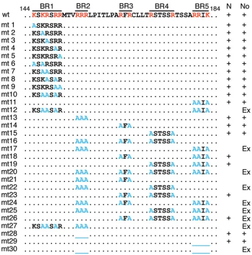
FIG 1Amino acid sequences of the wild type (wt) and mutants (mt) of AAP2 near the C terminus and their roles as an NLS and/or NoLS. The sequence of amino acid positions from 144 to 184 in the wild-type AAP2 is shown at the top, followed by the sequences of a total of 30 AAP2 mutants with arginine/ lysine-to-alanine substitutions (mt1 to mt27) or deletions (mt28 to mt30). The 5 BR clusters are indicated by lines above the wild-type sequence. The red and light-blue letters show basic amino acids and alanine mutations, respec-tively. The blue underlines indicate deletions. The dots in the sequences indi-cate residues that are the same as those of the wild type. The presence of a fully functional NLS (N) and/or NoLS (No), determined by immunofluorescence microscopy (Fig. 3), is indicated on the right. A plus in the N column indicates that the protein is observed exclusively in the nucleus regardless of its sub-nuclear localization. A plus in the No column indicates that the protein is strongly associated with the nucleolus. The mutants that showed a nucleolar-exclusion pattern are indicated by Ex.
on November 7, 2019 by guest
http://jvi.asm.org/
[image:3.585.297.545.65.316.2]AU1-tagged AAP2 and fibrillarin, an endogenous nucleolar pro-tein, colocalize in the nucleolus in HeLa cells (3). We first inves-tigated whether the N-terminally FLAG-tagged AAP2 we used for this study also localizes in the nucleolus as expected. To this end, HeLa cells were transfected with pCMV-FLAG-AAP2 and immu-nostained with anti-FLAG and anti-nucleostemin antibodies. The FLAG-tagged wild-type AAP2 was found to localize exclusively to the nucleus with significant nucleolar enrichment (Fig. 3A). The AAP2 signals were also found tightly associated with nucleoste-min-positive nuclear bodies (Fig. 3A). The AAP2 signals in the nucleolus detected by immunostaining were stronger in the pe-riphery than in the center, exhibiting a ring-shaped pattern with a central hollow. As discussed below, this nucleolar staining pattern is most likely an artifact caused by overexpression of a nucleolar-localizing protein (20). After trypsin treatment of the fixed cells, FLAG staining was visible throughout the nucleolus (Fig. 4). This observation confirmed that the FLAG peptide fused with AAP2 at the N terminus does not interfere with its intracellular localiza-tion. The functional integrity of the FLAG-tagged AAP2 had been confirmed previously (12).
Basic amino acid clusters in the C terminus of AAP2
consti-tute redundant multipartite NLSs and NoLSs.The C-terminal
region AAP2144 –184, where we found that the AAP2 NLS and NoLS reside, contains five BR clusters separated by 3 to 7 amino acids:
KSKRSRR (AAP2BR1), RRR (AAP2BR2), RFR (AAP2BR3), RS TSSR (AAP2BR4), and RRIK (AAP2BR5), from the N terminus to the C terminus (Fig. 1). To dissect the functional roles of these basic amino acids in the nuclear and nucleolar localization of AAP2, we constructed plasmids expressing a series of mutant AAP2 proteins by site-directed mutagenesis using the pCMV-FLAG-cmAAP2 plasmid (Fig. 1). Each mutant AAP2 was ex-pressed in HeLa cells by plasmid DNA transfection and analyzed for its intracellular localization by immunofluorescence micros-copy (Fig. 3).
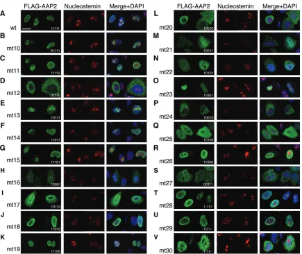
We first created AAP2 mutants 1 to 12 (Fig. 1, mt1 to mt12) to investigate the roles of AAP2BR1 and AAP2BR5, which carry the classical monopartite NLS motif, KRSR, and the putative NoLS motif, RRIK, respectively (basic amino acid residues in the motifs are underlined). Removal of positive charges from AAP2BR1 (AAP2mt1 to -mt10) or from AAP2BR5 (AAP2mt11) did not abolish the ability of mutant AAP2s to translocate to the nucleus with strong nucleolar enrichment and robust accumulation in the nucleostemin-positive nuclear bodies (Fig. 3BandC). Removal of positive charges from AAP2BR1 and AAP2BR5 in conjunction (AAP2mt12), however, resulted in substantial, if not complete, nu-clear exclusion and nucleolar exclusion (Fig. 3D). This first set of experiments indicated that there are at least two NLSs that involve either AAP2BR1 or AAP2BR5 within AAP2144 –184. We then created an additional set of 18 AAP2 mutants, 13 to 30 (Fig. 1, mt13 to mt30), to further investigate the role of each AAP2BR in nuclear and nucle-olar localization. In these mutants, the positive charge in one or more AAP2BRs was diminished by alanine substitutions or deletions in various combinations. The 7 AAP2 mutants that had only 1 mutant AAP2BR and 4 intact AAP2BRs (mt10, mt11, mt13, mt14, mt15, mt28, and mt29) were all found exclusively in the nucleus with obvi-ously enhanced association with the nucleolus (Fig. 3B,C,EtoG,T, andU). In keeping with this observation, GFP-AAP2144 –184fusion mutants that had four intact AAP2BRs were observed exclusively in the nucleus, with strong enrichment in the nucleolus (Fig. 5AtoC). All 13 mutants that had 3 or fewer intact AAP2BRs exhibited attenu-ated or complete loss of nucleolar association, and among them, 9 (mt16, mt17, mt20, mt21, mt22, mt24, mt25, mt27, and mt30) showed various degrees of impaired nuclear transport and cytoplas-mic retention (Fig. 3DandHtoV). AAP2 is a small protein that can passively cross the nuclear envelope through the nuclear pore com-plexes. The cytoplasmic signals that are stronger than the nuclear signals in AAP2mt12, -mt20, and -mt24, therefore, might indicate the presence of a nuclear export signal (NES) in AAP2. However, we have not observed any nuclear-cytoplasmic shuttling of the AAP2 protein during the course of AAV2 particle production in HEK 293 cells by plasmid transfection, where AAP2 consistently remains located in the nucleolus or nucleostemin-positive nuclear bodies (L. F. Earley and H. Nakai, unpublished observation). In addition, three different NES prediction programs (21–23) failed to identify any potential CRM1-binding NES in the AAP2 amino acid sequence. Thus, the above-mentioned observations are most likely due primarily to vari-ous degrees of impairment of nuclear import caused by different mutations, although the involvement of an unidenti-fied NES cannot be totally ruled out. Taken together, these observations indicate that the NLS and NoLS in AAP2 are re-dundant and that various combinations of 4 AAP2BRs out of 5 AAP2BRs within the AAP2144 –184region are able to constitute a functional multipartite NLS-NoLS.
FIG 2Intracellular localization of GFP or bacterial-galactosidase fused with the AAP2144 –184region at the C or N terminus, respectively. HeLa or HEK 293
cells were transiently transfected with plasmid pCMV-GFP-AAP2144 –184(A),
pCMV-GFP (B), pAAV-CMV-lacZ-AAP2144 –184(C), or pAAV-CMV-lacZ
(D), using PEI. pCMV-GFP-AAP2144 –184and pAAV-CMV-lacZ-AAP2144 –184
are plasmids expressing the GFP-AAP2144 –184fusion protein and the
-galac-tosidase–AAP2144 –184fusion protein, respectively, under the control of the
CMV promoter. For HeLa cells, the signals were detected by immunofluores-cence microscopy using corresponding antibodies, except for GFP, for which direct fluorescence was imaged. For HEK 293 cells, X-Gal staining was per-formed. Scale bars, 20m.
on November 7, 2019 by guest
http://jvi.asm.org/
[image:4.585.39.286.66.326.2]Copresence of AAP2BR1 and AAP2BR2 is an important but
not absolute requirement for the AAP2 NLS.The
immunofluo-rescence microscopy analysis described above revealed that all 10 AAP2BR mutants that exhibited impaired nuclear translocation and various degrees of cytoplasmic retention had either or both AAP2BR1 and AAP2BR2 mutations, and all 10 combinatorial mutations involving AAP2BR1 or AAP2BR2 impaired nuclear transport of AAP2 to various degrees. A statistical comparison of various combinations of two intact AAP2BRs and the presence of a fully functional NLS in the AAP2 mutants revealed that the com-bination of AAP2BR1 and AAP2BR2 is strongly associated with nuclear localization (Table 1). The GFP fusion experiment also demonstrated that AAP2BR1 by itself is not sufficient for active nuclear transport and that copresence of AAP2BR1 and AAP2BR2 is required for nuclear accumulation (Fig. 5DtoH). When we assessed the NLS role of each AAP2BR based on its ability to
re-store a fully functional NLS to AAP2 mutants (see Materials and Methods), we found that all 5 AAP2BRs can contribute to the facilitation of nuclear localization of AAP2. These observations indicate that, although all 5 AAP2BRs have an NLS role, the pres-ence of AAP2BR1 and AAP2BR2 in conjunction plays the most influential role in nuclear targeting. Due to the redundant nature of the AAP2 NLS, either one of these two AAP2BRs is dispensable if all 4 of the other AAP2BRs are present, as evidenced by the nuclear localization of AAP2mt10 and -mt13.
All the AAP2BRs can contribute to nucleolar localization
and function in a context-dependent manner.The NoLS role of
each AAP2BR was investigated based on the functional restora-tion of nucleolar-excluded AAP2 mutants, as detailed in Materials and Methods. This analysis revealed an obvious NoLS role in AAP2BR1, -2, -4, and -5. For example, the NoLS role of AAP2BR2 could be demonstrated by the observation that, when the mutated
FIG 3Intracellular localization of various AAP2 mutants with arginine/lysine-to-alanine substitutions or deletions within the BR clusters, AAP2BR1 to AAP2BR5. HeLa cells were transiently transfected with a plasmid expressing the wild-type or a mutant AAP2 with an N-terminal FLAG tag. The cells were fixed 48 h posttransfection, immunostained with anti-FLAG antibody (green) and anti-nucleostemin antibody (red), and counterstained with DAPI (blue), before being imaged on a Zeiss LSM 710 confocal microscope with a 100⫻objective. Representative cell images are shown for the wild type and each mutant. (A) Wild-type AAP. (B) AAP2mt10, showing a staining pattern representing all the AAP2BR1 mutants, AAP2mt1 to -mt10 (the data for AAP2mt1 to -mt9 are not shown). (C to V) AAP2mt11 to -mt30. Formation of multiple nuclear bodies containing both AAP2 and nucleostemin was evident in many cells expressing the wild-type AAP2 (A) and the AAP2 mutants showing an intracellular localization pattern similar to that of the wild type (B, C, F, G, T, and U). The identity and the role of these speckled structures have yet to be determined. The 5-digit numbers in the bottom right corner of each FLAG-AAP2 panel indicate mutations introduced in each AAV2BR in each mutant (1, wild type; 0, alanine mutation; -, deletion). For example, 01110 (mt12) indicates BR1⫺/BR2⫹/BR3⫹/BR4⫹/BR5⫺. Scale bar, 20m.
on November 7, 2019 by guest
http://jvi.asm.org/
[image:5.585.76.511.63.429.2]AAP2BR2 was corrected back to the wild-type sequence in the nucleolar-excluded AAP2mt17 (Fig. 3I), nucleolar retention was restored, as evidenced with AAP2mt11 (Fig. 3C). A comparison between AAP2mt19, showing modest nucleolar accumulation, and AAP2mt26, showing nucleolar exclusion, which differ only by the presence or absence of AAP2BR3, revealed that AAP2BR3 also functions as a weak NoLS (Fig. 3KandR). These results indicate that effective nucleolar accumulation is a consequence of the co-operation of the redundant NoLSs residing in the five AAP2BRs
in a given combination of AAP2BRs. Interestingly, AAP2mt26 (AAP2BR1⫹/BR2⫹/BR3⫺/BR4⫺/BR5⫺, where basic amino acids in the BRs are retained [plus] or mutated to alanine [minus]) exhibited nearly complete nuclear localization with nucleolar ex-clusion (Fig. 3R), while GFP-AAP2144 –184mt26, carrying
AAP2144 –184 with the same AAP2BR mutations, and
GFP-AAP2144-156, in which GFP was fused with a 13-mer peptide con-taining only AAP2BR1 and AAP2BR2, showed nuclear and nucle-olar accumulation (Fig. 5FandH). The same discrepancy in the pattern of nucleolar accumulation was observed in AAP2mt22 (Fig. 3N) and GFP-AAP2144 –184mt22 (Fig. 5E). It has previously been shown that adding a nonapeptide containing 9 basic amino acids to the C terminus of GFP allows nucleolar accumulation of the protein, not through a specific NoLS motif, but presumably through nonspecific, electrostatic interactions with negatively charged nucleolar components (9). There are 8 basic amino acids within the AAP2BR1-AAP2BR2 segment, and there are 10 basic residues in AAP2mt22 (Fig. 1). Therefore, the extra positive elec-tric charges added to GFP likely explain why the GFP-AAP2144-156, GFP-AAP2144 –184mt22, and GFP-AAP2144 –184mt26 fusion pro-teins were still capable of nucleolar retention while AAP2mt26 was not. Taken together, although there is no inconsistency in the NLS roles of AAP2BR1 and AAP2BR2 between the AAP2 mutants and the GFP fusion proteins, their NoLS roles are context dependent. Capsid assembly-promoting functions of AAP2BR mutants. A series of studies have demonstrated that nucleolar localization of viral components is important for the AAV life cycle (24–27). Indispensable roles of the nucleolus in the virus life cycle have also been demonstrated in other viruses (28–30). Having created a panel of AAP2 mutants whose intracellular localizations are char-acterized, we addressed how aberrant localization of AAP2 might affect its assembly-promoting function. To this end, we
per-FIG 4Immunofluorescence microscopic analysis of a FLAG-tagged AAP2 fused with GFP in HeLa cells with or without proteinase treatment. (A to C) HeLa cells were transiently transfected with pCMV-FLAG-cmAAP2-GFP ex-pressing the wild-type full-length AAP2 fused with a FLAG tag and GFP. In-tracellular localization of the fusion protein was analyzed with or without trypsin treatment by anti-FLAG antibody immunostaining (A), direct detec-tion of GFP fluorescence (B), and anti-GFP antibody immunostaining (C) under a fluorescence microscope. (D) Merged images. Scale bar, 20m.
FIG 5Microscopic assessment of the abilities of the AAP2BRs to target GFP to the nucleus and the nucleolus. The method used was the same as that forFig. 2. (A to F) GFP-AAP2144 –184mt13 to -mt26 are GFPs fused with the 41-amino-acid-long AAP144 –184region containing their corresponding AAP2BR mutations. (G
and H) GFP-AAP2144-150and GFP-AAP2144-156are GFPs fused with AAP2BR1 only (7 amino acids) and AAP2BR1-AAP2BR2 only (13 amino acids), respectively.
Scale bar, 20m.
on November 7, 2019 by guest
http://jvi.asm.org/
[image:6.585.58.267.65.222.2] [image:6.585.74.511.445.683.2]formed a transcomplementation assay in which AAV2 VP3-only particles were produced in HEK 293 cells in the presence of the wild-type AAP2 or a series of AAP2 mutants and quantified AAV2 particle yields using an AAV2 intact-particle-specific ELISA. There was a strong correlation between aberrant intracellular lo-calization of AAP2 and attenuated AAV2 capsid production (Fig. 6A). In particular, AAP2 mutants with nucleolar exclusion con-sistently showed a 3-log-unit or more reduction of capsid produc-tion compared to the wild type. Interestingly, AAP2mt8, -mt9, -mt10, and -mt13, which showed exclusive nuclear localization with enhanced nucleolar association similar to that of the wild type, exhibited a more than 20- to 100-fold decrease in capsid production (Fig. 6A). Thus, normal intracellular localization of AAP2 is a prerequisite but is not sufficient for effective capsid production. The impaired function observed in AAP2 mutants showing wild-type-like intracellular localization implies that AAP2BRs are not only the redundant organelle-targeting signals, but also play a direct or indirect role in the capsid assembly pro-cess, which we address below.
AAP2BRs maintain AAP2 expression levels for effective
cap-sid assembly.During the microscopic analysis, we had noticed
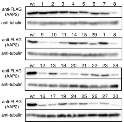
that the numbers of transfected HeLa cells and the signal intensi-ties in many AAP2 mutants were lower than those from the wild type to various degrees. Since AAP2BR mutations may have af-fected not only intracellular localization, but also protein expres-sion levels, we performed a quantitative Western blot assay on lysates from HEK 293 cells transfected with either the wild-type or mutant pCMV-FLAG-cmAAP2 plasmid. The molecular mass of the FLAG-tagged AAP2 protein was determined to be 27 kDa by SDS-PAGE (26 kDa for AAP2), which is slightly higher than a calculated molecular mass of 24 kDa. Mutation or deletion of the AAP2BR regions decreased AAP2 protein expression levels to var-ious degrees between 0.02 and 0.64 compared to 1.0 for the wild type (Fig. 6Aand7). The expression levels of all the mutants show-ing aberrant localization were reduced substantially, with the rel-ative levels ranging from 0.02 to 0.14. The reduction of protein levels was particularly pronounced in the nucleolar-excluded mu-tants. The AAP2BR1 or AAP2BR2 mutants showing localization similar to that of the wild type had greater quantities of protein on average than the mutants showing aberrant localization; however, they also showed attenuated AAP2 expression ranging from 0.03 to 0.64. Importantly, the results showed a very nice correlation between intracellular AAP2 levels, nucleolar accumulation of AAP2, and AAV capsid production. When AAP2 mutants re-tained the ability to localize normally (these mutants are indicated by the horizontal black line inFig. 6A), AAV capsid yields were positively correlated with the AAP2 mutant protein levels, with a Pearson correlation coefficient of 0.94 (Fig. 6B). When AAP2 mu-tants were excluded from the nucleolus, AAV capsid yields were disproportionately lower than the AAP2 mutant protein levels (these mutants are indicated by the horizontal gray line inFig. 6A). Although the underlying mechanism has yet to be elucidated, these results indicate that AAP2BRs play a pivotal role in maintaining the AAP2 levels for effective capsid assembly. The AAP2BRs’ role in this respect well explains why some of the AAP2BR1 or AAP2BR2 mutants showing wild-type localization failed to produce capsids effectively.
The AAP2 NoLS is important for nucleolar accumulation of
AAV2 VP3.The data presented above provided insights into the
AAP2BRs’ functional roles in intracellular localization of AAP2
TABLE 1 Associations between various combinations of two AAP2BRs and the presence of an NLS in AAP2 144 –184 AAP2BR Association with [no./total (%)] a: AAP2BR1 AAP2BR2 AAP2BR3 AAP2BR4 AAP2BR2 7/7 (100) ( P18 ⫽ 0.002; P25 ⫽ 0.001–0.017) AAP2BR3 4/7 (57) ( P18 ⫽ 1.000; P25 ⫽ 0.186–1.000) 4/5 (80) b( P18 ⫽ 0.211; P25 ⫽ 0.014–1.000) AAP2BR4 4/7 (57) ( P18 ⫽ 1.000; P25 ⫽ 0.186–1.000) 4/5 (80) b( P18 ⫽ 0.211; P25 ⫽ 0.014–1.000) 3/6 (50) ( P18 ⫽ 1.000; P25 ⫽ 0.317–1.000) AAP2BR5 4/7 (57) ( P18 ⫽ 1.000; P25 ⫽ 0.186–1.000) 4/4 (100) b( P18 ⫽ 0.031; P25 ⫽ 0.002–1.000) 3/5 (60) ( P18 ⫽ 0.696; P25 ⫽ 0.113–1.000) 3/5 (60) ( P18 ⫽ 0.696; P25 ⫽ 0.113–1.000) aEighteen AAP2 mutants (AAP2mt10 to -mt27) were categorized into two groups according to the presence or absence of an NLS as defined in the legend to Fig. 1. There were 9 NLS ( ⫹ ) and 9 NLS ( ⫺ ) AAP2 mutants. An unconditional exact test was performed to statistically evaluate the association between the presence or absence of a given combination of 2 AAP2BRs and the presence or absence of an NLS. The null hypothesis is that there is no association between AAV2BRs and NLSs. Two types of P values ( P18 and P25 ) are provided, as described in Materials and Methods. Shown are the frequencies of NLS ( ⫹ ) AAP2 mutants among all the mutants that carry a given combination of two different AAP2BRs. bThe statistical analysis for the combination does not give a conclusive result due to the wide range of P25 values across the statistically significant cutoff value of 0.05.
on November 7, 2019 by guest
http://jvi.asm.org/
and capsid assembly; however the data did not address the AAP2 role as a VP3 transporter. Since the nucleolus has been thought to be important for AAV2 capsid assembly and nucleolar-excluded AAP2BR mutants showed a substantially reduced ability to pro-duce viral capsids, it is possible that AAP2BR mutants would not associate with VP3, leading to impaired nuclear transport and/or failure to accumulate VP3 in the nucleolus even if VP3 is actively localized to the nucleus. To address this, HeLa cells were trans-fected with pCMV-AAV2VP3 and pCMV-FLAG-cmAAP2 (either
the wild type or AAP2mt26), and intracellular localization of AAP2 and VP3 was analyzed by immunofluorescence microcopy at 48 h posttransfection. As Sonntag et al. have previously shown, VP3 diffusely localizes to the cytoplasm and nucleoplasm but re-mains outside the nucleus unless AAP2 is present (3) (Fig. 8Aand B). In the presence of AAP2mt26, which had the ability to exclu-sively translocate to the nucleus but was not able to accumulate in the nucleolus, VP3 was seen predominantly in the nucleoplasm, colocalizing with AAP2mt26 and leaving only a small quantity of VP3 proteins in the cytoplasm. Importantly, VP3 failed to accu-mulate in the nucleolus (Fig. 8C). We have also found, using a plasmid carrying an AAV2capgene that does not express AAP2, that expression of the AAV2 VP1, VP2, and VP3 proteins does not result in nucleolar localization of any of the VP proteins in HeLa cells when AAP2 is not coexpressed (Earley and Nakai, unpub-lished). Taken together, these observations indicate that the NoLS in AAP2 plays a primary role in targeting capsid VP proteins to the nucleolus.
FIG 6AAV2 VP3 capsid production titers and wild-type and mutant AAP2 protein expression levels in HEK 293 cells. HEK 293 cells seeded on 6-cm dishes were transfected with pAAV2-RepVP3, pCMV-FLAG-cmAAP2 expressing either the full-length wild-type AAP2 or a full-length AAP2 mutant with an AAP2BR mutation(s), and pHelper, using PEI (12). The cells were harvested at 48 h posttransfection and made into a 100-l crude cell lysate in HEPES-buffered saline after three cycles of freezing and thawing. The AAV2 particle concentration in each crude lysate was quantified by an A20 antibody-based ELISA. (A) Viral titers relative to the titer obtained with the wild-type AAP2 (black bars). A relative titer of 1.0 corresponds to 7.2⫻1012particles/ml. The thick horizontal black line
between the graph and thexaxis labels indicates the wild type and AAP2 mutants that showed exclusive nuclear localization with enhanced nucleolar association. The thick horizontal gray line indicates the mutants showing nucleolar exclusion. The data were collected from biologically triplicate experiments. *,P⬍0.05 (two-tailed Welch’sttest) compared to the wild-type values. In a separate experiment, HEK 293 cells seeded on 6-cm dishes were transfected with pCMV-FLAG-cmAAP2 (the wild type or mutant) using PEI. The cells were harvested 48 h posttransfection, and mutant AAP2 protein expression levels relative to the level of the wild type were determined by a quantitative Western blot analysis using biologically duplicate samples (gray bars). The error bars represent standard errors of the mean (SEM) (black bars) or the difference between each value and the mean value (gray bars). (B) Correlations between AAP2 protein levels of the wild type and mutants indicated by the thick horizontal black line in panel A and AAV2 VP3 capsid production titers are shown in a scatter plot. All values are relative to the levels of the wild type.
FIG 7Western blot analysis of the wild-type and mutant AAP2 proteins ex-pressed in HEK 293 cells. Cells seeded on 6-cm dishes were transfected with pCMV-FLAG-cmAAP2 (wild type or mutant) using PEI. The same quantity of crude lysates prepared at 48 h posttransfection were separated on an 8% SDS-PAGE gel and blotted onto a membrane, which was then probed with both anti-FLAG and anti-␣-tubulin antibodies. The AAP2 mutant numbers are shown above the lanes.
FIG 8Intracellular localization of AAV2 VP3 in the presence or absence of wild-type AAP2 or in the presence of the nuclear-targeted, nucleolar-excluded AAP2 mutant, AAP2mt26. HeLa cells were transfected with the following plasmids: pCMV-AAV2VP3 only (A), pCMV-AAV2VP3 and pCMV-FLAG-cmAAP2 (wild type) (B), and pCMV-AAV2VP3 and pCMV-FLAG-cmAAP2mt26 (C). The cells were fixed at 48 h posttransfection and immunostained with FLAG, anti-AAV VP1/VP2/VP3, and anti-nucleostemin antibodies. Scale bar, 20m.
on November 7, 2019 by guest
http://jvi.asm.org/
[image:8.585.98.494.66.188.2] [image:8.585.59.268.452.657.2] [image:8.585.298.545.560.658.2]DISCUSSION
The NLS in AAP2 had remained unidentified in a series of studies reported by Kleinschmidt’s group (1–3), which prompted us to seek to determine the amino acid residues responsible for nuclear translocation of AAP2. The consensus sequences of the classical, importin-␣-binding NLSs have been well defined. The monopar-tite NLSs have the consensus sequence K(K/R)X(K/R), while the bipartite NLSs show a consensus sequence composed of two short basic amino acid clusters separated by a linker sequence, such as (K/R)(K/R)X10 –12(K/R)3/5, where the C-terminal side cluster con-tains at least three basic amino acids within 5 consecutive residues (14,18,19). Many noncanonical, importin-␣-binding NLSs have also been identified, including bipartite NLSs with a long linker of up to 29 residues (14,18,19). In light of this knowledge about various types of NLSs, one could assume that AAP2 has one clas-sical monopartite NLS in AAP2BR1 (KSKRSRR) and/or one non-canonical bipartite NLS with a long linker in AAP2BR1-X30 -AAP2BR5 (RRIK). Our experimental data, however, do not support this assumption. None of the AAP2BRs were capable of serving as a monopartite NLS. Substantial nuclear enrichment required the presence of AAP2BR1-AAP2BR2 (KSKRSRR-X3 -RRR) or at least four AAP2BRs when either AAP2BR1 or
AAP2BR2 is absent (i.e.,
AAP2BP1-AAP2BP3-AAP2BP4-AAP2BP5 and AAP2BP2-AAP2BP3-AAP2BP4-AAP2BP1-AAP2BP3-AAP2BP4-AAP2BP5). Thus, we identified at least three different motifs that can function as an NLS and overlap within the AAP2144 –184region. This redundant nature of the AAP2 NLS explains why a pervious investigation concluded that AAP2BR1 was not involved in nuclear localization even though the region contained a classical NLS (1).
In nucleolar-localizing proteins, an NLS and an NoLS can re-side in separate locations, but they often corere-side in a region highly rich in lysine and arginine residues. The NLS and NoLS of AAP2 that we identified is a joint NLS-NoLS, because all the AAP2BR regions play either, or both, of the NLS and NoLS roles. Many such joint NLS-NoLSs have been reported as NLSs in the literature, mainly due to incomplete investigation for nucleolar accumulation, and therefore, joint NLS-NoLSs have been over-looked in many instances (17). A recent attempt to extract com-monalities of 48 human NoLSs, including joint NLS-NoLSs, has revealed that NoLSs are highly basic; are predominantly located near the N or C terminus of proteins, in␣-helices or coils and rarely in-strands; and are solvent accessible (17). In this regard, the AAP2144 –184region containing the joint NLS-NoLSs is consis-tent with these general trends. That is, in addition to the high proportion of basic amino acids, the NLS-NoLS resides near the C terminus; the AAP2144 –184residues are exclusively in␣-helices (49%) or coils (51%) according to the Jpred3 prediction (31); and the proportions of buried residues in the AAP2144 –184region pre-dicted by Jpred3 are 37%, 10%, and 2% at prediction thresholds of 25%, 5%, and 0%, respectively, which are lower than in the full-length protein control in the study reported by Scott et al. (17) The AAP2144 –184region also contains the previously identified (K/R) (K/R)X(K/R) NoLS motif (15). Through a literature search, we could identify a joint NoLS that resembles the AAP2 NLS-NoLS. Liu et al. reported that a stretch of 18 amino acid residues, 2744KKKMKKHKNKSEAKKRKI2761, is part of a joint NLS-NoLS found in 1A6/downregulated in metastasis (1A6/DRIM), a large nucleolar-targeting protein (2,785 amino acids) involved in rRNA processing (32). This amino acid sequence is reminiscent of154R
RRLPITLPARFRCLLTRSTSSRTSSARRIK184, one of the com-plete joint NLS-NoLSs that we identified in AAP2. Although the observations obtained from the arginine/lysine-to-alanine mu-tagenesis experiments were not exactly the same in the Liu et al. study and ours, both identified basic amino acid clusters that play a dual NLS-NoLS role. In addition, both studies showed that a decrease in electric charge in the signals resulted in nucleolar ex-clusion. As demonstrated with AAP2 and many other nucleolar-localizing proteins (17), NLSs and NoLSs are inherently difficult to clearly define, particularly when they reside within the same region with a portion of the signal playing a dual role.
How NoLSs perform their role in nucleolar targeting has not been fully understood. Unlike NLSs, NoLSs do not have well-defined consensus sequences, although they share common char-acteristics to some degree, as described above. The nucleolus does not have any compartmentalizing membranous structures, and therefore, nucleolar accumulation presumably does not require sophisticated machinery that actively targets the organelle, like the nuclear transport machinery. Rather, nucleolar accumulation of proteins has been thought to be mediated by a retention mecha-nism through interactions with nucleolar components, such as RNA and other nucleolar proteins (33). RNA-binding motifs have been identified in many nucleolar proteins, including nucleolin, fibrillarin, and viral RNA-binding proteins (34–36). Nucleophos-min, a putative shuttle protein that transports cargos between the cytoplasm and the nucleolus (37–40), has been shown to mediate nucleolar retention of other proteins through the interaction be-tween two highly acidic regions (120EDAESEDEEEED132and161D
EDDDDDDEEDDDEDDDDDDFDDEEAEE188) in
nucleophos-min and positively charged anucleophos-mino acid regions in the other proteins (40). Thus, nucleolar accumulation is most likely medi-ated by electrostatic interaction rather than by a defined set of particular amino acid sequences (9). Concordant with this notion, we could only vaguely define the AAP2 NoLS, as opposed to its NLSs, and found that the abundance of positively charged AAP2BR regions, rather than a specific amino acid sequence or a specific combination of AAP2BRs, confers on AAP2 the ability to translocate from the nucleoplasm to the nucleolus. This is in line with the notion that the nucleolar accumulation of AAP2 results from nonspecific interaction with negatively charged nucleolar components and also raises the possibility that the degree of accumulation is determined by the net electric charge of the AAP2144 –184region. The electric charge of AAP2144 –184is 15, and at least 12 has been found to be required for efficient nucleolar accumulation of the AAP2BR mutants (Fig. 1). The net electric charges of the AAP2144 –184-corresponding regions of various serotype AAPs (AAP1 to -13) (1) vary from 8 to 15, with AAP5 being the lowest and AAP2 and AAP3B being the highest. Assuming that nucleolar accumulation is determined by the abundance of the net positive charges within this region, AAP5 would likely be devoid of an NoLS and excluded from the nucleolus. If this is true, AAP5 would not be able to transport AAV5 capsid VP proteins to the nucleolus, the organelle that is believed to be important for AAV capsid assembly, at least for AAV2 (24–27). How would AAV5 viral capsid proteins assem-ble if they are not transported to the nucleolus? Although our study demonstrates that the nucleolar localization of AAP2 strongly correlates with the ability of AAV2 to form capsids, it is intriguing to speculate that the nucleolar accumulation of AAP2 might merely be a coincidental, nonspecific consequence
on November 7, 2019 by guest
http://jvi.asm.org/
associated with a function(s) other than nucleolar targeting and that this targeting might not be functionally important. Such nonspecific nucleolar accumulation with no functional role has been demonstrated in the human histone H2B protein (9). Future studies on the subcellular localizations of AAPs derived from various serotypes will address this potential par-adox and elucidate the biological significance of the presence of an NoLS in AAPs. Nonetheless, our observations corroborate the driving role of the electric charge in nucleolar targeting and support the idea that NoLSs pay a lower cost for the acquisition of their role by intermingling their signals with NLSs that al-ready have high electric charges. This is particularly advanta-geous to small viruses that have a limited capacity to store genetic information.
Surprisingly, we could identify at least three complete NLSs that overlap within the same AAP2144 –184 region. They are AAP2BR1 plus -BR2, AAP2BR1 plus -BR3 plus -BR4 plus -BR5, and AAP2BR2 plus -BR3 plus -BR4 plus -BR5. Redundant NLSs, as either separate NLSs or overlapping NLSs, have been identified in many proteins, including those derived from viruses. For ex-ample, the polyomavirus large T antigen has two separate NLSs, VSRKRPR and PKKARED, each of which mediates nuclear local-ization in the absence of the other (41, 42); the nonstructural protein 1 of influenza A virus (NS1A) has a functionally compe-tent monopartite NLS, DRLRRDQKSLR, and a classical bipartite NLS-like sequence, KRKMARTARSKVRRDKMAD (43), which also conveys nucleolar localization; and the bovine adenovirus 3 33,000-molecular-weight (33K) protein has a 40-residue-long re-gion where two karyopherin-binding motifs, an importin-␣ 5-binding motif and a transportin 3-5-binding motif, overlap (44). Utilization of different nuclear transport pathways through the interactions between an NLS and multiple karyopherins have also been demonstrated in human immunodeficiency virus (HIV) Rev protein (45). The straightforward inference about the NLS redun-dancy is that it ensures and promotes efficient nuclear entry by increasing the avidity for the nuclear transport system and/or by using more than one nuclear entry pathway. However, there are also many instances in which the amino acid residues composing an NLS perform functions distinct from those in nuclear translo-cation. For example, the unconventional NLS in the nucleopro-tein (NP) of the influenza A virus plays a crucial role in nuclear import of the viral genomic RNA (46), and the second NLS in the transcription factor Fli-1 also functions as a DNA-binding do-main (47). In fact, DNA-binding domains frequently overlap NLSs (47), and this has led to speculation about the possible co-evolutionary origins of the two motifs (48,49). In the case of AAP2, our present and previous studies provide clues to under-standing the necessity of redundancy. In our previous study, we used a directed-evolution approach to experimentally and com-putationally evolve functionally competent mutant AAP2s from a randomly mutagenized library (12). We demonstrated that the functionally optimal AAP2 mutants harbor a strongly basic and hydrophilic heptapeptide, (R/S)7, in the AAP2BR1 region that re-markably resembles the native sequence, KSKRSRR (12). Since all the AAP2 mutants used in the previous study had a completely functional NLS-NoLS composed of AAP2BR2, -3, -4, and -5, AAP2BR1 is most likely not simply a redundant sequence com-posing an NLS-NoLS but has an important non-NLS-NoLS role. In keeping with this notion, mutations in this region could sub-stantially reduce AAP2 protein levels without affecting its
intra-cellular localization and could result in significantly impaired cap-sid assembly (AAP2mt8, -mt9, and -mt10). The reduction of capsid production by an AAP2BR1 mutation has also been re-ported by Naumer et al. (1). Although the mechanism for the decreased protein expression has yet to be determined, it could be either due to intrinsic instability caused by protein misfolding or due to decreased protein-protein or protein–nucleic-acid interac-tions that stabilize AAP2. Taken together, the redundancy of the NLS-NoLS in AAP2 is likely a necessity for optimal capsid pro-duction and presumably results from their alternative functional roles.
In summary, this study identifies a joint NLS-NoLS near the C terminus of AAP2. The identified basic amino acid region is im-portant, not only for nuclear and nucleolar localization, but also for maintaining AAP2 levels, indicating a non-NLS-NoLS role in the region. The system described here can readily be applied to AAPs derived from other serotypes. Further studies on interac-tions between AAP, capsid, and cellular proteins will help us un-derstand the mechanism of AAV capsid assembly and may lead to improved AAV vector production for gene therapy.
ACKNOWLEDGMENTS
We thank Stefanie Kaech Petrie and Aurelie Snyder at the OHSU Ad-vanced Light Microscope Core for their technical expertise, Hiroyuki Ido for technical assistance in molecular cloning of plasmids, Michael R. La-sarev for his advice on statistical analyses, and Michael S. Chapman for critical reading of the manuscript.
This work was supported by Public Health Service grants (R01 DK078388, R01 NS088399, T32 GM071338, and T32 AI007472) and a Sponsored Research Fund from Takara Bio Inc.
REFERENCES
1.Naumer M, Sonntag F, Schmidt K, Nieto K, Panke C, Davey NE, Popa-Wagner R, Kleinschmidt JA. 2012. Properties of the adeno-associated virus assembly-activating protein. J Virol86:13038 –13048. http://dx.doi.org/10.1128/JVI.01675-12.
2.Sonntag F, Kother K, Schmidt K, Weghofer M, Raupp C, Nieto K, Kuck A, Gerlach B, Bottcher B, Muller OJ, Lux K, Horer M, Kleinschmidt JA. 2011. The assembly-activating protein promotes capsid assembly of dif-ferent adeno-associated virus serotypes. J Virol85:12686 –12697.http: //dx.doi.org/10.1128/JVI.05359-11.
3.Sonntag F, Schmidt K, Kleinschmidt JA.2010. A viral assembly factor promotes AAV2 capsid formation in the nucleolus. Proc Natl Acad Sci U S A107:10220 –10225.http://dx.doi.org/10.1073/pnas.1001673107. 4.Chook YM, Suel KE.2011. Nuclear import by karyopherin-s:
recogni-tion and inhibirecogni-tion. Biochim Biophys Acta1813:1593–1606.http://dx.doi .org/10.1016/j.bbamcr.2010.10.014.
5.Kalderon D, Roberts BL, Richardson WD, Smith AE. 1984. A short amino acid sequence able to specify nuclear location. Cell39:499 –509. http://dx.doi.org/10.1016/0092-8674(84)90457-4.
6.Robbins J, Dilworth SM, Laskey RA, Dingwall C.1991. Two interde-pendent basic domains in nucleoplasmin nuclear targeting sequence: identification of a class of bipartite nuclear targeting sequence. Cell64: 615– 623.http://dx.doi.org/10.1016/0092-8674(91)90245-T.
7.Dingwall C, Laskey RA.1991. Nuclear targeting sequences—a consen-sus? Trends Biochem Sci 16:478 – 481. http://dx.doi.org/10.1016/0968 -0004(91)90184-W.
8.Ben-Efraim I, Gerace L.2001. Gradient of increasing affinity of importin
for nucleoporins along the pathway of nuclear import. J Cell Biol152: 411– 417.http://dx.doi.org/10.1083/jcb.152.2.411.
9.Musinova YR, Lisitsyna OM, Golyshev SA, Tuzhikov AI, Polyakov VY, Sheval EV.2011. Nucleolar localization/retention signal is responsible for transient accumulation of histone H2B in the nucleolus through electro-static interactions. Biochim Biophys Acta1813:27–38.http://dx.doi.org /10.1016/j.bbamcr.2010.11.003.
10. Wang Y, Chen B, Li Y, Zhou D, Chen S.2011. PNRC accumulates in the nucleolus by interaction with B23/nucleophosmin via its nucleolar
on November 7, 2019 by guest
http://jvi.asm.org/
ization sequence. Biochim Biophys Acta1813:109 –119.http://dx.doi.org /10.1016/j.bbamcr.2010.09.017.
11. Szebeni A, Herrera JE, Olson MO.1995. Interaction of nucleolar protein B23 with peptides related to nuclear localization signals. Biochemistry 34:8037– 8042.http://dx.doi.org/10.1021/bi00025a009.
12. Kawano Y, Neeley S, Adachi K, Nakai H.2013. An experimental and computational evolution-based method to study a mode of co-evolution of overlapping open reading frames in the AAV2 viral genome. PLoS One 8:e66211.http://dx.doi.org/10.1371/journal.pone.0066211.
13. Nakai H, Fuess S, Storm TA, Muramatsu S, Nara Y, Kay MA.2005. Unrestricted hepatocyte transduction with adeno-associated virus sero-type 8 vectors in mice. J Virol79:214 –224.http://dx.doi.org/10.1128/JVI .79.1.214-224.2005.
14. Lange A, Mills RE, Lange CJ, Stewart M, Devine SE, Corbett AH.2007. Classical nuclear localization signals: definition, function, and interaction with importin␣. J Biol Chem282:5101–5105.http://dx.doi.org/10.1074 /jbc.R600026200.
15. Weber JD, Kuo ML, Bothner B, DiGiammarino EL, Kriwacki RW, Roussel MF, Sherr CJ.2000. Cooperative signals governing ARF-mdm2 interaction and nucleolar localization of the complex. Mol Cell Biol20: 2517–2528.http://dx.doi.org/10.1128/MCB.20.7.2517-2528.2000. 16. Viranaicken W, Gasmi L, Chaumet A, Durieux C, Georget V, Denoulet
P, Larcher JC.2011. L-Ilf3 and L-NF90 traffic to the nucleolus granular component: alternatively-spliced exon 3 encodes a nucleolar localization motif. PLoS One 6:e22296. http://dx.doi.org/10.1371/journal.pone .0022296.
17. Scott MS, Boisvert FM, McDowall MD, Lamond AI, Barton GJ.2010. Characterization and prediction of protein nucleolar localization se-quences. Nucleic Acids Res38:7388 –7399.http://dx.doi.org/10.1093/nar /gkq653.
18. Lange A, McLane LM, Mills RE, Devine SE, Corbett AH.2010. Expand-ing the definition of the classical bipartite nuclear localization signal. Traf-fic11:311–323.http://dx.doi.org/10.1111/j.1600-0854.2009.01028.x. 19. Kosugi S, Hasebe M, Matsumura N, Takashima H, Miyamoto-Sato E,
Tomita M, Yanagawa H.2009. Six classes of nuclear localization signals specific to different binding grooves of importin␣. J Biol Chem284:478 –
485.http://dx.doi.org/10.1074/jbc.M807017200.
20. Svistunova DM, Musinova YR, Polyakov VY, Sheval EV.2012. A simple method for the immunocytochemical detection of proteins inside nuclear structures that are inaccessible to specific antibodies. J Histochem Cy-tochem60:152–158.http://dx.doi.org/10.1369/0022155411429704. 21. Kosugi S, Yanagawa H, Terauchi R, Tabata S.2014. NESmapper:
accu-rate prediction of leucine-rich nuclear export signals using activity-based profiles. PLoS Comput Biol.10:e1003841.http://dx.doi.org/10.1371 /journal.pcbi.1003841.
22. Fu SC, Imai K, Horton P.2011. Prediction of leucine-rich nuclear export signal containing proteins with NESsential. Nucleic Acids Res39:e111. http://dx.doi.org/10.1093/nar/gkr493.
23. Prieto G, Fullaondo A, Rodriguez JA.2014. Prediction of nuclear export signals using weighted regular expressions (Wregex). Bioinformatics30: 1220 –1227.http://dx.doi.org/10.1093/bioinformatics/btu016.
24. Wistuba A, Kern A, Weger S, Grimm D, Kleinschmidt JA. 1997. Subcellular compartmentalization of adeno-associated virus type 2 assem-bly. J Virol71:1341–1352.
25. Bevington JM, Needham PG, Verrill KC, Collaco RF, Basrur V, Trempe JP.2007. Adeno-associated virus interactions with B23/Nucleophosmin: identification of sub-nucleolar virion regions. Virology 357:102–113. http://dx.doi.org/10.1016/j.virol.2006.07.050.
26. Qiu J, Brown KE.1999. A 110-kDa nuclear shuttle protein, nucleolin, specifically binds to adeno-associated virus type 2 (AAV-2) capsid. Virol-ogy257:373–382.http://dx.doi.org/10.1006/viro.1999.9664.
27. Johnson JS, Samulski RJ.2009. Enhancement of adeno-associated virus infection by mobilizing capsids into and out of the nucleolus. J Virol 83:2632–2644.http://dx.doi.org/10.1128/JVI.02309-08.
28. Hiscox JA.2002. The nucleolus—a gateway to viral infection? Arch Virol 147:1077–1089.http://dx.doi.org/10.1007/s00705-001-0792-0. 29. Pederson, T. 2011. The nucleolus. Cold Spring Harb Perspect Biol
3:a000638.http://dx.doi.org/10.1101/cshperspect.a000638.
30. Matthews D, Emmott E, Hiscox J.2011. Viruses and the nucleolus, p 321–345.InOlson M (ed), The nucleolus. Springer, New York, NY. 31. Cole C, Barber JD, Barton GJ.2008. The Jpred 3 secondary structure
prediction server. Nucleic Acids Res36:W197–W201.http://dx.doi.org/10 .1093/nar/gkn238.
32. Liu J, Du X, Ke Y.2006. Mapping nucleolar localization sequences of 1A6/DRIM. FEBS Lett580:1405–1410.http://dx.doi.org/10.1016/j.febslet .2006.01.064.
33. Olson MO, Dundr M.2005. The moving parts of the nucleolus. His-tochem Cell Biol123:203–216.http://dx.doi.org/10.1007/s00418-005 -0754-9.
34. Hiscox JA.2007. RNA viruses: hijacking the dynamic nucleolus. Nat Rev Microbiol5:119 –127.http://dx.doi.org/10.1038/nrmicro1597.
35. Schmidt-Zachmann MS, Nigg EA.1993. Protein localization to the nu-cleolus: a search for targeting domains in nucleolin. J Cell Sci105:799 – 806.
36. Aris JP, Blobel G.1991. cDNA cloning and sequencing of human fibril-larin, a conserved nucleolar protein recognized by autoimmune antisera. Proc Natl Acad Sci U S A88:931–935.http://dx.doi.org/10.1073/pnas.88 .3.931.
37. Fankhauser C, Izaurralde E, Adachi Y, Wingfield P, Laemmli UK.1991. Specific complex of human immunodeficiency virus type 1 Rev and nu-cleolar B23 proteins: dissociation by the Rev response element. Mol Cell Biol11:2567–2575.
38. Borer RA, Lehner CF, Eppenberger HM, Nigg EA.1989. Major nucle-olar proteins shuttle between nucleus and cytoplasm. Cell56:379 –390. http://dx.doi.org/10.1016/0092-8674(89)90241-9.
39. Valdez BC, Perlaky L, Henning D, Saijo Y, Chan PK, Busch H.1994. Identification of the nuclear and nucleolar localization signals of the pro-tein p120. Interaction with translocation propro-tein B23. J Biol Chem269: 23776 –23783.
40. Adachi Y, Copeland TD, Hatanaka M, Oroszlan S. 1993. Nucleolar targeting signal of Rex protein of human T-cell leukemia virus type I specifically binds to nucleolar shuttle protein B-23. J Biol Chem268: 13930 –13934.
41. Richardson WD, Roberts BL, Smith AE.1986. Nuclear location signals in polyoma virus large-T. Cell 44:77– 85. http://dx.doi.org/10.1016/0092 -8674(86)90486-1.
42. Howes SH, Bockus BJ, Schaffhausen BS.1996. Genetic analysis of polyo-mavirus large T nuclear localization: nuclear localization is required for productive association with pRb family members. J Virol70:3581–3588. 43. Melen K, Kinnunen L, Fagerlund R, Ikonen N, Twu KY, Krug RM,
Julkunen I.2007. Nuclear and nucleolar targeting of influenza A virus NS1 protein: striking differences between different virus subtypes. J Virol 81:5995– 6006.http://dx.doi.org/10.1128/JVI.01714-06.
44. Kulshreshtha V, Ayalew LE, Islam A, Tikoo SK.2014. Conserved argi-nines of bovine adenovirus-3 33K protein are important for transportin-3 mediated transport and virus replication. PLoS One9:e101216.http://dx .doi.org/10.1371/journal.pone.0101216.
45. Arnold M, Nath A, Hauber J, Kehlenbach RH.2006. Multiple importins function as nuclear transport receptors for the Rev protein of human immunodeficiency virus type 1. J Biol Chem281:20883–20890.http://dx .doi.org/10.1074/jbc.M602189200.
46. Cros JF, Garcia-Sastre A, Palese P. 2005. An unconventional NLS is critical for the nuclear import of the influenza A virus nucleoprotein and ribonucleoprotein. Traffic 6:205–213. http://dx.doi.org/10.1111/j.1600 -0854.2005.00263.x.
47. Cokol M, Nair R, Rost B.2000. Finding nuclear localization signals. EMBO Rep1:411– 415.http://dx.doi.org/10.1093/embo-reports/kvd092. 48. Hu W, Philips AS, Kwok JC, Eisbacher M, Chong BH.2005. Identifi-cation of nuclear import and export signals within Fli-1: roles of the nu-clear import signals in Fli-1-dependent activation of megakaryocyte-specific promoters. Mol Cell Biol 25:3087–3108. http://dx.doi.org/10 .1128/MCB.25.8.3087-3108.2005.
49. LaCasse EC, Lefebvre YA. 1995. Nuclear localization signals overlap DNA- or RNA-binding domains in nucleic acid-binding proteins. Nucleic Acids Res23:1647–1656.http://dx.doi.org/10.1093/nar/23.10.1647.