Nikunj Shah,aAndreas J. Hülsmeier,aNina Hochhold,aMichel Neidhart,bSteffen Gay,bThierry Henneta
Institute of Physiology and Zurich Center of Integrative Human Physiology, University of Zurich, Zurich, Switzerlanda
; Center of Experimental Rheumatology, University Hospital Zurich and Zurich Center of Integrative Human Physiology, Zurich, Switzerlandb
Collagens, the most abundant proteins in animals, also occur in some recently described nucleocytoplasmic large DNA viruses
such as
Mimiviridae
, which replicate in amoebae. To clarify the impact of viral collagens on the immune response of animals
exposed to
Mimiviridae
, we have investigated the localization of collagens in
Acanthamoeba polyphaga
mimivirus particles and
the response of mice to immunization with mimivirus particles. Using protein biotinylation, we have first shown that viral
colla-gen encoded by open reading frame L71 is present at the surface of mimivirus particles. Exposure to mimivirus collacolla-gens elicited
the production of collagen antibodies in DBA/1 mice immunized intradermally with mimivirus protein extracts. This
anti-body response also targeted mouse collagen type II and was accompanied by T-cell reactivity to collagen and joint inflammation,
as observed in collagen-induced arthritis following immunization of mice with bovine collagen type II. The broad distribution of
nucleocytoplasmic large DNA viruses in the environment suggests that humans are constantly exposed to such large virus
parti-cles. A survey of blood sera from healthy human subjects and from rheumatoid arthritis patients indeed demonstrated that 30%
of healthy-subject and 36% of rheumatoid arthritis sera recognized the major mimivirus capsid protein L425. Moreover,
whereas 6% of healthy-subject sera recognized the mimivirus collagen protein L71, 22% of rheumatoid arthritis sera were
posi-tive for mimivirus L71. Accordingly, our study shows that environmental exposure to mimivirus represents a risk factor in
trig-gering autoimmunity to collagens.
N
ucleocytoplasmic large DNA viruses (NCLDVs) represent a
growing group of giant viruses found in various types of
aquatic environments (
1
). NCLDVs include
Poxviridae
,
Asfarviri-dae
,
Iridoviridae
,
Ascoviridae
,
Phycodnaviridae
,
Mimiviridae
, and
Marseilleviridae
(
2
). The
Paramecium bursaria
chlorella virus 1
(PBCV-1) was the first large DNA virus characterized at the
mo-lecular level and shown to harbor a complex genome of 330 kbp
(
3
). But the largest NCLDVs described to date belong to the
Mimi-viridae
, which occur in fresh and saline environments and
repli-cate within amoebae (
4
).
Acanthamoeba polyphaga
mimivirus was
the first member of the
Mimiviridae
isolated from a cooling water
tower and was characterized in 2004 (
5
). Other members of
Mimi-viridae
include megavirus isolated from a marine environment
(
6
), mamavirus (
7
), and moumouvirus (
8
).
Mimiviridae
feature
large capsids exceeding 400 nm in diameter and harbor large
ge-nomes of more than 1 Mbp. The gege-nomes of NCLDVs encode
structural proteins and enzymes usually not found in viruses, such
as aminoacyl-tRNA synthetases, DNA repair enzymes, potassium
ion channel, protein kinases, and glycosyltransferases (
5
,
9
,
10
).
Interestingly,
Mimiviridae
also express multiple collagen genes
during their infectious life cycle in amoebae. For example,
mimi-virus expresses seven collagen genes, namely, L71, R196, R239,
R240, R241, L668, and L669, already by 6 h postinfection (
11
).
Even the virophage Sputnik includes two collagen genes among its
predicted 21 open reading frames (ORFs) (
12
). The functional
relevance of these collagens is, however, presently unknown. First
analysis of mimivirus proteins indicated that collagen is
hydroxy-lated in the same way as animal collagen (
13
). Cryo-electron
mi-croscopy and atomic force mimi-croscopy studies failed to reveal any
collagen-like structures in mimivirus (
14
,
15
) although the dense
fibers surrounding mimivirus capsids have been suggested to
rep-resent cross-linked glycosylated collagen (
14
).
The ubiquitous distribution of NCLDVs in aquatic
environ-ments (
16
,
17
) suggests that humans are constantly exposed to
such viruses. Mimivirus cannot replicate in animal cells but can be
internalized by phagocytosis by mouse and human macrophages
(
18
). The uptake of mimivirus particles by human macrophages
potentially leads to virus antigen presentation and thereby to the
generation of antibodies against virus proteins. Considering the
structural similarity between animal and
Mimiviridae
collagens,
we made the hypothesis that antibodies generated against
Mimi-viridae
collagens may cross-react with animal collagens and
thereby contribute to an autoimmune response to collagenous
structures in animals previously exposed to
Mimiviridae
. The
present study provides evidence supporting this hypothesis by
showing that arthritis can be triggered in mice immunized with
mimivirus particles and by revealing the increased occurrence of
antibodies against mimivirus collagen in rheumatoid arthritis
pa-tients.
MATERIALS AND METHODS
Ethics statement.All mouse experiments were performed in compliance with the Swiss Animal Protection Ordinance and were approved by the local veterinary authority (Kantonales Veterinäramt Zürich, Switzer-land). The human sera tested in this study were part of previously existing collection and experimental protocol approved by the Kantonale Ethik-Kommission Zürich (KEK).
Giant virus infection and protein extraction.Acanthamoeba polyphaga
and mimivirus were provided by Didier Raoult (CNRS UMR6020, Uni-versité de la Méditerranée, Marseille). Marseillevirus (2) was isolated from a water sample collected from the Lake Zurich. Amoebae were routinely
Received24 October 2013Accepted24 October 2013
Published ahead of print30 October 2013
Address correspondence to Thierry Hennet, thennet@access.uzh.ch.
Copyright © 2014, American Society for Microbiology. All Rights Reserved.
doi:10.1128/JVI.03141-13
on November 7, 2019 by guest
http://jvi.asm.org/
cultured as a monolayer in peptone-yeast-glucose (PYG) medium at 28°C as previously described (5). Mimivirus and marseillevirus were added at a multiplicity of infection (MOI) of 10 to amoebae, and newly formed virus was collected from the culture supernatant at 2 days postinfection. Virus particles were suspended in a mixture of 0.5 M Tris-HCl, pH 8.5, 0.2% CHAPS (3-[(3-cholamidopropyl)-dimethylammonio]-1-propanesul-fonate), 2 mM Tris-(2-carboxyethyl) phosphine (TCEP), and 6 M guani-dine hydrochloride and incubated at 65°C for 10 min. After the mixture was cooled to room temperature, iodoacetamide was added to a final concentration of 3 mM, and the mixture was further incubated at room temperature for 40 min. After dithiothreitol (DTT) was added to a final concentration of 15 mM, protein extracts were centrifuged at room tem-perature at 17,000⫻g, and proteins in the supernatant were precipitated with 12% trichloroacetic acid.
Surface biotinylation of mimivirus proteins.Purified mimivirus par-ticles were suspended in phosphate-buffered saline (PBS), and sulfo-N-hydroxysuccinimide-biotin (sulfo-NHS-biotin; Thermo Scientific, Wal-tham, MA, USA) was added to a final concentration of 1 mg/ml. Virions were rotated for 30 min at room temperature, and the reaction was quenched by the addition of an equal volume of 100 mM glycine in PBS. Virions were pelleted and washed twice with 100 mM glycine in PBS, and proteins were extracted with guanidine hydrochloride as described above. Extracts were diluted 10-fold in PBS– 0.1% CHAPS containing proteinase inhibitors (Calbiochem Proteinase Inhibitor Cocktail III; Merck Milli-pore, USA) and subjected to avidin cartridge purification (ABSciex, Framingham, MA, USA). The cartridge was successively washed with 500
l of PBS– 0.1% CHAPS, followed by 1 ml of 650 mM NaCl in 20 mM phosphate buffer (pH 7.2), 0.1% CHAPS, 1 ml of PBS, 0.1% CHAPS, and finally, 1 ml of 0.1% CHAPS in H2O. Biotinylated proteins were then eluted with 800l of 0.4% trifluoroacetic acid– 0.1% CHAPS. Proteins were precipitated with trichloroacetic acid and subjected to SDS-PAGE. Individual protein bands were excised and subjected to in-gel tryptic di-gestion, as previously described (19,20), followed by liquid chromatog-raphy-mass spectrometry (LC-MS) protein identification. LC-MS data were analyzed using Mascot (version 2.3.02; Matrix Science, London, United Kingdom). Mascot was set up to search a Swiss-Prot concatenated target-decoy database (accessed 11 January 2011; 1,049,100 entries), as-suming that enzyme trypsin is used for protein digestion. Mascot was searched with a fragment ion mass tolerance of 0.60 Da and a parent ion tolerance of 10.0 ppm. An iodoacetamide derivative of cysteine was spec-ified in Mascot as a fixed modification. Oxidation of methionine and biotinylation of lysine were specified in Mascot as variable modifications. Scaffold (version 3.4.9; Proteome Software Inc., Portland, OR, USA) was used to statistically validate tandem MS (MS/MS)-based peptide and pro-tein identifications.
Collagen-induced arthritis model.DBA/1 mice were purchased from Charles River (Germany) and bred and maintained in the animal facility of the Institute of Physiology, University of Zurich. All experiments were performed in compliance with the Swiss Animal Protection Ordinance and approved by the local veterinary authority (Kantonales Veterinäramt Zürich, Switzerland). Collagen-induced arthritis was established as de-scribed previously (21). Briefly, 6- to 8-week-old mice were immunized intradermally in the tail with either PBS, bovine collagen type II (Chon-drex, USA), mimivirus L71 collagen-like protein, mimivirus protein ex-tract, or marseillevirus protein extract emulsified in Complete Freund’s Adjuvant (CFA; Chondrex, USA). Each mouse received either 50l of PBS, 100 to 120g of bovine collagen type II, 150g of recombinant L71 collagen-like protein, 120 to 150g of mimivirus proteins, or 120 to 150
g of marseillevirus protein emulsified 1:1 in CFA in a total volume of 50
l. Thirty days later mice received a booster injection of the same amount of antigen emulsified 1:1 in Incomplete Freund’s Adjuvant (IFA; Chon-drex, USA). Development of arthritis was monitored daily for 75 days postimmunization. Severity was scored on a level of 0 (no inflammation) to 4 (most severe inflammation) per limb per mouse, thus allowing a maximum score of 16 per mouse (21).
Anticollagen type II antibodies.Mouse blood sera were collected by heart puncture. Anti-mouse CII antibody titers were measured in blood serum by enzyme-linked immunosorbent assay (ELISA; Chondrex, USA) as per the manufacturer’s instructions.
Histology.Limbs were skinned and fixed overnight in 10% neutral buffered formalin. Tissues were further decalcified using Immunocal so-lution (Quartett, Germany) for 4 to 5 days, dehydrated, and paraffin em-bedded. Sections of 5m were mounted on glass slides and stained with hematoxylin and eosin (H&E).
Recall assay.Axillary, lateral axillary, superficial inguinal, and popli-teal lymph nodes from mice were collected 8 to 10 days after booster immunization. Aliquots of 100,000 cells in 100l of complete RPMI 1640 medium were stimulated with antigens and incubated for 48 h in a CO2
incubator at 37°C with 5% CO2. Cells were stimulated in 100l of either
medium alone as a negative control or concanavalin-A (Sigma, Switzer-land) at 3g/ml as a positive control. T-cell proliferation-grade dena-tured mouse collagen type II at 1 mg/ml (Chondrex, USA), T-cell prolif-eration-grade denatured bovine collagen type II at 1 mg/ml (Chondrex, USA), and heat-denatured mimivirus collagen L71 at 1.5 mg/ml were used as antigens. After 48 h, 1Ci of [3H]thymidine (Perkin-Elmer, USA) per
well was added and incubated for 16 to 18 h, and cells were harvested on 96-well glass filters (Perkin-Elmer, USA). Radioactivity was counted using a 96-well scintillation beta-counter (Wallac, Perkin-Elmer).
Anti-mimivirus ELISA.Mimivirus proteins were coated in microtiter plates at 0.1g per well in 100l of PBS overnight at 4°C. Plates were washed three times with PBS– 0.05% Tween and blocked with PBS– 0.05% Tween–1% bovine serum albumin at 37°C for 2 h. Plates were washed, 100l of diluted human and rabbit sera was added, and the samples were further incubated at room temperature for 1 h. After three wash steps, 100l of 1:5,000-diluted biotinylated human or anti-rabbit IgG antibody (BD Biosciences, Switzerland) was added for 2 h. Plates were washed, and 100l of 1:1,000-diluted streptavidin-horserad-ish peroxidase (HRP) conjugate (BD Biosciences) was added for 1 h in dark. Plates were washed, incubated for 2 min with 50l of 3,3=,5,5= -tetramethylbenzidine (TMB) substrate (BD Biosciences) before the reac-tion was stopped with 25l of 2 N H2SO4. Color development was
mea-sured at 440 nm.
Immunoprecipitation of mimivirus proteins.Aliquots of 25l of human serum were incubated with 30l of protein G-Sepharose 4 Fast Flow (GE Healthcare, Switzerland) beads along with 80l of PBS on a rotating shaker for 1 h at 4°C. After centrifugation at 500⫻gfor 5 min at 4°C, supernatants were discarded, and beads were incubated with 20g of mimivirus protein extract in 80l of PBS and further incubated on a rotating shaker for 30 min at 4°C. Beads were washed three times in PBS, and antigen-antibody complexes were eluted from the beads by the addi-tion of 40l of 0.1 M glycine, pH 2.7. After neutralization by the addition of 20l of 1 M Tris-HCl, pH 9, eluates were separated by SDS-PAGE. Slices of polyacrylamide gel excluding IgG chains were excised and sub-jected to in-gel tryptic digest as previously described (20), and peptides were identified by tandem mass spectrometry as above.
Cloning, bacterial expression, and purification. The mimivirus ORFs L71 and L425 were custom synthesized (Genescipt, USA) and sub-cloned into the expression vector pET16b (Merck Millipore, Switzerland) linearized with XhoI and HindIII (for L71) or with XhoI and BamHI (for L425). His6-tagged recombinant proteins were expressed after
transfor-mation intoEscherichia coliBL21(DE3) cells (Novagen, Switzerland) un-der induction of 0.2 mM isopropyl--D-thiogalactopyranoside (IPTG) at
32°C for 1.5 h. Recombinant proteins were purified over Ni-Sepharose 6 Fast Flow (GE Healthcare, Switzerland) gravity flow columns.
Western blotting.Aliquots of 15g of recombinant mimivirus L71 and L425 proteins were subjected to SDS-PAGE and transferred to poly-vinylidene difluoride (PVDF) membrane (Bio-Rad). A His6-tagged
15-kDa fragment of the human GLT25D2 protein (22) was used as a negative control. A 36-kDa His6-tagged fragment of human collagen type III en-compassing 114 G-X-Y repeats and lacking N and C propeptides was
on November 7, 2019 by guest
http://jvi.asm.org/
provided by Christoph Rutschmann, Institute of Physiology, University of Zurich. Blots were blocked in a solution of 1% polyvinylpyrrolidone (Sigma, Switzerland) and 5% dry milk overnight at 4°C, washed three times for 5 min with TBS– 0.1% Tween, and incubated with human serum diluted 1:4,000 for 2 h at room temperature. After four washes of 5 min each, blots were incubated with anti-human IgG-HRP (Promega, Swit-zerland) at a 1:7,500 dilution at room temperature for 1 h. Blots were developed with SuperSignal chemiluminescent substrate (Thermo Scien-tific).
Statistical analysis.One-way analysis of variance (ANOVA) with Dunnett’s multiple comparison (GraphPad Prism) was performed to compare experimental groups.
RESULTS
Surface localization of mimivirus collagen.
To assess the possible
localization of collagens at the surface of mimivirus, we used a
biotinylation approach on mimivirus particles. Biotinylated
mimivirus proteins were captured on streptavidin beads, eluted,
and identified by tandem mass spectrometry, which revealed 60
surface proteins including the collagen protein L71 (
Table 1
). The
L71 protein has 945 amino acids with four collagen domains
en-compassing 561 amino acids (
Fig. 1
). The first collagenous
do-main of L71 includes a stretch with 73% sequence identity to a
major human collagen type II T-cell epitope identified in
rheuma-toid arthritis (
23
,
24
). Other identified surface proteins comprised
the capsid protein L425, the putative glucose-methanol-choline
(GMC)-type oxidoreductase R135, and the thioredoxin
domain-containing protein R362 among several uncharacterized proteins
(
Table 1
).
[image:3.585.316.525.68.119.2]Mimivirus proteins promote arthritis in mice.
Considering
the surface expression of collagen L71, we have addressed the
po-tential of mimivirus and recombinant L71 protein to induce joint
inflammation in DBA/1 mice using the standard protocol for
col-lagen-induced arthritis (
21
), which closely resembles rheumatoid
arthritis in humans. Bovine collagen type II was used as a positive
control to induce arthritis. A protein extract of the giant virus
marseillevirus, which lacks collagen-like proteins, was used as a
negative control. Intradermal immunization of bovine collagen
type II and mimivirus protein extracts led to joint inflammation,
as assessed by visual inspection and histological examination of
limb tissues. Mice immunized with mimivirus proteins reached
clinical scores of 6, whereas those immunized with bovine
colla-gen type II reached a score of 12 by 75 days (
Fig. 2A
). In contrast,
immunization with recombinant L71 protein alone and
immuni-zation with marseillevirus proteins failed to elicit joint
inflamma-tion (
Fig. 2A
). Altered cartilage integrity and synovial hyperplasia
were evident in joints of mice immunized with bovine collagen
TABLE 1Mimivirus surface proteins identified by biotinylationORF Protein annotationa
% Coverage
R459 Uncharacterized protein 92
L724 Uncharacterized protein 69
L725 Uncharacterized protein 51
R489 Uncharacterized protein 49
R714 Uncharacterized protein 42
L485 Uncharacterized protein 41
L330 Uncharacterized protein 36
R305 Uncharacterized protein 36
R727 Uncharacterized protein 35
R345 Uncharacterized protein 35
L53 Uncharacterized protein 33
R457 Uncharacterized protein 33
R362 Thioredoxin domain-containing protein 26
L488 Uncharacterized protein 24
R346 Uncharacterized protein 24
L586 Uncharacterized protein 23
L829 Uncharacterized protein 23
R623 Uncharacterized protein 22
L550 Uncharacterized protein 22
L425 Capsid protein-1 19
L647 Uncharacterized protein 19
R306 Uncharacterized protein 19
L645 Uncharacterized protein 19
L591 Uncharacterized protein 19
L719 Uncharacterized protein 18
R463 Uncharacterized protein 18
R705 Uncharacterized protein 17
L585 Uncharacterized protein 17
L454 Uncharacterized protein 17
R653 Uncharacterized protein 16
R610 Uncharacterized protein 14
L629 Uncharacterized protein 14
R710 Uncharacterized protein 13
L399 Uncharacterized protein 12
R443 Thioredoxin domain-containing protein 12
R253 Uncharacterized protein 11
L324 Uncharacterized protein 11
R307 PP2C-like domain-containing protein 11
L492 Uncharacterized protein 11
R596 Probable FAD-linked sulfhydryl oxidase 10
R135 Putative GMC-type oxidoreductase 9.5
L442 Uncharacterized protein 9.5
R347 Uncharacterized protein 9.5
R622 Putative tyrosine-protein phosphatase 9
L609 Uncharacterized protein 8.6
L612 Uncharacterized protein 7.5
L309 Uncharacterized protein 6.2
R252 Uncharacterized protein 6.1
R526 Putative alpha/beta hydrolase 6.1
R692 Uncharacterized protein 4.8
L236 Uncharacterized protein 4.7
L448 Uncharacterized protein 4.5
L264 Uncharacterized WD repeat-containing protein 4.5
R588 Uncharacterized protein 3.3
L605 Structural PPIase-like protein 3
R553 Uncharacterized protein 2.7
L71 Collagen-like protein 1 2.5
L357 Uncharacterized protein 2.4
R643 Uncharacterized protein 2.2
L397 Uncharacterized protein 2.1
aMimivirus surface proteins were identified by mass spectrometric peptide sequencing
after biotinylation and analyzed by Mascot software. Results were validated by Scaffold (version 3.4.9; Proteome Software, Inc., Portland, OR), and peptide identifications were accepted if they established⬎80% probability as specified by the Peptide Prophet algorithm (34). Protein identifications were accepted if they could be established at ⬎99% probability and contained at least two identified peptides as specified by Protein Prophet algorithm (35). GMC, glucose-methanol-choline; PP2C, protein phosphatase 2C; PPIase, peptidyl-prolyl isomerase; FAD, flavin adenine dinucleotide.
FIG 1Domain organization of mimivirus L71 protein. The four collagen domains of L71 are shown as gray boxes with the number of G-X-Y repeats given inside. The asterisk shows the position of the sequence motif similar to the epitope human collagen type II recognized as immunodominant in rheu-matoid arthritis (24). The sequence of this human collagen type II (Hu CII) T-cell epitope encompassing amino acids 259 to 273 is shown aligned with the corresponding sequence of mimivirus L71 (Mi L71) amino acids 101 to 115.
on November 7, 2019 by guest
http://jvi.asm.org/
[image:3.585.43.289.77.630.2]type II and, to a lesser extent, in mice immunized with mimivirus
proteins (
Fig. 2B
).
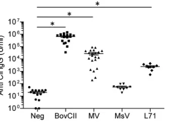
The breakdown of immune tolerance induced by bovine
colla-gen and mimivirus proteins was confirmed by the detection of
elevated serum titers of anti-mouse collagen type II IgG in mice
immunized with bovine collagen type II and mimivirus proteins,
whereas mice immunized with PBS and marseillevirus did not
show elevated anti-mouse collagen type II IgG titers (
Fig. 3
). Mice
immunized with recombinant L71 protein also showed
signifi-cantly elevated anti-mouse collagen type II IgG titers yet by an
order of magnitude lower than the titers observed in mice
immu-nized with mimivirus proteins (
Fig. 3
). The lower antibody
re-sponse achieved with L71 immunization may account for the lack
of joint inflammation seen in these mice. The cross-reactivity of T
cells was investigated in recall assays (
25
). Cells isolated from
draining lymph nodes of the immunized mice were found to
pro-liferate in response to
in vitro
presentation of denatured fragments
of mouse collagen type II (
Fig. 4A
), bovine collagen type II (
Fig.
4B
), and mimivirus collagen protein L71 (
Fig. 4C
), thereby
con-firming the presence of autoreactive T cells after immunization
with mimivirus proteins. The proliferative response to collagen
was strongest for cells isolated from mice immunized with
mimi-virus proteins. This finding was surprising since mice immunized
with bovine collagen type II showed the highest score for limb
inflammation and for anticollagen IgG titers. The strong
prolifer-ative response of T cells from mimivirus protein-immunized mice
may reflect the higher antigenicity of mimivirus collagen
consid-ering its peptide sequence divergence from mammalian collagen
sequences.
Immunity to mimivirus in humans.
To determine whether
humans are commonly exposed to mimivirus, we first examined
the presence of antibodies against mimivirus in 100 healthy
sub-jects by ELISA using whole mimivirus proteins as antigens.
Reac-tivity to mimivirus proteins was variable; 58 human sera showed
significant IgG titers in the 5% range of titers observed in the sera
of rabbits previously immunized with mimivirus proteins (
Fig. 5
).
To identify the major mimivirus proteins recognized by human
sera, we coupled the IgG fraction of sera from healthy subjects
(healthy-subject sera) and rheumatoid arthritis patients
(rheuma-toid arthritis sera) to protein G-Sepharose beads, which were
fur-FIG 2Joint inflammation in DBA/1 mice immunized with mimiviruspro-teins. (A) Clinical severity of arthritic limbs in the groups of mice immunized with PBS (), bovine collagen type II (), recombinant L71 protein (), marseillevirus proteins (}), and mimivirus proteins (Œ) are shown as means⫾ standard errors of the means. The arrow shows the time point of booster im-munization. Data represent three independent experiments with 10 to 21 mice per group. (B) Representative H&E-stained sections of hind limbs by day 75 after immunization showing cartilage damage and synovial hyperplasia in mice immunized with bovine collagen type II (BovCII) and mimivirus protein (MV). No signs of pathology were visible in PBS-immunized negative-control mice (Neg). Scale bar, 100m.
FIG 3Anti-collagen type II IgG titers in DBA/1 mice immunized with mimi-virus proteins. Levels of serum IgG measured by ELISA against endogenous mouse collagen type II (CII) in mice immunized with PBS (Neg), bovine collagen type II (BovCII), mimivirus proteins (MV), marseillevirus proteins (MsV), or recombinant L71 protein (L71). Data represent three independent experiments with 10 to 21 mice per group. Horizontal bars show means⫾ standard errors of the means (*,P⬍0.01).
on November 7, 2019 by guest
http://jvi.asm.org/
[image:4.585.335.506.65.187.2] [image:4.585.78.244.66.585.2]ther incubated with preparations of mimivirus proteins.
Mimivi-rus proteins retained on the IgG-protein G beads were identified
by mass spectrometric peptide sequencing after trypsin digestion.
The major capsid protein L425 was found in all samples, followed
by the putative GMC-type oxidoreductase R135 and core protein
L410, which were found in 7 of 10 samples (
Table 2
). Interestingly,
the most frequent mimivirus proteins recognized by human sera
were surface proteins according to our surface biotinylation study
(
Table 1
). Mimivirus collagens did not appear among the proteins
recognized by serum. This absence may be related to the
abun-dance of lysine in mimivirus collagens, thereby yielding very short
tryptic peptides that remained below the detection range of mass
spectrometric peptide sequencing. The recognition of multiple
mimivirus proteins by human serum confirmed the exposure of
humans to mimivirus.
To further validate the occurrence of antibodies against
spe-cific mimivirus proteins in human sera, reactivity toward
mimi-virus L425 and L71 proteins was analyzed by Western blotting.
The major capsid L425 and collagen L71 proteins were expressed
as His
6-tagged recombinant proteins in
E. coli
and purified on
Ni
2⫹-Sepharose columns. Pools of 100 healthy-subject sera and
100 rheumatoid arthritis sera were probed against the
recombi-nant L425 and L71 mimivirus proteins. We examined the
reactiv-ity of sera toward surface collagen L71 since this protein was not
detected among the mimivirus proteins captured by immobilized
serum IgG in our previous experiment. For the 100
healthy-sub-ject and 100 rheumatoid arthritis sera tested, respectively, 30 and
36 sera recognized the capsid L425 protein (
Fig. 6A
). This result
confirmed that exposure to mimivirus is common in the human
population. The detection of IgG against the mimivirus capsid
protein L425 in 30% of tested sera suggests repeated antigenic
challenge, probably caused by repeated contact with mimivirus.
Reactivity of human sera toward mimivirus collagen was more
discriminatory. Whereas only 6 healthy-subject sera recognized
the mimivirus collagen L71, 22 rheumatoid arthritis sera were
positive for the mimivirus collagen L71 (
Fig. 6B
). To exclude
non-specific cross-reactivity of human sera toward polypeptides
con-taining (G-X-Y)
ncollagen domains, we tested the recognition of
L71-positive sera for a fragment of human collagen type III
en-compassing 114 G-X-Y repeats and lacking N and C propeptides.
None of the 28 human sera positive for mimivirus L71 recognized
the 36-kDa (G-X-Y)
114construct (
Fig. 7
), thereby demonstrating
the specificity of the antibody response to mimivirus L71 collagen.
Accordingly, this work confirmed that the reactivity to mimivirus
collagen was 3.5 times more frequent in the pool of rheumatoid
arthritis sera than in the sera from healthy subjects, which showed
limited reactivity.
DISCUSSION
The present study demonstrated that mice generated autoreactive
anticollagen antibodies after immunization with mimivirus
pro-teins including viral collagens. A possible relationship between
exposure to mimivirus collagen and the development of
autoim-munity was corroborated by the occurrence of IgG against
mimi-virus collagen among rheumatoid arthritis patients. These
find-ings suggested that repeated exposure to mimivirus leads to
antibody formation to virus collagen and to a breakdown of
im-mune tolerance for endogenous collagens. Giant viruses like
mimivirus are ubiquitous in the environment (
16
,
17
), thereby
supporting the frequent contact of humans with such viruses.
Mimivirus is most likely ingested by water uptake and captured by
dendritic cells and macrophages lining the gastrointestinal
mu-cosa. Alternatively, virus particles may enter the airways as aerosol
and be taken up by alveolar macrophages. In fact, mimivirus can
be phagocytized by human and mouse macrophages although the
virus cannot replicate in these cells (
18
). In line with this
observa-FIG 4Autoreactive T-cell response in DBA/1 mice immunized with mimivirus proteins. (A) Recall responses in cells isolated from draining lymph nodes of mice immunized with PBS (Neg), bovine collagen type II (BovCII), or mimivirus proteins (MV) after stimulation with denatured mouse collagen type II. (B) Recall responses after stimulation with denatured bovine collagen type II. (C) Recall responses after stimulation with denatured fragmented recombinant mimivirus protein L71. Data represent means⫾standard errors of the means of groups of 3 mice (*,P⬍0.01).FIG 5Reactivity of human sera toward mimivirus proteins. Anti-mimivirus IgG titers in sera of 100 healthy subjects were measured by ELISA after dilution to 1:100 and expressed as a ratio to IgG titers measured in 1:1,000-diluted sera from rabbits previously immunized with mimivirus particles. Data represent means⫾standard errors of the means from four analyses.
on November 7, 2019 by guest
http://jvi.asm.org/
[image:5.585.129.462.66.166.2] [image:5.585.114.471.618.693.2]tion, mimivirus infection has been related to pneumonia in
iso-lated cases, although without evidence for virus particles in disease
cases (
26
,
27
). This putative pathogenicity, however, does not
pre-clude a more general effect of mimivirus on priming an
autoim-mune response.
The sequence similarity between a stretch of mimivirus L71
and human collagen type II (
Fig. 1
) supports a possible
cross-reactivity of antibodies due to antigenic mimicry. This was indeed
confirmed in our study by the detection of anti-mouse collagen
type II IgG in mice immunized with recombinant L71 protein. A
similar case of antigenic mimicry occurs in
Campylobacter jejuni
infection, which causes gastroenteritis but can lead to
Guillain-Barré syndrome when antibodies against
Campylobacter
lipooli-gosaccharides cross-react with endogenous GM1 gangliosides on
nerve cells (
28
). Likewise, the detection of antibodies toward
mimivirus L425 capsid protein in some
Francisella tularensis
-in-fected patients suggested cross-reactivity of mimivirus antigens
with other microorganisms (
29
). But, surprisingly, no reactivity to
mimivirus antigens was found in sera from healthy subjects in the
study of Pelletier et al. (
29
).
[image:6.585.38.551.78.160.2]The main factors involved in the pathogenicity of rheumatoid
arthritis could be either genetic or environmental. Rheumatoid
arthritis is an autoimmune disease with a significant
environmen-tal component, as supported by twin studies (
30
,
31
). Repeated
contact to collagen antigens found in the environment may
pro-mote the development of cross-reactive anticollagen antibodies
and to inflammation in collagen-rich tissues. Antibodies against
collagens can recognize either the triple helical conformation or
the peptide sequences in the triple helical domain or in
telopep-tides. The specificity of antibodies against collagen depends on the
activation of a humoral response alone or on a combination of
cell-mediated and humoral responses (
32
). The reactivity of
hu-man serum to mimivirus collagen L71 shown by Western blotting
indicates that epitopes based on amino acid sequence and not
TABLE 2Mimivirus proteins recognized by human serum IgGORF Protein annotation
Mascot score for sera from:a
Healthy subjects Rheumatoid arthritis subjects
L425 Capsid protein 68 601 463 68 387 983 271 258 70 327
R135 Putative GMC-type oxidoreductase 370 394 153 774 255 163 161
L410 Core protein 223 284 179 685 123 133 97
R345 Putative regulator of chromosome condensation 52 123 145 28 58 350 185 77 217
R349 Uncharacterized protein 28 28 30 26 26 35 26 28 32
aScores for viral proteins recognized by five healthy subject sera and five rheumatoid arthritis sera are listed in columns for each serum tested. Values indicate Mascot scores
representing the probability of positive matches for the recognized proteins. Scores above 25 were significant at aPvalue of⬍0.05.
FIG 6Recognition of mimivirus proteins by human sera. (A) Representative Western blots of sera from healthy subjects (HS) and rheumatoid arthritis (RA) patients recognizing mimivirus capsid protein L425. (B) Representative Western blots of sera from healthy subjects (HS) and rheumatoid arthritis (RA) patients recognizing mimivirus collagen L71. Sera were diluted 1:4,000. Positions of recombinant L425 and L71 proteins in the blots are shown at the left of each panel using an anti-His6antibody (His6). A 15-kDa fragment of His6-tagged human GLT25D2 protein was used as a negative control (Neg).
on November 7, 2019 by guest
http://jvi.asm.org/
[image:6.585.135.449.416.682.2]three-dimensional (3D) conformations are being recognized.
This notion was supported by finding no reactivity of the
L71-positive human sera for the human collagen type III (G-X-Y)
114polypeptide used as a negative control. The lack of recognition for
the (G-X-Y)
114polypeptide also indicated that the reactivity
to-ward L71 was specific to mimivirus exposure and not the result of
cross-reactivity to collagen domain-containing proteins, such as
those found in some Gram-positive bacteria. We did find that
mimivirus L71 protein was immunogenic and led to the
pro-duction of anti-mouse collagen type II IgG, but immunization
of DBA/1 mice with recombinant L71 protein failed to induce
arthritis. This recombinant protein likely did not mimic the
native conformation of collagen-like proteins, which is
essen-tial for arthritogenicity. In fact, denaturation of collagen prior
to immunization abrogates the arthritic response (
33
).
Based on these results and in view of the structural similarity
between mimivirus collagen and human collagen sequences, we
propose that giant viruses expressing collagen represent a
poten-tial environmental risk factor contributing to the development of
rheumatoid arthritis. A systematic survey of mimivirus
distribu-tion in the environment will contribute to a better appreciadistribu-tion of
the environmental risk associated with such giant viruses in
rela-tion to the geographical incidence of rheumatoid arthritis.
ACKNOWLEDGMENTS
We are grateful to Lubor Borsig, Daniel Rhyner, Carole Oertli, Jürg Cabal-zar, Christoph Rutschmann, and Charlotte Burger for technical assis-tance. We are grateful to the Functional Genomics Center Zurich for its support with mass spectrometric analysis.
This work was supported by a Swiss National Science Foundation Grant 310030-129633 to T.H.
REFERENCES
1.Wilson WH, Van Etten JL, Allen MJ.2009. ThePhycodnaviridae: the story of how tiny giants rule the world. Curr. Top. Microbiol. Immunol.
328:1– 42.http://dx.doi.org/10.1007/978-3-540-68618-7_1.
2.Colson P, Pagnier I, Yoosuf N, Fournous G, La Scola B, Raoult D.2013.
“Marseilleviridae,” a new family of giant viruses infecting amoebae. Arch.
Virol.158:915–920.http://dx.doi.org/10.1007/s00705-012-1537-y. 3.Van Etten JL, Burbank DE, Kuczmarski D, Meints RH.1983. Virus
infection of culturable chlorella-like algae and development of a plaque assay. Science219:994 –996.http://dx.doi.org/10.1126/science.219.4587 .994.
4.Moliner C, Fournier PE, Raoult D.2010. Genome analysis of microor-ganisms living in amoebae reveals a melting pot of evolution. FEMS
Mi-crobiol. Rev. 34:281–294. http://dx.doi.org/10.1111/j.1574-6976.2009 .00209.x.
5.Raoult D, Audic S, Robert C, Abergel C, Renesto P, Ogata H, La Scola B, Suzan M, Claverie JM.2004. The 1.2-megabase genome sequence of Mimivirus. Science 306:1344 –1350. http://dx.doi.org/10.1126/science .1101485.
6.Arslan D, Legendre M, Seltzer V, Abergel C, Claverie JM.2011. Distant Mimivirus relative with a larger genome highlights the fundamental fea-tures ofMegaviridae. Proc. Natl. Acad. Sci. U. S. A.108:17486 –17491. http://dx.doi.org/10.1073/pnas.1110889108.
7.Colson P, Yutin N, Shabalina SA, Robert C, Fournous G, La Scola B, Raoult D, Koonin EV.2011. Viruses with more than 1,000 genes: Mama-virus, a newAcanthamoeba polyphagamimivirus strain, and reannotation of Mimivirus genes. Genome Biol. Evol.3:737–742.http://dx.doi.org/10 .1093/gbe/evr048.
8.Yoosuf N, Yutin N, Colson P, Shabalina SA, Pagnier I, Robert C, Azza S, Klose T, Wong J, Rossmann MG, La Scola B, Raoult D, Koonin EV.
2012. Related giant viruses in distant locations and different habitats:
Acanthamoeba polyphagamoumouvirus represents a third lineage of the
Mimiviridaethat is close to the megavirus lineage. Genome Biol. Evol.
4:1324 –1330.http://dx.doi.org/10.1093/gbe/evs109.
9.Van Etten JL, Gurnon JR, Yanai-Balser GM, Dunigan DD, Graves MV.
2010. Chlorella viruses encode most, if not all, of the machinery to glyco-sylate their glycoproteins independent of the endoplasmic reticulum and Golgi. Biochim. Biophys. Acta1800:152–159.http://dx.doi.org/10.1016/j .bbagen.2009.07.024.
10. Wang IN, Li Y, Que Q, Bhattacharya M, Lane LC, Chaney WG, Van Etten JL.1993. Evidence for virus-encoded glycosylation specificity. Proc. Natl. Acad. Sci. U. S. A.90:3840 –3844.http://dx.doi.org/10.1073/pnas.90 .9.3840.
11. Legendre M, Santini S, Rico A, Abergel C, Claverie JM.2011. Breaking the 1000-gene barrier for Mimivirus using ultra-deep genome and tran-scriptome sequencing. Virol. J. 8:99. http://dx.doi.org/10.1186/1743 -422X-8-99.
12. La Scola B, Desnues C, Pagnier I, Robert C, Barrassi L, Fournous G, Merchat M, Suzan-Monti M, Forterre P, Koonin E, Raoult D.2008. The virophage as a unique parasite of the giant mimivirus. Nature455:100 – 104.http://dx.doi.org/10.1038/nature07218.
13. Luther KB, Hulsmeier AJ, Schegg B, Deuber SA, Raoult D, Hennet T.
2011. Mimivirus collagen is modified by bifunctional lysyl hydroxylase and glycosyltransferase enzyme. J. Biol. Chem.286:43701– 43709.http: //dx.doi.org/10.1074/jbc.M111.309096.
14. Xiao C, Chipman PR, Battisti AJ, Bowman VD, Renesto P, Raoult D, Rossmann MG.2005. Cryo-electron microscopy of the giant Mimivirus. J. Mol. Biol.353:493– 496.http://dx.doi.org/10.1016/j.jmb.2005.08.060. 15. Kuznetsov YG, Xiao C, Sun S, Raoult D, Rossmann M, McPherson A.
2010. Atomic force microscopy investigation of the giant mimivirus. Vi-rology404:127–137.http://dx.doi.org/10.1016/j.virol.2010.05.007. 16. Boughalmi M, Saadi H, Pagnier I, Colson P, Fournous G, Raoult D, La
Scola B.2013. High-throughput isolation of giant viruses of the
Mimiviri-daeandMarseilleviridaefamilies in the Tunisian environment. Environ.
Microbiol.15:2000 –2007.http://dx.doi.org/10.1111/1462-2920.12068.
FIG 7Specific recognition of mimivirus collagen L71 by human sera. Representative Western blots of L71-positive sera from healthy subjects (HS) and rheumatoid arthritis (RA) patients recognizing mimivirus collagen L71 but not a fragment of human collagen type III containing 114 G-X-Y repeats (CIII). Sera were diluted 1:4,000. Positions of recombinant L71 and CIII proteins in the blots are shown at the left of the panel using an anti-His6antibody (His6).
on November 7, 2019 by guest
http://jvi.asm.org/
[image:7.585.138.450.64.192.2]17. La Scola B, Campocasso A, N=Dong R, Fournous G, Barrassi L, Flau-drops C, Raoult D.2010. Tentative characterization of new environmen-tal giant viruses by MALDI-TOF mass spectrometry. Intervirology53:
344 –353.http://dx.doi.org/10.1159/000312919.
18. Ghigo E, Kartenbeck J, Lien P, Pelkmans L, Capo C, Mege JL, Raoult D.
2008. Ameobal pathogen mimivirus infects macrophages through phago-cytosis. PLoS Pathog.4:e1000087.http://dx.doi.org/10.1371/journal.ppat .1000087.
19. Shin BK, Wang H, Yim AM, Le Naour F, Brichory F, Jang JH, Zhao R, Puravs E, Tra J, Michael CW, Misek DE, Hanash SM. 2003. Global profiling of the cell surface proteome of cancer cells uncovers an abun-dance of proteins with chaperone function. J. Biol. Chem.278:7607–7616. http://dx.doi.org/10.1074/jbc.M210455200.
20. Shevchenko A, Tomas H, Havlis J, Olsen JV, Mann M.2006. In-gel digestion for mass spectrometric characterization of proteins and pro-teomes. Nat. Protoc.1:2856 –2860.http://dx.doi.org/10.1038/nprot.2006 .468.
21. Brand DD, Latham KA, Rosloniec EF.2007. Collagen-induced arthritis. Nat. Protoc.2:1269 –1275.http://dx.doi.org/10.1038/nprot.2007.173. 22. Schegg B, Hülsmeier AJ, Rutschmann C, Maag C, Hennet T.2009. Core
glycosylation of collagen is initiated by two(1-O)galactosyltransferases. Mol. Cell. Biol.29:943–952.http://dx.doi.org/10.1128/MCB.02085-07. 23. Snir O, Backlund J, Bostrom J, Andersson I, Kihlberg J, Buckner JH,
Klareskog L, Holmdahl R, Malmstrom V.2012. Multifunctional T cell reactivity with native and glycosylated type II collagen in rheumatoid ar-thritis. Arthritis Rheum. 64:2482–2488. http://dx.doi.org/10.1002/art .34459.
24. Backlund J, Carlsen S, Hoger T, Holm B, Fugger L, Kihlberg J, Burkhardt H, Holmdahl R.2002. Predominant selection of T cells spe-cific for the glycosylated collagen type II epitope (263–270) in humanized transgenic mice and in rheumatoid arthritis. Proc. Natl. Acad. Sci. U. S. A.
99:9960 –9965.http://dx.doi.org/10.1073/pnas.132254199.
25. Becher B, Durell BG, Miga AV, Hickey WF, Noelle RJ.2001. The clinical course of experimental autoimmune encephalomyelitis and inflammation is controlled by the expression of CD40 within the central nervous system. J. Exp. Med.193:967–974.http://dx.doi.org/10.1084/jem.193.8.967.
26. La Scola B, Marrie TJ, Auffray JP, Raoult D.2005. Mimivirus in pneu-monia patients. Emerg. Infect. Dis. 11:449 – 452. http://dx.doi.org/10 .3201/eid1103.040538.
27. Vanspauwen MJ, Franssen FM, Raoult D, Wouters EF, Bruggeman CA, Linssen CF.2012. Infections with mimivirus in patients with chronic obstructive pulmonary disease. Respir. Med.106:1690 –1694.http://dx .doi.org/10.1016/j.rmed.2012.08.019.
28. Yuki N, Susuki K, Koga M, Nishimoto Y, Odaka M, Hirata K, Taguchi K, Miyatake T, Furukawa K, Kobata T, Yamada M.2004. Carbohydrate mimicry between human ganglioside GM1 andCampylobacter jejuni li-pooligosaccharide causes Guillain-Barre syndrome. Proc. Natl. Acad. Sci. U. S. A.101:11404 –11409.http://dx.doi.org/10.1073/pnas.0402391101. 29. Pelletier N, Raoult D, La Scola B.2009. Specific recognition of the major
capsid protein ofAcanthamoeba polyphagamimivirus by sera of patients infected byFrancisella tularensis. FEMS Microbiol. Lett.297:117–123. http://dx.doi.org/10.1111/j.1574-6968.2009.01675.x.
30. Jarvinen P, Aho K.1994. Twin studies in rheumatic diseases. Semin. Arthritis Rheum.24:19 –28.http://dx.doi.org/10.1016/0049-0172(94)90096-5. 31. Svendsen AJ, Holm NV, Kyvik K, Petersen PH, Junker P.2002. Relative
importance of genetic effects in rheumatoid arthritis: historical cohort study of Danish nationwide twin population. BMJ324:264 –266.http://dx .doi.org/10.1136/bmj.324.7332.264.
32. Lynn AK, Yannas IV, Bonfield W.2004. Antigenicity and immunoge-nicity of collagen. J. Biomed. Mater. Res. B Appl. Biomater.71:343–354. http://dx.doi.org/10.1002/jbm.b.30096.
33. Stuart JM, Townes AS, Kang AH.1982. Nature and specificity of the immune response to collagen in type II collagen-induced arthritis in mice. J. Clin. Invest.69:673– 683.http://dx.doi.org/10.1172/JCI110495. 34. Keller A, Nesvizhskii AI, Kolker E, Aebersold R.2002. Empirical
statis-tical model to estimate the accuracy of peptide identifications made by MS/MS and database search. Anal. Chem.74:5383–5392.http://dx.doi .org/10.1021/ac025747h.
35. Nesvizhskii AI, Keller A, Kolker E, Aebersold R.2003. A statistical model for identifying proteins by tandem mass spectrometry. Anal. Chem.75:
4646 – 4658.http://dx.doi.org/10.1021/ac0341261.
on November 7, 2019 by guest
http://jvi.asm.org/