Adaptor Protein To Disrupt Inflammatory Signaling
David A. Griffiths,a,bHussein Abdul-Sada,a,bLaura M. Knight,a,bBrian R. Jackson,a,bKathryn Richards,a,bEmma L. Prescott,a,b A. Howard S. Peach,cG. Eric Blair,aAndrew Macdonald,a,bAdrian Whitehousea,b
School of Molecular and Cellular Biology, Faculty of Biological Sciences,a
and Astbury Centre for Structural Molecular Biology,b
University of Leeds, Leeds, United Kingdom; Department of Plastic Surgery, Leeds Teaching Hospitals NHS Trust, St. James’s University Hospital, Leeds, United Kingdomc
Merkel cell carcinoma (MCC) is a highly aggressive nonmelanoma skin cancer arising from epidermal mechanoreceptor Merkel cells. In 2008, a novel human polyomavirus, Merkel cell polyomavirus (MCPyV), was identified and is strongly implicated in MCC pathogenesis. Currently, little is known regarding the virus-host cell interactions which support virus replication and vi-rus-induced mechanisms in cellular transformation and metastasis. Here we identify a new function of MCPyV small T antigen
(ST) as an inhibitor of NF-B-mediated transcription. This effect is due to an interaction between MCPyV ST and the NF-B
essential modulator (NEMO) adaptor protein. MCPyV ST expression inhibits IB kinase␣(IKK␣)/IKK-mediated IB
phos-phorylation, which limits translocation of the NF-B heterodimer to the nucleus. Regulation of this process involves a previously
undescribed interaction between MCPyV ST and the cellular phosphatase subunits, protein phosphatase 4C (PP4C) and/or protein
phosphatase 2A (PP2A) A, but not PP2A A␣. Together, these results highlight a novel function of MCPyV ST to subvert the innate
immune response, allowing establishment of early or persistent infection within the host cell.
M
erkel cell carcinoma (MCC) is an aggressive neuroendocrine carcinoma of the skin (1). Similar to other skin cancers, prolonged UV exposure is a known MCC risk factor, and inci-dence also increases sharply in the elderly (2). MCC is also dra-matically increased upon the loss of immunocompetence and is more frequent in AIDS patients and organ transplant recipients (3,4). As such, this etiology is reminiscent of other virus-induced cancers.In 2008, Merkel cell polyomavirus (MCPyV) was identified as clonally integrated in⬃80% of MCCs (5). It is the most recently discovered virus known to cause human cancer and the first hu-man polyomavirus strongly linked with tumor induction (6,7). Like other polyomaviruses, the MCPyV genome comprises early and late gene regions separated by a noncoding regulatory region. The early region encodes large T antigen (LT), small T antigen (ST), and 57kT proteins produced by alternative splicing (5).
A monoclonal viral integration pattern is observed in MCC, indicating that MCPyV infection and integration occur prior to clonal expansion of tumor cells (5). Additionally, truncating mu-tations in the integrated MCPyV LT render the virus replication defective, thus dismissing any passenger role of the virus in MCC tumorigenesis (6). Furthermore, ST and N-terminal regions of LT remain unaffected by tumor-derived mutations, suggesting that they also contribute to MCC tumorigenesis. This is supported by experiments showing that depletion of the T antigens leads to cell cycle arrest and cell death in MCPyV-positive MCC cell lines (8). This highlights that MCPyV ST and LT are important factors in regulating virus replication and play critical roles in human cell transformation. However, the molecular mechanisms implicating the MCPyV T antigens in virus replication and cancer develop-ment are yet to be fully elucidated.
Previous studies with related polyomaviruses, such as simian virus 40 (SV40), show that ST is required for upregulation of early viral promoter activity and also stimulates LT-mediated activa-tion of the late viral promoter (9, 10). Furthermore, although SV40 ST expression alone is not capable of transforming cells,
coexpression of SV40 ST and LT, together with the telomerase catalytic subunit hTERT and the oncogenic version of H-RAS, are sufficient to generate fully transformed human cells capable of tumor formationin vivo(11). SV40 ST activity is due to an inter-action with protein phosphatase 2A (PP2A), the major cellular serine/threonine phosphatase. It associates with PP2A A and C subunits, displacing the regulatory B subunit, resulting in changes to PP2A substrate specificity and subsequent control of signaling pathways (12,13).
In contrast, recent evidence suggests that MCPyV ST is onco-genic (14). Expression of ST alone is sufficient to transform rodent cells to anchorage- and contact-independent growth and also in-duces serum-free proliferation of human cells. Moreover, specific depletion of MCPyV ST is sufficient to inhibit MCPyV-positive MCC cell growth. Surprisingly, however, these effects were inde-pendent of the conserved interaction with PP2A (14). Uniquely, MCPyV ST deregulates cap-dependent translation by maintain-ing the hyperphosphorylated state of the eukaryotic translational regulatory protein, 4E-binding protein 1 (14). In addition, it tar-gets the cellular ubiquitin ligase SCFFwb7, which stabilizes MCPyV LT and several cellular oncoproteins, such as c-Myc and cyclin E (15).
NF-B is a pivotal family of transcription factors that regulate expression of numerous target genes with diverse roles ranging from inflammation and immunity to cell death or proliferation (16). The family consists of five structurally homologous mem-bers, forming a variety of homo- and heterodimers, including the
Received5 August 2013 Accepted4 October 2013
Published ahead of print9 October 2013
Address correspondence to Adrian Whitehouse, a.whitehouse@leeds.ac.uk, or Andrew Macdonald, a.macdonald@leeds.ac.uk.
Copyright © 2013, American Society for Microbiology. All Rights Reserved.
doi:10.1128/JVI.02159-13
on November 7, 2019 by guest
http://jvi.asm.org/
most common heterodimer of RelA. NF-B is found in an inhib-ited cytoplasmic form, maintained in this conformation by virtue of an interaction with members of the inhibitor ofB (IB) family of proteins (17). NF-B is activated in response to inflammatory stimuli by cytokines, such as tumor necrosis factor alpha (TNF-␣) or by pattern recognition receptors (PRRs) that detect pathogen-associated molecular patterns (PAMPS). Engagement of the TNF-␣receptor (TNFR) or PRRs initiates a coordinated signaling cascade, resulting in the activation of the IB kinase (IKK) com-plex (18–20). This complex contains two catalytic kinase compo-nents, IKK␣and IKK, as well as a noncatalytic regulatory sub-unit, NF-B essential modulator (NEMO) or IKK␥. NEMO acts as a scaffold for the IKK complex and serves to recruit it to up-stream signaling complexes. The activated IKK complex phospho-rylates IB, resulting in its rapid degradation, allowing the re-leased NF-B heterodimer to translocate to the nucleus and activate transcription of proinflammatory cytokines and upregu-late expression of type I interferons (21).
Given the essential role for NF-B activation in both the in-flammatory and antiviral responses, it has emerged as a prime target for viral subversion. Virus proteins antagonize all stages of the NF-B signaling pathway (22). Both the hepatitis C virus core and human papillomavirus E7 proteins inhibit IKK activation, preventing IB degradation (23, 24). Human cytomegalovirus (CMV) and molluscum contagiosum poxvirus target NEMO to disrupt IKK activation. While the poxvirus MC159 protein inhib-its IKK activation through an interaction with NEMO, conversely the CMV M45 protein redistributes NEMO to autophagocytic bodies, where it is degraded (25,26). The African swine fever virus prevents NF-B activation downstream of the IKK complex by encoding an IB orthologue, replacing the host inhibitor upon IKK-induced degradation (27,28). Alternatively, the myxovirus-encoded nuclear factor colocalizes with NF-B in the nucleus to prevent induction of NF-B-dependent genes (29). Interestingly, SV40 ST has been shown to upregulate NF-B activation in a PP2A-dependent manner, although some proinflammatory tar-gets, such as IL-8, are downregulated in the presence of SV40 ST (30). Intriguingly, however, the PP2A-independent transforming ability of MCPyV ST (14) suggests that alternative mechanisms of modulating NF-B by MCPyV may be important.
In this study, we show that MCPyV ST negatively regulates NF-B-mediated transcriptional activation. This effect is me-diated through a novel interaction between MCPyV ST and NEMO. Specifically, MCPyV ST inhibits IKK␣/IKK-induced IB phosphorylation, limiting translocation of NF-B into the nucleus. Regulation of this process involves a previously unde-scribed interaction between MCPyV ST and the cellular phospha-tase subunits, PP4C and/or PP2A A. These findings have impor-tant implications for virus replication and MCPyV-induced tumorigenesis, as MCPyV ST may interfere with the host innate immune response to enhance viral replication and persistence in the host cell.
MATERIALS AND METHODS
Plasmids, antibodies, and cells.The MCPyV ST was PCR amplified from MCC tumor genomic DNA using primers 5=-GGG GGT ACC ATG GAT TTA GTC and 5=-GGG CCC GGG CTA GAA A and cloned into the multiple cloning site of pEGFP-cI to generate pEGFP-ST. FLAG-tagged MCPyV small T antigens were also amplified and cloned into pCDNA5/ frt/To to create iST-FLAG. The green fluorescent protein (GFP)-ST
car-boxy-terminal truncation series was PCR amplified from pEGFP-ST and cloned into the multiple cloning site of pEGFP-cI. pEGFP-ST R7A was PCR amplified from pEGFP-ST, incorporating the desired mutation in the forward primer. pGFP-ST⌬95-111 and pGFP-ST⌬111-128 deletion mutants were produced using the QuikChange site-directed mutagenesis kit (Stratagene), as directed by the manufacturer’s instructions. Primer sequences for these deletion and site-directed mutations are available upon request. The MCPyV early promoter region was PCR amplified from MCC genomic DNA using the following primers: 5=-CAT CCT GAA AAA TAA ATA AGG ATA CTT ACT C and 5=-ATA ACA ATT AGG AGC AAT CTC CA. Primers incorporated KpnI and SmaI restriction sites, allowing the promoter to be inserted 5=of the luciferase gene in pGL3-Basic, to generate pGL3-Early. Expression vectors for EE-PP2A Aand FLAG-PP4C were kindly provided by Stefan Strack, University of Iowa, and Marilyn Goudrealt, University of Toronto, respectively. Antibodies against NEMO (Abcam), LC3 (Cell Signaling), lamin B (Calbiochem), GAPDH (glyceraldehyde-3-phosphate dehydrogenase; Abcam), phos-pho-IB (Cell Signaling), phospho-IKK␣/IKK (Cell Signaling), p50 (Santa Cruz), p65 (Santa Cruz), FLAG (Sigma-Aldrich), Glu-Glu (Ab-cam), GFP (Living Colors), glutathioneS-transferase (GST; Sigma-Al-drich), and-actin (Sigma-Aldrich) were purchased from their respective suppliers. Western blotting and immunofluorescence analysis were car-ried out using specific antibodies at 1:1,000 and 1:250 dilutions, respec-tively.
HEK293, Huh7, and Flp-In 293 cells were maintained in Dulbecco’s modified Eagle’s medium (DMEM) containing 10% fetal bovine serum (FBS) and 1% penicillin-streptomycin. MCC13 and MKL-1 cell lines were maintained in RPMI 1640 media supplemented with 10% FBS and 1% penicillin-streptomycin. To generate the i293-ST inducible cell line, Flp-In 293 cells were cotransfected with iST-FLAG and pPGK/Flip/ObpA using Lipofectamine 2000 (Invitrogen) and selected by hygromycin B re-sistance at 100g/ml. ST-FLAG expression was induced with 2g/ml doxycycline hyclate for up to 48 h.
Immunoblotting.Cells were lysed in RIPA buffer (50 mM Tris-HCl at pH 7.6, 150 mM NaCl, 1% NP-40) and supplemented with a protease inhibitor cocktail (Roche). Proteins were separated by SDS-PAGE before transfer onto nitrocellulose membrane (Hybond C Extra; Amersham Bio-sciences). Membranes were probed with the appropriate primary and horseradish peroxidase (HRP)-conjugated secondary antibodies. Pro-teins were detected using EZ-ECL enhancer solution (Geneflow).
Quantitative real-time PCR arrays.Total RNA was harvested from uninduced or induced i293-ST cells by TRIzol extraction, before incuba-tion with chloroform and precipitaincuba-tion by addiincuba-tion of isopropanol. RNA was treated for genomic DNA contamination using a DNase-free kit (Am-bion), before purification using an RNeasy minikit (Qiagen). The SA Bio-sciences RT2first-strand kit (C-03; SA Biosciences) was used to produce cDNA. SA Biosciences RT2profiler PCR array system kits were used to perform the arrays, with plates containing signal transduction and cancer pathway finder primer sets. Quantitative real-time PCR was performed using a 7900 HT (ABI), with the manufacturer’s software used to analyze results as previously described (31).
Determination of secreted IL-8 and CCL20 levels by ELISA.HEK293 and MCC13 cells transfected with a plasmid expressing enhanced GFP (EGFP)-ST or empty vector control were treated with 10 ng/ml TNF-␣, and supernatants were collected 24 h posttreatment. Levels of secreted interleukin-8 (IL-8) and CCL20 were detected by enzyme-linked immu-nosorbent assay (ELISA) using the manufacturer’s protocol (R&D Sys-tems).
Luciferase reporter assays.i293-ST and MCC13 cells were transfected with 50 ng of reporter plasmid expressing firefly luciferase under the con-trol of the NF-B elements from the concanavalin A promoter (32), a cyclic AMP responsive promoter (pCRE), or a tandem AP-1 element re-porter using polyethyleneimine (PEI) (Polysciences Inc.) (33). Where ap-propriate, cells were cotransfected with 1g of MCPyV ST expression plasmid or cellular protein (0.5g) (e.g., MyD88). Empty plasmid was
Griffiths et al.
on November 7, 2019 by guest
http://jvi.asm.org/
added to ensure each transfection received the same amount of total DNA. To normalize for transfection efficiency, 10 ng pRLTK Renilla luciferase reporter plasmid was added to each transfection. Where necessary, at 24 h posttransfection, cells were treated with 10 ng/ml TNF-␣, 50 ng/ml tetra-decanoyl phorbol acetate (TPA), or 20 ng/ml IL-1␣for a further 12 h. Samples were lysed in passive lysis buffer, and activity was measured using a dual luciferase reporter assay (Promega), as previously described (34).
Coimmunoprecipitation assays.Assays were performed as previous described (35,36). For GST pulldown screens, 293 cells were cotrans-fected with 1.5g of EGFP or EGFP-ST and various GST-tagged expres-sion vectors (described in reference32) using Lipofectamine 2000. After 24 h, cells were lysed in RIPA buffer, and lysates were bound to glutathi-one-Sepharose 4B beads (GE Healthcare) for 2 h at 4°C. Beads were washed and then analyzed by immunoblotting using GST- or GFP-spe-cific monoclonal antibodies (Sigma and Living Colors, respectively).
To perform epitope tag-based coimmunoprecipitation assays, 293 cells were transfected with the appropriate vectors. Cellular lysates were harvested after 48 h and then incubated with protein A-agarose beads, GFP-TRAP A beads (Chromotek), anti-FLAG M1 affinity gel (Sigma-Aldrich), or EE-specific antibody (Abcam) bound to protein A-agarose beads for 2 h at 4°C before being washed in phosphate-buffered saline (PBS). Beads were resuspended and analyzed by immunoblotting with the appropriate antibodies.
Endogenous coimmunoprecipitations were performed using unin-duced or inunin-duced i293-ST cells. After 48 h of induction, precleared cellular lysates were then incubated with no antibody, a control antibody, or a NEMO-specific polyclonal antibody (Abcam) for 2 h prior to addition of protein A-agarose beads, as previously described (37). After an additional 2 h of incubation, beads were washed, and precipitated proteins were analyzed by immunoblotting with a FLAG-specific antibody.
Immunofluorescence.293, Huh7, and MCC13 cells grown on glass coverslips were transfected with EGFP or EGFP-ST expression vectors. After 24 h, cells were then fixed with 4% paraformaldehyde, permeabil-ized in 1% Triton X-100, and blocked in PBS-1% bovine serum albumin (BSA) for 1 h. Cells were labeled with the appropriate primary antibodies and then incubated with the appropriate Alexa Fluor-conjugated second-ary antibody, as previously described (38,39). Cells were viewed on a Zeiss 510 meta laser scanning confocal microscope under a 63⫻oil immersion objective lens, and images were analyzed using the LSM imaging software (Zeiss).
Inhibition of autophagy assay.293 cells were transfected with 2g of pEGFP-cI or pEGFP-ST. After 12 h, the appropriate wells were either mock treated or treated with either an inhibitor cocktail (pepstatin [100
M] and leupeptin [3.64M]) or 3-methyladenine (5 nM) for 12 h at 37°C. Cell lysates were analyzed by immunoblotting using the appropriate antibodies.
SILAC-based immunoprecipitations.iST-293 and i293-FLAG cells were grown in R0K0DMEM containing12CL-arginine and12CL-lysine (“light” DMEM) and R6K4DMEM containing13C
L-arginine and13C L-lysine (“heavy” DMEM), respectively. All stable isotope labeling by amino acids in cell culture (SILAC)-based media was supplied by Dundee Cell Products and supplemented with 10% dialyzed fetal calf serum (Dundee Cell Products). iST-293 and i293-FLAG cells were then induced for 48 h, and cells were lysed in 5 ml ice-cold RIPA buffer. Cellular lysates were then incubated with anti-FLAG M1 affinity gel (Sigma-Aldrich) for 2 h at 4°C, before being washed in PBS. For each sample, beads were resus-pended in 40l 2⫻protein solubilizing buffer [50 mM Tris-HCl (pH 6.8), 2% (wt/vol) SDS, 20% (vol/vol) glycerol, 50g/ml bromophenol blue, 10 mM dithiothreitol (DTT)] and boiled at 95°C for 5 min, and each amount of samples was then mixed, separated by one-dimensional SDS-PAGE (4 to 12% Bis-Tris Novex minigel; Invitrogen), and visualized by colloidal Coomassie blue staining (Novex; Invitrogen). The entire protein gel lane was excised and cut into 6 gel slices. Each gel slice was then subjected to in-gel digestion with trypsin, and liquid chromatography-tandem mass spectrometry (LC-MS/MS) analysis (University of Bristol
Proteomics Facility) was used to identify and quantify the precipitated proteins as previously described (40,41).
NF-B pathway activation.i293-ST cells were induced for 42 h and then incubated with serum-free DMEM for 6 h to eliminate nonspecific activation of the NF-B pathway. Cells were then incubated with 100 ng/ml TNF-␣(InvivoGen) in 10% DMEM. Cell lysis was performed in PBS-1% Triton X-100, and lysates were separated into nuclear and cyto-plasmic fractions as previously described (42). Proteins were then sub-jected to immunoblotting, using appropriate antibodies.
RESULTS
MCPyV ST regulates transcription of cellular genes associated
with innate immunity.Polyomavirus ST have important
func-tions in viral replication and transformation (10,11). To analyze the putative functions of MCPyV ST, expression profiling analysis was performed using a Flp-In 293 cell line capable of inducible MCPyV ST expression, termed i293-ST (Fig. 1Ai). The MCPyV ST coding region was initially PCR amplified from genomic DNA isolated from an MCC tumor, incorporating a carboxy-terminal FLAG tag. Sequencing analysis showed that the MCPyV ST gene amplified is identical to the MCC350 isolate (5), with the excep-tion of 3 single nucleotide polymorphisms, none of which result in amino acid changes (data not shown).
Quantitative reverse transcription-PCR arrays were performed using cDNA reversely transcribed from RNA isolated from unin-duced or inunin-duced i293-ST cells. MCPyV ST expression resulted in a marked downregulation of genes associated with the immune response, in particular markers of innate immunity, such as CCL20, CXCL-9, IL-8, and TANK (Fig. 1Aii). A clear trend of the genes affected shows that many are associated with the NF-B pathway, either involved in the activation of NF-B or targeted by NF-B-specific transcriptional activation (Fig. 1Aii, green or red, respectively).
To independently confirm these findings with regard to MCC, we initially assessed the effect of MCPyV ST expression on innate immune expression markers in the MCPyV-negative MCC13 cell line. MCC13 cells were transfected with either control EGFP or EGFP-ST expression constructs (50 to 60% transfection rate) and then stimulated NF-B activation using TNF-␣. ELISAs were then performed, measuring secreted levels of CCL20 and IL-8. Results showed that in TNF-␣-stimulated cells, levels of both CCL20 and IL-8 were downregulated significantly in the presence of MCPyV
ST (Fig. 1B). To confirm that the EGFP-ST expression construct
was functional, we assessed its ability to stimulate expression from the MCPyV early promoter, as previously shown by other polyo-mavirus ST (9). Results demonstrate that upon EGFP-ST expres-sion, MCPyV early promoter activity was upregulated almost 3-fold compared to the pEGFP-cI-transfected control, demon-strating that the EGFP-ST fusion protein was functional. More-over, similar results were also observed in induced i293-ST cells
(Fig. 1Biii). Moreover, we also assessed total levels of IB, as an
indicator of NF-B activation, in MCPyV-negative (MCC13) and MCPyV-positive (MKL-1) MCC cell lines. Immunoblot analysis was performed on MCC13 and MKL-1 cells using an IB-specific antibody, and densitometry-based quantification demonstrated a reduction of approximately 60% in IB levels in the MCPyV-positive MKL-1 cell line (Fig. 1C). Together, these data support the expression profiling, confirming that MCPyV ST downregu-lates NF-B-targeted transcription.
MCPyV ST regulates NF-B signaling downstream from
re-ceptor complexes.We next determined whether MCPyV ST
on November 7, 2019 by guest
http://jvi.asm.org/
cifically regulates NF-B activity and, if so, which part of the pathway is targeted. First, to analyze the effect of MCPyV ST on NF-B-mediated transcription, expression of a luciferase reporter gene driven by NF-B was monitored after stimulation of the NF-B pathway, in the absence or presence of MCPyV ST. Inde-pendent experiments were performed in the i293-ST cell and the MCC MCPyV-negative MCC13 cell line transfected with control EGFP or EGFP-ST expression constructs. In both the induced and EGFP-ST-transfected MCC13 cell lines, MCPyV ST expression strongly inhibited NF-B-driven luciferase expression in response to known NF-B activating agents, TNF-␣, TPA, and IL-1␣(Fig. 2A). In contrast, MCPyV ST had no inhibitory effects on TPA-stimulated cyclic AMP (cAMP) response element (CRE) or acti-vating protein 1 (AP-1)-responsive reporter constructs (Fig. 2B). These results indicate that the inhibitory effect of MCPyV ST is specific to the NF-B pathway.
To initially investigate the possible molecular mechanism of
MCPyV ST-mediated inhibition, upstream components of the NF-B pathway were overexpressed in control versus MCPyV ST-expressing i293-ST and MCPyV-negative MCC13 cells, and NF-B-driven luciferase expression was measured. In both cell lines, MCPyV ST significantly reduced NF-B-driven luciferase in re-sponse to overexpressed Toll-like receptor (TLR) adaptor pro-teins MyD88 and TRIF and the ubiquitin ligases TRAF2 and TRAF6 (Fig. 2C). These data suggest that MCPyV ST subverts NF-B signaling downstream from receptor complexes.
MCPyV ST interacts with NEMO.Our results indicate that
MCPyV ST prevents NF-B activation by a mechanism indepen-dent of activated receptor complexes. A plausible alternative is that MCPyV ST interacts with another target protein within the pathway. Therefore, to identify this binding partner, 293 cells were cotransfected with EGFP or EGFP-ST and various GST-tagged proteins, including kinases (MEKK3), inhibitors (Abin2), or reg-ulatory proteins (NEMO) associated with the NF-B pathway,
FIG 1MCPyV ST downregulates cellular gene expression associated with the NF-B pathway. (A) (i) The MCPyV ST coding region was PCR amplified, incorporating a C-terminal FLAG tag from an MCC tumor, and inserted into pCDNA5/frt/To to yield iST-FLAG. A stable inducible cell line was subsequently produced via site-specific DNA recombination within a Flp-In 293 cell line, termed i293-ST. To verify correct inducible expression of the MCPyV ST fusion protein, i293-ST cells remained uninduced or were incubated for either 24 or 48 h in the presence of doxycycline hyclate. After induction, cell lysates were analyzed by immunoblotting using a FLAG-specific antibody. (ii) RNA was extracted from uninduced or induced i293-ST cells prior to expression profiling using SA Biosciences RT2Profiler PCR array systems. Fold changes were calculated using the manufacturer’s software. (B) (i) The MCC13 cell line was transfected with plasmids expressing EGFP or EGFP-ST. The cells were treated with TNF-␣for 24 h, and levels of secreted IL-8 (i) and CCL20 (ii) were analyzed by ELISA (*,P⬍
0.05 versus control [ttest]). (iii) HEK 293 cells were transiently transfected with either pEGFP-cI or pEGFP-ST and pGL3-Early. In addition, uninduced (Uni) or induced (In) i293-ST cells were transfected with pGL3-Early. Cell lysates were harvested after 24 h and used in a luciferase assay to determine the amount of light emitted in relative fluorescence units (RFL). Results show the average luciferase emissions of triplicate measurements. SmT, small T. (C) (i) Cellular lysates from MCC13 (MCPyV negative) and MKL-1 (MCPyV positive) were analyzed by immunoblotting using a total IB-specific antibody. GAPDH was used as a measure of equal loading. (ii) Densitometry quantification of the Western blots was carried out using ImageJ software and is shown as a percentage of relative densitometry normalized to the loading control GAPDH (n⫽3).
Griffiths et al.
on November 7, 2019 by guest
http://jvi.asm.org/
[image:4.585.136.450.64.387.2]and coimmunoprecipitations were performed using GST-Sephar-ose 4B beads. Results show enhanced binding between MCPyV ST and the GST-tagged NF-B essential modulator (NEMO); in con-trast, no other interactions were observed with other tagged pro-teins associated with the NF-B pathway (Fig. 3A). To confirm the interaction observed between MCPyV ST and NEMO, a series of additional coimmunoprecipitations were performed. Cell lysates expressing EGFP or EGFP-ST and either GST or GST-tagged NEMO were incubated with protein A-agarose beads alone or conjugated with a control or GFP-specific antibodies. Results show that MCPyV ST interacted with NEMO (Fig. 3B). To address potential overexpression artifacts, coimmunoprecipitations were also performed in uninduced versus induced i293-ST cells using a NEMO-specific antibody. Immunoblot analysis using a FLAG-specific antibody also showed a weak but consistent interaction between MCPyV ST and endogenous NEMO (Fig. 3C).
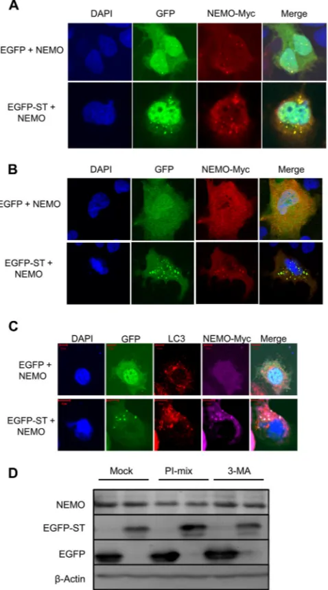
We next determined whether MCPyV ST colocalized with NEMO. The MCPyV-negative MCC cell line, MCC13, and Huh7 cells were transfected with plasmids expressing either EGFP or EGFP-ST, in the presence of a NEMO-myc expression vector, and colocalization was visualized by direct GFP fluorescence (Fig. 4A
andB). Huh7 cells were utilized instead of 293 cells in this exper-iment to allow clear visualization of cytoplasmic structures. Re-sults from both cell lines show the subcellular localization of NEMO in EGFP-expressing cells residing throughout the cyto-plasm and nucleus, with small discrete foci present in both com-partments as previously described (43). In contrast, expression of EGFP-ST resulted in an increase in the number and size of the cytoplasmic puncta, which exhibited significant colocalization of NEMO with a proportion of MCPyV ST. This was particularly evident in the EGFP-ST-transfected MCC13 cell line. In addition, confocal imaging suggests that the puncta are cytoplasmic, but we cannot rule out the presence of some nuclear puncta. Together, these data provide the first evidence of an interaction between MCPyV ST and NEMO.
MCPyV ST does not promote the degradation of NEMO.The
distinct cytoplasmic puncta containing NEMO and MCPyV ST are reminiscent of autophagosomes (44). Interestingly, human cytomegalovirus (HCMV) targets NEMO for autophagosomal degradation, curtailing the cellular inflammatory response (26). To investigate whether MCPyV ST targets NEMO to autophago-somes for degradation, we assessed whether the cytoplasmic
FIG 2MCPyV ST specifically downregulates NF-B activity, downstream of receptor complexes. (A) Uninduced or induced i293-ST (i) or MCC13 cells expressing EGFP or EGFP-ST (ii) were transfected with a reporter plasmid expressing firefly luciferase under the control of the NF-B elements from the concanavalin A promoter. At 24 h posttransfection, cells were treated with 10 ng/ml TNF-␣, 50 ng/ml TPA, or 20 ng/ml IL-1␣for 12 h. Samples were then analyzed for luciferase activity. To normalize transfection efficiency, 10 ng pRLTK Renilla luciferase reporter plasmid was used. Dox, doxycycline. (B) Cotrans-fections were performed as described above, using NF-B, the cyclic-AMP responsive promoter, and tandem AP-1 responsive reporter plasmids, and samples were analyzed for luciferase activity. (C) Uninduced or induced i293-ST (i) or MCC13 cells expressing EGFP or EGFP-ST (ii) were cotransfected with a concanavalin A promoter reporter plasmid and plasmids of the indicated cellular proteins (1g of each), and samples were analyzed for luciferase activity, *,P⬍
0.05 versus control (ttest).
on November 7, 2019 by guest
http://jvi.asm.org/
[image:5.585.137.451.66.394.2]puncta containing MCPyV ST and NEMO observed colocalized with the autophagosome marker, microtubule-associated protein 1A/1B-light chain 3 (LC3) (45).Figure 4C shows that LC3 was observed in cytoplasmic foci, distinct from the foci containing MCPyV ST and NEMO.
Endogenous NEMO levels were also assessed in the presence of MCPyV ST. 293 cells expressing EGFP or EGFP-ST were left un-treated or incubated for 12 h with an autophagy inhibitor, a lyso-somal protease inhibitor (PI) mixture, or 3-methyladenine
(3-MA). Results show that endogenous NEMO levels remained unchanged in the presence of EGFP or MCPyV ST. Moreover, no discernible changes in NEMO levels were observed after the addi-tion of autophagy inhibitors (Fig. 4D). To confirm the function-ality of the autophagy inhibitor 3-MA, Beclin1 levels were as-sessed. 3-MA reduced the levels of Beclin1 back to untreated levels in the presence of the autophagy inducer rapamycin (data not shown). Together, these results indicate that NEMO is not tar-geted to autophagosomes nor degraded by MCPyV ST.
FIG 3MCPyV ST interacts with NEMO. (A) 293 cells were cotransfected with either EGFP or EGFP-ST and the indicated GST-tagged eukaryotic expression vectors. After 24 h, cell lysates were incubated with GST-Sepharose 4B beads, and interacting proteins were immunoblotted with GFP-specific antibodies. (B) 293 cells were cotransfected with EGFP or EGFP-ST in the presence of either GST or GST-NEMO expression vectors. (i) Transfected cell lysates were probed with GFP-, FLAG-, and GST-specific antibodies to serve as a loading control (inputs). (ii) Transfected cell lysates were then incubated with either beads alone control, control antibody (Ab), or GFP-TRAP affinity beads, and bound protein was immunoblotted with a GST-specific antibody in addition to a GFP-specific antibody as a precipitation control. (C) (i) i293-ST cells remained uninduced or were incubated with doxycycline for 48 h. Cellular lysates were immunoblotted with a FLAG-specific antibody. (ii) Cellular lysates were then incubated with polyclonal NEMO-specific antibody bound to protein A-Sepharose beads, and precipitated proteins were then immunoblotted with a FLAG-specific antibody in addition to monoclonal NEMO-specific antibody as a precipitation control.
Griffiths et al.
on November 7, 2019 by guest
http://jvi.asm.org/
[image:6.585.134.447.63.527.2]MCPyV ST prevents phosphorylation of IB and inhibits
nuclear translocation of NF-B. After demonstrating that
MCPyV ST targets NF-B activity downstream of receptor com-plexes and establishing that MCPyV ST interacts with but does not degrade NEMO, we speculated that MCPyV ST may inhibit nu-clear translocation of NF-B, resulting in the observed inhibition of NF-B-dependent transcription. Furthermore, as IB phos-phorylation and subsequent release from NF-B are mediated by interactions between NEMO and upstream kinases, we also
envis-aged that the interaction between MCPyV ST and NEMO may interfere with the phosphorylation status of cytoplasmic IB upon NF-B stimulation, prolonging IB binding to NF-B and ren-dering it inactive.
The effect of MCPyV ST on NF-B activation, following TNF-␣stimulation, was therefore investigated. i293-ST cells re-mained uninduced or MCPyV ST expression was induced for 42 h, prior to 6 h serum starvation, to prevent nonspecific activation of the NF-B pathway; NF-B activation was then stimulated us-ing TNF-␣. Nuclear and cytoplasmic fractions were then sub-jected to immunoblotting and densitometry-based quantification to analyze p50, RelA, and phosphorylated IB levels. Phosphory-lated IB was not detected in either the nuclear or cytoplasmic samples from unstimulated cells (Fig. 5A), showing no NF-B activation in the absence of TNF-␣. In contrast, TNF-␣ -stimu-lated cells, which remained uninduced, showed high levels of phosphorylated IB in cytoplasmic fractions. Strikingly, stimu-lated cells expressing MCPyV ST showed a significant reduction of approximately 60% in cytoplasmic phosphorylated IB levels. Furthermore, nuclear levels of p50 and RelA were also markedly lower, approximately 50%, in TNF-␣-activated cells expressing MCPyV ST, compared with uninduced cells (Fig. 5AandB). This suggests that MCPyV ST inhibits IB phosphorylation through an interaction with NEMO, reducing the nuclear translocation of NF-B subunits and subsequent activation of NF-B responsive genes.
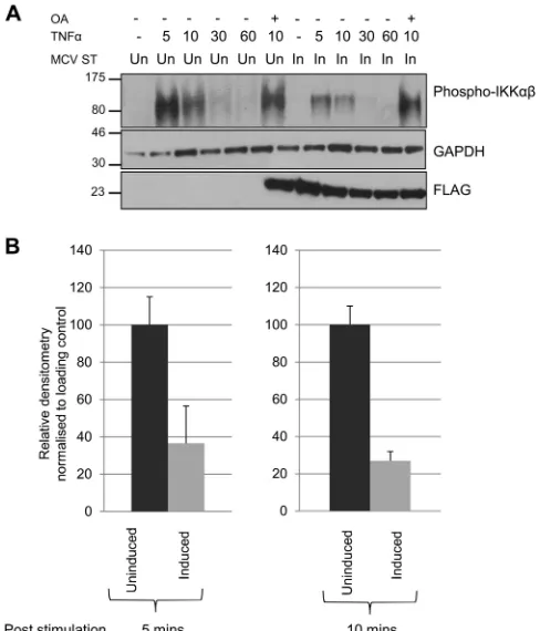
MCPyV ST inhibits IKK␣/IKKphosphorylation.The IKK
complex plays an essential role in NF-B signaling (21). It phos-phorylates inhibitory IB proteins, resulting in their proteasomal degradation, in turn releasing NF-B proteins and allowing their nuclear translocation. Activation of the IKK complex depends on phosphorylation of its two catalytic subunits, IKK␣and IKK. Having demonstrated that MCPyV ST interacts with the IKK component NEMO, and also affects the phosphorylation status of IB, we next assessed the phosphorylation status of IKK␣and IKKin uninduced and MCPyV ST-expressing cells upon TNF-␣ stimulation. Treatment with TNF-␣led to a rapid but transient increase in IKK␣/IKKphosphorylation (Ser176/177) (Fig. 6A), returning to basal levels by 60 min in uninduced cells. In contrast, MCPyV ST expression led to a marked reduction in IKK␣/IKK phosphorylation levels (Fig. 6A). Densitometry-based quantifica-tion shows this reducquantifica-tion is between 60 and 70% in the presence of MCPyV ST (Fig. 6B). Importantly, cells treated with the broad-spectrum phosphatase inhibitor okadaic acid increased IKK␣/ IKKphosphorylation in MCPyV ST-expressing cells, suggesting cellular phosphatases are involved in MCPyV ST-mediated regu-lation of the IKK complex.
SILAC-based immunoprecipitations identify PP4C and PP2A
Aas novel MCPyV ST binding partners.Having established
that MCPyV ST expression results in a marked reduction in phos-phorylated IKK␣and IKKlevels, and that this reduction can be inhibited using the broad-spectrum phosphatase inhibitor oka-daic acid, the interaction with MCPyV ST and cellular phospha-tases was further examined. Polyomavirus ST-PP2A interactions are well characterized (46); however, surprisingly, the interaction between MCPyV ST and PP2A A␣is not required for MCPyV ST-mediated cellular transformation (14). Therefore, SILAC-based pulldown assays were employed to identify additional MCPyV ST cellular phosphatase-interacting partners. This tech-nique facilitates the detection of lower abundance proteins, in
FIG 4A proportion of MCPyV ST colocalizes with NEMO. Following trans-fection of Huh7 (A) and MCC13 (B) cells with either pEGFP or pEGFP-ST in the presence of pNEMO-myc, after 24 h, cells were fixed and permeabilized, and GFP fluorescence was analyzed by direct visualization, whereas NEMO was identified by indirect immunofluorescence using a Myc-specific antibody. (C) A similar experiment was repeated, but Huh7 cells were stained with LC3-and Myc-specific antibodies. (D) 293 cells were transfected with pEGFP-cI or pEGFP-ST. After 12 h, the appropriate wells were either mock treated or treated with either a protease inhibitor (PI) cocktail or 3-methyladenine for 12 h. Cell lysates were then analyzed by immunoblotting using the indicated antibodies. DAPI, 4=,6-diamidino-2-phenylindole.
on November 7, 2019 by guest
http://jvi.asm.org/
[image:7.585.45.284.69.499.2]particular those interacting in substoichiometric amounts or binding with lower affinity in multiprotein complexes (47,48). i293-ST cells expressing the MCPyV ST-FLAG-tagged protein were metabolically labeled in “heavy” media containing13C iso-topes of arginine and lysine, while FLAG-expressing control cells were grown in “light” media containing the12C isotopes. Cell lysates were then incubated with FLAG affinity beads, and precip-itated proteins were identified by LC-MS/MS analysis. False pos-itives immunoprecipitated with the FLAG control were dis-counted, allowing true MCPyV ST interacting partners to be identified. MCPyV ST precipitated two additional cellular phos-phatase subunits, PP4C and PP2A A(Fig. 7A). PP2A A␣was also precipitated but around 40 times less efficiently than the other subunits. Surprisingly, NEMO was not precipitated in this eximent, but this was probably due to the experiment being per-formed in unstimulated cells. Moreover, the interaction between NEMO and MCPyV ST may be transient and not direct, which reduces its potential to be identified in this experiment.
To verify the putative interactions between MCPyV ST and the cellular phosphatases, coimmunoprecipitations were performed in 293 cells transfected with either EGFP or EGFP-ST in the pres-ence of control, FLAG-PP4C, or EE-PP2A Aexpression vectors. For PP4C immunoprecipitations, cell lysates were incubated with protein A-agarose beads alone or conjugated with a control or FLAG-specific antibodies, and precipitated proteins were identi-fied using a GFP-specific antibody. For PP2A A, cell lysates were incubated with GFP-TRAP beads, and immunoprecipitated pro-teins were identified using an EE-specific antibody. Results show that MCPyV ST interacts with both PP4C and PP2A Asubunits
(Fig. 7BandC).
To elucidate whether MCPyV ST expression leads to the redis-tribution of PP4C or PP2A A subunits into the cytoplasmic puncta associated with NEMO, FLAG-PP4C or EE-PP2A Awas expressed in Huh7 cells expressing either EGFP or EGFP-ST and NEMO-Myc. Results demonstrate that in the presence of MCPyV ST, PP4C is present in the puncta containing NEMO; in contrast,
FIG 5MCPyV ST inhibits phosphorylation of IB and prevents nuclear translocation of the NF-B heterodimer. (A) i293-ST cells remained uninduced (Un) or induced (In) for MCPyV ST expression. Cells were then serum starved before TNF-␣was used to stimulate activation of the NF-B pathway. Nuclear and cytoplasmic fractions were then isolated and analyzed by immunoblotting to detect phosphorylation of IB and levels of nuclear translocation of p65 and p50 using the indicated antibodies. (B) Densitometry quantification of the Western blots was carried out using ImageJ software and is shown as a percentage of relative densitometry normalized to the loading control GAPDH. Standard deviations of 3 replicated experiments are shown.
Griffiths et al.
on November 7, 2019 by guest
http://jvi.asm.org/
[image:8.585.135.451.65.459.2]no colocalization is observed in EGFP control cells (Fig. 8A). However, it is difficult to draw the same conclusion regarding PP2A A, as there is a colocalization between PP2A A and NEMO in control cells, although MCPyV ST does colocalize with PP2A Aand NEMO (Fig. 8B). This suggests that MCPyV ST may manipulate cellular phosphatases to modulate the NF-B pathway.
To determine whether an enhanced interaction is observed between NEMO and the cellular phosphatases in the presence of MCPyV ST, coimmunoprecipitation assays were performed. 293 cells were transfected with either EGFP or EGFP-ST in the pres-ence of NEMO-Myc, in addition to either an empty FLAG or EE control vector or a FLAG-PP4C or EE-PP2A Aexpression vec-tor. Cell lysates were then incubated with a polyclonal Myc-spe-cific antibody to precipitate NEMO, and the amount of EGFP, EGFP-ST, or cellular phosphatase proteins also precipitated was identified using GFP-, FLAG-, and EE-specific antibodies. Results show that in the presence of EGFP-ST, an increase in precipitated FLAG-PP4C or EE-PP2A Ais observed compared with the pres-ence of control EGFP alone. Moreover, an increase in precipitated EGFP-ST is also observed in the presence of both cellular phos-phatases, particularly PP4C (Fig. 8C). This suggests that MCPyV
ST can enhance the interaction between the cellular phosphatases and NEMO.
An interaction between MCPyV ST, NEMO, and the cellular
phosphatase subunits PP4C and/or PP2A A, but not PP2A A␣,
is required for MCPyV ST-mediated downregulation of NF-
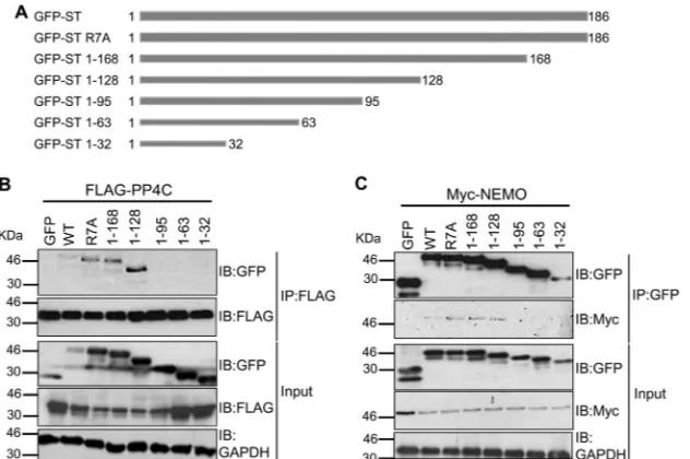
B-targeted transcription.To identify the domains within MCPyV
ST which are required for the interaction between NEMO and the cellular phosphatase PP4C, a carboxy-terminal truncation series of MCPyV ST was produced (Fig. 9A). In addition, the site-di-rected mutant of MCPyV ST, R7A, was utilized, which has previ-ously been reported to abolish the interaction between MCPyV ST and PP2A A␣(14). Coimmunoprecipitation assays were then per-formed with each truncation and R7A mutant. Initially, 293 cells were transfected with an EGFP control vector, EGFP-ST, the EGFP-R7A mutant, or each EGFP carboxy-terminal truncation in the presence of a FLAG-PP4C expression vector. Cell lysates were then incubated with FLAG affinity resin to precipitate FLAG-PPC4, and Western blot analysis was performed using a GFP-specific antibody to assess the presence of interacting GFP-ST fu-sion proteins. Results show that the R7A mutant, which inhibited the interaction between MCPyV ST and PP2A A␣, still associated with PP4C. However, analysis of the MCPyV ST carboxy-terminal truncation series showed that a region containing residues 95 to 128 is required for PP4C binding (Fig. 9B). A similar experiment was also performed to map the MCPyV ST interaction domain required for NEMO binding. 293 cells were transfected with an EGFP control vector, EGFP-ST, the EGFP-R7A mutant, or each EGFP carboxy-terminal truncation in the presence of a NEMO-myc expression vector. Cell lysates were then incubated with GFP-TRAP affinity beads, and Western blot analysis was performed using a Myc-specific antibody to assess the precipitation of NEMO-myc. Results again showed that the R7A mutant was still able to interact with NEMO, suggesting that an interaction with PP2A A␣is not required for the interaction between NEMO and MCPyV ST. In contrast, deletion of residues 95 to 128 abolished the interaction with NEMO (Fig. 9C). Together, these data suggest that the interaction between MCPyV ST, NEMO, and PP4C re-quires residues 95 to 128 within MCPyV ST, although it must be noted that these large-scale truncations of MCPyV ST may affect other functions of the ST protein. Moreover, it also suggests that PP2A A␣is not required for the interaction between NEMO or PP4C and MCPyV ST.
To analyze the functional significance of residues 95 to 128 of MCPyV ST in more detail, two further internal deletion mutants were produced. These specifically deleted either residues 95 to 111 or residues 111 to 128 within the MCPyV ST protein but retained the carboxy terminus (Fig. 10A). Similar coimmunoprecipitation analyses were performed as described for the carboxy truncation mutants above (Fig. 10BandC). Results showed that deletion of residues 111 to 128 (⌬111-128 mutant) had no effect on NEMO or PP4C binding. However, residues 95 to 128 of MCPyV ST are required for the interaction between MCPyV ST and both NEMO and PP4C. Moreover, we also assessed the ability of the R7A,⌬ 95-111, and⌬111-128 mutants to interact with PP2A A(Fig. 10D). Cell lysates were incubated with GFP-TRAP beads, and immuno-precipitated proteins were identified using an EE-specific anti-body. Results showed that wild-type MCPyV ST and the R7A mu-tant interacted with PP2A A, but residues 95 to 111 are also required for the interaction between MCPyV ST and PP2A A. Together, these data suggest that residues 95 to 111 are required
FIG 6Cellular phosphatases are required to inhibit IKK␣/IKK phosphory-lation. (A) i293-ST cells remained uninduced or induced for MCPyV ST ex-pression. Cells were then serum starved before TNF-␣was used to stimulate activation of the NF-B pathway. Cells were lysed directly into SDS sample buffer, and lysates were immunoblotted to detect phosphorylation of IKK␣/ IKK(Ser176/177). GAPDH was used as a loading control, and FLAG blotting detected expression of MCPyV ST. OA, okadaic acid. (B) Densitometry quan-tification of the Western blots at 5 and 10 min time points was carried out using ImageJ software and is shown as a percentage of relative densitometry normal-ized to the loading control GAPDH. Standard deviations of 3 replicated exper-iments are shown.
on November 7, 2019 by guest
http://jvi.asm.org/
[image:9.585.41.285.66.351.2]for the interaction between MCPyV ST, NEMO, and the cellular phosphatase subunits PP4C and PP2A A.
Importantly, to confirm that the⌬95-111 and⌬111-128 mu-tants retained other MCPyV ST functions, we assessed their ability to stimulate expression from the MCPyV early promoter, as pre-viously shown inFig. 1Biii. Results demonstrate that both mutants upregulated the MCPyV early promoter almost 3-fold, similar to wild-type ST, compared with the pEGFP-cI-transfected control
(Fig. 10E). This demonstrates that the ⌬95-111 and ⌬111-128
mutants do retain other MCPyV ST functions.
We next determined the effect of the MCPyV ST⌬95-111- and ⌬111-128-specific deletions and the R7A mutant on the ability of MCPyV ST to downregulate NF-B-targeted transcription. To this end, expression of the luciferase reporter gene driven by NF-B was monitored after stimulation of the NF-B pathway, in
the absence or presence of control EGFP, EGFP-ST, EGFP-ST R7A, EGFP-ST⌬95-111, and ⌬111-128 mutants (Fig. 10F). As previously described, MCPyV ST expression strongly inhibited NF-B-driven luciferase expression in response to TNF-␣. A sim-ilar inhibition was observed for the R7A mutant, which suggests that PP2A A␣is not required for MCPyV ST-mediated downregu-lation of NF-B-targeted transcription. In contrast, analysis of MCPyV ST⌬95-111 and⌬111-128 demonstrated that the⌬ 95-111 mutant, which failed to interact with PP4C and PP2A Aand NEMO, was unable to downregulate NF-B-targeted transcrip-tion. This suggests that the interaction between the cellular phosphatases PP4C and/or PP2A A, NEMO, and MCPyV ST is required for the MCPyV ST-mediated suppression of NF-B signaling.
Together, these data suggest that a possible mechanism of
FIG 7MCPyV ST interacts with cellular phosphatase subunits PP4C and PP2A A. (A) iST-293 and i293-FLAG cells were grown in “light” and “heavy” DMEM, respectively. Induced cellular lysates were then incubated with anti-Flag M1 affinity gel, and precipitated proteins were separated by one-dimensional SDS-PAGE. LC-MS/MS analysis was then performed to identify precipitated proteins. (B) 293 cells were cotransfected with EGFP or EGFP-ST in the presence of either control or FLAG-PP4C expression vectors. (i) Transfected cell lysates were probed with GFP- and FLAG-specific antibodies to serve as a loading control (Input). (ii) Transfected cell lysates were then incubated with either beads alone control, control antibody, or FLAG affinity gel, and bound protein was immunoblotted with a GFP-specific antibody in addition to FLAG-specific antibody as a precipitation control. (C) 293 cells remained untransfected or were cotransfected with EGFP or EGFP-ST in the presence of an EE-PP2A Aexpression vector. (i) Transfected cell lysates were probed with GFP- and EE-specific antibodies to serve as a loading control (Input). (ii) Transfected cell lysates were then incubated with GFP-TRAP affinity beads, and bound protein was immunoblotted with an EE-specific antibody.
Griffiths et al.
on November 7, 2019 by guest
http://jvi.asm.org/
[image:10.585.135.453.63.454.2]MCPyV ST-mediated NF-B-targeted transcriptional repression is through the activity of specific cellular phosphatases and that dephosphorylation of the IKK complex may be an important tar-get for this inhibition.
DISCUSSION
Herein we identify MCPyV ST as a novel inhibitor of NF-B in response to a variety of inflammatory stimuli. This is the first immunomodulatory function ascribed to any polyomavirus ST which may provide protection against the localized immune re-sponse during the MCPyV life cycle and MCPyV-mediated tumorigenesis. Through expression profiling, we first
demon-strated a general downregulation of NF-B activation and NF- B-responsive gene transcription.
Although this downregulation was initially identified using an inducible MCPyV ST expression system based on a 293 cell line, we have also confirmed a downregulation of total levels of IB in an positive MCC cell line compared with an MCPyV-negative MCC cell line and also downregulation of NF- B-medi-ated transcription using 293 and MCC cell lines. Interestingly, these results are supported by digital subtractive data sets identi-fying cellular genes differentially expressed between MCPyV-pos-itive and MCPyV-negative MCC tumors (49). Although, the arti-cle did not focus on the NF-B pathway, data sets show a similar
FIG 8MCPyV ST colocalizes with NEMO and cellular phosphatase subunits PP4C and PP2A Ain discrete cytoplasmic puncta. (A) Huh7 cells were transfected with either EGFP or EGFP-ST in the presence of Myc-NEMO and FLAG-PP4C expression vectors. After 24 h, cells were fixed and permeabilized, and GFP fluorescence was analyzed by direct visualization, whereas NEMO and PP4C were identified by indirect immunofluorescence using Myc- and FLAG-specific antibodies, respectively. (B) A similar experiment was repeated, but cells were transfected with either EGFP or EGFP-ST in the presence of EE-PP2A A expression vectors. GFP fluorescence was analyzed by direct visualization, whereas endogenous NEMO and PP2A Awere identified by indirect immunofluo-rescence using NEMO- and EE-specific antibodies, respectively. (C) 293 cells were cotransfected with EGFP (i) or EGFP-ST (ii) in the presence of NEMO-myc and either control or FLAG-PP4C or EE-PP2A Aexpression vectors. (i) Transfected cell lysates were probed with GFP-, Myc-, FLAG-, or EE-specific antibodies to serve as a loading control (Inputs). Transfected cell lysates were then incubated with Myc affinity gel, and bound protein was immunoblotted with a GFP-, FLAG-, or EE-specific antibody in addition to a NEMO-specific antibody as a precipitation control.
on November 7, 2019 by guest
http://jvi.asm.org/
[image:11.585.139.447.63.478.2]downregulation of NF-B-associated genes, such as NF-B1A, IL1R1, IKBKB, IRAK1, NF-B1, and TRAF2. Moreover, these data were also validated by ELISA and reporter gene assays, dem-onstrating significant downregulation of NF-B-targeted tran-scription in the presence of MCPyV ST in both 293 and MCC cell lines.
NF-B pathway inhibition is a common mechanism used by viruses to subvert the host innate response, preventing expression of cytokines, chemokines, and proteins involved in antigen pre-sentation (22). This aids the establishment of infection and en-ables virus replication to proceed. Further investigation into the mechanism of MCPyV ST-mediated NF-B inhibition indicated that MCPyV ST acts downstream of receptor complexes and in-teracts with NEMO at the IKK complex. This complex is a junc-tion point that integrates signaling from numerous upstream pathways. By inhibiting IKK, it is assumed that MCPyV ST would be able to block signals emanating from endocytic and cytosolic PRRs, in addition to inflammatory receptors, such as TNFR. This represents an effective strategy for immune evasion, where expres-sion of one protein, namely, ST, is able to ablate an immune re-sponse to MCPyV infection. Moreover, NEMO controls NF-B activation in response to a wider range of stimuli. Of particular interest, NEMO is necessary for NF-B activation in response to DNA damage. As levels of DNA damage are increased during polyomavirus replication (50), it is plausible that ST may also inhibit these signaling events.
NEMO is an attractive target for virus subversion of host re-sponses, and various studies highlight the strategies utilized to inhibit IKK activation. Kaposi’s sarcoma-associated herpesvirus (HPV) ORF71 protein binds NEMO, regulating NF-B both pos-itively and negatively (51). Similarly, HPV E7 protein also inhibits
activation of NF-B by attenuating the IB complex, although the molecular mechanism is unknown (24). Conversely, human T-cell leukemia virus type 1 (HTLV-1) tax protein also interacts with NEMO, resulting in stimulation of NF-B signaling (52). Viruses are therefore able to gain advantages by either stimulating or in-hibiting the NF-B pathway. Activation can result in increased cell proliferation, which provides molecular components essential to virus replication.
Our findings suggest that MCPyV ST binds NEMO to nega-tively regulate NF-B, helping MCPyV to evade the host antiviral response. MCPyV ST expression prevents phosphorylation of IB␣, rendering NF-B inactive in the cytoplasm and unable to translocate into the nucleus and activate its transcriptional targets. Specifically, both subunits of the NF-B heterodimer (p50 and RelA) show a marked decrease in the nucleus when stimulated with TNF-␣in the presence of MCPyV ST. The interaction be-tween MCPyV ST and NEMO does not result in NEMO degrada-tion, as observed with HCMV infection (26), but we observed a clear localization of MCPyV ST, NEMO, and the cellular phos-phatase PP4C and PP2A A subunits in discrete cytoplasmic puncta. Further analysis suggests that MCPyV ST preferentially interacts with NEMO to prevent IKK␣/IKKphosphorylation necessary for effective NF-B activation, and this is dependent on cellular phosphatases. Interestingly, contrary to the progress made in elucidating the mechanisms which regulate the activation of the IKK complex, the processes which regulate deactivation of the IKK complex are poorly understood. To date, only a few cellular phosphatases have been implicated in the regulation and termina-tion of NF-B activity in a cell-, pathway-, or substrate specific manner (53–55). Of particular relevance, both PP2A and more recently PP4C have been shown to dephosphorylate and
inacti-FIG 9Mapping the domains within MCPyV ST which are required for the interaction with NEMO and PP4C. (A) Schematic representation of the EGFP-tagged MCPyV ST R7A and carboxy-terminal truncation series. (B) 293 cells were cotransfected with either an EGFP control vector, EGFP-ST, EGFP-ST R7A, or each EGFP carboxy-terminal truncation in the presence of a FLAG-PP4C expression vector. Transfected cell lysates were probed with GFP-, FLAG-, or GAPDH-specific antibodies to serve as a loading control (Input). Transfected cell lysates were then incubated with FLAG affinity gel, and bound protein was immuno-blotted (IB) with GFP-specific antibodies in addition to FLAG-specific antibody as a precipitation control. (C) 293 cells were cotransfected with either an EGFP control vector, EGFP-ST, EGFP-ST R7A. or each EGFP carboxy-terminal truncation in the presence of a NEMO-myc expression vector. Transfected cell lysates were probed with GFP-, Myc-, or GAPDH-specific antibodies to serve as a loading control (Input). Transfected cell lysates were then incubated with GFP-TRAP affinity gel, and bound protein was immunoblotted with Myc-specific antibodies in addition to GFP-specific antibody as a precipitation control.
Griffiths et al.
on November 7, 2019 by guest
http://jvi.asm.org/
[image:12.585.137.453.66.276.2]vate the IKK complex (55, 56), suggesting that these are ideal cellular phosphatases for MCPyV ST to recruit in order to mediate inhibition of the NF-B pathway. In addition to the catalytic sub-units of the IKK complex, NEMO is itself phosphorylated on mul-tiple residues by several proteins kinases (57). These phosphory-lation events serve to regulate NEMO function and inflammatory signaling. It will be of interest to determine whether NEMO is also a target for ST-associated protein phosphatases in MCPyV-ex-pressing cells.
In contrast to MCPyV, SV40 promotes NF-B activation via PP2A, although expression of proinflammatory cytokines and major histocompatibility complex class I molecules is also down-regulated (30). The interaction of SV40 ST with PP2A results in increased protein kinase C signaling and upregulated Akt activity. This leads to activation of NF-B and confers cellular resistance to
apoptosis (58). Recent work comparing MCPyV-negative and MCPyV-positive tumors has shown that both tumor sets are in-distinguishable in terms of histological and clinical features. How-ever, no MCPyV-positive tumors present phosphatidylinositol 3-kinase (PI3K)/pAKT activation, while a subset of MCPyV-neg-ative tumors contains PI3K-activating mutations (59). Moreover, MCPyV ST-induced cellular transformation is independent of the conserved polyomavirus ST interaction with PP2A. It is possible that MCPyV-mediated tumorigenesis occurs via an alternative cellular signaling pathway or as a result of other genetic abnormal-ities. Therefore, we employed a quantitative SILAC-based immu-noprecipitation technique coupled to mass spectrometry to facil-itate the detection of proteins in substoichiometric amounts or binding with lower affinity in multiprotein complexes (47). Re-sults show that MCPyV ST interacts with the PP2A A␣subunit as
FIG 10An interaction between MCPyV ST, NEMO, and the cellular phosphatase subunits PP4C and/or PP2A Ais required for MCPyV ST-mediated downregulation of NF-B-targeted transcription. (A) Schematic representation of the EGFP-tagged MCPyV ST internal deletions of residues 95 to 111 and 111 to 128. (B) Coimmunoprecipitation assays were performed as described in the legend toFig. 9Busing 293 cells cotransfected with an EGFP control vector, EGFP-ST, or either the EGFP-ST⌬95–111 or⌬111–128 mutant in the presence of a FLAG-PP4C expression vector. WT, wild type. (C) Coimmunoprecipitation assays were performed as described in the legend toFig. 9Cusing 293 cells cotransfected with an EGFP control vector, EGFP-ST or either the EGFP-ST⌬95–111 or⌬111–128 mutant in the presence of a Myc-NEMO expression vector. (D) Coimmunoprecipitation assays were performed using 293 cells cotransfected with an EGFP control vector, EGFP-ST, or R7A or either the EGFP-ST⌬95–111 or⌬111–128 mutant in the presence of an EE-PP2A Aexpression vector. Transfected cell lysates were probed with EE-, GFP-, or GAPDH-specific antibodies to serve as a loading control (Input). Transfected cell lysates were then incubated with GFP-TRAP affinity gel, and bound protein was immunoblotted with an EE-specific antibody in addition to GFP-specific antibody as a precipitation control. (E) HEK 293 cells were transiently transfected with pEGFPcI, pEGFP-ST, R7A,⌬95–111 mutant,⌬111–128 mutant, or pGL3-Early. Cell lysates were harvested after 24 h and used in a luciferase assay to determine the amount of light emitted in relative fluorescence units (RFL). Results show the average luciferase emissions of triplicate measurements. (F) MCC13 cells expressing EGFP, EGFP-ST, EGFP-ST R7A, EGFP-⌬95–111 mutant, or⌬111–128 mutant were transfected with a reporter plasmid expressing firefly luciferase under the control of the NF-B elements from the concanavalin A promoter. At 24 h posttransfection, cells were treated with 10 ng/ml TNF-␣for 12 h. Samples were then analyzed for luciferase activity. To normalize transfection efficiency, 10 ng pRLTK Renilla luciferase reporter plasmid was used, and values were normalized relative to the densitometry expression levels of the EGFP and EGFP-ST vector signals. *,P⬍0.05 versus GFP plus TNF-␣(ttest).
on November 7, 2019 by guest
http://jvi.asm.org/
[image:13.585.125.460.64.380.2]previously reported (14); however, a stronger association was ob-served with PP4C and PP2A Asubunits. SV40 ST binds the PP2A A␣subunit, while murine polyomavirus ST is known to interact with both PP2A subunits (60). Current evidence suggests that PP2A Aisoforms act as major tumor suppressor. Although ex-pressed at lower levels in human cells than A␣isoforms, they are more commonly mutated in a number of different cancers (61,
62). Interaction with this subunit may implicate alternative cellu-lar signaling pathways associated with MCC. More surprising was the association between MCPyV ST and the catalytic subunit of PP4. Although polyomavirus ST antigens have been reported to make contact with the PP2A C subunit, due to A subunit interac-tions (63), binding to the catalytic subunit of other major cellular phosphatases has not been previously demonstrated. This raises the question whether MCPyV ST and PP4C directly interact or if PP4C is part of a complex containing MCPyV ST and PP2A A. Of possible relevance is the finding that protein phosphatase 5 (PP5) interacts with the PP2A A and B subunits (64). PP5 differs from the other serine/threonine phosphatases, as its regulatory and catalytic regions are encoded within a single polypeptide chain (65). However, the PP5 catalytic region is related to the PP2A catalytic subunit and therefore can be incorporated into a heterotrimeric complex with the PP2A A and B subunits (64). As incorporation of the catalytic subunit of PP4 in this manner has not been reported, these results may implicate a novel function for PP2A complexes as well as polyomavirus ST proteins. Interest-ingly, a similar mechanism may be employed by G protein, beta-subunit like (GL), a member of the WD repeat-containing fam-ily which is involved in various intracellular signaling events. Recent studies suggest that GL interacts with PP2A and PP6 and functions as a negative regulator of NF-B signaling by recruiting protein phosphatases to the IKK complex (66). Further work will distinguish the roles of each of the phosphatase subunits bound by ST; however, these data do raise the exciting possibility that dis-tinct pools of ST protein complexes within the cell, each bound by a different phosphatase and performing a specific function, may exist. In this regard, the use of the R7A mutation, which ablates binding to PP2A A␣, has been essential to exclude this phospha-tase from the immunomodulatory effects described in this study. More refined mutations that are able to distinguish between PP2A Aand PP4C will allow further dissection of the function of these phosphatases in the MCPyV life cycle.
In summary, these findings highlight a novel and important role of MCPyV ST to subvert the innate immune response, allowing establishment of early or persistent infection within the host cell.
ACKNOWLEDGMENTS
We are indebted to Stefan Strack, University of Iowa, and Marilyn Gou-drealt, University of Toronto, for kindly providing reagents. We also thank Julia Newton-Bishop for help with sample collection, Jamel Mank-ouri (Royal Society Research Fellow), and Gareth Howell, Leeds Bioim-aging and Flow Cytometry Facility, for useful advice.
This work was supported in part by the British Skin Foundation, a BBSRC DTG studentship and research development fellowship, Cancer Research United Kingdom, Yorkshire Cancer Research, and a College of Pharmacy, University of Basrah, Ministry of Higher Education and Sci-entific Research Iraq, scholarship.
REFERENCES
1.Becker JC, Schrama D, Houben R.2009. Merkel cell carcinoma. Cell. Mol. Life Sci.66:1– 8.
2.Agelli M, Clegg LX.2003. Epidemiology of primary Merkel cell carci-noma in the United States. J. Am. Acad. Dermatol.49:832– 841. 3.Engels EA, Frisch M, Goedert JJ, Biggar RJ, Miller RW.2002. Merkel cell
carcinoma and HIV infection. Lancet359:497– 498.
4.Penn I, First MR. 1999. Merkel’s cell carcinoma in organ recipients: report of 41 cases. Transplantation68:1717–1721.
5.Feng H, Shuda M, Chang Y, Moore PS.2008. Clonal integration of a polyomavirus in human Merkel cell carcinoma. Science319:1096 –1100. 6.Shuda M, Feng H, Kwun HJ, Rosen ST, Gjoerup O, Moore PS, Chang Y.2008. T antigen mutations are a human tumor-specific signature for Merkel cell polyomavirus. Proc. Natl. Acad. Sci. U. S. A.105:16272–16277. 7.zur Hausen H.2008. Novel human polyomaviruses—Re-emergence of a well known virus family as possible human carcinogens. Int. J. Cancer
123:247–250.
8.Houben R, Shuda M, Weinkam R, Schrama D, Feng H, Chang Y, Moore PS, Becker JC.2010. Merkel cell polyomavirus-infected Merkel cell carcinoma cells require expression of viral T antigens. J. Virol.84:
7064 –7072.
9.Bikel I, Loeken MR.1992. Involvement of simian virus-40 (Sv40) small T-antigen in transactivation of Sv40 early and late promoters. J. Virol.
66:1489 –1494.
10. Cicala C, Avantaggiati ML, Graessmann A, Rundell K, Levine AS, Carbone M.1994. Simian-virus-40 small-T antigen stimulates viral DNA replication in permissive monkey cells. J. Virol.68:3138 –3144. 11. Moens U, Van Ghelue M, Johannessen M.2007. Oncogenic potentials of
the human polyomavirus regulatory proteins. Cell. Mol. Life Sci.64:1656 – 1678.
12. Sablina AA, Hahn WC.2008. SV40 small T antigen and PP2A phospha-tase in cell transformation. Cancer And Metastasis Rev.27:137–146. 13. Arroyo JD, Hahn WC.2005. Involvement of PP2A in viral and cellular
transformation. Oncogene24:7746 –7755.
14. Shuda M, Kwun HJ, Feng H, Chang Y, Moore PS.2011. Human Merkel cell polyomavirus small T antigen is an oncoprotein targeting the 4E-BP1 translation regulator. J. Clin. Invest.121:3623–3634.
15. Kwun HJ, Shuda M, Feng H, Camacho CJ, Moore PS, Chang Y.2013. Merkel cell polyomavirus small T antigen controls viral replication and oncoprotein expression by targeting the cellular ubiquitin ligase SCF(Fbw7). Cell Host Microbe14:125–135.
16. Richards KH, Macdonald A.2011. Putting the brakes on the anti-viral response: negative regulators of type I interferon (IFN) production. Mi-crobes Infect.13:291–302.
17. Gilmore TD, Herscovitch M.2006. Inhibitors of NF-kappa B signaling: 785 and counting. Oncogene25:6887– 6899.
18. Karin M, Ben-Neriah Y.2000. Phosphorylation meets ubiquitination: the control of NF-kappa B activity. Annu. Rev. Immunol.18:621– 663. 19. Wu CJ, Conze DB, Li T, Srinivasula SM, Ashwell JD.2006. Sensing of
Lys 63-linked polyubiquitination by NEMO is a key event in NF-kappaB activation. Nat. Cell Biol.8:398 – 406.
20. Scheidereit C.2006. IkappaB kinase complexes: gateways to NF-kappaB activation and transcription. Oncogene25:6685– 6705.
21. Hayden MS, Ghosh S.2004. Signaling to NF-kappa B. Genes Dev.18:
2195–2224.
22. Le Negrate G.2012. Viral interference with innate immunity by prevent-ing NF-kappaB activity. Cell. Microbiol.14:168 –181.
23. Joo MS, Hahn YS, Kwon MJ, Sadikot RT, Blackwell TS, Christman JW.
2005. Hepatitis C virus core protein suppresses NF-kappa B activation and cyclooxygenase-2 expression by direct interaction with I kappa B kinase beta. J. Virol.79:7648 –7657.
24. Spitkovsky D, Hehner SP, Hofmann TG, Moller A, Schmitz ML.2002. The human papillomavirus oncoprotein E7 attenuates NF-kappa B acti-vation by targeting the I kappa B kinase complex. J. Biol. Chem.277:
25576 –25582.
25. Randall CM, Jokela JA, Shisler JL.2012. The MC159 protein from the molluscum contagiosum poxvirus inhibits NF-kappaB activation by in-teracting with the IkappaB kinase complex. J. Immunol.188:2371–2379. 26. Fliss PM, Jowers TP, Brinkmann MM, Holstermann B, Mack C, Dick-inson P, Hohenberg H, Ghazal P, Brune W. 2012. Viral mediated redirection of NEMO/IKKgamma to autophagosomes curtails the inflam-matory cascade. PLoS Pathog.8:e1002517.
Griffiths et al.