EFFICACY OF CINNAMON AND GARLIC AS ENDODONTIC
IRRIGANTS AGAINST ENTEROCOCCUS FAECALIS-
AN IN VITRO STUDY
Dissertation submitted to
The Tamil Nadu Dr. M G R Medical University
In the partial fulfillment of the degree of
MASTER OF DENTAL SURGERY
Branch IV
CONSERVATIVE DENTISTRY AND ENDODONTICS
I take this opportunity to thank my teacher and mentor Dr S Rajesh MDS, Professor, Guide and Head of the Department, Department of Conservative Dentistry and Endodontics, Sree Mookambika Institute of Dental Sciences, Kulasekharam, Kanyakumari District, for his help, supportand patient guidance during this study and also right from the time I joined for this course. His immense
knowledge and remarkable insight into the subject of Conservative Dentistry and
Endodontics has made the topic easier to understand and remember.
I would also like to acknowledge and thank Dr Vijay Mathai, MDS Professor ,Department of Conservative Dentistry and Endodontics, Sree Mookambika Institute of Dental Sciences, Kulasekharam, for being a source of inspiration and motivation in continuing the study.
I am deeply indebted to Dr K C Mariamma MDS, Reader, Department of Conservative Dentistry and Endodontics, Sree Mookambika Institute of Dental Sciences, Kulasekharam, for her timely help, support and encouragement throughout the study.
My heartfelt and sincere thanks to Dr Mano Christaine MDS, Reader. His nature of allowing a great deal of freedom in work and adhering strictly at the same
time to the guiding principles and high teaching standards has been a source of
encouragement to me and beneficial to my professional improvement as a
post-graduate student.
DrJothish Ravi MDS, Senior Lecturer. Without his help and guidance during
style. and Dr. T.S. Manoj Kumar BDS for their valuable help and guidance.
I am grateful to V.Gopalakrishnan, Head of the Department of Microbiology, Pankajakasthuri Research centre, Trivandrum for patiently explaining and helping me to understand and perform the experiment for the study.
I am thankful to Mr.Sharath Babu for providing me with his timely statistical
analysis involved in this study.
I gratefully acknowledge my batch mate Dr Manu Unnikrishnan and my
fellow post graduates, Dr Suji Susan, Dr Rajalakshmy, Dr Sarah Christopher and
Dr Rahul S for their motivation and encouraging words.
I would like to thank all my family members for their prayers, constant
enthusiasm and immense support.
The small endeavour of mine would not have been possible without the help
and effort from Print Land Printers, Nagercoil. I thank them for their sincere support.
CONTENTS
SL NO: INDEX PAGE NO:
1. List of Abbreviations I
2. List of Tables II
3. List of Graphs III
4. List of Figures IV
5. Abstract V-VII
6. Introduction 1-5
7. Aims & Objectives 6
8. Review of Literature 7-32
9. Materials & Methods 33-39
10. Results and Observations 40-42
11. Discussion 43-49
12. Summary & Conclusion 50-51
13. Tables VIII-XI
14. Graphs XII-XIII
15. Figures XIV-XXIII
E.faecalis
-
Enterococcus Faecalis
Ca (OH)2
-
Calcium Hydroxide
NaOCl
-
Sodium Hypochlorite
CHX
-
Chlorhexidinedigluconate
EDTA
- Ethylene di-amine tetra acetic acid
MTAD - Mixture of tetracycline, acid and detergent
µm
-
Micro meter
ANOVA
-
Analysis of Variance
SEM
-
Scanning Electron Microscopy
ATCC
-
American Type Culture Collection
MBC
-
Minimum bactericidal concentration
MIC
-
Minimum inhibitory concentration
CFU
-
Colony forming unit
BHI
-
Brain heart infusion broth
Table 1
Mean values of number of viable colonies of bacteria in each group at 10-1 dilution
Table 2
Mean values of number of viable colonies of bacteria in each group at10 -3
dilution
Table 3
Mean values of number of viable colonies of bacteria in each group at 10-6 dilution
Table 4
Multiple comparison of mean number of viable colonies of bacteria in the different groups
Table 5
Mean values of number and percentage of samples with growth of bacteria in each group at 10-1 dilution
Table 6
Mean values of number and percentage of samples with growth of bacteria in each group at 10-3 dilution
Table 7
Mean values of number and percentage of samples with growth of bacteria in each group at 10-6 dilution
Table 8
Graph 1
Mean values of number of viable colonies of bacteria in each group at 10-1 dilution
Graph2
Multiple comparison of mean number of viable colonies of bacteria in the different groups
Graph3
LIST OF FIGURES
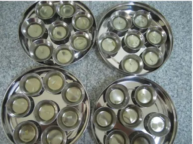
1 Specimens stored in normal saline after biomechanical preparation 2 Specimens immersed in streptococcus selection broth for autoclaving 3 Specimens subjected to autoclaving
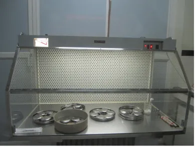
4 Laminar flow
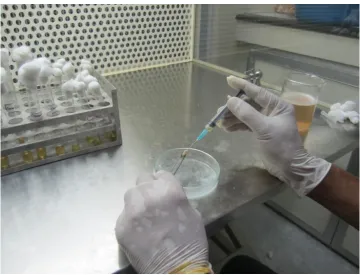
5 Inoculation of specimens with E.faecalis suspension using tuberculin syringes 6 Specimens kept for incubation
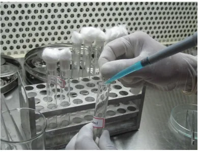
7 SEM picture showing E.faecalis colonies 8 Irrigation of specimens with the test irrigants 9 Sample preparation using H files.
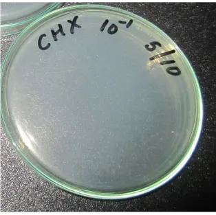
10 Collection of dentinal shavings in test tube containing normal saline 11 Agar plates showing complete inhibition by 5.25% NaOCl
12 Agar plate showing complete inhibition by 2% CHX
CINNAMON AND GARLIC AS ENDODONTIC IRRIGANTS AGAINST ENTEROCOCCUS FAECALIS – AN IN VITRO STUDY
ABSTRACT
Introduction
Eliminating microorganism from root canal system is possible only by a thorough chemomechanical preparation which is accomplished by proper instrumentation along with irrigants and intracanal medicaments. However complete sterilization of pulp space is not always achieved due to extremely complex anatomy. Persistent endodontic infections are mainly due to retention of microorganisms in the dentinal tubules. Enterococcus faecalis and Candida albicans constitute 77% of the persistent asymptomatic infections. It is important to appreciate that while hand and rotary instruments produce shape, it is the irrigants that clean the root canal system. Irrigants not only are important for the removal of debris and dentinal chips produced during cleaning and shaping, but are of clinical importance in the eradication of the radicular infection.
Aims & Objectives:
Fifty freshly extracted intact human mandibular premolars were decoronated and biomechanically prepared using 15-50 K files. The specimens were stored in normal saline until autoclaving. Specimens were autoclaved in separate steel containers containing BHI broth. The specimens were inoculated with E.faecalis suspension and incubated for 21 days in an incubator. The broth was replaced on alternate days. After 21 days the specimens were retrieved and divided into five groups (N=10). One tooth was subjected for SEM evaluation to confirm the penetration of bacteria. Five groups were treated with the respective irrigating solutions for 5 minutes. The sample preparation was done using H files and transferred into test tubes containing 10 ml sterile normal saline (10-1). Three dilutions 10-1,10-3 and 10-6 were used for the count. From this one ml from each dilution was pipetted on to a sterile 100 mm diameter in duplicate. To each of these plates 15 ml of agar medium, melted and cooled to 45 0C, was added, mixed well and allowed to solidify. These plates were incubated for two days at 37° C.After incubation the number of colonies was counted in suitable plates. The number of the colonies multiplied by the dilution factor gives the total number of CFU in the scrapings per tooth.
Results
The present study was done to evaluate the antimicrobial efficacy of Cinnamon and Garlic as endodontic irrigants against E.faecalis when compared to NaOCl and CHX. The study concluded that 5.25% NaOCl and 2% CHX showed complete inhibition where as Cinnamon and Garlic showed inhibition which suggest that they have an antimicrobial action but not up to the extent of NaOCl and CHX.
Clinical Implications
Successful endodontic treatment depends on healthy periradicular tissue. The importance of bacteria in pulpal pathosis has been demonstrated in the past (1). Primary endodontic infections are polymicrobial and are dominated by obligatory anaerobic bacteria (2). Eliminating microorganism from root canal system is possible only by a thorough chemomechanical preparation (3) which is accomplished by proper instrumentation along with irrigants and intracanal medicaments (4). However complete sterilization of pulp space is not always achieved due to extremely complex anatomy (5). Even after meticulous chemomechanical preparation bacteria can still be recovered from canals (6). Persistent endodontic infections are mainly due to retention of microorganism in the dentinal tubules. Enterococcus faecalis is the primary organism detected in persistent asymptomatic infections (7). Enterococcus faecalis is a facultative anaerobic gram positive rod which can invade the dentinal tubules (5) endure prolonged periods of starvation and possess certain virulence factors and lytic enzymes (8,9). Its mode of action is through biofilm formation which helps it resist destruction by enabling the bacteria to become 1000 times more resistant to most commonly used irrigants and intracanal medicaments(8).
Thorough chemomechanical preparation plays a key role in the success of endodontic treatment. It is important to appreciate that while hand and rotary instruments produce shape, it is the irrigants that clean the root canal system. Irrigants not only are important for the removal of debris and dentinal chips produced during cleaning and shaping, but are of clinical importance in the eradication of the radicular infection (10).
2. Mechanically flushes out the debris 3. Nontoxic and biocompatible
4. Dissolves necrotic and vital pulp tissue 5. Serves as a lubricant
6. Removes the smear layer 7. Low surface tension
NaOCl is a widely used irrigant as it covers most of the requirements for endodontic irrigants than any other known compound. Hypochlorite has the unique capacity to dissolve the necrotic tissue and the organic components of smear layer. Studies conducted so far point out to evidence that is strongly in favour of NaOCl as the main irrigant (11,12). But it has some disadvantages such as high toxicity, unpleasant taste, corrosive to instruments, inability to remove the inorganic portion of smear layer and reduction in elastic modulus and flexural strength of dentin (13). The difference in cell structure, thickness and endotoxin formation of facultative gram positive anaerobes makes it resistant to low concentration of NaOCl when compared to gram negative organisms. Increasing the concentration has its consequences. The purpose of introducing 17% EDTA into the irrigation regimen was to remove the smear layer which will facilitate penetration of irrigant to greater depth of the dentinal tubule.
dermatitis, desquamative gingivitis, discolouration of the teeth and tongue and dysgeusia (12). Due to the potential risk of adverse effects of systemic applications, and the ineffectiveness of systemic antibiotics in necrotic or pulpless teeth and the periapical tissues, the local application of antibiotics emerged as an effective mode of delivering antibiotics to infected root canals. Some of the antibiotic based irrigants are MTAD and Tetraclean (14). Other irrigants recently introduced are HEBP, EDTA-T, Chlorine dioxide, Silver diamine fluoride, Triclosan and Gantrez, Ozonated water, Photon activated disinfection (10). The constant increase in antimicrobial resistance and side effects caused by synthetic drugs has prompted researchers to look for herbal alternatives. Scientist have realized an immense potential in natural products made from medicinal plants to serve as alternate source of combating infections in human beings (15,16).
Herbs in dentistry have become more popular due to easy availability, cost effectiveness, low toxicity and lack of microbial resistance and increased shelf life (13). The changing trends from conventional irrigants to herbal extracts began in 2003 when propolis was compared with saline and NaOCl as root canal irrigants (17). Later Morinda citrofolia juice was used in conjunction with EDTA as a possible alternative to NaOCl (17). Other herbal alternatives such as Triphala, Green tea polyphenols were also recently evaluated as endodontic irrigants (16).
phytochemicals (15). Extracts of these herbs have proven potency against oral pathogens and are a genuine part of many of commercially available oral rinses and pastes.
Garlic is one of the greatest health tonics and has proven medicinal properties. It contains a substance called allicin which is equivalent to that of penicillin (1mg of allicin is equated to that of 15 IU of penicillin) (20). It also contains sulphur containing compounds like alliin, ajoene, diallylsulfide, dithiin, S-allylcysteine, enzymes, vitamin B, proteins and minerals. Also contains nonsulfur compound responsible for imparting antitumour, antioxidant and antimicrobial properties which can detoxify the body and has potent antioxidant activities. Allicin can destroy cell wall and cell membrane of root canal bacteria (21). Garlic inhibit the growth of oral pathogens such as Streptococcus mutans (21) and P.gingivalis and hence used in the management of dental infections such as periodontitis (21, 22). Studies report that a mouthwash containing 10% garlic in quarter Ringer solution produced a drastic reduction in oral bacteria (21).
AIM
To evaluate the antimicrobial efficacy of Cinnamon and Garlic as endodontic irrigants against E.faecalis with comparison of 5.25% NaOCl and 2% CHX.
OBJECTIVES
1. To evaluate the antimicrobial efficacy of Cinnamon against E.faecalis.
2. To evaluate the antimicrobial efficacy of Garlic against E.faecalis.
3. To evaluate the antimicrobial efficacy of NaOCl against E.faecalis.
4. To evaluate the antimicrobial efficacy of CHX against E.faecalis.
5. To compare the antimicrobial efficacy of herbal extracts with NaOCl.
6.To compare the antimicrobial efficacy of herbal extracts with CHX.
R.R white et al (1997) (24) determined the residual antimicrobial activity of 2% CHX and 12% CHX after canal irrigation. After biomechanical preparation the canals were filled with sterile water and samples were collected using sterile paper points at 6,12,24,48 and72 hrs. Antimicrobial activity was determined by determining the zone of inhibition in agar plates inoculated with Streptococcus mutans. Antimicrobial activity was present in all 2% CHX treated teeth throughout the 72hrs testing period and in most teeth, in relatively lower concentration for 6 to 24hrs after irrigation with 0.12% CHX.
L.B.Peters et al (2000) (25) developed a test model to quantify the penetration of bacteria into dentinal tubules. Model consists of two compartments separated by a bovine dentin specimen with pulpal side facing the incubated chamber of the test model. One compartment contained the test organisms such as E.faecalis and A.israelli and the other filled with sterile broth that was evaluated for growth of the test organism. The depth of bacterial penetration was measured with or without smear layer using both histological and quantitative recovering grinding technique after 6 wks of exposure. E.faecalis penetrated dentin significantly deeper than A. israelii. After removal of smear layer with EDTA, E.faecalis penetrated significantly deeper than in dentin penetrated with saline only or with a combination of saline and sodium hypochlorite.
B.P.F.A Gomez et al (2001) (27) compared the antimicrobial activity of 0.5%, 1%, 2.5%, 4%, 2.5% NaOCl and 2 forms of chlorhexidine gluconate (gel and liquid) in three concentration (0.2%,1%,and 2%) in the elimination of E. faecalis. A broth dilution test using 24-well cell culture plates was performed and contact time taken for the irrigants to kill the bacterial cells was recorded. The tubes considered to have positive bacterial growth were confirmed by gram staining, colony morphology on BHI agar + blood and by use of biochemical identification kit. CHX liquid (0.2%) CHX gel (2%) produced negative cultures within 30s and 1minute respectively. When used at 0.2% concentration CHX gel destroyed bacterial cells after 2hrs of contact as opposed to 15minutes when used at 1% concentration. High concentration of NaOCl took less time to inhibit bacterial growth than lower concentration.
Bettina Basrani et al (2002) (28) evaluated the substantive antimicrobial activity of different medicaments such as 2% CHX gel, 0.2% CHX gel, 2% CHX solution + 2.5% CHX containing controlled release device for 7 days. After medication canals were inoculated with E.faecalis for 21 days. Dentin samples were collected with Gates-Glidden drills and bacterial growth were assessed with spectrophotometric analysis after 72 hrs of incubation. Mean optical densities were significantly lower for groups with 2% CHX when compared with those of controls. Canal dressing for 1 week with 2% CHX showed residual antimicrobial activity against E.faecalis.
cervical third. Better cleaning was found in the cervical and middle third for all groups with the worst result in the apical third.
Onyeagba R. A .et al (2004) (30) studied antimicrobial effects of ethanolic extracts of garlic, ginger and lime against Staphylococcus aureus, Bacillus species, E.coli and Salmonella species. The entire test organism was susceptible to undiluted lime juice. The aqueous and ethanolic extracts of garlic and ginger did not inhibit any of these organisms. The highest zone of inhibition was observed with a combination of extracts on Staphylococcus aureus. Salmonella species were resistant to almost all the extracts except lime.
I. M. Saleh et al (2004) (31) investigated the ability of different endodontic sealers and Ca (OH)2 to kill E.faecalis in experimentally infected dentinal tubules. After biomechanical preparation the canals were filled with GP and AH plus, Grossmans sealer, Ketac Endo, Apexit, Roekoseal Automix, Roekoseal Automix with an experimental primer or Ca (OH) 2 only. After 7 days the canals were reestablished with sterile Peeso reamers size 2. Dentin powder was removed using Peeso reamers size 5. Antimicrobial efficacy determined by reduction in CFU. AH Plus and Grossman sealer killed bacteria. Other endodontic sealers, as well as Ca (OH) 2 were less effective.
the CFU below the limit of detection after 10s in case of A.naesulundii and C .albicans. E.faecalis proved significantly more resistant to NaOCl. But at higher concentrations less time was required though 5.25% NaOCl was not completely effective after 1minute.
Katharine. R. Carson et al (2005) (33) compared the antimicrobial activity of 6% and 3% NaOCl, 2% and 0.12% CHX, 0.01% and 0.005% doxycycline against four microorganisms such as Prevotella intermedia, Streptococcus sanguis, Lactobacillus acidophilus, Peptostreptococcus micros associated with primary endodontic infections. Agar diffusion test was used to determine the antimicrobial activities of the irrigants. MIC analysis was performed using macrodilution method. 0.01% and 0.005% doxycycline had significantly greater zones of inhibition than 6% NaOCl for P.micros, P.intermedia, S.sanguis. 6% NaOCl showed significantly greater zone of inhibition than 3% NaOCl for all endopathogens tested. 0.12% CHX had smaller zone of inhibition.
Brian. D. Barnhart et al (2005) (35) evaluated the cytotoxicity of NaOCl, IKI, Betadine scrub, Ca(OH)2, chlorine dioxide on cultured gingival fibroblast using the CYQuant assay. Human gingival fibroblasts were grown in Dulbeccos modified eagle medium containing 10% fetal bovine serum at 37°c and 5% CO2. Cells were split plated for 24h to allow attachment. Irrigants were tested at different concentrations. IKI and Ca (OH) 2 were well tolerated by human gingival fibroblast.
Russell. S. Eddy et al (2005) (36) investigated the antibacterial efficacy of Chlorine dioxide (10% clidox -S), 13.8% Bioclenz, 5.25% Clorox, Saline against E.faecalis using dentinal tubule disinfection method. The dentin disk were incubated for 30 minutes and were then frozen, pulverized, serially diluted in phosphate buffered saline, and plated on BHI plates in triplicate. Antimicrobial efficacy determined by reduction in CFU. NaOCl was effective at eliminating E.faecalis than any of other irrigants. Bioclenz & Clidox-s were significantly better than saline, which was ineffective at eliminating E.faecalis.
Melissa L. Ruff et al ( 2006) (38) compared the antifungal efficacy of 6% NaOCl, 2%CHX, 17% EDTA and Biopure MTAD against Candida albicans inoculated root canals using dentinal tubule disinfection method. Aliquots from the experimental teeth were placed on Sabourauds dextrose agar plates and antifungal activity determined by reduction in CFU and significantly superior to biopure MTAD and 17% EDTA. MTAD was significantly superior to 17% EDTA.
M. S. Clegg et al (2006) (39) evaluated the effectiveness of 6%, 3%, 1%, 2% CHX, 1% NaOCl followed by Biopure MTAD, sterile phosphate buffered solution on apical dentin biofilms. Samples collected from patients were cultured on hemisections of root apices to generate a polymicrobial biofilm. Each biofilm was separately immersed in irrigants. SEM evaluation was done to determine the presence and morpholgy of bacteria. Dentin shavings were obtained using No 2 sterile carbide burs. Antimicrobial activity determined by reduction in CFU. 2% CHX was not capable of disrupting the biofilm. Viable bacteria could not be cultured from specimens exposed to 6% NaOCl, 2% CHX or 1% NaOCl followed by Biopure MTAD. 6% NaOCl was the only irrigant capable of both rendering bacteria and physically removing the biofilm.
Isabelle Portenier et al (2006) (41) investigated the antibacterial activity of MTAD and CHX towards two strains of E. faecalis and the inhibitory effect of dentin and bovine serum albumin (BSA) on the antibacterial activity. Survival of bacteria exposed to medicaments in the presence or absence of inhibitors was monitored in an in vitro model. Full concentration MTAD and 0.2% CHX killed both strains. Combining CHX with cetrimide further reduced the time required for killing. The presence of dentin or BSA caused a marked delay in killing by both medicaments.
V. B. Berber et al (2006) (42)evaluated the efficacy of 0.5%, 2.5% and 5.25% NaOCl as intracanal irrigant associated with hand and rotary instrumentation techniques against E.faecalis using dentinal tubule disinfection method. Sampling of the canal was done using three sterile paper points. Dentinal shaving were also collected by discing the tooth into three thirds and dentin chips collected using ISO 018, ISO 021, ISO O25, ISO 029 sterile diamond tipped conical burs. Antimicrobial efficacy determined by reduction in CFU. At all depths and thirds of the root canals and for all techniques used 5.25% NaOCl showed better antimicrobial efficacy than 2.5% NaOCl.
eliminated in 30s by all antimicrobial agents, with or without agitation in contrast with the facultative and aerobe strain.
Seung - Eunn yang et al (2006) (44) determined the effects of smear layer and CHX treatment on the adhesion of E. faecalis to bovine dentin using 40 dentin blocks. Blocks were immersed in a suspension of E. faecalis for 3hrs after treating the groups with saline (5mts), 17% EDTA (5mts), 2% CHX, (7 days), EDTA and CHX (7 days). The bacteria adhering to the dentin surface were counted by examination using a SEM. Significant amount of bacteria was retained on the samples treated with saline. Smallest amount of bacteria adhered to samples treated with 2% CHX for 7 days.
John. M. Williams et al (2006) (45) compared the detection of E.faecalis by Real Time quantitative PCR, Reverse transcription – PCR (RT-PCR), and cultivation during endodontic treatment. Intracanal samples were collected upon access, post-instrumentation /irrigation and post calcium hydroxide treatment from 15 primary and 14 refractory infections involving 29 single rooted teeth. The bacterium was up to 3 times more prevalent in refractory than primary infections at each collection step. Real time PCR detected significantly more E- faecalis positive teeth and samples than cultivation. Real time PCR and reverse transcription PCR are more sensitive methods than cultivation for detecting E. faecalis in endodontic infections.
interactions between E.faecalis & F. nucleatum play a major role in endodontic infections.
Bettina. R. Basrani et al (2007) (47) determined the minimum concentration of NaOCl required to form a precipitate with 2% CHX. X ray photon spectrometry and time of flight secondary ion mass spectrometry (TOF-SIMS) were used to qualify and quantify the precipitate. A color change and precipitate were induced in 2% CHX by 0.023% and 0.19% NaOCl respectively. Both XPS and TOF-SIMS showed the presence of para- chloroaniline in an amount directly related to concentration of NaOCl used.
Bradley. M. Newberry et al (2007) (48) compared the antimicrobial effect of 3% NaOCl , 17% EDTA and Biopure MTAD as final rinse on eight strains of E.faecalis using dentinal tubule disinfection method. No 4 sterile carbide round bur was used to prepare three sample of dentin shavings. MIC and MLC were calculated for each strains of E.faecalis using both tube dilution methods. MTAD inhibited most strains of E. faecalis growth when diluted 1:8192 times and killed most strains of E.faecalis when diluted 1:512 times.
At deeper bur depth NaOCl remained significantly more antimicrobial. In all three dilutions doxycycline produced larger zone of inhibition.
Luciano Giardino et al (2007) (50) evaluated the antimicrobial efficacy of 5.25%NaOCl, Biopure MTAD ,Tetraclean against E. faecalis biofilm generated on cellulose nitrate membrane filters. Membrane filters were transferred into tubes containing antimicrobial solution test agent and incubated for 5, 30, 60mts at 20°c. Antimicrobial efficacy determined by reduction in CFU. 5.25% NaOCl was effective after 5mts when compared with other test group. Same effect was produced by Tetraclean only after 60mts. Tetraclean reduced 90% bacteria load after 5mts and >99.9% after 30mts. Bacterial load reduction using Biopure MTAD has not been significant after 5mts but much lower after 30mts than Tetraclean and 5.25% NaOCl.
Caio. C. R. Ferraz et al (2007) (51) compared the antimicrobial efficacy of 0.2%, 1% & 2%, CHX gel, 0.2%, 1% and 2% CHX solution, 0.5%, 1%, 2.5%, 4%, 5.25%, NaOCl against S.Aureus , E.faecalis, S.Sanguis , S.Sobrinus, Actinomyces naeslundii, porphyromonas gingivalis, P. endodontalis, Prevotella intermedia and Prevotella denticola. Antimicrobial efficacy determined by zone of inhibition. The largest inhibition zones were produced with 2% CHX gel being significantly different from the inhibition zones produced by all NaOCl concentration including 5.25%.
infected root canals with 5.25% NaOCl/15% EDTA. The combination of 1.3% NaOCl/Biopure MTAD left nearly 50% of the canals contaminated with E. faecalis.
Mathew. J. Royal et al (2007) (53) compared the effectiveness of 5.25% NaOCl, MTAD and 2% CHX in the rapid disinfection of polycaprolactone –based root canal filling material (Resilon pellets). Resilon pellets and GP Pellets were contaminated with E. faecalis and disinfected with 5.25% NaOCl, MTAD and 2% CHX. Samples were placed in centrifuge tubes containing BHI agar and incubated. At 1, 3, 7 days broth were checked for turbidity and scored for growth. Gram staining was done to identify bacterial species. 5.25% NaOCl, MTAD and 2% CHX were effective in the rapid disinfection of Resilon and GP pellets and a 1minute immersion was sufficient for disinfection.
Monika Marending et al (2007) (54) evaluated the impact of different irrigation sequence of 2.5% NaOCl (exposure time 24 mts) and 17% EDTA (exposure time 3 mts) on the elastic modulus and flexure strength of standardized human root dentin bars prepared from extracted upper third molar . Specimens after exposure to irrigants were subjected to 3-point bending test using a universal testing machine. Modulus of elasticity and flexural strength were evaluated. 24hr exposure to NaOCl caused a significant drop in flexural strength compared with water or EDTA treated groups whereas the elastic modulus remain unaffected .Short exposure to EDTA did not affect the mechanical dentin parameters.
intraradicular infections and chronic apical periodontitis. Bacterial samples were taken before treatment (S1) after chemomechanical preparation using CHX (S2) and after 7 day dressing with Ca(OH)2 /CHX paste (S3). Bacterial count and growth were determined by 16S Ribosomal RNA gene sequencing analysis. 0.12% CHX solution as an irrigant significantly reduced the number of intracanal bacteria but failed to render the canal free of cultivable bacteria in one half of the cases. Application of a 7 day intracanal dressing further increased significantly the number of cases yielding negative cultures.
William. J. Nudera et al (2007) (56) in this study determined the minimum inhibitory and bactericidal concentration of Triclosan and Triclosan with Gantrez against Prevotella intermedia, Fusobacterium nucleatum, Actinomyces naeslundii, Porphyromonas gingivalis and E. faecalis. The MIC of both test solutions was determined for each of the 5 microorganism using microtiter serial dilutions. MBC was also determined by streaking the samples on agar plates. Addition of Gantrez demonstrated bactericidal activity of Triclosan. Both Triclosan and Triclosan with Gantrez demonstrated bactericidal activity against the 5 specific endodontic pathogens.
Sunita Bansod et al (2008) (58) evaluated the anti fungal activity of garlic oil, neem oil, jitra oil, lemongrass oil, dalchine oil((cinnamon), nilgri oil, clove oil, cardamom, mint oil, tulsi oil, ajwain, ashwagandha and zinger oil against aspergillus fumigatus and aspergillus niger. Maximum antimycotic activity was demonstrated by oils of Cymbopogon martini, Eucalyptus globulus and Cinnamomum zylenicum.
Zohreh Ahangari et al (2008) (59) compared the antimicrobial effects of 2.5% NaOCl, 2% CHX against E. faecalis contaminated root canals of human extracted teeth. SEM evaluation was done for 2 samples to confirm sufficient penetration of microorganism into dentinal tubules. After biomechanical preparation dentinal shaving were taken using H files and transported into test tubes containing BHI and incubated at 370 for 96 hrs. Antimicrobial efficacy determined by reduction in CFU. In MTAD group, one sample showed bacterial growth. Bacterial growth was present in 5 samples in 2.5% NaOCl group and in 4 samples of CHX group.
Md. Mahfuzul Hoque et al (2008) (19) compared the antimicrobial activity of ethanol, aqueous extracts, essential oils of cloves and cinnamon extract against food borne pathogens Listeria monocytogenes, staphylococcus aureus, E.coli, Salmonella enteritidis, Vibrio parahaemolyticus and Bacillus cereus and 5 food spoilage bacteria. Pseudomonas aeroginosa, P.putida, Alealigenes faecalis, and Aeromonas hydrophila. Ethanolic extract of clove was potentially active against E.coli, L.monocytogenes. S.aureus, V. parahemolyticus, Pseudomonas, Aeromonas, and Allaligenes faecalis. The aqueous extract of clove was active against S. aureus strains and V. parahaemolyticus. The ethanolic extract of cinnamon was active only against staphylococcus aureus. The essential oil of clove and cinnamon showed maximum inhibition for A. hydrophila.
Sandeep Singh et al (2009) (62) compared the antimicrobial efficacy of Biopure MTAD and 2.5% NaOCl irrigation in infected root canals following single visit endodontic procedure. Single canal anterior and premolar teeth that had pulpal or periapical pathology were selected. Biomechanical preparation along with 2.5% NaOCl and 0.9% saline and Biopure MTAD was done for 5 minutes. Microbiological samples were taken using sterile paper points before and after biomechanical preparation. Antimicrobial efficacy determined by reduction in CFU. 2.5% NaOCl resulted in significantly greater number of anaerobic organisms when compared to Biopure MTAD.
S. Shobana et al (2009) (63) evaluated the antibacterial activity of two varieties of garlic against enteric pathogens such as E. coli, Proteus mirabilis, Salmonella typhi, Shigella flexineri and Enterobacter aerogenes. Ethanolic extracts of sativum was found to be highly effective against all the bacteria tested. Enterobacter aeurogenosa was not susceptible to aqueous extract of both garlic varieties.
Ya Shen et al (2009) (65) in this study developed a biofilm model that closely mimicked in vivo biofilm and to determine its susceptibility to endodontic antimicrobial irrigants by con-focal laser scanning microscopy. Collagen coated hydroxyapatites and uncoated hydroxyapatite disk were inoculated with dispersed subgingival plaque for 3 wks. Biofilm were subjected to 1, 3 and 10 minute exposure to CHX plus and 2% CHX. The collagen coated hydroxyapatite biofilm was thicker than hydroxyapatite biofilm. Less bacteria were killed in C-HA biofilm than in HA model.
Rosina Khan et al (2009) (66) evaluated the antimicrobial activities of the crude ethanolic extracts of five plant Acalia nilotica, Syzgium aromaticum and cinnamum zeylanicum, Eucalyptus globulus against multidrug resistant strains of E. coli, Klebsiella pnuemoniae, Candida Albicans and ATCC strains of Streptococcus bovis, Pseudomonas aerogenosa, Salmonella typhimurium and Ecoli. Extracts of A.nilotica, C.zeylanicum and S aromoticum showed the most potent activity against all the microorganism. E.faecalis, S. aureus. S. bovins and S.mutans were the most susceptible to all the plant extracts tested. S. typhimurium, K. pneumoniae, E coli, p. aeruginosa and C.Albicans were found to be sensitive to extracts of A.nilotica,,C..zeylanicum, S. aromaticum.
Proteus species. Antibacterial efficacy maintained at room temperature was for maximum 7 days. At - 200C the activity was maintained for 90 days.
Bishnu joshi et al (2009) (67) evaluated the antibacterial property of ethanolic extracts of different medicinal plants Ocimum sanctum, Cinnamomum zeylanicum, Xanthoxylum armatum and Origanum majorina against 10 medically important bacterial strains namely Bacillus subtilis , Bacillus cereus, Bacillus thuringiensis, Staphylococcus aureus, Pseudomonas species, Proteus species, Salmonella typhi, E. coli, Shigella dysentriae, Klebsiella pneumonia. Antibacterial activity was determined by agar well diffusion method. Among all the pathogens, all gram positive bacteria were inhibited by all 4 plant extract. Gram negative bacteria found to be resistant. Exceptionally Salmonella typhi showed zone of inhibition against extract of Ocimum sanctum.
Luciano Giardino et al (2009) (68) evaluated the antimicrobial effect of MTAD, Tetraclean, Cloreximid and 5.25% NaOCl on three common endodontic pathogen (E. faecalis, Porphyromonas gingivalis and Prevotella Intermedia). Antibacterial activity was determined by agar plate diffusion method. 5.25% NaOCl showed a high antimicrobial activity against anaerobic bacteria. MTAD and Tetraclean showed a high action against both, strictly anaerobic and facultative anaerobic bacteria. Chlorhexidine + cetrimide showed the lowest antibacterial activity against both facultative and strictly anaerobic bacteria.
Ram kumar Pundir et al (2010) (18) evaluated the antimicrobial activity of ethanolic extracts of Snzygium aromaticum and Allium sativum against food associated bacteria (Bacillus subtilis , B. megaterium , B. polymyxa, B. sphaericus, Staphylococcus aureus , E.coli) and molds (penicillium oxalicum, Aspergillus flavus A. luchuenis, Rhizopus stolonifer , Scopulariopsis species and Mulcor species). Antimicrobial activity determined by agar well diffusion method. MIC& MBC was also determined. Garlic and clove showed antibacterial activity and zone of inhibition was in the range of 20 mm-31mm.
U. B. Owne Ureghe et al (2010) (69) studied the inhibitory activity of garlic and lime on seven bacteria isolated from 240 extracted carious teeth. Agar well diffusion method was used in the susceptibility testing and MIC was calculated. Considerable antibacterial activity of garlic and lime were noted against the seven bacteria (Streptococcus mutans, Lactobacillus acidophillus, Norcardia asteroides, Pseudomonas aeroginosa, Actinomycosis viscosus, Staphylococcus aureus and Veillonella alcaligens.
Carmen Maria Ferrer-Luque et al (2010) (71) evaluated the antimicrobial activity of Maleic acid and combinations of Cetrimide with chelating agents against E. Faecalis biofilm. E.faecalis biofilm grow in the MBFC high- through put device for 24hrs and exposed to irrigating solution. Maleic acid showed antimicrobial activity against E.faecalis biofilm both alone or in association with Cetrimide from 30s onwards and the combination of EDTA and citric acid with cetrimide eradicated biofilm after 1minute of contact.
Saurabh S .Chandran et al (2010) (72) evaluated the antifungal efficacy of 5.25% NaOCl, 2% CHX,17% EDTA with and without the inclusion of an antifungal agent (1% clotrimazole) against Candida albicans inoculated root canals using dentinal tubule disinfection method. Aliquots from the experimental teeth were cultured on Sabouraud agar and antimicrobial efficacy determined by reduction in CFU. 5.25% NaOCl exhibited superior antifungal efficacy compared with 2% CHX and17% EDTA. 5.25% NaOCl and 2% CHX with clotrimazole showed significantly greater antifungal properties than 17% EDTA with clotrimazole.
Maria Teresa Arias-Moliz et al (2010) (74) assessed the antimicrobial efficacy of Cetrimide and CHX alone , in combination and alternating form in eradicating E.faecalis biofilm grown in the MBFC-high through put device for 24hrs. Contact time tested were 30s, 1 minute, 2 minute. Cetrimide eradicated E.faecalis biofilm at concentration of 0.5%, 0.0312% and 0.0078% at 30s, 1 minute & 2 minute contact times respectively. CHX did not eradicate the biofilm at any concentration or time. The associated use of Cetrimide and CHX provided better results than their applications as single agent and the alternating application was significantly more effective than the combined mode of application.
Luis E. Chavez de paz et al (2010) (75) in his study combined confocal microscopy, a mini flow cell system and image analysis to test in situ the effect of antimicrobials and alkali on biofilm of E.faecalis, Lactobacillus paracaesi, Strerptococcus anginosus, Streptococcus gordonii isolated from root canals with persistent infections . Biofilm formed for 24hrs were exposed for 5mts to alkali, 2.5% CHX, 50mmol/L EDTA, 1% NaOCl. The biofilm were characterized by using fluorescent markers targeting all membrane integrity and metabolic activity. NaOCl affected the membrane integrity in all microorganisms and removed most biofilm cells. Exposure to EDTA affected the membrane integrity in all organisms but failed to remove more than a few cells in biofilms of E.faecalis, L.paracasei, S.sanginosus. CHX had a mild effect on the membrane integrity of E.faecalis and removed only 50% of its biofilm cells.
remaining viable bacteria. Growth determination was done by formation of colonies in the agar plates. 1.3% or 2.5% NaOCl is ineffective in eliminating strains of E. faecalis at less than 40mts. 5.25% NaOCl was 100% effective at 40mts.
J. Prabhakar et al (2010) (77) evaluated the antimicrobial efficacy of Triphala, Green tea polyphenols, MTAD and 5% NaOCl against E. faecalis biofilm formed on tooth substrate. Zone of inhibition, MIC&MBC of test samples were also determined. At the end of third and sixth week the tooth with the biofilm formed were treated for 10 minutes with the test solution. Sample were taken by scraping the biofilm and inoculated on agar plates. Maximum inhibition was observed by 5% NaOCl (2mts) followed by MTAD (2mts) compared with Triphala and GTP. Three week biofilm showed complete inhibition of bacterial growth when treated with Triphala, MTAD and NaOCl, but samples treated with GTP and saline showed presence of bacterial growth. Six week biofilm showed growth when treated with Triphala, GTP and MTAD where as NaOCl showed complete inhibition.
F. G Pappen et al (2010) (78) investigated the antibacterial action of MTAD + 0.01% Cetrimide, 1 (Twen 80 replaced by 0.01% CTR in MTAD) MTAC-2 (Tween 80 replaced by 0.1% CTR) MTAD-D (MTAC without the tween 80 and no CTR) against planktonic cultures and mixed species in vitro biofilm model using direct exposure test. Tetraclean was more effective than MTAD against E.faecalis in planktonic culture and in mixed species in vitro biofilm. CTR improved the antimicrobial properties of the solution.
conventional hypochlorite Solution. Thus NaOCl solutions in concentrations of 1%, 2%, 4% and 5.8% were tested at room temperature 37oC and 45oC on bovine muscle tissue with and without agitation by ultrasonic and sonic energy and pipetting . Percentage of weight loss of tissue specimens were calculated before and after treatment and contact angle on dentin at concentration of 1% & 5.8%. Weight loss of the tissue increased with the concentration of NaOCl. The effect of agitation on tissue dissolution was greater than that of temperature; continuous agitation resulted in fastest tissue dissolution. Hypochlorite with added surface active agent had the lowest contact angle on dentin.
Isabela N. Rocas et al (2010) (80) studied the bacteria in endodontic treatment procedure by using a combined ribosomal RNA- based reverse transcriptase polymerase chain reaction and reverse capture checker board hybridization approach. Samples were taken from infected canals of teeth with apical periodontitis before treatment (S1) and after chemomechanical preparation with NaOCl & after medication with Ca (OH)2 paste. Presence of bacteria was screened by DNA based single PCR assay. RNA extracts were subjected to RT-PCR and were surveyed for the presence of 28 targeted bacteria by checker board method. Bacteria were found in all (S1)) sample, detectable level of ribosomal RNA was seen in 60% of cases after chemomechanical preparation and 53% after intracanal medication.
maximum antibacterial activity against 2 week E.faecalis biofilm. Triphala and GTPS showed significantly better antibacterial activity.
Moeen Mahmoud Al Weshah et al (2011) (81) investigated the effect of 2% CHX on the hardness of different levels of root dentin when used as root canal irrigant. 20 extracted teeth were endodontically treated and sectioned into 3 sections cervical, middle and apical and three sections were mounted on an acrylic resin disc shape mould. Microhardness was measured using a Wallace microhardness appliance at 5 locations at 1 mm distance from the root canal before and after irrigation with 2% CHX for an hour. No significant difference in the decrease of dentin hardness was noticed.
Zahed Mohammadi et al (2011) (82) evaluated the antibacterial substantivity of a new sodium hypochlorite based root canal irritant ( HYPOCLEAN) Tetraclean, 5.25% NaOCl against E.faecalis infected root canals using dentinal tubule disinfection method. Dentin shavings were taken on 0, 7, 14, 21 and 28 days using sterile slow-speed round burs with increasing diameter of ISO sizes 025, 027, 029, 031 and 033 respectively. Antimicrobial efficacy determined by reduction in CFU. The substantivity of Tetraclean was significantly higher than both Hypoclean and NaOCl solutions and retained in the root canal dentin for at least 28 days. The modified NaOCl solution showed more effective antibacterial activity than 5.25% NaOCl at all experimental periods.
soya agar and colonies counted using digital colony counter. Miswak could be a good natural substitute to NaOCl, while propolis showed results comparable to that of saline.
Ashish Saraf et al (2011) (84) evaluated the antibacterial activity of two different Cinnamon species C.zeylanicum (commercial variety) and C.flexuous against human pathogenic bacteria Staphylococcus aureus, Klebsiella pneumoniae, Bacillus subtilis. Maximum inhibition was shown by C.zeylanicum against gram positive bacteria S.aureus. C. flexuous showed inhibition but was inactive against Klebsiella pneumonia. Bacillus subtilis was least inhibited.
Zohreh Dalisrani et al (2011) (85) compared the antimicrobial effect of chamoline, garlic, clove, cinnamon, sage, thyme against Candida albicans. Non growth halo in disks containing chamoline, garlic, clove, cinnamon, sage and thyme was observed. Cinnnamon, garlic, chamoline, sage, clove, thyme had inhibitory effects on Candida albicans.
effect at a concentration of 0.082%. Concentration of NaOCl should increase approximately 70 fold to demonstrate the same antibacterial effect as NS.
Melahat Gorduysus et al (2011) (87) compared the antimicrobial effects of various endodontic irrigants 2% NaOCl, 2% CHX, 2.4% Iodine pottasium iodide, 17% EDTA and bioactive glass, against four selected microorganisms E. faecalis, E coli, S. aureus, S.pyogenes. MIC and MBC were determined. 17% EDTA was most effective against S.pyogenes, S.aureus, E. faecalis in 24 hrs. NaOCl displayed activity equivalent to that of IKI on E.coli, S. aureus and E. faecalis.
M N Shahani et al (2011) (88) compared the antimicrobial substantivity of 2% CHX,1% Povidone iodine, 2.5% hydrogen peroxide followed by 2% NaOCl alone as irrigants in instrumented root canals up to 72 hrs. After biomechanical preparation of the canals endodontic paper point were placed for 2mts and removed and stored in cryogenic vials at refrigerator temperature. Procedure was repeated after 12, 24, 48 & 72 hr. Antimicrobial activity determined by zone of inhibition. 2% CHX showed antimicrobial substantivity lasting for 72 hr followed by 1% Povidone iodine and 2% NaOCl.
Ponmurgan Karuppiah et al (2012) (89) evaluated the antibacterial effect of allium sativm, clove and zingiber officinale against multidrug resistant clinical pathogens causing nosocomial infection. All bacterial isolate were susceptible to crude extracts of both plant extracts. Except enterobacter species and klebsiella species, all other isolates were susceptible. The highest zone was observed with garlic against Pseudomonas aeroginosa.
within the root canal system. After biomechanical preparation the samples were irrigated with saline followed by 2%CHX, 50% citric acid followed by 2% CHX, 14% EDTA followed by 2%CHX. The chemical identity and quantification of the PCA in the formed precipitate was determined using gas chromatography/ mass spectrometry. All experimental group contained PCA. Citric acid used as the intermittent irrigant had the least amount of PCA formation in the canal system.
MATERIALS
K- FILES - MAILLEFER, DENTSPLY 5.25% NaOCl - NICE CHEMICALS Pvt, Limited. 2%CHX - VIJAY DENTOCARE
17%EDTA - VISTA DENTAL PRODUCTS Normal saline - BAXTER Pvt. Limited
Broaches - MILTEX COMPANY Hedstrome files - MAILLEFER, DENTSPLY Gates gliddden drills - MANI
Diagnostic sensitivity agar - HI - MEDIA, MUMBAI ATCC 29212 strains of E.faecalis - HI- MEDIA, MUMBAI Streptococcus selection broth - HI -MEDIA, MUMBAI Sheep Blood Agar - HI - MEDIA, MUMBAI McConkeys Agar - HI- MEDIA, MUMBAI Petridishes - BOROSIL COMPANY Micropippetes - BOROSIL COMPANY Test tubes - BOROSIL COMPANY
EQUIPMENTS USED
Airotor hand piece - NSK PANA-AIR Micromotor handpiece - DTMUSA, NSK
Autoclave - All AMERICAN 41 QUART
ELECTRIC STERILIZER MODEL# 75X
Steam distillation unit - CORNING COMPANY Rotator - REMI MAKE COMPANY Water bath - BESTON MAKE COMPANY Incubator -BESTON MAKE COMPANY Laminar flow - ROTEK COMPANY
SELECTION OF TEETH AND CANAL PREPARATION
In the first step of the study, the anatomical crown of all the teeth were cut away at Cemento enamel junction(CEJ) perpendicular to the long axis of the teeth using a water cooled diamond wheel bur on an airturbine hand piece at 300000 rpm. The remaining roots measured 12-18 mm. The exploration of the radicular canal was accomplished with no 10 and no 15K file to make sure that the roots had only one canal and it was patent. The preparation of the entrance of radicular canal was done with rotary instrument gates glidden drills of sizes 2 and 3. An appropriate sized barbed broach was used to extirpate the pulp.No15 size K file was inserted into the canal so that the tip of the file was visible at the apical foramen. Then the working length was determined one mm short of the file penetration into the canal. K files of sizes 15-50 were used to biomechanically prepare the root canal. The canals were recapitulated and irrigated with 5.25% NaOCl, 2% CHX, 17% EDTA and final rinse was with normal saline. Subsequent to the canal preparation the apical foramen of all the specimens were sealed with cyanoacrylate glue to prevent bacterial microleakage.
Specimens were placed in steel containers containing BHI broth and subjected to autoclave at 121ºc at 15 PSI for 20 minutes for sterilization. Subsequent to sterilization all the specimens were transported and manipulated under laminar flow using sterile instruments and equipments.
PREPARATION OF E.FAECALIS SUSPENSION
In order to get a controlled and standard suspension of the organism the following procedure was adopted:
sub-flask. This was incubated at 37° c for 24 hours. Enumeration of live bacteria (CFU) was carried out as follows:
1 ml of the broth culture was added to 9 ml of sterile normal saline and mixed well to get a dilution of 1 in 10 (10-1). The broth culture in the conical flask was kept in the refrigerator at 8° C. From the 10-1 dilution 1 ml was added to 9 ml of sterile normal saline to get a dilution of 1 in 100 (10-2). This serial dilution was continued until a dilution of 10-6. 1 ml from each dilution was pipetted into a sterile petri dish in duplicate. 20 ml of Diagnostic Sensitivity Agar melted, cooled and was added to each plate, mixed well and allowed to solidify. The plates were incubated at 37°C for 24 hours. The plates were examined for growth. The plates in which the number of colonies is between 30 and 300 were selected. The average number of colonies of duplicate plates was calculated. This number was multiplied by the dilution factor to get the number of colony forming units in 1 ml of the original broth culture. The average number of CFU of the dilution 10-5 was 38.The concentration of the original broth culture was computed to 3.8 x 10⁵ CFU per ml. For injecting into the tooth a suspension of bacteria containing 10⁵ CFU per ml was used. For preparing this 10 ml, original broth culture was diluted to 38 ml with sterile normal saline.
AQUEOUS EXTRACTION PROCEDURE
TOOTH INOCULATION WITH E.FAECALIS
The root canals were inoculated with E.Faecalis suspension using sterile 1ml tuberculin syringes and specimens were separately placed in steel containers containing 2ml of broth. The steel containers containing the specimens were kept in incubators at 37ºc for 21 days. After 21 days all the specimens were retrieved and each specimen was transferred into test tubes containing 3ml of saline and was shaken three times for 30 seconds each time on a rotator to remove the excess culture medium. In addition large amount of bacteria present on the surface of the specimen were removed during rinsing and irrigation. One sample was subjected for SEM evaluation to confirm the penetration of microorganism into the dentinal tubule.
The contaminated samples were divided into five groups, each containing ten teeth. Test irrigating solutions were used as follows.
Group 1 - Normal Saline Group 2 - 5.25% NaOCl Group 3 - 2% CHX Group 4 - Cinnamon Group 5 - Garlic
each. Dentinal shavings were collected using no 40 H file in an aseptic condition. Shavings were transferred into test tubes containing 10 ml sterile normal saline (10-1). One ml from the above was diluted to 10 ml with sterile normal saline (10-2). One ml from the above was diluted to 10 ml with sterile normal saline (10-3). One ml from the above was diluted to 10 ml with sterile normal saline (10-4). One ml from the above was diluted to 10 ml with sterile normal saline (10-5). One ml from the above was diluted to 10 ml with sterile normal saline (10-6).
Three dilutions from the above viz., 10-1, 10-3 and 10-6 were used for the count. From this one ml from each dilution was pipetted on to a sterile 100 mm diameter in duplicate. To each of these plates 15 ml of agar medium, melted, cooled and was added mixed well and allowed to solidify. These plates were incubated for two days at 37° C. After incubation the number of colonies was counted in suitable plates. The number of the colonies multiplied by the dilution factor gives the total number of CFU in the scrapings per tooth.
TO CHECK WHETHER CONTAMINATION BY ANY OTHER BACTERIA HAS NOT TAKEN PLACE
In order to ascertain that contamination by any other bacteria has not taken place during the procedures the following test were carried out:
morphology of E. faecalis. Only one type of colonies was found on each plate. By Gram’s staining typical gram positive cocci in chains were observed.
Antimicrobial activity was determined by evaluating the CFU in three dilutions 10-1, 10-3, 10-6. The no of viable colonies and percentage of inhibitory effect was recorded in different dilutions, tabulated and are shown in tables 1, 2, 3, 5, 6, 7.
STATISTICAL ANALYSIS OF THE RESULTS
The statistical analysis was performed using SPSS software (Statistical Package for Social Sciences) version 16.0. The data was interpreted at a confidence interval of 95%. Analysis of variance (ANOVA) was used to compare the antimicrobial activity of herbal extracts with comparison of standards. Post Hoc test followed by Scheffe Test was performed for multiple comparisons of the specimens.
(P value < 0.05 considered as significant difference).
TABLE 1 shows mean values of number of viable colonies of bacteria in each group at 10-1 dilution.
The statistical test shows significant difference in the multiple comparison between the groups but there is no significant difference between group 2 compared with group 3 at 0.05 (95% confidence interval)
TABLE 2 shows mean values of number of viable colonies of bacteria in each group at 10-3 dilution.
TABLE 3 shows mean values of number of viable colonies of bacteria in each group at 10-6 dilution.
The statistical test shows significant difference between group1 and other groups, but there is no significant difference between groups 2, 3, 4, 5 in the multiple comparison groups, but no significance between group 2 and group 3.
TABLE 4 shows multiple comparison of mean number of viable colonies of bacteria in the different groups.
The statistical test shows significant difference in the multiple comparison between the groups but there is no significant difference between group 2 compared with group 3 at 0.05 (95% confidence interval)
TABLE 5 shows mean values of number and percentage of samples with growth of bacteria in each group at 10-1 dilution.
The statistical test shows significant value comparing group 1 with other groups, but no significance between group 2 and group 3.
TABLE 6 shows mean values of number and percentage of samples with growth of bacteria in each group at 10-3 dilution.
The statistical test shows significant value comparing group 1 with other groups, but no significance between group 2 and group 3.
The statistical test shows significant value comparing group 1 with other groups, but no significance between group 2 and group 3.
TABLE 8 shows multiple comparisons of mean values of number and percentage of samples with growth of bacteria in between groups
The statistical test shows significant difference when comparing group 1 with other groups, group 2 with other groups, group 3 with other groups, group 4 with other groups, group 5 with other groups.
GRAPH 1 The statistical test shows significant value comparing group 1 with other groups, but no significance between group 2 and group 3.
GRAPH 2 The statistical test shows significant difference in the multiple comparison between the groups but there is no significant difference between group 2 compared with group 3 at 0.05 (95% confidence interval)
GRAPH 3 The statistical test shows significant difference when comparing group 1 with other groups, group 2 with other groups, group 3 with other groups, group 4 with other groups, group 5 with other groups.
Interpretation of Results
Long term success in endodontic treatment depends on complete debridement and disinfection of the pulp space. Despite a thorough mechanical preparation pulp remnants, debris, bacteria may be present in the irregularities of root canal system (92). Hence it is highly desirable that instrumentation should be accompanied by irrigation and intracanal medicaments for the elimination of microorganisms (4). In this present study a modification of Haapasalo and orstavik model was used for assessing the antimicrobial efficacy of endodontic irrigants in dentinal tubule disinfection (5). The model was further modified by using extracted human teeth rather than bovine teeth. This modification was appropriate because of the marked difference in diameter between the canals of bovine and human teeth (28). Quantitative estimation of CFU in the dentinal tubules after disinfection was included in the present study which was a modification of Haapasalo and orstavik model (93). Persistent endodontic infections are mainly due to retention and recolonization of microorganisms in the dentinal tubule (7). E.faecalis was selected as the test organism because it is a facultative organism that is non- fastidious, easy to grow and efficiently and rapidly colonizes the tubules (94). It has been used extensively in endodontic research because it has been detected in 63% of post treatment diseases (95) and due to the high level of resistance to a wide range of antimicrobial agents. E.faecalis has been used as the test organism to determine the efficacy of endodontic irrigants in the previous in vitro studies. ATCC 29212 strains of E.faecalis which has been the standard strain used in the previous studies was selected for the present study (96).
needed for the organism to penetrate deeper into root dentin and there was delayed bacterial penetration when root cementum is left intact (97). In this study the cementum was left intact to simulate clinical conditions (36). The minimum instrumentation size required for the penetration of irrigants in the apical third is 30 (98). The canals were enlarged up to 50 K file in the present study. E.faecalis can penetrate dentinal tubules to a depth of 300-400 µm within 3 weeks. Prolonged incubation period increased the number of infected dentinal tubules but depth of penetration of bacteria increases slowly with time (5). Hence in this study the teeth were inoculated with the organism and incubated for 21 days.
Ingrowth or progress of bacteria into the dentinal tubules could be delayed or prevented by the presence of a smear layer. Etching of dentin before exposure results in deeper penetration (99). 17% EDTA was used in this study for removing the smear layer in the experimental specimens before autoclaving and inoculation. Another important factor for the survival of bacteria is the availability of a nutrient source (100). The teeth were immersed in the streptococcus selection broth and the broth was replaced on alternate days during the 21 day incubation period. Subsequent change of the broth allowed the microorganism to rearrange in biofilms which is a structure known to confer resistance of microbial cells to different antimicrobial agents (40). Another reason for the replacement of the broth was to avoid medium saturation (28). Most of the previous studies evaluated the presence of bacteria using SEM or light microscopy (101). In the present study SEM evaluation was done to confirm the presence of bacteria.
done in a previous study in 2007 (102). The methodology adopted for enumeration of CFU (Pour plate method) was in resemblance with the study conducted by Lynn et al (93).
1. In the previous study there is direct contact of microorganism with the antimicrobial agent, bacterial suspension were mixed with the antimicrobial agent where as in the present study the dentinal tubules are inoculated with the organism to simulate the clinical condition.
2. The inhibitory effect of dentin on the bactericidal effect of the irrigant has been documented (105).
An important limitation of many studies when evaluating the endodontic microbiota refers to sample preparation. In comparison to the study conducted by Berber et al 5.25% NaOCl eliminated ATCC 29212 strains of E.faecalis in a 10 minute contact time (42). The methodology adopted in the latter study is similar to the present study except for the sample preparation. Burs of different sizes were used to procure dentinal shavings unlike H files used in the present study. Bacterial inhibition at greater depths was assessed using burs of different sizes whereas H files were indicative of bacterial inhibition to a limited depth. Increasing the concentration is undesirable because it is an irritant to periapical tissue (106). Other undesirable effects of NaOCl are its unpleasant taste, high toxicity, corrosive to instruments, reduces the elastic modulus and flexural strength of dentin (13).
evidenced by the studies conducted by Oncag et al 2001 proving that 2% CHX was effective against E.faecalis in a 5 minute contact time (109). Another important property of 2% CHX is substantivity (110). A study proved that 2% CHX application after 10 minutes of application produced antibacterial effects for up to 12 weeks (111). Though it has broad antimicrobial activity, it lacks tissue dissolving property. Allergic reactions such as contact dermatitis, desquamative gingivitis, discolouration of teeth and tongue and dysgeusia have also been reported (12). Studies have evaluated the genotoxicity and have proved that 2% CHX is biocompatible.
Even after thorough chemomechanical preparation bacteria can still be recovered from canals (6). To combat the emerging antimicrobial resistance and considering the undesirable side effects of synthetic drugs, herbal alternatives might prove to be advantageous. Approximately 60% to 80% of the world population relies on traditional medicines for the treatment of common illness (112). Medicinal plants are known to produce certain bioactive molecules which react with other organisms in the environment inhibiting bacterial or fungal growth (113). Contrary to synthetic drugs antimicrobials of plant orgin are not associated with many side effects and have an enormous therapeutic potential to heal many infectious diseases (112). An important characteristic of plant extract and their components is their hydrophobicity which enabled them to partition the lipids of bacterial cell membrane and mitochondria disturbing the cell structures and rendering them more permeable. Extensive leakage from the bacterial cells or the exit of critical molecules and ions will lead to death (67).
and not the nutrients (114). Test irrigants Cinnamon and Garlic showed reduction in CFU in all the samples tested at 10-1 dilution. Complete inhibition was observed in 2 samples of garlic and 1 sample of Cinnamon. These results are indicative of potent antimicrobial action by the test irrigants. Saline showed innumerable colonies in the same dilution. Comparing the antimicrobial activity of herbal extracts Garlic had fewer colonies than Cinnamon and hence more antibacterial.
Garlic (Allium Sativum) is a bulbous perennial medicinal plant which belongs to the family Liliaceae. The antimicrobial activity is attributed to thiosulphinates. Studies reported that if extract is free from thiosulphinates the antimicrobial activity will be lost (113). Garlic can be used on microorganism that has particularly developed resistance to antibiotics. This is concluded in a previous study (2001) which explained that garlic oil and 4 diallyl sulphides showed in vitro activity against antibiotic resistant Pseudomonas aeruginosa and Staphylococcus aureus (115). Multidrug resistant E.faecalis can be made vulnerable by the medicinal properties of garlic. Studies proved that extracts of garlic are bactericidal and are effective against E.coli, S.aureus B.cereus, Salmonella, listeria, proteus and Streptococcal species (116). Garlic contains many sulphur containing compounds and one of which is the odourless molecule alliin. When the plant is cut or damaged alliin comes into contact with the enzyme alliinase, which convers alliin into alliicin. The later is responsible for the typical smell and medicinal properties of garlic. In microorganisms allicin interferes with lipid sysnthesis and RNA production. The target enzyme with which alliicin interacts has been identified as acetyl-CoA Synthetase. Studies have proved that allicin has antifungal and anti thrombotic effects (116).