A DISSERTATION ON
STUDY OF
RECONSTRUCTIVE STRATEGIES
RECONSTRUCTIVE STRATEGIES
RECONSTRUCTIVE STRATEGIES
RECONSTRUCTIVE STRATEGIES F
F
F
FOR
OR
OR
OR LOWER ONE
LOWER ONE
LOWER ONE
LOWER ONE
THIRD LEG SOFT TISSUE DEFECT
THIRD LEG SOFT TISSUE DEFECT
THIRD LEG SOFT TISSUE DEFECT
THIRD LEG SOFT TISSUE DEFECT
In partial fulfillment of the
regulations for the award of the degree of
MASTER OF CHIRURGIE
(MCh.) Degree
BRANCH – III – PLASTIC SURGERY
THE TAMILNADU
DR. M.G.R. MEDICAL UNIVERSITY
CHENNAI, TAMIL NADU.
DECLARATION
I solemnly declare that this dissertation “RECONSTRUCTIVE
STRATEGIES FOR LOWER ONE THIRD LEG SOFT TISSUE
DEFECT” was prepared by me under the able guidance and supervision of our
Professor & HOD, Department of Plastic & Reconstructive Surgery, Madurai Medical College and Government Rajaji Hospital, Madurai between August
2010 and January 2013.
This is submitted to The Tamil Nadu Dr. M.G.R. Medical
University, Chennai, in partial fulfilment of the requirement for the award of
MASTER OF CHIRURGIE, M.Ch. PLASTIC SURGERY, degree
Examination to be held in AUGUST 2013.
Date :
CERTIFICATE
This is to certify that this dissertation entitled
“RECONSTRUCTIVE STRATEGIES FOR LOWER ONE THIRD LEG
SOFT TISSUE DEFECT” Submitted by Dr. E.R.SRINIVAS Post Graduate,
Department of Plastic & Reconstructive Surgery, Madurai Medical College to
The Tamil Nadu Dr. M.G.R. Medical University, Chennai, in partial fulfilment
of the requirement in the award of degree of MASTER OF CHIRURGIE IN
PLASTIC SURGERY, Branch – III, for the AUGUST 2013 examination is
a bonafide research work carried out by him under our direct supervision and
guidance during the years 2010 - 2013.
PROF. DR. C. BALASUBRAMANIAN, M.S., M.Ch.,
Head of the Department,
Department of Plastic & Reconstructive Surgery, Govt. Rajaji Hospital &
ACKNOWLEDGEMENT
I thank our respected Dean of Madurai Medical College and Medical Superintendent of Government Rajaji Hospital, Madurai for permitting me to use the hospital facilities for this study.
I am very grateful to my humble, enthusiastic teacher, advisor
PROF. C. BALASUBRAMANIAN, M.S., M.Ch, Head of the Department of
Plastic & Reconstructive Surgery, for his constant guidance, encouragement and enthusiasm throughout the period of this study.
I am indebted to our beloved PROF. R. RAJA MUTHAIAH, Associate Professor, Department of Plastic & Reconstructive Surgery for his able guidance, thoughtfulness and tolerance during the entire course of my study.
I deeply express my respects and gratitude to our Associate Professors DR. S. GNANASEKARAN, DR. P. PARTHASARATHY and all the
Assistant Professors in our department – DR. V. RAVICHANDRAN , DR. P. SURESH KUMAR,DR.S.ARAM,DR.V.JEYAKODISH AND DR. K. RAJA for their guidance, suggestions, critical comments, execution and assistance throughout the study course, helping me to sail steadily.
Devi, my juniors,DR. T. THIRUMALAISWAMY , DR. G. ARUN &DR SENTHIL
KUMAR for their comments, criticism and continued help during this study.
I sincerely thank my patients without whom this study would not be possible.
Above all, I thank my PARENTS and THE ALMIGHTY for the blessings they have showered upon me in all the deeds of my life.
CONTENTS
S. No. TITLE Page No.
01. INTRODUCTION 1
02. AIM OF STUDY 4
03. REVIEW OF LITERATURE 5
04 SURGICAL ANATOMY 9
04. MATERIALS AND METHODS 18
06 SURGICAL PRINCIPLES 22
05. OBSERVATION AND RESULTS 40
06. DISCUSSION 52
07. CONCLUSION 60
08. CLINICAL PHOTOGRAPHS
09. PROFORMA
10. BIBLIOGRAPHY
11. MASTER CHART
12. ANTIPLAGIARISM CERTIFICATE
INTRODUCTION
Increasingly, urban trauma is becoming a major health care issue. Large emergency departments are inundated with patients with multiple injuries, requiring state-of-the-art care. Most of these complex injuries involve trauma to the extremities, often due to motor vehicle accidents. In a study by MacKenzie et al. It was shown that lower extremity injuries accounted for about 40% of the charges for motor vehicle trauma treatment in a given year. Hospital-based studies reveal that disabilities persist for a long time with mean time taken to return to work ranges from 42 months to120 months.1
Coverage of soft tissue defect of the leg presents unique defects requiring the ingenuity of the surgeon in planning flaps for stable coverage.
Though well established norms are in place regarding the time and nature of cover, it requires a team effort, practicing it with involvement of the
orthopaedic surgeon and allied specialities like vascular surgeons, general surgeons.
The relatively unprotected antero-medial portion of tibia results in exposed bone after trauma, which requires specialized soft tissue cover.2, 3
Treatment of lower extremity trauma has evolved over the last two
decades to the point that many that would require amputation are now routinely salvaged.6
Plastic surgeons role becomes not only important in covering a raw area, but also in providing a functional lower limb with an acceptable aesthetic result.
Though we live in an era of zero delay work, microvasuclar transfer and a single stage work up, due to circumstances beyond our control, it is still
necessary to revisit the older methods which are reliable, comparable and easily reproduced.7, 8, 9
The study was done to reflect our work and thus to enhance our quality of work to produce good results with a few complications as possible.
There is need to challenge the concept, that distally based flaps are inferior to proximally based flaps just as the dogma that skin flap survival depends on rigid length to width ratios has been refuted. Adjusting all other factors, the true critical factor of flap viability is the nature of their intrinsic blood supply rather than any arbitrary orientation or configuration in either case.10
The patient expectations, an understanding of quality of existing source vessels in the given region and local anatomical constraints should be
We must periodically reassess our own work, chart our future
AIM
1. To evaluate various reconstructive options for management of lower 1/3rd leg soft tissue defect and to highlight their merits and demerits
2. To establish a definitive time based protocol in managing these patients
REVIEW OF LITERATURE
The first written report on injuries of the lower extremity is found in Roman war surgery books, in which amputation is suggested as the elective treatment for serious damage.
• Pierre-Joseph Desault (1744-1795), Paris, coined the term debridement”.7 • Trueta - Infection could be avoided if all devitalized tissue was excised.7 • Trentz-O et al described priorities of treatment in open tibial fractures 7
1. Resuscitation
2. Restoration of vascularity 3. Debridement
4. Skeletal stabilization 5. Second look debridement
6. Coverage with a local or free muscle flap within 96 hours.
7. Reconstruction of bone defect by bone grafting, callus distraction or bone segment transport with fixator.
• Mcgregor and Morgan defined the distinction between axial and random
pattern flaps.11
• In 1854, Hamilton described the cross-leg flap, introducing a new method
• Filatov, Gillies and Hugo Ganzer in 1920s described the tubed pedicled
flaps.14, 15, 16
• Ger in 1968 described the first muscle flap and the concept of
musculocutaneous flap revolutionizing lower extremity reconstruction.17 • Marko Godina in 1980 introduced and developed the concept of
Emergency free tissue transfer.18
• Barclay in 1982 and Amarante in 1986 used distally based
fasciocutaneous flaps based on lower perforators of the peroneal and posterior tibial arteries.4, 19
• Cormack and Lamberty described in detail the arterial anatomy of skin
flaps in 1986 classifying Fasciocutaneous flaps.20
• The propeller flap concept described by Hyakusoku in 1991 for
reconstruction of elbow defects in post burns contracture was extended by Teo to include flaps based on peforators which rotated 180 and could be used in lower extremities. 21
• Masquelat in 1992 described neurofaciocutaneous flaps, these elegant
flaps provided an alternative approach to defects which usually requires microsurgical reconstruction.22
• A further step in lower extremity reconstruction finally was achieved with
.
• Byrd et al. found that the overall complication rate for wounds closed
within the first week of injury was 18% compared to 50% complication rates for wounds closed in the subacute phase of 1-6 weeks. 9
• Godina et al found that the least complication rates were in wounds
closed within 72 hours of injury.24
• Yaremchuk et al postulated that serial, complete debridement is more
important than timing of soft tissue coverage.10
• Platelet counts increase four fold in the subacute phase after injury
contributing to increased complication rate according to Choe IE et al.25 • Laughlin et al reviewed the functional outcome in eight patients with
grade IIIB, and six with Grade IIIC injuries and found out that the recovery period was long.26
• The use of tissue expansion in the lower extremity has not been used
frequently It is generally useful for healed, chronic defects as placement near open wounds results in more complications (Borgest et al).27
• Graf P et al found distally based fasciocutaneous flaps to be an alternative
for free flaps.16
• Cross leg flaps were presented in a study from Central Hospital, China.
• Tolhurst, Haesekar and Zeeman demostrated 15% greater survival length
in flaps that included fascia.29
• "Vacuum Assisted Closure" (VAC) is a non-invasive negative pressure
healing process indicated in the treatment of chronic wounds associated with unfavourable local or systemic factors.
Continuous sub-atmospheric suction pressure (125 mmHg) was applied to the wound site. 125 mm hg appears to be the optimal, utilizing an
alternating pressure cycle of five minutes suction followed by two
minutes off suction. The cyclical application alters the cytoskeleton of the wound bed cells which trigger a cascade of intracellular signals that
increase rate of cell division and subsequent formation of granulation tissue. The wounds were closed primarily, covered with split-thickness skin grafts, or a regional flap. 30, 31, 32
According to retrospective study by Parrett et al., published in 2006 there is reduction from 42 % to 11% of free flap use with the advent of VAC • Hyperbaric Oxygen
HBO2 has several specific biological actions which enhance wound
healing process. Hyper-oxygenation of tissue, vasoconstriction, down regulation of inflammatory cytokines, up-regulation of growth factors, antibacterial effects, potentiation of antibiotics, and leukocyte effects. 33,
SURGICAL ANATOMY OF THE LEG
The wound coverage of lower one third of leg is a challenging problem because of its anatomical features. The tibia and fibula are vulnerable to injury, open fractures being more common due to paucity of soft tissues around them. Most muscles become tendons at this level hence flap cover becomes
mandatory in case of soft tissue loss. The peroneal, anterior and posterior tibial are the three major arteries of the leg and are in closed compartments without significant communication between them.
Detailed knowledge on blood supply of muscles and mapping of
perforators has widened resources available for plastic surgeon in carrying out reconstructive surgeries in lower one third of leg.
The leg can be divided into four compartments
A. Anterior compartment
This consists of the following muscles. 1. Tibialis Anterior
2. Extensor Hallucis Longus 3. Extensor Digitorum Longus 4. Peroneus Tertius.
B. Lateral compartment
Muscles include 1. Peroneus Longus 2. Peroneus Brevis.
The artery(s) of this compartment are peroneal artery and anterior tibial artery. The nerve is superficial peroneal nerve.
C. Superficial posterior compartment
Muscles include 1. Gastrocnemius 2. Soleus
3. Plantaris 4. Popliteus
The arteries are sural, peroneal and posterior tibial and nerve is the tibial nerve.
D. Deep posterior compartment
Muscles include
1. Flexor hallucis longus 2. Flexor digitorum longus 3. Tibialis posterior
THE BLOOD SUPPLY OF THE LOWER LEG:
In 90% of cases the popliteal artery runs obliquely inferolaterally to the lower edge of popliteus where it divides into anterior and posterior tibial arteries. This course can be marked on the surface by a line drawn from the junction of the middle and lower thirds of the thigh 2.5cm medial to the midline of the back of the limb, running downwards and slightly laterally to reach the midline between the femoral condyles. From here the course of the vessel continues inferolaterally along the same line to the level of the tibial tuberosity.
The anterior tibial artery passes between the tibial and fibular heads of tibialis posterior to pass over the upper edge of the interosseous membrane to reach the anterior compartment. It descends in front of the interosseous membrane and lies on the tibiat in the lower third of the leg. The anterior tibial artery is initially medial to the deep peroneal nerve, but descends behind it, it is once more medial to it at the ankle. Near the ankle it gives origin to the medial and lateral malleolar arteries before passing midway between the malleoli onto the dorsum of the foot as the dorsalis pedis artery.
continues downward behind flexor digitorum longus and becoming superficial, crosses the lower end of the tibia parallel to and 2.5cm in front of the medial border of the Achilles tendon. At the ankle joint it passes deep to the flexor retinaculum, midway between the medial malleolus and the medial tubercle of the calcaneus and divides, deep to abductor hallucis, into the medial and lateral plantar arteries.
The peroneal artery arises from the posterior tibial artery 2.5 cm below the lower edge of the popliteus muscle. It inclines laterally to descend along the medial crest of the fibula deep to or in the substance of flexor hallucis longus and ends behind the tibiofibular syndesmosis. It sends a nutrient artery to the fibula, and is linked to the posterior tibial artery by a communicating branch which lies on average 6.5 cm above the tip of the fibular malleolus. Nearer the ankle (approximately 5 cm above the fibular tip) it gives off a perforating branch which pierces the interosseous membrane and descends in front of the tibiofibular syndesmosis to the anastomose around the ankle.37, 38, 39, 40
The median superficial sural artery
heads of gastrocnemius is variable but is usually about half way down the lateral head of gastrocnemius. It follows the lateral edge of the Achilles tendon where it anastomoses with branches of the peroneal artery and posterior tibial artery. 40, 41
Variations: The anterior tibial artery is often diminished in caliber but never
entirely absent. The middle portion may be greatly reduced in which case the perforating branch of the peroneal artery joins the distal portion (5% of 2458 cases). When greatly increased in size the anterior tibial supplies the plantar surface of the foot.
The posterior tibial may reach only as far as the distal third of the leg where it is then reinforced by a large communicating branch from the peroneal. It may end as a nutrient vessel to the tibia or in supplying a muscle, or it may even be absent, in which case its territory is supplied by a particularly well-developed peroneal artery. Rarely may it be increased in caliber.
The peroneal artery is never absent but is occasionally much reduced in size. Anatomically its size is inversely proportionally to that of the other arteries in the leg, and as described above it may feed the distal parts of the anterior or posterior tibial arteries. Interestingly it is the vessel least affected by
Location of septocutaneous perforators. 29
Code
N - Number of Perforator LM - Lateral Malleolus FH - Fibular Head
PTA - Posterior Tibial Artery PA - Peroneal Artery
Surface markings for the 3 main vessels are
A. Posterior tibial artery
A line from tibial tuberosity to the mid malleolar point. The vascular axis lies 4.5 cm medial and parallel to this line or 1.5 cm from medial border of tibia.
B. Anterior Tibial artery and Peroneal artery
perforators of the leg as depicted by Bhattacharya is taken as the safe limit of dissection inferiorly. 29
Cormack and Lamberty classification: The types include 29
A - Has a fascial plexus B - Has a single perforator
C - Has multiple perforators and segmental source artery
Mathes and Nahai's Classification of fasciocutaneous flaps based on its
blood supply 29
A - Direct cutaneous pedicle to the fascia B - Septocutaneous perforator
C - Musculocutaneous perforator
Variants of Fasciocutaneous flaps 29
1. Antegrade (superiorly based) 2. Retrograde (inferiorly based) 3. Deepithelized turn over flaps 4. Islanded perforator based flaps
MATERIALS AND METHODS
This study was conducted in the Department of Plastic Surgery, Government Rajaji Hospital, Madurai over a period of 30 months from Aug 2010 to Jan 2013.
Only cases with soft tissue defect of lower 1/3rd leg requiring flap cover i.e defects with tendon, bone or implant exposed or in patients undergoing staged procedures were included in this study. A total of 73 patients were included in the study.
Timing of coverage was classified into18
Acute - within 72 hours
Subacute - 3 days to 6 weeks
Chronic - Greater than 6 weeks
Defects were classified according to their site as per the usual norms of upper third, mid third and lower third.
Inclusion criteria
All patients with post traumatic soft tissue defects of the lower 1/3rd leg who required a soft tissue cover were included in the study
Exclusion criteria
who were not willing to participate in the study and for whom skin graft was planned were excluded from the study.
Methodology: All the patients included in the study were admitted to the
trauma ward under the care of the attending orthopedician and received first aid. They were then resuscitated to minimize bleeding, restore airway and correct shock. 5, 8
Detailed history was taken on the mechanism of injury; the time since injury and history of neurological deficits. Then all the patients were subjected to a full general and local clinical examination to rule out other coexisting injuries and to assess the site and size of the defect, the presence or absence of exposed bone, tendons or neurovascular structures, the degree of wound
contamination and the condition of surrounding skin. A complete vascular and neurological examination with comparison to the other healthy limb was
performed. Laboratory investigations necessary for surgical fitness were done. X-rays and hand held Doppler studies were done to identify and classify the fracture and assess vascular status. 5, 8, 12
All patients were taken up for wound toilet and debridement on the day of admission. Skeletal stability was achieved if necessary with external
fixators, illizarov ring fixators, plates or K-wires as deemed appropriate by the orthopedic surgeon. To control the infection the wound pus culture and
dressed daily with a saline dressing. Once the wounds were free of infection the soft tissue cover was planned. The appropriate reconstructive technique was selected for every patient according to the reconstructive ladder putting into consideration the site, size and type of the defect, the condition of local tissues, previous surgical procedures in the injured limb, future planned surgical
procedures and the patient’s general condition. 12, 42, 43
All the patients received postoperative care including proper antibiotic therapy, analgesics in the post-operative period, elevation of the limb to prevent oedema and monitoring of the flap- colour, temperature and capillary refill. First look dressing of the skin graft was done on the 5th postoperative day. Assisted ambulation was allowed for the patients whenever possible at the end of the 5th postoperative day. Dependable weight bearing was allowed at the end of the 7th postoperative day depending on the presence of bone fractures and the method of bone fixation. Sutures were removed on the 10th post operative day and the patients were transferred back to the orthopedic surgeon for further treatment.43, 44
functionality, return to work and aesthetic appeal. Follow up periods varied from 6 months to two years depending on the patient’s compliance.44, 45
Functionality – 2points
Return to work – 2 points
Esthetic appeal – 1 point
Scores of 4 & 5 indicates good satisfaction.
2 & 3 indicate fair satisfaction.
1 indicate poor satisfaction
Data was collected in the form of a proforma which included epidemiological data, clinical data, wound area measurements and operative surgical
SURGICAL PRINCIPLES
The initial assessment and treatment of patients with lower extremity trauma should be in accordance with the Advanced Trauma Life Support (ATLS) guidelines.
The second phase of management begins when the patient arrives in the operating room. Debridement is initiated with the pneumatic tourniquet. Large bone fragments with significant soft tissue attachment are maintained and small, free fragments are removed. A second debridement is performed with the tourniquet deflated. and the wound is irrigated.44, 45
GUSTILO ANDERSON CLASSIFICATION of open fractures (1976) . 46
Type I - Open fracture with a clean wound, < 1cm in length.
Type II - Open fracture with a laceration >1cm long and without extensive soft
tissue damage or avulsion.
Type III - Subtype (1984)
A - Adequate periosteal cover of a fractured bone despite extensive soft tissue laceration or damage; high energy trauma irrespective of size of wound.
C - Associated arterial injury requiring repair, irrespective of degree of soft tissue injury
Byrd, spicer and Ciney enlarged the classification of open tibial fractures into four types in order in increasing energy of injury.9
Type I fractures represents low energy forces causing an oblique fracture of the tibia with a relatively clean cutaneous wound less than 2 cm length.
Type II fracture indicate moderate energy forces causing either a displaced fracture or a comminuted tibia fracture with a skin wound greater than 2 cm in length.
Type III fracture results from high energy forces causing a significantly displaced or severely comminuted fracture or segmental fractures with extensive associated skin loss and devitalized muscle.
Type IV fracture pattern indicates extreme energy forces, a history of crush or degloving injury or vascular injury requiring repair.
As surgeons gained more experience in treating these complex injured wound, the need to objectively assess and predict the functional outcome was felt and that led to the development of a wide variety of scoring systems, the notable of which are
• Mangled extremity severity score (MESS)
• Limb salvage Index (LSI)
Johansen reported the Mangled extremity severity score (MESS) in 1990. The MESS evaluates 4 important variables47
Skeletal / soft-tissue injury
Low energy (stab; simple fracture; pistol gunshot wound): 1 Medium energy (open or multiple fractures, dislocation): 2 High energy (high speed MVA or rifle GSW): 3
Very high energy (high speed trauma + gross contamination): 4
Limb ischemia
Pulse reduced or absent but perfusion normal: 1* Pulseless; paresthesias, diminished capillary refill: 2 Cool, paralyzed, insensate, numb: 3*
Shock
Systolic BP always > 90 mm Hg: 0 Hypotensive transiently: 1
Persistent hypotension: 2
Age (years)
< 30: 0 30-50: 1 > 50: 2
MESS score of greater than or equal to 7 had a 100% predictable value for amputation.
The GHOISS assessed the severity of the injury to the limb separately to each of the three components of the limb: the ‘covering tissues’ i.e., skin and fascia, the ‘skeleton’ i.e., bones and joints, and the ‘functional tissues’ i.e., muscles, tendons and nerve units. Treatment may be influenced by several systemic factors and outcomes were given two points each, and the final score is arrived by adding all the individual scores together. Salvage should be attempted if score is less than or equal to 14, primary amputation considered in those with score of 17 or above and those in between should be assessed by an
experienced team on case-to-case basis.48
LEAP study concludes that although the published scores were successful in predicting amputations, scoring systems are not predictive of functional
recovery among patients who have undergone successful limb reconstruction. Practitioners should exercise caution when interpreting scores in the context of potential recovery from high-energy trauma.49
large fragments, however the large fragments with soft tissue attachments can be salvaged eliminating the need for a second bone grafting procedure.
Various options available for reconstruction of the lower extremity are:
A. Skin Graft B. Skin Flap
C. Fasciocutaneous flaps D. Adipofascial flap E. Muscle flap with SSG F. Propellar flaps
G. Free flaps I. Cross leg flaps
Technique of Flap rising
A. Skin grafts
Skin grafts are applicable only if there is a healthy vascular recipient bed or if the periosteum over the bone is intact.
B. Skin flaps
Local skin flaps like transposition flaps are suitable only for small to medium defects. With understanding of the anatomy better Fasciocutaneous flaps came into vogue and are more reliable and a larger flap can be harvested.
C. Fasciocutaneous flaps
Fasciocutaneous (FC) flaps have been well investigated and tried out in leg defect. FC flap can be used in the ipsilateral limb locally as well as distally in the form of cross leg flap.50, 51
'The perforator plus' technique combines the advantages of providing
additional blood supply and safeguarding the venous return.
The 'perforator plus' peninsular flap has prior identified perforator at the base and, therefore, gives freedom to make a back cut without any fear of
compromising blood supply. A back cut moves the pivot point closer to the defect thereby facilitating better movement of flap and thus easing the tension on the distal edge. This principle is applicable to rotation, transposition,
Selection of fasciocutaneous flaps for lower leg defects
- Distally based fasciocutaneous flaps based on lower perforators of the peroneal and posterior tibial arteries.
- lateral supramalleolar flap - Reverse sural artery flap.
D. Adipofascial flaps
Adipofascial flaps have become extremely popular in the last decade in the reconstruction of lower leg defects. Adipofascial flaps are like fasciocutaneous flaps, as the vascular basis is same in both flaps. The basic advantage of
E. Muscle flaps
They are compound flaps which constitute muscle, fascia, subcutaneous fat and skin combined as a single unit of tissue based on one or more vascular pedicles. Blood supply to these flaps comes primarily from the muscle and reaches the skin by vessels which pierce the muscle pass through the fascia to ramify in the subcutaneous tissues.17, 53, 54, 55
Mathes and Nahai classified Muscles into five types based on the number of vascular pedicles entering the muscle.54
Muscle flap for distal third leg defects include - Peroneus brevis
- Tibialis anterior
- Extensor hallucis longus - Distally based Soleus
Peroneus brevis Muscle Flap
The blood supply is provided by segmental branches, usually three or four in number, arising from peroneal artery.
The distal 10 to 12 cm of the muscular attachment of the fibula can be released without compromising the blood supply to the muscle flap. The muscle flap, nearly 10cm long by 3cm wide, can be swung forward to cover the upper half of the lateral malleolus and the adjoining part of the fibula. A split-thickness skin graft is used for epithelial cover. There will be no functional loss by using this muscle flap if the peroneus longus is preserved.56
F. Neurofasciocutaneous flaps
Small arteries and veins usually accompany the cutaneous nerves and they send perforators to overlying skin was Masquelat concept
The reverse flow sural fasciocutaneous flap is based on median superficial sural artery. Good circulation is ensured by the constant anastomoses with 3 to 5 septocutaneous perforators from peroneal artery.
If the accompanying arteries of lesser saphenous vein are included, the success rate of the flap increases as these give off cutaneous perforators along its
to tip of lateral malleoli, to locate the arc of rotation, avoiding injury to the more distal septocutaneous perforators. After measuring the size of the defect, the cutaneous island to be transferred is marked out on middle or distal third of leg, depending on the length of pedicle necessary to reach the wound. Keeping the pedicle centralized with regard to flap, skin incision made from proximal to distal. Proximally the sural nerve and accompanying short saphenous vein identified, ligated, cut and included in the flap. The fascia must be included in both the skin island and the pedicle dissection. Later via a skin tunnel or the skin bridge the flap is transferred. Primary closure of donor site defect is done if it is less than 4cm in diameter or else covered split skin graft.57, 58, 59
G. Propellar flaps
They are basically fasciocutaneous flap containing the skin, subcutaneous tissue and the deep fascia based on a single musculocutaneous perforator.
The perforator vessels are located through an exploratory initial incision. The approach to the pedicle could be supra-facial or sub-fascial, with the latter being generally easier and less bloody. . The pedicle should be cleared of all muscular side branches and all the fascial strands that could potentially cause kinking of the pedicle. Raising the rest of the flap is quick and
straightforward. The flap is carefully lifted from the wound bed, attached only by its pedicle, and rotated around this pedicle into the defect in which ever rotational direction that is most comfortable for the venae comitantes. The secondary defect is either closed primarily or using a skin graft.60, 61
The 'Throw over flap': A modification of the propeller flap for reconstruction
of non-adjacent soft tissue defects.
H. Free flaps
In 1973, Daniel and Taylor reported the free transfer of groin skin and subcutaneous tissue by use of microvasuclar anastomoses.
Commonly used free fasciocutaneous flaps are Antero-lateral thigh flap, Radial artery forearm flap, Dorsalis pedis flap, Scapular, Parascapular, Lateral arm flap, and Posterior calf fasciocutaneous flap.
Muscle and myocutaneous free flaps commonly used for lower limb reconstruction are latissimus dorsi, gracilis, tensor fascia lata and rectus abdominis flaps.
Composite osteocutaneous free flaps used for one stage reconstruction are radial artery osteocutaneous flaps, fibula flap and deep circumflex
osteocutaneous free flap. It is usually the Grade IIIb fractures of the leg, that needs free flap cover. 18, 63, 64, 65
Antero Lateral Thigh Free Flap
This is a versatile soft tissue flap. Skin paddle available is the largest, up to 25 X 30 cms in adult and the donor site morbidity is very low. ALT can be used as a chimeric flap i.e. vastus lateralis muscle on one branch and on separate
Pertinent anatomy: The anterolateral thigh flap is a fasciocutaneous flap based on the septocutaneous or musculocutaneous perforators of the descending branch of the lateral circumflex femoral artery.
A satisfactory perforator is generally found within 3 cm radius of the midpoint of a line connecting the anterior superior iliac spine with the superolateral border of the patella. More than half of the perforators transverse the substance of the vastus lateralis muscle. The descending branch of the lateral circumplex femoral artery and its venae-comitantes lie between the vastus-lateralis and rectus-femoris muscle.
Operative procedure
Patient in supine position. Draw a circle of 3 centimeters radius at midpoint on the line joining anterior superior iliac spine and supero-lateral border of patella. Identify cutaneous perforators with hand held Doppler along this line / circle. Mark skin paddle length and width as per defect. Design an elliptical flap, include the main perforator area.
Look for perforators, both direct septo-cutaneous or musculocutaneous. Dissect musculocutaneous perforators carefully through muscle up to the main pedicle. Take lateral skin incision through the fascia-lata, tag fascia to skin. Dissect from below upwards, by dissecting the fascia-lata and skin paddle off the underlying vastus lateralis muscle until the inferior-most perforator is reached.
The perforator is carefully dissected from its surrounding muscle. If a single perforator to be used, a small cuff of muscle should be included with the flap to avoid twisting of the pedicle. This is not necessary if two perforators are included in the flap.
Dissect the descending branch of the lateral circumflex femoral artery and vein superiorly to their branches to the rectus-femoris, which should be preserved. Verify perfusion, then clip and divide the pedicle. Undermine medially and laterally in the suprafascial plane for closure of wound. Close the skin in two layers. If the donor area is large skin graft may be necessary.65, 66
Latissimus dorsi Muscle Free Flap
perforating branches of the intercostal and lumbar arteries. The extramuscular pedicle length is about 9cm on average.
Innervation- Thoracodorsal nerve is the motor nerve, it arises from the posterior cord of the brachial plexus and is derived from the sixth, seventh and eighth cervical nerve roots.
Surgical procedure -Mid-lateral or supine position with a sandbag. A longitudinal incision is made from the axilla to the posterior iliac crest. The skin paddle should be marked. Then, the lateral border of the muscle is exposed. The key point of the technique is the dissection at the anterior border to expose the vascular pedicle. The pedicle must be carefully dissected, and the muscle should be released from its origin.
I. Cross leg flaps
In some clinical situations where local fasciocutaneous and myocutaneous flaps are unavailable and when a free flap has failed because of technical errors or damaged vasculature, a cross-leg flap is the best choice. The inclusion of fascia in the flap makes length-to-breath ratio 3: 1 perfectly safe. The use of external fixator avoids the problem of immobilization and facilitates its uses in whom free tissue transfer may not be optimal.
The indications may be markedly broadened especially in the centres with no access to microsurgery. It offers the possibility of salvaging limbs that are otherwise non-reconstructable. It is a backup procedure in an urgent situation and supplies a large quantity of skin. Advantages of cross-leg flap include ease of dissection, versatility, shorter operating time, minimal donor site morbidity and replacement of like tissue.17, 28
Usual Technical faults
Patient Selection
1. Poor general condition 2. Malnutrition, anaemia
3. Systemic disease, smoking, drug addiction
Intraoperative
1. Poor flap planning
2. Failure to identify deep fascia and its incorporation in the flap 3. Failure of suturing the deep fascia to the dermis
4. Coarse tissue handling
5. Locating the pedicle away from the vascular axis
6. Unnecessary undermining of the Pedicle by blunt dissection. 7. Twist and kink at the pedicle
8. Suturing under tension 9. Pressure dressing
10. Failure to put a drain under the flap
Postoperative management
1.Frequent monitoring is essential 2. Elevate leg to combat edema
3. Look for haematoma, evacuate if detected 4. Prevent infection
5. Distal flap should be inspected twice a day and dressed on alternate day
Identification of early signs of flap necrosis
1. Decreased temperature 2. Discolouration
5. Pinprick revealing dark blood
Follow up
1. Educating patient about how to take care of the flap
2. Review by surgeon at least 15 days once in the first 3 months and every two months for two years thereafter.
3. Weight bearing allowed gradually after 3 months
4. The appearance of sensation is variable. Starts appearing around 6 months and takes almost 2 years.
Donor site morbidity
There is no functional loss. The grafted area gradually becomes soft and supple but seldom matches with the adjacent normal skin. It is usually acceptable to the patients.
OBSERVATIONS AND RESULTS
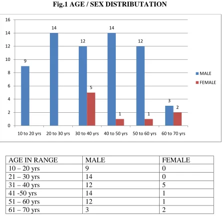
Fig.1 AGE / SEX DISTRIBUTATION
The age of patients ranged from 10 to 70 years in this study. Common age group affected is between 21 to 30 years and 41 to 50 years, 19% each, n=14.
Male to female ratio is 7: 1 (M = 64, F = 9)
9
14
12
14
12
3 5
1 1
2
0 2 4 6 8 10 12 14 16
10 to 20 yrs 20 to 30 yrs 30 to 40 yrs 40 to 50 yrs 50 to 60 yrs 60 to 70 yrs
MALE FEMALE
AGE IN RANGE MALE FEMALE
10 – 20 yrs 9 0
21 – 30 yrs 14 0
31 – 40 yrs 12 5
41 -50 yrs 14 1
51 – 60 yrs 12 1
[image:46.595.74.521.129.562.2]The most common indication for flap cover was exposed tibia (71%), followed by exposed tendon 21% and exposed Implant 8%.
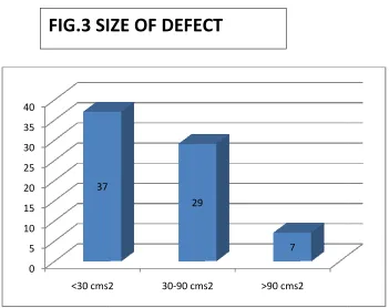
The most common size of defect was small i.e, less than 30 cms2 (51%), followed by medium sized defects 30 to 90 cms2 (40%) & Large defects
greater than 90 cms2 (9%)
[image:47.595.73.490.96.268.2]0 10 20 30 40 50 60 TIBIA 0 5 10 15 20 25 30 35 40 <30 cms2 37
FIG.2 EXPOSED PART / INDICATION FOR FLAP COVER
FIG.3 SIZE OF DEFECT
The most common indication for flap cover was exposed tibia (71%), followed by exposed tendon 21% and exposed Implant 8%.
The most common size of defect was small i.e, less than 30 cms2 (51%), followed by medium sized defects 30 to 90 cms2 (40%) & Large defects
greater than 90 cms2 (9%)
TIBIA TENDO ACHILLES IMPLANT 52
15
6
<30 cms2 30-90 cms2 >90 cms2 29
[image:47.595.117.468.360.638.2]7
FIG.2 EXPOSED PART / INDICATION FOR FLAP COVER
SIZE OF DEFECT
The most common indication for flap cover was exposed tibia (71%),
followed by exposed tendon 21% and exposed Implant 8%.
The most common size of defect was small i.e, less than 30 cms2 (51%), followed by medium sized defects 30 to 90 cms2 (40%) & Large defects
NAME OF FLAP
No. OF
CASES IBFTL-INFERIORLY BASED FASCIOCUTANEOUS
TRANSPOSITION FLAP - LATERAL SIDE
33 (45%)
IBFTM-INFERIORLY BASED FASCIOCUTANEOUS
TRANSPOSTION FLAP - MEDIAL SIDE
3 (4%)
IBFST-INFERIORLY BASED FASCIOCUTANEOUS SLIDING
TRANSPOSTION FLAP
6 (8%)
RSNFP- REVERSE SURAL NEUROFASCIOCUTANEOUS
PEDICLED FLAP
24 (33%)
RSNFI-REVERSE SURAL NEUROFASCIOCUTANEOUS
ISLAND FLAP
2 (3%)
PBM-PERONEUS BREVIS MUSCLE FLAP 1 (1%)
PF-PROPELLAR FLAP 1 (1%)
ALTFF-ANTERO – LATERAL THIGH FREE FLAP 2 (3%)
LDFF-LATISSIMUS DORSI FREE FLAP 1 (1%)
The most commonly performed procedure is the inferiorly based
fasciocutaneous flaps (45%), followed by reverse fasciocutaneous flaps (32%)
33
3 6
24
2 1 1 2 1
0 5 10 15 20 25 30 35
[image:48.595.73.520.71.313.2]IBFTL IBFTM IBFST RSNFCP RSNFCI PBMF PF ALTFF LDMFF
FIG.4 No. OF CASES / FLAPS
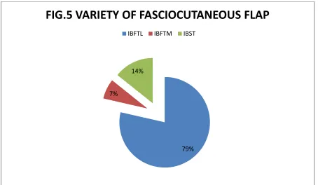
VARIETY OF FASCIOCUTANEOUS FLAP NO. OF
CASES
IBFTL-INFERIORLY BASED FASCIOCUTANEOUS TRANSPOSITION FLAP - LATERAL SIDE
33
IBFTM-INFERIORLY BASED FASCIOCUTANEOUS TRANSPOSTION FLAP - MEDIAL SIDE
3
IBFST-INFERIORLY BASED FASCIOCUTANEOUS SLIDING TRANSPOSTION FLAP
6
Inferiorly based fasciocutaneous flap from lateral side(79%) was the most commonly performed fasciocutaneous flap because of the presence of reliable and constant perforator.
79% 7%
[image:49.595.72.526.127.391.2]14%
FIG.5 VARIETY OF FASCIOCUTANEOUS FLAP
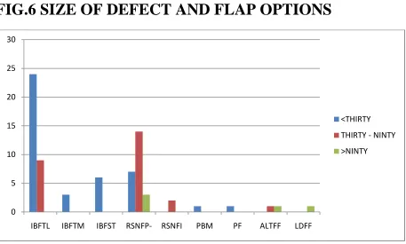
FIG.6 SIZE OF DEFECT AND FLAP OPTIONS
Inferiorly based Fasciocutaneous flaps is the most common procedure performed for small to medium sized defect.
Neurofasciocutaneous flaps are excellent choice for medium to large size defect. We have done a muscle flap for smaller defect
Propeller flap was done in one patient with small defect. Free flaps were done in three patients with large sized defects.
0 5 10 15 20 25 30
IBFTL IBFTM IBFST RSNFP- RSNFI PBM PF ALTFF LDFF
<THIRTY THIRTY - NINTY >NINTY
SIZ E
IBFT L
IBFT M
IBFST RSNFP RSNFI PBM PF ALTFF LDFF <30 24 3 6 7 0 1 1 0 0
30-90
9 0 0 11 2 0 0 1 0
>90 5 1 1
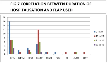
DAYS IN HOSPITAL
IBFLT IBFTM IBFST RSNFP RFNFI PB PF ALTFF LDFF
0-10 16 2 3 5 1 1
10-20 7 1 2 12 1 1 1 1
20-30 7 1 6 1
30-40 3 1
The average duration of hospitalization was least for fasciocutaneous flaps – (57% of patients were discharged within 10 days) and longest for pedicled Neurofaciocutaneous flaps and free flaps (2 to 5 weeks).
0 2 4 6 8 10 12 14 16 18
IBFTL IBFTM IBFST RSNFP- RSNFI PBM PF ALTFF LDFF
[image:51.595.72.518.125.391.2]0 to 10 11 to 20 21 to 30 31 to 40
Oedema and infection were the common complications encountered 23 and 18 % respectively.
46%
3% 2% 2% 1% 5% 23%
[image:52.595.77.496.70.381.2]18%
Fig.8 COMPLICATIONS
nil partial necrosis dehiscense complete necrosis graft loss superficial necrosis oedema minor infection
COMPLICATIONS No. OF CASES
NIL 43 (46%)
PARTIAL NECROSIS 3 (3%)
DEHISCENSE 2 (2%)
COMPLETE NECROSIS 2 (2%)
GRAFT LOSS 1 (1%)
SUPERFICIAL NECROSIS 5 (5%)
OEDEMA 21 (23%)
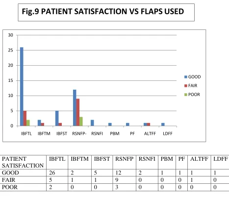
Of the 46 patients who rated the reconstruction as Good, 26 (57%) had
underwent distally based fasciocutaneous flap from lateral side, 12 (26%) had underwent distally based reverse neurofasciocutaneous flap of them rated the reconstruction as good, 2 islanded RSA, 1 muscle flap, 1 propellar flap, 1 ALT and 1 LD.
Of the 5 patients who had rated the reconstruction as poor 3(60%) had
underwent distally based reverse neurofasciocutaneous flap and 2 (40%) distally based fasciocutaneous flap.
0 5 10 15 20 25 30
IBFTL IBFTM IBFST RSNFP- RSNFI PBM PF ALTFF LDFF
GOOD FAIR POOR
PATIENT
SATISFACTION
IBFTL IBFTM IBFST RSNFP RSNFI PBM PF ALTFF LDFF
GOOD 26 2 5 12 2 1 1 1 1
FAIR 5 1 1 9 0 0 0 1 0
[image:53.595.68.535.87.495.2]POOR 2 0 0 3 0 0 0 0 0
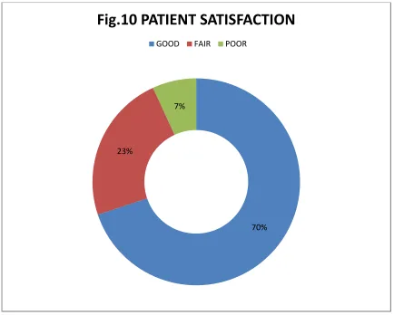
70% of patients graded the reconstruction as Good, 23% as Fair and 7% as poor.
70% 23%
[image:54.595.75.509.98.446.2]7%
Fig.10 PATIENT SATISFACTION
GOOD FAIR POOR
PATIENT SATISFACTION No. OF CASE
GOOD
51 FAIR
17 POOR
Of the 73 patients, 64 were
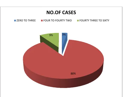
operated in the chronic phase and 2 (3%) in the acute phase.
ZERO TO THREE
PHASE IN DAYS 0-3
[image:55.595.73.486.135.459.2]4-42 43-60
Fig.11 PHASE OF COVERAGE
Of the 73 patients, 64 were operated in the sub-acute phase (88%), 7 (9%) were operated in the chronic phase and 2 (3%) in the acute phase.
3%
88% 9%
NO.OF CASES
FOUR TO FOURTY TWO FOURTY THREE TO SIXTY
NO. OF CASE 2 (3%) 64 (88%) 7 (9%)
Fig.11 PHASE OF COVERAGE
acute phase (88%), 7 (9%) were
COMPLICATION IBFTL IBFTM IBFST RSNFP RSNFI PBM PF ALTFF LDFF
NIL 24 1 1 11 1 1 1 1 1
PARTIAL NECROSIS
2 0 0 1 0 0 0 0 0
COMPLETE NECROSIS
1 0 0 1 0 0 0 0 0
DEHISCENSE 2 0 0 0 0 0 0 0 0
GRAFT LOSS 0 0 0 0 1 0 0 0 0
SUPERFICIAL NECROSIS
3 0 0 2 1 0 0 0 0
OEDEMA 7 0 2 11 0 0 0 1 0
MINOR INFECTION
6 0 1 9 0 1 0 1 0
IBFTL NIL WAS-53%,PARTIAL NECROSIS WAS 5%,COMPLETE
NECROSIS-2%,DEHISCENSE-4%,GRAFT LOSS-0%,SUPERFICIAL NECROSIS-7%,EDEMA-16%,MINOR INFECTION-13%
IBFTM NIL-50%, PARTIAL NECROSIS-50%
IBFST NIL-50%, MINOR INFECTION-50%
RSNFP NIL – 36%, PARTIAL NECROSIS-3%, COMPLETE NECROSIS-3%,
SUPERFICIAL NECROSIS-6%, EDEMA-23%, MINOR INFECTION-29%
RSNFI FLAP NIL-34%, SUPERFICIAL NECROSIS-33%, GRAFT LOSS-33%
PF AND LDFF NIL WAS 100%
PBM, ALTFF FLAPS NIL-50%, MIN0R INFECTION-50%
0 5 10 15 20 25 30
IBFT IBFTM IBFST RSNFP RSNFI PBM PF ALTFF LDFF
NIL PARTIAL NECROSIS DEHISCENSE COMPLETE NECROSIS GRAFT LOSS SUPERFICIAL NECROSIS EDEMA MINOR INFECTION
Fig.13 COMPARISON OF PHASE OF COVER vs COMPLICATION
PERCENTAGE
FROM 0 – 3 days:
FROM 4-42 days: NIL-56%, SUPERFICIAL NECROSIS
NECROSIS – 3%, OEDEMA
>42 days: PARTIAL NECROSIS 14%, GRAFT LOSS
NECROSIS-29%, MINOR INFECTION
0 5 10 15 20 25 30 35 40 45
COMPLICAIONS 0-3
NIL 1
PARTIAL NECROSIS
0 DEHISCENSE 0 COMPLETE
NECROSIS
0 GRAFT LOSS 0 SUPERFICIAL
NECROSIS
0 OEDEMA 1 MINOR
INFECTION
0
Fig.13 COMPARISON OF PHASE OF COVER vs COMPLICATION
-50%, OEDEMA 50%
56%, SUPERFICIAL NECROSIS – 7%, PARTIAL 3%, OEDEMA-26%, MINOR INFECTON-21%
PARTIAL NECROSIS 14%, GRAFT LOSS-14%, COMPLETE 29%, MINOR INFECTION-14% DEHISCENSE-29%
ZERO-THREE FOUR-FOTY TWO >FORTY TWO
4-42 >42
42 0
2 1
1 1
2 0
1 0
4 1
20 0
16 1
Fig.13 COMPARISON OF PHASE OF COVER vs COMPLICATION
Discussion
The wound coverage of lower one third of leg is a challenging problem because of its anatomical features. The tibia and fibula are vulnerable to injury, open fractures being more common due to paucity of soft tissues around them. And as most muscles become tendons at this level, flap cover becomes mandatory in the event of trauma.
The basic objectives in reconstruction of leg defect are: a. Good and early healing of bone
b. Good movement of contiguous joints c. An aesthetically acceptable stable cover
Early return to work and restoration of near normal functionality should be the aim of reconstruction of the lower extremity.
Our study was a prospective descriptive study which studied 73 patients who underwent reconstruction of lower one third leg soft tissue defect during the study period from August 2010 to January 2013.
INCIDENCE - YEARLY
ETIOLOGICAL INCIDENCE
1. Road Traffic Accident - 69 patients 2. Fall from Height - 3 patients 3. Train Traffic Accident - 1 patient
The etiological indications for Lower one third leg soft tissue defect in this study showed Road traffic accidents to be the most common cause at 94 %.
In concordance with Fabio and Santanelli73, Road traffic accidents
continue to be the major cause of soft tissue defect in a developing country like ours just as in the developed nations.
In this study the age of the patients varied from 10 years to70 years with the mean age of 30 years. In the series of Gururaj and Suri MP et al (25-35 years mean age).2
Common age group affected is between 21 to 30 years and 41 to 50 years, 19% (n=14) each.
In this study 87% of those operated were males while other studies have quoted 64% (Gururaj and Akthar et al). Male to female ratio is 7: 1 (M = 64, F = 9) 2, 3 Most patients presented with injury on the right side (59%)
The most common indication for flap cover was exposed tibia (71%) which is the same in other studies as well.1
followed by medium sized defects (40%) and 9% with defect > 90 cms2.
Almost 88% (n=64) of patients in this study were stablised with external fixator despite conclusive studies by Trabulsy et al fixators et al proving nonreamed locked nails were more effective than external fixators.43
Again this may reflect availability rather than personal preference.
In this series maximum number of flaps was done in the sub-acute phase – 88% and the least in the acute phase 3%, chronic phase being 10%.
This is in total contrast to literature elsewhere where early cover is recommended. (Godina et al, Byrd et al).9, 18
The reasons for the decreased immediate cover were: 1. Co existing head injury taking priority for management. 2. Lack of immediate referral by orthopaedicians.
3. Doubtful vascularity of the limb.
4. Co existing wounds on the leg requiring skin grafting.
5. Co morbid illnesses with patients on Aspirin for ischaemic heart disease. 6. Delayed skeletal stabilisation where internal fixation was used.
7. Shortage of plastic surgery team members.
The results from other studies showed that immediate wound
done to perform immediate reconstruction. These results are in agreement with previous studies.
We emphasize the importance of co-operation at the time of primary surgery between orthopaedic and plastic surgeon to preserve access to potential flaps. The technique of bony fixation of the tibia may prevent the use of this flap, especially in the presence of external fixation pins which may injure perforating vessels or tether the flap, restricting its range of transposition. The most commonly performed procedure is the inferiorly based
fasciocutaneous flap (57%), followed by reverse fasciocutaneous flaps (36%)
Inferiorly based Fasciocutaneous flap from lateral side is the most common procedure (45%) performed for small to medium sized defect as the perforator is constant and reliable in the lower lateral aspect of the leg.
Neurofasciocutaneous flaps are excellent choice for medium to large size defect. Peroneus muscle flap was done for smaller defect.
Propeller flap was executed in one patient with small defect. Three patients with large defects underwent free flaps
(Two patients – Antero-Lateral thigh free flap)
(One patient – Latissimis dorsi muscle free flap with skin graft)
techniques like perforator flaps and neurocutaneous flaps there is a resurgence of interest in non microsurgical reconstructive options. This is of special significance in a resource challenged centre like ours. In this study we have attempted to explore the above mentioned reconstructive strategies for lower 3rd leg reconstruction.
However the indications and the criterion of selection of a particular technique for a particular defect are not well established and is rather a matter of personal judgement.
51% (n=37) of patients had small sized defects, 40% (n=29) had medium sized defects and only 6% (n=7) presented with large defects.
The size of the defect and the experience of the centre in microvasuclar surgery was a significant factor in deciding reconstructive options
Perforator plus technique : While raising the local fasciocutaneous flap we
always tried to include the perforator at the base of the flap, which was identified pre-operatively with hand held Doppler.52
The average duration of hospitalization was least for fasciocutaneous flaps. (57% of patients who underwent fasciocutaneous flaps were discharged or transferred to ortho ward before 10th day) and longest for pedicled
Oedema (n=21) and Infection (n=17) was the most common complication in this series, it was managed by conservative measures – Anti-oedema measures, appropriate antibiotics / Irrigation, but one case necessitated a sequestrectomy in the operation theatre.
Partial flap loss in three patients ( 2 Reverse sural artery neurofasciocutaneous flaps and 1 distally based fasciocutaneous flap) was managed in 2 ways
1. Where bone was not exposed, wound was allowed to granulate after removing the necrosed part and later covered with split skin graft.
2. Where bone was exposed, the patient was taken to the operation theatre and the flaps were adjusted after shifting the pedicle further distally as needed. Total flap loss in 2 cases( 1 Reverse sural artery neurofasciocutaneous flaps and 1 distally based fasciocutaneous flap) were covered with a skin graft after
allowing it to granulate after making drill holes in the exposed bone and the other reconstructed with alternate flap cover- Reverse sural artery flap. Resuturing or strapping was done for two patients with minimal dehiscence Complications were greatest in the subacute phase, the chronic cases
surprisingly mirrored the early phase, perhaps owing to adequate preparation with repeat debridements, sequestrectomy, antibiotic cover and wound
Complication rate was least in those cases given early cover, highest in the sub acute phase and in chronic cases the complication rate was comparable to acute phase.
The complication rates for the acute and sub acute phases were correlating with Byds's series where he had complication rates of 18% and 50% respectively.9 This once again emphasises the need for early cover. 59
The patients were asked to rate the reconstruction. 93 % of the patients in this study were satisfied with the surgery and the outcome. As expected local flaps had a high satisfaction rates while distant flaps had fair or poor satisfaction rates, but we have to take into consideration that these patients had significantly more severe injuries than those who underwent local and regional flaps, hence identification of these patients and early education regarding the possible functional outcomes will mentally prepare the patient for the long road ahead and significantly improve the long term functional outcome after such difficult reconstruction.
With the knowledge of perforators supplying the lower third leg, perforator flaps are now being done. They are to be done with equal care as though performing a micro-vascular procedure.
Though free tissue transfer has revolutionised coverage of lower 1/3rd leg
Fasciocutaneous flaps and reverse neurofasciocutaneous flaps still have well established roles to play in lower extremity reconstruction.
Limb reconstructive is a long and complicated process in which unlike other surgical emergencies protocols are still evolving and evidenced based
guidelines are not available.
CONCLUSION
1. Though Free flaps are the gold standard for coverage of lower 1/3 leg soft tissue defects, distally based fasciocutaneous flaps and distally based reverse neurofaciocutaneous flaps are still very useful in a set up like ours where sophisticated instruments, prolonged theatre time, back-up anesthesia team for re-exploration is not available all the time , and also because of the long wait list of trauma patients for surgery as ours is a tertiary care centre. Fasciocutaneous flaps are reliable, safe, and fast to learn.
Merits and Demerits of various flaps
Merits of Muscle flaps
1. Obliterate dead space
2. Increase perfusion and resistance
to infection
3. Provide functional innervated
coverage
4. Cover exposed vital structures 5. Superior adherence to deep
irregular complex wound base
Demerits of Muscle flaps
1. Functional loss
2. Skin graft over muscle less appealing
3. Loss of skin graft
4. Difficulty in monitoring muscle flap 5. Difficult dissection
6. Atropies over time
Merits of fasciocutaneous flaps
1. Simple concept
2. Easy dissection and lesser operating time
3. Minimal bleeding
4. Similar texture, thickness and color 5. Axial vessels protected
6. Perforating and conducting vessels readily seen
7. Less bulky than musculo-cutaneous flaps
8. No functional disturbance 9. Donor site readily grafted
10. Length-breadth ratio more flexible 11.Reliable results
12.Staged procedures can be carried out at a later date
13.No need for special set up, training or instruments
14.Easy post-op care
Demerits of Fasciocutaneous flaps
1. Flap donor site grafting 2. Loss of graft
3. Lesser arc of movement (except in reverse neurofasciocutaneous flaps)
4. Does not fill cavity
5. Not resistant to infection as compared to muscle flaps 6. If surrounding skin is damaged
the skin may not be available for reconstruction
7. Secondary procedures like pedicle division and flap
thinning may be necessary as in reverse neurofasciocutaneous flaps
Merits of propeller flap
- Greater arc of rotation - up to 180 degrees. - Rest as those of fasciocutaneous flap
Merits of Free flaps
1. While planning for free flaps, the size or geometry of the defect is not an issue.
2. The recipient vessels can be sourced away from the zone of injury.
3. There is no additional scarring in surrounding area
4. In a well-planned surgery, the success rate reaches 98%.
5. According to need of the defect, particular flaps can be choosed
•
Demerits of Free flaps
1. Technically challenging 2. Microscope and other
sophisticated instruments are required
3. Long operative time 4. Back-up team is required 5. Donor site morbidity
6. Rigorous monitoring in the posr-op period
Demerits of propeller flap
- Technically challenging
- Needs experience in using hand held Doppler
- Rest as those of fasciocutaneous flap
Cross leg flaps. As of today, cross leg flaps are used in special salvage
situations like, when a recipient artery is not available for free tissue transfer or microsurgical facilities are not available. Requires a long period of
immobilization, hence chances of contracture formation and deep vein thrombosis are very high. We have not done cross leg flaps in our institute during the study period.
2. Most cases in this study were operated on in the sub-acute phase which had the highest complication rates, indicating the need for early reconstruction. We emphasize combined team approach along with orthopedician and general surgeons, and planning initiated in the trauma ward itself. Flap coverage is best done within 72 hours of injury.
Overall early surgery significantly reduces patient’s morbidity, decreased hospital stay and early return to work
Algorithm for Reconstruction of lower leg soft tissue defects
-IBFCF (L) -RSA -Free flap
-Muscle flap -IBFCF (L) -RSA
-RSA -Free flap -Cross leg flap
-Propellar
Depending on the size of the defect
Medium Large
-RSA - IBFCF (L) - RSA
-Propellar - RSA - Propellar
-Free flap - Propellar - Free flap
- Free flap
IBFCF (L) – Inferiorly based fasciocutaneous flap from lateral side
RSA – Reverse sural artery neurofasciocutaneous flap
Depending on the site of the defect
Inferiorly based fasciocutaneous Transposition flap from Lateral side
Inferiorly based fasciocutaneous Transposition flap from Lateral side
Inferiorly based fasciocutaneous Transposition flap from Lateral side
Inferiorly based fasciocutaneous Transposition flap from Lateral side
Inferiorly based fasciocutaneous Transposition flap from Lateral side
Sliding fasciocutaneous Transposition flap from Lateral side
Sliding fasciocutaneous Transposition flap from Lateral side
Sliding fasciocutaneous Transposition flap from Lateral side
Pedicled Reverse sural artery Neurofasciocutaneous flap
Pedicled Reverse sural artery Neurofasciocutaneous flap
Pedicled Reverse sural artery Neurofasciocutaneous flap
Islanded Reverse sural artery Neurofasciocutaneous flap
Islanded Reverse sural artery Neurofasciocutaneous flap
Islanded Reverse sural artery Neurofasciocutaneous flap(contd..,)
Peroneus brevis muscle flap with Skin graft
Propellar flap
Antero-Lateral Thigh Free Flap
Antero-Lateral Thigh Free Flap
Latissimus dorsi Muscle Free Flap
Common Presentation
Bone Exposed
Common Presentation contd..,
Tendon Exposed
Common Presentation contd..,
Implant Exposed
Complications
Infection + Dehiscence
Partial Necrosis
Complications Contd..,
Superficial necrosis
Superficial necrosis
Partial necrosis
Dehiscence
aDEPARTMENT OF PLASTIC & RECONTRUCTIVE SURGERY
MADURAI MEDICAL COLLEGE, MADURAI
RECONSTRUCTIVE STRATEGIES FOR LOWER ONE THIRD LEG SOFT TISSUE DEFECT
PROFORMA
Name : Age: Sex:
Address : IP No:
DOA :
DOS :
DOD :
Complaints :
Smoking :
Co-morbid illness :
Etiology :
Local Examination :
Side - Right/Left
Size- CM2
Exposure – Tibia/ Tendo Achilles/Implant/Others
Fracture – Tibia/ Both bones/Calcaneum
Bone Loss – CMs
Sensation- Yes/no
Nerve injury – Yes/no
Infection – Yes/no
Debridement :
Date –
Details –
Fixation :
Date –
Details –
Reconstruction :
Date –
Details –
Bone reconstruction:
Complication:
BIBLOIGRAPHY
1. MacKenzie EJ, Cushing BM, Jurkovich GJ, et al. Physical impairment and functional outcomes six months after severe lower extremity fractures. J Trauma 1993; 34(4):528–539.
2. Gururaj G. Injuries in India: a national perspective. In: Burden of disease in India: Equitable development--- Healthy future. New Delhi: National Commission on Macroeconomics and Health, Ministry of Health and Family Welfare. Government of India: 2005:325—47. 3. World Health Organization, Centre for Neurotrauma. Prevention,
critical care and rehabilitation of neurotrauma - perspectives and future statergies. Geneva: World Health Organisation; 1995.
4. Barclay TL, Cardose E, Sharpe DT, Crockett DJ. Repair of Lower leg injuries with fasciocutaneous flaps - BJPS 1982; 35:127-132.
5. Bhattacharya V. Fasciocutaneous flaps, plastic and Reconstructive surgery: Current trends (proceedings of CME programme at National Conference of APSI, Calcutta 1988 P-36-40).
6. Kumar P. Bhasker K.G., Chittoria R. Thomas PC. Flaps in lower limb trauma: current status IJPS 2000; 33: 30-37.
8. Brown RF. The management of traumatic tissue loss in the lower limb,