“CORRELATION BETWEEN
CLINICAL ACTIVITY, ENDOSCOPIC SEVERITY & LABORATORY PARAMETERS IN ULCERATIVE COLITIS”
DISSERTATION SUBMITTED FOR
DM MEDICAL GASTROENTEROLOGY
BRANCH- IV
AUGUST 2013
CERTIFICATE
This is to certify that this dissertation entitled ”Correlation between clinical activity, endoscopic severity & lab parameters in ulcerative colitis” submitted by DR. R.Poppy Rejoice to the faculty of Medical Gastroenterology, The Tamilnadu Dr.MGR Medical University,
Guindy, Chennai-600032 in partial fulfillment of the requirement for the
award of DM Degree, Branch IV (Medical Gastroenterology) is a
bonafide work carried out by her under my direct supervision and
guidance.
Prof.Dr. S.Jeevan Kumar,M.D., D.M
Professor and HOD
Department of Digestive Health and Diseases
Govt. Peripheral Hospital, Annanagar Kilpauk, Chennai.
Prof.Dr.P.Ramakrishnan,MD.,D.L.O
Dean
Kilpauk Medical College, Kilpauk, Chennai.
CORRELATION BETWEEN CLINICAL
ACTIVITY, ENDOSCOPIC SEVERITY
ACKNOWLEDGEMENTS
I am greatly indebted to my guide Dr. S. Jeevan Kumar, M.D.,D.M., Professor of Medical Gastroenterology, Department of Digestive Health and Diseases, Govt. Kilpauk Medical College, Chennai,
for giving a chance to undertake this dissertation work under his
guidance. Also I express my deep sense of gratitude for his
encouragement, directions, periodical discussions, rigorous reviews and
precious suggestions for shaping my dissertation. I also thank him for
giving me the permission to do this dissertation in Govt. Peripheral
Hospital, Anna Nagar, Chennai-102.
I express my gratitude to Dr.P.Ganesh, M.D.,D.M., Associate Professor, Department of Digestive Health and Diseases, Govt. Kilpauk
Medical College, for his kind encouragement and review of my work,
besides providing me with all the required facilities.
I am extremely grateful to Dr.P.Ramakrishnan, M.D.,D.L.O ,
Dean, Govt. Kilpauk Medical College for granting me the permission to
do this dissertation in Kilpauk Medical College Hospital, Chennai.
Assistant Professors in the Department of Digestive Health and Diseases,
who have guided me a lot.
I am thankful to my colleagues Dr. A.R.Akilandeswari,
Dr.Jayakumar jayakrishnan, Dr.G.Sathya, Dr.A.Senthil vadivu,
Dr.R.Vinoth Kumar, Dr.S.Mukundan, Dr.V. Anand, Dr.P.Thirumal,
Dr.A.Santi selvi, Dr.M.Tarakeshwari, Dr. D.Babu vinish, Dr.Balaji,
Dr.Vishnu, Dr.Vaishnavi, Dr.Sajeeth, Dr.Sukumar, Dr.Kayalvizhi, who
have helped me a lot in this dissertation.
I am extremely thankful to Dr.R.Ravanan Ph.D., Associate
professor, statistics, Presidency college, for the help in the statistical
analysis of my dissertation work.
I thank all the referring institutions and doctors for their trust and
timely referral of needy patients to our department. I thank all the patients
who have ungrudgingly lent themselves to undergo this study, without
which this study would not have seen the light of the day.
I also thank all the paramedical staffs of Govt. Peripheral Hospital,
CONTENTS
S.No. Title Page No.
1. Chapter 1 Introduction 1
2. Chapter 2 Review of Literature 4
3. Chapter 3 Aim of the study 21
4. Chapter 4 Materials and methods 22
5. Chapter 5 Results and statistical analysis
27
6. Chapter 6 Discussion 41
7. Chapter 7 Conclusion 49
8. Chapter 8 Bibliography
9. Appendix A Proforma
10. Appendix B Master Chart
INTRODUCTION
Ulcerative colitis (UC) is a chronic inflammatory disease of the
bowel, characterized by multiple relapses & remission ,which leads to
significant morbidity and health care cost and is also challenging to the
treating gastroenterologist.
All chronic diarrhoea and rectal bleeding during the year 1960 s
were diagnosed as amoebic colitis or infective diarrhoea (1). Ulcerative
colitis in India was first reported by Tandon and Chuttani et al (2).
Various studies from India as well as from south India cites the
increasing trends in ulcerative colitis (3).
UC affects the mucosal layer of the colon and produces multiple
ulcerations, diffuse inflammation and desquamation of the colonic
epithelium. The etiology of UC is unknown; however, genetic,
immunologic, and environmental factors has a role in the pathogenesis of
UC. The disease had a bimodal distribution. Both young adults & elderly
It was found that 9% have severe, 71% have moderate & 20% of patients
with mild disease activity at the time of presentation.
Diagnosis of UC is based on clinical, endoscopic and histo
pathological findings. Mucosal assessment by colonoscopy is the best
option in the diagnosis of ulcerative colitis . Newer diagnostic tools like
chromoendoscopy ,NBI ,EUS have a definite role in the management of
UC (4).
Severity of the disease is determined by assessing clinical
symptoms, elevation in ESR or CRP & by fecal calprotectin. Dysplasia,
colorectal malignancy, fulminant colitis were some of the feared
complications of UC.
Response to therapy varies from patient to patient. Treatment is to
prevent complications and to provide a better quality of life. Mucosal
healing is the goal of therapy since it is associated with a decrease in
colectomy rates and other complications like dysplasia and decrease in
the incidence of relapse and use of corticosteroids.
The pattern of mucosal healing varies with different drugs used in
UC. Various studies provides insight into the value of achieving mucosal
Until recently, limited data from India is available about mucosal
healing with various drugs in the management of UC. However, with the
recent development of Multi-Matrix System (MMX) mesalamine, there
are mucosal healing data from trials in UC (6).
Therefore the present study has been undertaken to asses the
correlation of clinical disease activity with that of endoscopic activity
REVIEW OF LITERATURE
DR. Samuel Wilks first described Ulcerative colitis as idiopathic
colitis and found that this disease is different from bacillary dysentery
(7). In India UC was first reported by Tandon and Chuttani et al.
EPIDEMIOLOGY
UC is one of the most prevalent gastrointestinal diseases (8). The
prevalence of UC ranges from 37 - 246 /100,000 persons (9). It is a
common disease in most of the industrialized countries in the world. The
highest incidence is reported from Scandinavia and Scotland and then
from England and North America (10). It was thought to be uncommon in
the developing countries but there is an increasing incidence &
prevalence of UC in Hispanics and Asians & they are more likely to have
UC than Chrons disease (11). UC has a bimodal age distributation,
primarily in the third decade & again in the 7th decade. Breast feeding,
smoking and appendisectomy appears to have a protective role against
UC (12,13).
INDIAN SCENARIO
population (14). Another prevalence study from Singapore revealed that
the prevalence of UC was three times higher in Indians when compared
with that of the Chinese (15) .In another report from UK in migrant
South Asian group ,a higher prevalence of UC was noted in this group
than in the Europeans(16).
PATHOPHYSIOLOGY
The interaction of genetic, environmental and immune factors play
a role in the development of UC. Genetic factors were well demonstrated
in twin studies. Genome-wide association studies showed that IL23R,
HLA-DR1 & major histocompatibility complex (MHC) class II were
associated with UC (17,18). Failure in the immune tolerance leads to
dysfunction of epithelial barrier function & activate immune system to
secrete various cytokines and chemokines resulting in inflammation.
Environmental factors like mycobacterial infection, dysbiosis (19)
of intestinal microflora, diet like wheat, maize, cow milk were postulated
to contribute to UC. Breast feeding, cigarette smoking and
Humoral and cellular immunity has a role in the pathogenesis of
UC. Immune dysregulation is the likely factor.60 - 85% 0f UC patients
have auto antibodies.
Psychogenic factors also has a role in the pathogenesis of UC.
PATHOLOGY
The disease starts from rectum and extends proximally with
involvement of the mucosa in a continous and symmetrical manner but
transmural involvement is rare. This feature differentiates UC from
crohns disease. But in patients treated with enemas rectal sparing is seen.
Periappendiceal involvement is another finding in UC.
DISTRIBUTION
In UC, inflammation begins above the anorectum and extends
above in a diffuse manner. When inflammation is restricted to
The rectum - proctitis
From rectum to splenic flexure - left-sided colitis
Beyond the splenic flexure - extensive colitis
MACROSCOPY
In mild disease the mucosa becomes hyperaemic. Edema and
granularity are other features. When it becomes severe, it progress to
hemorrhagic lesions with ulcers which increase in size & involve the
lamina propria. Recurrent, relapses results in epithelial regeneration &
leads to pseudopolyps . In chronic disease, mucosa become atrophy &
have no specific feature .It is characterised by shortening & narrowing
of the colon & also results in acute dilatation of the colon.
MICROSCOPY
In early stage of the disease edema, congestion ,acute inflammatory
cell infiltration occurs. Cryptitis, crypt abscesses, goblet cell mucin
depletion, mucopus are other features of UC. In chronic disease
distortion of architecture of the crypt occurs , atrophy of crypts, increase
in the space between crypts, irregular epithelial mucosa, basal aggregates
of lymphoid cells & an infiltration of chronic inflammatory cells are
THE DISTINCTION OF UC FROM CROHNS DISEASE
Inflammatory change in the terminal ileum (‘‘backwash ileitis’’),
involvement of the ileum or proximal gastrointestinal tract distinguishes
UC from Crohn’s disease. The inflammation in ulcerative colitis is
continuous and mucosal-based, rarely extending beyond the most
superficial layers of the submucosa. The inflammation in Crohn’s disease
is almost always patchy, and importantly, it is transmural and aggregated.
The aggregate may be lymphoid follicles, or in a minority of cases, it is
granulomatous.
NATURAL HISTORY
UC is characterized by periods of remission and relapse.
A population-based cohort study revealed that , at the time of
presentation, 9% had severe disease, 71% & 20% had moderate and mild
disease respectively. When they were followed at 3 to 7 years after
initial diagnosis, they found that 25% were in remission, continuous
disease activity was seen in 18% and intermittent relapses in 57%. At 25
years of follow-up, 90% had an intermittent course (21). The annual risk
CLINICAL PRESENTATION
Depends upon the anatomic location and severity of disease the
clinical symptoms varies. Rectal bleeding is the commonest finding
& may be massive, or associated with expulsion of mucopus. Diarrhoea
is related to decreased rectal compliance. Tenesmus, urgency are other
features. Constipation & vague pain abdomen can also occur. Patients can
have constitutional symptoms, like fever, fatigability, joint pain, and loss
of weight.
Proctitis is characterised by the presence of inflammation
confined to the distal 15 cms of rectum .It is most commonly seen, and
usually the mildest form of ulcerative colitis; constituting about 25% to
30% of cases .Patients with proctitis have bleeding per rectum, urgency,
and sometimes constipation, because of late transit of fecal matter in the
proximal colon. Other features like fever and other constitutional
symptoms are uncommon.
Procto sigmoiditis and left sided colitis, accounts for about fourty
Left sided colitis is characterised by inflammation upto the splenic
flexure.
Extensive colitis and pancolitis have inflammation in the transverse
colon and right colon. Patients presents with loose stools with bleeding
per rectum, urgency and tenesmus. Abdominal pain may be localized
or diffuse.
Toxic megacolon is the severe form of UC, characterised by
inflammation involving the superficial mucosa and also into the
submucosa & muscule layers. Fever, cramps, distention and rebound
tenderness are other features.
EXTRAINTESTINAL MANIFESTATIONS
The commonest extraintestinal finding in IBD is arthritis.
Involvement of the skin, eyes, mouth, joints & liver can occur. It can
occur even prior, during or after exacerbations of intestinal
disease (22). Peripheral arthropathy, cutaneous lesion & eye
involvement were associated with severity of the disease.
DIAGNOSIS
ulcerative colitis from crohns disease as well as to exclude other
diseases. Colonoscopy can define the distribution, severity, and disease
activity and also to evaluate the response to therapy ,to determine the
course of medical and surgical management. Multiple biopsies are taken,
at least two from the terminal ileum, cecum, ascending, transverse
descending and sigmoid colon, and also from the rectum even if the
mucosa appears normal. In long standing disease colonoscopy plays an
integral part in dysplasia and colorectal cancer surveillance (23).
Narrow band imaging (NBI), chromoendoscopy can be used to
improve the yield from mucosal biopsies (24). Even then in 4% to 6% of
IBD, it is difficult to differentiate UC and CD and it is termed as
IBD-unclassified (IBDU) (25).
ASSESSMENT OF DISEASE EXTENT AND SEVERITY
Multiple clinical and endoscopic scoring methods are there to
classify the severity of UC. The simplest clinical scoring system, is based
on stool frequency, presence of fever, tachycardia, anemia, & elevated
Truelove and Witts Classification of the Severity of UC
Mild
<4 stools/day, with or without only small amounts of blood
No fever
No tachycardia
Mild anemia
ESR < 30 mm/hr
Moderate
Intermediate between mild & severe
Severe
>6 stools/day, with blood
Fever > 37.5 C
Heart rate > 90 beats/min
Anemia with Hb level < 75% of normal
ESR > 30 mm/hr
Other scoring systems were the Baron score, Mayo score,
Powell-Tuck, Rachmilewitz endoscopic index, and the UC Disease Activity
Index (27-29). Most of the scoring system are similar in their description
ENDOSCOPIC ASSESMENT OF DISEASE ACTIVITY IN UC
SEROLOGICAL MARKERS
Perinuclear antineutrophil antibody (pANCA) is positive in
60-70% patients with UC. Combination of positive pANCA & negative
anti–Saccharomyces cerevisiae antibody (ASCA) had a sensitivity,
specificity and positive predicitive value of 57%, 97% & 92.5%
respectievely. Elevated ESR, CRP also indicates severe disease.
Endoscopic Assessment
0 Normal
1 vascular pattern is lost
2 Granularity, non friable
3 Friable on touch
DIFFERENTIAL DIAGNOSIS
A lot of inflammatory & non inflammatory conditions can mimic
UC. They are Cronhs colitis, Infective colitis, Ischemic colitis, Amoebic
colitis, Microscopic colitis and Pseudomembranous colitis.
WHAT IS MUCOSAL HEALING
In UC, IBD task force defined mucosal healing as the absence of
friable mucosa, blood or erosions & absence of ulcers in all visualized
sites of colonic mucosa (30). This is the endpoint for assessing disease
activity. A population-based cohort study from Norway revealed that the
presence of healing of mucosa 1 year following the diagnosis was
significantly associated with a reduced need for colectomy at 5 years and
reduced need for corticosteroids.
TREATMENT
The goals of treatment are control of symptoms, induction of
remission, healing of endoscopic lesions, and prevention of
complications.
Current management of UC has 3 main approaches: lifestyle
5-aminosalicylates (5-ASA) is still considered to be the first-line of
treatment for induction & maintenance of remission (31). Oral
aminosalicylates are useful in both proximal & distal colitis. Topical
agents are effective in distal colitis. 40% and 80% clinical response is
seen with sulfasalazine or its alternate forms in mild-to-moderate
ulcerative colitis. The response rate varies between clinical trials due to
different patient groups & end-point in the response.
In moderate to severe disease & in those who have no response
for first-line 5-ASA therapy, corticosteroids are used for induction. Oral
corticosteroids are not recommended for maintaining remission in
ulcerative colitis. With severe disease, IV corticosteroid is indicated.
Lack of improvement in 7 to 10 days is defined as failure of medical
therapy and is an indication for proctocolectomy (32).
Immunomodulators consist of thiopurine derivatives such as
6-mercaptopurine and azathioprine and methotrexate and cyclosporine.
They are used in steroid-dependent or in 5-ASA refractory UC.
BIOLOGICALS
Infliximab (Remicade) is a chimeric, tumor necrosis factor
monoclonal antibody, approved by the FDA both for induction
& maintenance of remission in severe disease. It is also indicated for the
treatment of extraintestinal manifestations of IBD, including ankylosing
spondylitis, pyoderma gangrenosum, and chronic uveitis (33). Patients
are started treatment with an induction dose of five mg/kg intravenously
at weeks zero, two, and six, then with maintenance infusions every
8 weeks. In partial responders, the dose can be raised to 10 mg/kg.
The efficacy of infliximab therapy in adults with UC was evaluated
in the, double-blind, placebo-controlled RCT termed Active Ulcerative
Colitis Trial 1 and 2 (ACT-1 and ACT-2) (34). Thus, treatment with
infliximab is helpful in inducing response in patients with
moderate-to-severe active UC and also maintains a response if treatment is
continued at 8-week intervals after the induction period.
Adaluzimab is not approved for treatment of UC. However,
open-label trials showed ADA is useful in induction & maintenance of
remission in active UC in patients intolerant or refractory to standard
the efficacy of induction and maintenance of clinical remission in patients
with moderate-to-severe UC.
Natalizumab is a humanized IgG4 monoclonal antibody against
lymphocyte adhesion molecules, α4 integrins currently is approved for
treating patients with Crohn's disease as a second line therapy; but its
use in UC is now under evaluation.
Other new anti-adhesion molecule is MLN-02 ( LDP-02),
a humanized IgG1 monoclonal antibody to α4β7 integrin. In a phase 2 trial,
2 infusions of 0.5 mg/kg of MNL-02 given at 29 days apart were effective
in attaining response at 6 weeks in moderately active UC.
SURGICAL MANAGEMENT
Approximately 25% to 35% of UC patients will require surgery for
their disease. Indications are severe, fulminant, steroid-refractory disease,
the presence of dysplasia on targeted or random biopsies, and the
detection of colorectal cancer. The most common surgical intervention
HEALTH MAINTANANCE
Osteopenia and osteoporosis are common problems in UC patients.
The risk of osteopenia and osteoporosis in UC is estimated to have a
prevalence as high as 70% (36). Patients with evidence of osteopenia
should be checked for vitamin D deficiency and treated accordingly.
Patients with osteoporosis should begin treatment with a bisphosphonate.
Nutritional deficiency like iron deficiency due to blood loss and
chronic inflammation; iron studies should be checked and repleted.
Folate and vitamin B12 levels are also important to assess, especially in
patients with anemia. Sulfasalazine impairs folate absorption, thus
patients on this drug should receive daily folate supplementation.
Colorectal cancer screening is a part of management in UC. Major
gastroenterological societies recommend regular annual surveillance
with colonoscopy in those who had pancolonic UC for 8 or more years
and 12 to 15 years of left sided colitis after initial diagnosis. 5-ASAs
may have a role in decreasing the risk of CRC in UC, but further studies
are needed.
A sizable percentage of UC patients have not been immunized
vaccinated for the following if they are not yet immune: hepatitis A,
hepatitis B, influenza, tetanus, streptococcal pneumonia (Pneumococcus),
diphtheria, pertussis, and varicella. Meningococcus and human papilloma
virus vaccines should be administered to target populations (adolescents
and young women, respectively). It is important to administer
vaccinations prior to the administration of immunomodulators, anti-TNF
therapy, or steroids.
Fertility is decreased for both men and women with IBD. In men,
ongoing therapy with immunomodulators (MTX, 6-MP) fertility rate is
decreased.
WHICH IS THE BETTER WAY TO DETERMINE RESPONSE TO THERAPY IN UC: SYMPTOMS OR ENDOSCOPIC
ASSESSMENT?
In clinical practice, the response to disease activity in UC is
determined by assessing clinical symptoms such as the presence or
absence of blood, the number of stools per day, and the presence or
absence of evidence of systemic toxicity. In addition, to this other means
Mucosal healing is the best ultimate goal, because it is associated
with long period of remission, steroid sparing and decreases colectomy
AIM AND OBJECTIVES OF THE STUDY
1. To evaluate whether the clinlcal disease severity in ulcerative
colitis and lab parameters reflect the degree of endoscopic activity.
2. To asses whether endoscopic findings during remission predicts
MATERIALS AND METHODS
This is a pospective study conducted in the Department of
digestive health and diseases, Govt. peripheral hospital, Annanagar,
Kilpauk Medical College, Chennai from April 2012-february 2013.
INCLUSION CRITERIA:
Patients with a clinical diagnosis of ulcerative colitis , who
underwent colonoscopic examination and had histological confirmation
of ulcerative colitis were included.
EXCLUSION CRITERIA:
Infectious enterocolitis
Colorectal cancer
Crohn's disease
Indeterminate colitis
Pregnancy
Children
History of colorectal operation
First the study protocol was designed and our institution ethical
committee approved the design of the study. Then the patients taken for
this study were explained about the whole study and an informed consent
was obtained.
The patients who came to our out patient department during the
study period with a diagnosis of ulcerative colitis were included. All the
particulars of the patients were obtained as per proforma attached here
with . Patients were interviewed about their demographic details first.
Then detailed history about clinical presentation , duration of illness, past
history, personal and family history, environmental and psychological
factors prior to the onset of disease were asked. A thorough clinical
examination was done. Complete hemogram, basic blood chemistry,
liver function test CRP were done as per proforma. The patients was also
looked for the presence of extra intestinal manifestations. Disease
severity was assesed using Truelove and Witts classification and they
were stratified into mild, moderate and severe disease respectively.
two liters of plain water was asked to drink in split doses, half in the
previous day evening and another half in the morning. Patients were
allowed to take only clean liquid following that.
Colonoscopy was done in the morning. Colonoscopy was done
with PENTAX video colonoscope . Endoscopic assessment and grading
was done for all patients who underwent colonoscopy (Baren et al).
Biopies were taken as required and sent to our pathologist for tissue
diagnosis.
Diagnosis of UC was made by combination of clinical, endoscopic
appearance and histopathological examination. Only histopathologically
confirmed cases from our hospital during the study period were taken for
this study.
Baseline clinical severity, laboratory parameters and endoscopic
grading were recorded for all these patients. These patients were treated
as per the standard treatment protocol . These patients were in follow up
and looked for clinical response.
Clinical remission was reflected by a normal frequency of bowel
movements and no bleeding per rectum. Once the patient was in clinical
colonoscopic examination at 3 and 6 months respectievely and the
treatment response was assesed based on mucosal healing (Baron et al
: 0-normal, 1-granular oedematous mucosa with absence of vascular
pattern, 2-bleeds on touch, 3-spontaneous bleed). Lab parameters were
also done during this time. Analysis was done to find the correlation
between clinical severity and endoscopic activity of the disease at
STATISTICAL ANALYSIS
While studying the relation between test results, clinical severity
and endoscopic grading ,the chi square test or Fishers exact test was used
when appropriate, and a multivariate analysis was performed using a
logistic regression analysis. Significance was assigned to any probability
value of less than 0.05.
The statistical software package SPSS for windows version (SPSS
Inc, Chicago III) was used to analyse the data. Mean and standard
deviations were used to summarize data for continous variables whereas
percentages were used for categorical variables.
RESULTS
In this study 14428 patients attended our OPD as new cases. Out of
which 158 had bleeding per rectum and diarrhea. 101 patients had history
and clinical features suggestive of Inflammatory bowel disease were
included. All patients who gave consent under went laboratory
examination and colonoscopic examination. 48 patients were excluded
due to various reasons like
Not given concent for colonoscopy 2
Histology Tuberculosis 3
Histology non specific colitis 22
Cronhs disease 2
Infective colitis 2
Ischemic colitis 1
DEMOGRAPHIC DETAILS
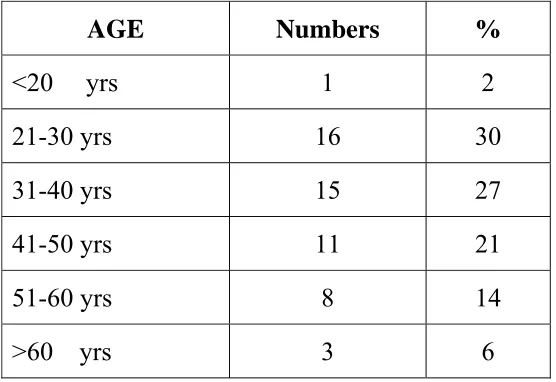
Table 1 Demographic profile
AGE Numbers %
<20 yrs 1 2
21-30 yrs 16 30
31-40 yrs 15 27
41-50 yrs 11 21
51-60 yrs 8 14
>60 yrs 3 6
Among the 53 patients 30 were male and 23 patients were female.
Sex ratio was 1.3:1. Age ranges from 16 to 64 years. 36 year was the
mean age . Peak age at which disease onset was noted was in the third
decade. All the patients were from the geographical location around north
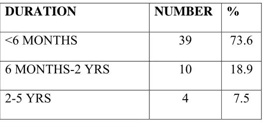
DURATION OF SYMPTOMS
Table 2Duration of symptoms
The average period of symptoms prior coming to the hospital
ranges from 15 days to 5 years. 73.6% had disease duration less than
6 months.
RISK FACTORS
Using the questionnaire the risk factors were analyzed. One patient
had family history of IBD. In this study 6% were previous smokers. 4%
had history of NSAID intake. None of them had previous history of
appendisectomy. None was on oral contraceptive pills.
DURATION NUMBER %
<6 MONTHS 39 73.6
6 MONTHS-2 YRS 10 18.9
CLINICAL PRESENTATION
The commonest clinical presentation was diarrhea, but nocturnal
symptom was present in 32%. Rectal bleeding was present in 69.8% of
cases. 9.4% had fever. Pain abdomen , tenesmus, urgency were other
features.
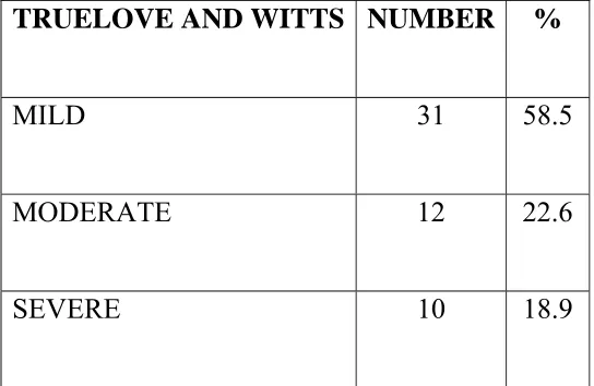
[image:36.612.181.453.356.533.2]CLINICAL ASSESMENT OF SEVERITY-TRUELOVE AND WITTS CLASSIFICATION
TABLE 3:CLINICAL SEVERITY OF DISEASE
According to Truelove and Witts classification ,mild disease was
commonly encounted in this study (58.5% ). 22.6% had moderate
disease. Severe disease was present in 18.9%.
TRUELOVE AND WITTS NUMBER %
MILD 31 58.5
MODERATE 12 22.6
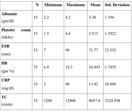
LABORATORY FINDINGS
TABLE 4:BASELINE LAB PARAMETERS
N Minimum Maximum Mean Std. Deviation
Albumin
(gm/dl) 53 2.2 4.2 3.30 1.194
Platelet count
(lakhs) 53 1.5 4.6 2.515 1.2922
ESR
(mm) 53 7 98 31.77 23.352
HB
(gm %) 53 6.0 14.3 10.443 1.7455
CRP
(mg/dl) 53 2 98 13.42 18.496
TC
(cmm) 53 1200 12900 8057.4 2324.598
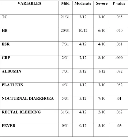
CORRELATION OF DISEASE ACTIVITY WITH CLINICAL & LABORATORY PARAMETER
Nocturnal diarrhoe (P=.01) and fever (P=.03) had a positive
TABLE 5:CLINICAL ACTIVITY & LAB PARAMETERS
VARIABLES Mild Moderate Severe P value
TC 21/31 3/12 3/10 .065
HB 20/31 10/12 6/10 .070
ESR 7/31 4/12 4/10 .061
CRP 2/31 7/12 8/10 .000
ALBUMIN 7/31 3/12 1/12 .072
PLATLETS 4/31 1/12 3/10 .082
NOCTURNAL DIARRHOEA 5/31 5/12 7/10 .01
RECTAL BLEEDING 31/31 4/12 2/10 .062
ENDOSCOPIC DISTRIBUTATION OF DISEASE
At initial presentation ,the endoscopic pattern was
Proctitis 35.8%
Proctosigmoiditis 26.4%
Left sided colitis 13.2%
Extensive colitis 5.6%
Pancolitis 18.9%
Proctitis was the commonest presentation in this study. None of the
patients had acute fulminant colitis.
ENDOSCOPIC DISTRIBUTATION AND CORRELATION OF
CLINICAL & LAB PARAMETERS
Tenesmus and urgency were associated with proctitis and
proctosigmoiditis. Presence of fever had a positive correlation with
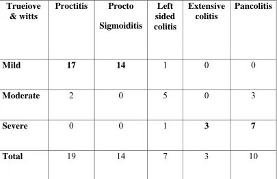
CLINICAL SEVERITY AND ENDOSCOPIC DISTRIBUTION OF
DISEASE
Distal colon is involved if the patient had a mild clinical activity.
Extensive colitis and pancolitis were associated with severe disease
[image:40.612.115.520.297.559.2]activity.
TABLE 6:CLINICAL & ENDOSCOPIC CORRELATION
Trueiove & witts
Proctitis Procto
Sigmoiditis
Left sided colitis
Extensive colitis
Pancolitis
Mild 17 14 1 0 0
Moderate 2 0 5 0 3
Severe 0 0 1 3 7
Total 19 14 7 3 10
Value Df
Asymp. Sig. (2-sided)
Pearson Chi-Square 63.692(a) 8 .000
Likelihood Ratio 64.304 8 .000
Linear-by-Linear
Association
37.457 1 .000
N of Valid Cases 53
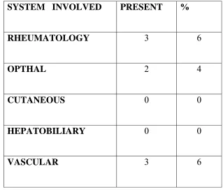
EXTRAINTESTINAL MANIFESTATIONS
15 % had extra intestinal manifestation in this study. Vascular
thrombosis, axial & peripheral arthropathy and uveitis were the
manifestations. None of them had neither cutaneous lesion nor primary
TABLE 7: EXTRAINTESTINAL MANIFESTATIONS
SYSTEM INVOLVED PRESENT %
RHEUMATOLOGY 3 6
OPTHAL 2 4
CUTANEOUS 0 0
HEPATOBILIARY 0 0
VASCULAR 3 6
TREATMENT GIVEN TO THE PATIENT
36 patients(67.9%) were treated with sulphasalazine,14 (26.4%)
were treated with sulphasalazine and prednisolone and 3(5.6%) with
azathioprine and prednisolone respectievely. Biologicals were not used.
All had clinical remission in this study.
CLINICAL VS ENDOSCOPIC REMISSION FOLLOWING TREATMENT
36 patients with mild to moderate disease were treated with
sulphasalazine therapy. During this time all of them were in clinical
remission. The endoscopic findings at 3 & 6 months were compared with
the baseline colonoscopic findings.
SULPHASALAZINE THERAPY AND MUCOSAL HEALING
No mucosal healing was noted in 80.5% and 70.5% at 3 and
6 months. A partial mucosal response was noted in19.5% and 25%
respectievely at three and six months. Sulphasalazine therapy was not
[image:43.612.120.517.406.654.2]associated with a complete mucosal healing in this study.
TABLE :8 ENDOSCOPIC RESPONSE & SULPHASALAZINE THERAPY
DURATION PARTIAL COMPLETE TOTAL
RESPONSE
NO
RESPONSE
3 MONTHS 7 0 7/36
19.5%
29/36
80.5%
6 MONTHS 9 0 9/36
25%
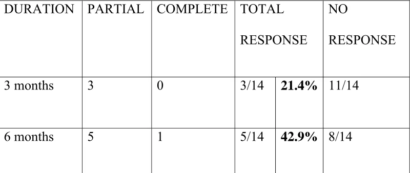
MUCOSAL HEALING IN SULPHASALAZINE AND PREDNISOLONE GROUP
Fourteen were treated with sulphasalazine, four gms per day and
oral prednisolone 40 mgs / day for 4 - 6 weeks, followed by decreasing
the dose of prednisolone. Colonoscopy at three months , response was
noted in 21.4%.All of them had a partial response only.
They were followed at 6 months both clinically and
endoscopically. All of them were in clinical remission but 42.9% had an
endoscopic response. Also 40% had a distal shift in distribution of
[image:44.612.117.517.462.631.2]disease.
TABLE :9 ENDOSCOPIC RESPONSE IN SULPHASALAZINE
WITH PREDNISOLONE
DURATION PARTIAL COMPLETE TOTAL
RESPONSE
NO
RESPONSE
3 months 3 0 3/14 21.4% 11/14
ENDOSCOPIC RESPONSE IN AZATHIOPRINE &
PREDNISOLONE GROUP
6 of them was initially in this group .Because of the intolerance to
drug, 3 of them moved to the other group. In this group, complete,
partial and nil response was observed in 33% each respectievely, both at
three and six months.
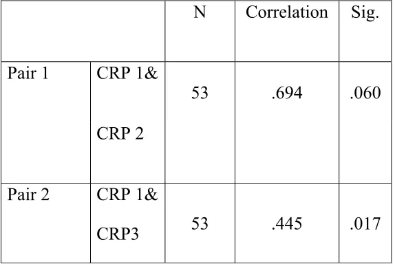
ROLE OF LABORATORY PARAMETERS DURING THE
FOLLOW UP PERIOD
All the lab parameters were also repeated during the follow up
period. But no correlation was noted between ESR, CRP with the clinical
disease activity during the third and sixth months. During which time,
even though all the patients were in clinical remission. they haven’t
T-Test
TABLE :10 Paired Samples Correlations
N Correlation Sig.
Pair 1 CRP 1&
53 .694 .060
CRP 2
Pair 2 CRP 1&
CRP3 53 .445 .017
COMPLICATIONS
Skin rash 2
Anemia 4
Obesity 3
Exacerbation of symptoms following endoscopy were seen in 4
patients.
No major adverse reactions were encountered during the study
DISCUSSION
Ulcerative colitis is a common disease in most of the industrialized
countries in the world, but it was thought to be uncommon in the
developing countries (37). The highest incidence is reported from
Scandinavia and Scotland and then by England and North America (38).
In the West, the incidence and prevalence of UC is 8–14 per 100,000 and
120 -200 per100,000 persons, respectively (39).
Limited community studies were available from India. One
community study from Punjab reveals the incidence of UC was
6.02/100000 per year and a prevalence rate of 44.3/100000
population.(40) but several hospital incidence rates were available from
India and it was 12/10,000 by Sood et al 1999.
But in this study the hospital incidence of UC was 36/10000. An
increasing trend was noted The temporal association of the increasing
incidence rates is related to the change in westernisation of lifestyles,
change in diet as well as in the environment caused by industrialization
Age ranges from 16 -64 years and the mean age was 36 years in
this study.The maximum number of patients were in the third decade.
Following were the various studies on UC, from India
Author Mean (yrs) Range (yrs)
Sood et al Ludhiana 31.7 14-65
Khosla et al Rohtak 24.4 (M) 38.0 (F)
Kapur P et al Delhi 13-65
Kochhar et al PGI 13-78
Chuttani et al MAMC 31.5 (M), 25.3 ( F) 9-56
Patients in India present mostly in 3rd and 4th decade and failed to
show a bimodal age pattern (42). But in the West a bimodal distribution
was noted characterized by a small late surge in the elderly, between
60 and 70 years.(43).
Males were affected slightly more than females, the ratio being
1.3:1. Another study by Tandon et al ,Delhi ,the sex ratio was 1.5:1.
Most studies from West did not showed any difference among gender
in the occurrence of UC. The male-to-female ratio of about 1 : 1 was
In this study, the average duration of symptoms prior coming to the
hospital ranges from 15 days to 5 years. 73.6% had disease duration less
than 6 months. Similar result was observed in a North Indian study and
the mean duration of symptoms before coming to hospital entry was
2.7 years.
Association of IBD among family, occurs in first-degree relatives
The relative risk of UC in a sibling was estimated to be around 7% and
17% based on western studies .Here also 1 patient (1.8%) had family
history of IBD.
CLINICAL ASSESMENT OF SEVERITY-TRUELOVE AND WITTS CLASSIFICATION
On the basis of Truelove and Witts classification majority had
mild disease (58.5%) at presentation. Moderate and severe disease were
noted in 22.6% & 18.9% respectievely . A recent review report says
that ulcerative colitis in India runs a mild course (45). The disease course
reported in Indian immigrants in England and Durban was mild to
ENDOSCOPIC DISEASE ACTIVITY
At initial presentation 35.8% had proctitis and 26.4% had
proctosigmoiditis. Distal disease was commonly noted in this study. Left
sided colitis and extensive colitis were observed in 13.2% & 5.7%
respectievely. Pancolitis was present in 18.9%. In a study from India half
of the patients had left sided colitis (47.5%), proctosigmoiditis in 25%
and pancolitis in 27.5%.
In one of the latest reviews from the West (47), 46% had
proctosigmoiditis, 17% left sided colitis and 37% had pancolitis.
According to a prospective, Norwegian study,(48) at initial presentation,
proctitis was present in 1/3 rd of patients, another one-third had left sided
colitis, in the remaining third there was proximal extension beyond the
splenic flexure. Pancolitis was seen in 25% of the patients.
The distribution of the disease varies according to age, ethnicity
and risk factors. A recent publication from Japan described that mild
colitis and proctitis were significantly larger in patients who had onset
of disease at older than 60 years, relatively to those who experienced
CLINICAL PRESENTATION & ENCOSCOPIC DISEASE ACTIVITY
Diarrhoea and rectal bleeding were the commonest clinical
presentation. Nocturnal diarrhea and fever had a positive correlation with
the endoscopic severity of the disease. They had either extensive colitis
or pancolitis. Kato and colleagues confirmed that severe clinical activity
was seen more commonly in patients who had severe disease activity in
the proximal colon compared with the rectum or sigmoid (50).
In this study, rectal bleeding and urgency were associated with
distal colonoscopic lesion. Similar result was seen in another study from
Tokyo (51).
LAB PARAMETERS & SEVERITY OF DISEASE
In this study patients with elevated CRP had either pancolitis or
extensive colitis. It had a positive correlation with the severity of the
disease. Similar result was seen in another study from Rochester.(52) In
another Japanese study ,CRP elevation reflects the activity of proximal
ENDOSCOPIC ASSESSMENT OF RESPONSE TO TREATMENT
Sulfasalazine is a 5-ASA compound , used as first line therapy for
the induction of remission in patients with mild to moderate UC. In this
study, sulphasalazine therapy in mild to moderate disease had 100%
clinical remission ,but not associated with a significant endoscopic
response. No mucosal healing was seen in 80.5% and 75% at 3 and
6 months. Only a partial mucosal response was noted in19.5% and 25%
respectievely at three and six months. Sulphasalazine therapy was not
associated with a complete mucosal healing in this study.
In another study, sulfasalazine ,3 to 6 g/day induces remission in
39% to 62% with mild to moderate UC. A dose-dependent response was
reported when it was used for induction in active UC.(43). In the
ASCEND I &II trials, mesalamine at 2.4 & 4.8 gm per day in mild
disease had similar efficacy, but in moderate active disease a higher
dose(4,8 gm) was more effective to induce mucosal healing. This dose
of mesalamine is comparable to 12 grams of sulphasalszine.
Recent studies have concluded that improved release formulations
of 5 ASA & more aggressive dosing schedules were needed for inducing
The poor response to sulphasalazine in this study can be attributed
to the lower dose 4 grams per day, used in this study.
In case of sulphasalazine and prednisolone combination therapy,
mucosal healing was seen in 21.4% & 42.9 % respectievely at 3 and
6 months in this study. The long-term remission rate in patients on
glucocorticoids for severe UC is reported as approximately 50% (43).
Another report says, with corticosteroids, 54% achieved complete
remission, 30% partial response, and 16% no response over first
30 days. (53).
In AZA and prednisolone group, mucosal healing was found in 66
%.No response was seen in 33%. One randomised controlled trial showed
that azathioprine along with glucocorticoids had a response rate of 79%
at one month. A varying result was given by another trial, the response
rate was 53% at six months.
Rutgeerts et al ,in his study Infliximab produces mucosal healing
Kohn et al ,in a small series reported that 77% clinical response
was seen after a single infliximab infusion of 5 mg/kg, & 80% of the
responders were in clinical remission after a period of 2 years.
ROLE OF CRP AND MUCOSAL HEALING
In this study, CRP that was done subsequently at 3 & 6 months
while the patients were in clinical remission doesn’t correlate with the
clinical activity. This might be because of the presence of endoscopic
disease activity. Solern et al,in his study elevated CRP is a marker of
endoscopic activity.(52)
COMPLICATIONS
No major complications were encounted during this study. Recent
data from IBSEN study with 10-year follow-up also did not observe a
significant increased risk of death in patients with UC.(55)
Mild exacerbation of clinical symptoms were noted following
colonoscopy in 4 patients. Recent colonoscopy is associated with a flare
CONCLUSION
1. This study fails to show a bimodal age distribution in UC.
2. On the basis of Truelove and Witts classification majority had a
mild disease.
3. Proctitis was the commonest colonoscopic finding in this study.
4. Mild clinical disease was associated with distal colitis and severe
disease was found to have either extensive or pancolitis.
5. Nocturnal diarrhea and fever were observed more commonly in
patients who had maximum disease activity in the proximal colon
compared with the rectum or sigmoid.
6. CRP elevation reflects the activity of proximal lesions .
7. In patients with mild to moderate disease none of the patients had a
complete mucosal healing response with sulphasalazine therapy
at 3 and 6 months respectievely .Sulphasalazine 4 grams may be a
8. Elevated CRP during the follow up period ,while the patient is in
clinical remission indicates the absence of mucosal healing. This
can be used as an indirect marker to predict future response.
9. Full colonoscopy is particularly important in the initial mapping of
disease extent and severity as well as to investigate any
discrepancy between clinical symptoms and endoscopic
appearance.
10. Mucosal healing as assessed by endoscopy is a useful tool for
evaluating and guiding response to therapy in patients with IBD.
11. Mucosal healing is an admirable goal that we should strive to
LIMITATIONS OF THIS STUDY
1. Dose of sulphasalazine is probably short of therapeutic range in
moderate UC.
2. Sample size were small in steroid and azathioprine group.
BIBILOGRAPHY
1. Podolski DK, Inflammatory bowel disease, NEJM 2002;347:
417-429.
2. Tandon BN et al, UC in north India; GUT 1965 oct;6:448.
3. Venkatraman .S. Ramakrishna BS et al, Risk of colorectal cancer
in UC , India J Gastroenterol Hepatol 2005;20:705-9
4. Grace Chan, David S. Fefferman, Richard J. Farrell, Endoscopic
Assessment of Inflammatory Bowel Disease: Colonoscopy /
Esophagogastroduodenoscopy; Gastroenterol Clin N Am 41 (2012)
271–290
5. Froslie KF, Jahnsen J, Moum BA, et al. Mucosal healing in
inflammatory bowel disease: results from a Norwegian
population-based cohort. Gastroenterology 2007;133:412–22.
6. Lichtenstein GR, Kamm MA, Boddu P, et al. Effect of once- or
twice-daily MMX mesalamine (SPD476) for the induction of
remission of mild to moderately active ulcerative colitis. Clin
Gastroenterol Hepatol 2007;5:95–102.
7. Wilks S, Ongman, Brown, Green, Longman & Roberts, Lectures
on pathologicalanatomy London 1859
8. Molodecky NA, Soon IS, Rabi DM, et al. Increasing incidence and
9. Loftus EV. Clinical epidemiology of inflammatory bowel disease:
incidence, prevalence,and environmental influences.
Gastroenterology 2004;126(6):1504–17.
10. Mendeloff AI, Calbins BM. The epidemiology of idiopathic
inflammatory bowel disease. In : Kurner JB, Shorter NG, eds.
Inflammatory bowel disease.Philadelphia : Lea and Febiger 1988; 3: 3-34.
11. Hou JK, El-Serag H, Thirumurthi S. Distribution and
manifestations of inflammatory bowel disease in Asians,
Hispanics, and African Americans: a systematic review. Am J
Gastroenterol 2009;104(8):2100–9.
12. Klement E, Cohen RV, Boxman J, et al. Breastfeeding and risk of
inflammatory bowel disease: a systematic review with
meta-analysis. Am J Clin Nutr 2004; 80(5):1342–52.
13. Andersson RE, Olaison G, Tysk C, et al. Appendectomy and
protection against ulcerative colitis. N Engl J Med
2001;344(11):808–14.
14. A Sood, V Midha, N Sood, A S Bhatia, G Avasthi Incidence and
prevalence of ulcerative colitis in Punjab, North IndiaGut
16. Probert CS, Jayanthi V, Huges AO, et al. Prevalence and family
risk of ulcerative colitis and Crohn’s disease: an epidemiological
study among Europeans and South Asians in Leicestershire. Gut
1993;34:1547–51.
17. Mayer L. Evolving paradigms in the pathogenesis of IBD. J
Gastroenterol 2010; 45(1):9.
18. Abraham C, Cho JH. Mechanisms of disease: inflammatory bowel
disease. N Engl J Med 2009;361(21):2066–78.
19. Sartor RB ;Microbial influences in IBD;Gastroenterology 2008
:134;577
20. Truelove SC,Richards WC;Biopsy studies in UC.BMJ
1956;4979:1315
21. Langholz E, Munkholm P, Nielsen OH, et al. Incidence and
prevalence of ulcerative colitis in Copenhagen county from 1962 to
1987. Scand J Gastroenterol 1991;26: 1247–56
22. Orchard TR,Wordsworth BP,Jewell DP;Peripheral arthropathies in
IBD.Their articular distributation & natural history.Gut
1998;42:387.
23. Cornaggia M, Leutner M, Mescoli C, et al. Chronic idiopathic
inflammatory bowel diseases: the histology report. Dig Liver Dis
24. Kudo T, Matsumoto T, Esaki M, et al. Mucosal vascular pattern in
ulcerative colitis: observations using narrow band imaging
colonoscopy with special reference to histologic inflammation. Int
J Colorectal Dis 2009;24:495–501.
25. Zhou N, Chen WX, Chen SH, et al. Inflammatory bowel disease
unclassified. J Zhejiang Univ Sci B 2011;12:280–6.
26. Truelove SC, Witts LJ. Cortisone in ulcerative colitis; final report
on a therapeutic trial. Br Med J 1955;ii:1041– 8.
27. Baron JH, Connell AM, Lemmard-Jones JE. Variation between
observers in describing mucosal appearances in proctocolitis. Br
Med J 1964;1:89–92.
28. Powell-Tuck J, Day DW, Buckell NA, et al. Correlations between
defined sigmoidoscopic appearances and other measures of disease
activity in ulcerative colitis. Dig Dis Sci 1982;27:533–7.
29. Sutherland LR, Martin F, Greer S, et al. 5-aminosalicylic acid
enema in the treatment of distal ulcerative colitis,
proctosigmoiditis, and proctitis. Gastroenterol 1987;92: 1894–8.
30. D'Haens G, Sandborn WJ, Feagan BG, et al. A review of activity
31. Sutherland L, Macdonald JK. Oral 5-aminosalicylic acid for
induction of remission in ulcerative colitis. Cochrane Database
Syst Rev 2006;2:CD000543.
32. Roses RE, Rombeau JL. Recent trends in the surgical management
of inflammatory bowel disease. World J Gastroenterol
2008;14:408–12.
33. Rutgeerts P, Vermeire S, Van Assche G. Biological therapies for
inflammatory bowel diseases. Gastroenterology 2009;136:1182–
97.
34. Hanauer SB. Review article: high-dose aminosalicylates to induce and maintain remissions in ulcerative colitis. Aliment Pharmacol Ther 2006;24(Suppl 3):37–40.
35. Gies N, Krocker KI, Wong K, et al. Treatment of ulcerative colitis
with adalimumab or infliximab: long-term follow-up of a
single-center cohort. Aliment Pharmacol Ther 2010;32:522–8.
36. Ali T, Lam D, Bronze MS, et al. Osteoporosis in inflammatory
bowel disease. Am J Med 2009;122:599–604.
37. Ekbom A, Helmick C, Zack M et al. The epidemiology of inflammatory bowel disease : A large, population based study in
Sweden. Gastroenterology 1991; 100: 350-8.
38. Cosnes et al Epidemiology and Natural History of Inflammatory
39. Mendeloff AI, Calbins BM. The epidemiology of idiopathic
inflammatory bowel disease. In : Kurner JB, Shorter NG, eds.
Inflammatory bowel disease.Philadelphia : Lea and Febiger 1988; 3: 3-34.
40. A Sood, V Midha, N Sood, A S Bhatia, G Avasthi Incidence and
prevalence of ulcerative colitis in Punjab,North India Gut
2003;52:1587–1590
41. Probert CS, Jayanthi V, Huges AO, et al. Prevalence and family
risk of ulcerative colitis and Crohn’s disease: an epidemiological
study among Europeans and South Asians in Leicestershire. Gut
1993;34:1547–51.
42. Ajit Sood*, Vandana Midha*, Neena Sood**, Sandeep Puri*, Vikas KaushalProfile of Ulcerative Colitis in a North Indian
HospitalJournal of Indian Academy of Clinical Medicine _ Vol. 5 _
No. 2 125
43. Mark T. Osterman,Gary R. Lichtenstein Ulcerative Colitis
Sleisinger and Fordtrans Gastrointestinal and Liver Diseases
chapter 112 :1975
46. Farmer RG, Easley KA, Rankin GB. Clinical pattern, natural
history and progression of ulcerative colitis : A long term follow up
of 116 patients. Dig Dis Sci 1993; 38: 1137-46.
47. Farmer RG, Easley KA, Rankin GB. Clinical pattern,natural
history and progression of ulcerative colitis : A long term follow up
of 116 patients. Dig Dis Sci 1993
48. Moum B, Ekbom A, Vatn MH, et al. Change in the extent of
colonoscopic and histological involvement in ulcerative colitis over
time. Am J Gastroenterol 1999;94:1564–1569.
49. Fujimoto T, Kato J, Nasu J, et al. Change of clinical characteristics
of ulcerative colitis in Japan: analysis of 844 hospital-based
patients from 1981 to 2000. Eur J Gastroenterol Hepatol
2007;19(3):229–35.
50. Kato J, Kuriyama M, Hiraoka S, et al. Is sigmoidoscopy sufficient
for evaluating inflammatory status of ulcerative colitis patients? J
Gastroenterol Hepatol 2011;26: 683–7.
51. Osada,Ohkusa T et al ,Correlations among total colonoscopic
findings,clinical symptoms and laboratory markers in UC .J
Gastroenterol Hepatol 2008 Dec23,supp 2 S262-7
52. Solern CA et al,Correlation of CRP with clinical, endoscopic,
histological and radiographic activity in IBD,Inflamm Bowel Dis
53. Faubion WA Jr, Loftus EV Jr, Harmsen WS, et al. The natural
history of corticosteroid therapy for inflammatory bowel disease: a
population-based study. Gastroenterology 2001;121:255–60.
54. Maneest Dave et al Mucosal Healing in Inflammatory Bowel
Disease—A True Paradigm of Success? Gastroenterol Hepatol (N
Y). 2012 January; 8(1): 29–38
55, Hoie O, Wolters FL, Riis L, et al. Low colectomy rates in
ulcerative colitis in an unselected European cohort followed for 10
years. Gastroenterology 2007;132:507–15.
56. Menees S et al,Does colonoscopy cause increased UC
symptoms,Inflamm Bowel Dis 2007;13:12-8
ANNEXURE – A
PROFORMANAME AGE SEX:
DDHD NO: DIAGNOSIS: DURATION:
EPIDEMIOLOGY Drug
Place Skin/Eye problem
Per capita income CAD
COMPLAINTS PAST HISTORY
Pain abdomen DM
Loose stools HTN
Nocturnal symptoms Surgery
Blood in stools FAMILY HISTORY
Fever IBD
Fatigue, tiredness PERSONAL HISTORY
Weight loss Smoker
Jaundice Alcahol abuse
GENERAL EXAMINATION INVESTIGATIONS
BMI HB
Temperature TC
Pulse DC
BP ESR
Pallor PLATELET
Skin lesions ALBUMIN
Jaundice RFT
Eyes CRP
Oral cavity STOOL
Joints TRUELOVE &WITTS
Peripheral vessels Mild
ABDOMEN Moderate
RECTAL Severe
Baseline(1) 3 months (2) 6 months (3)
COLONOSCOPY
ESR
CRP
HB
TC/DC
ANNEXURE – B
MASTER CHART
No Age sex
dura-tion
diar-rhoea
rectal
bleed fever platelet ESR 1 HB1 CRP 1 true luv colon 1 colon 2 TC HB
2 ESR2 CRP2
ESR 3
CRP 3
colon
3 extra treatment
1 54 m 2 4 2 0 1.5 7 11.7 6 S1 P1 P1 6500 11.9 8 7 7 5 P1 nil S
2 60 f 3 3 3 0 2.3 10 11 2 S1 P2 P2 5342 10.8 12 8 10 4 P1 nil S
3 30 f 3 4 1 0 1.8 11 10.8 8 S1 P1 P1 7800 11 11 8 8 6 P1 nil S
4 63 f 0.5 6 2 0 1.9 30 8.7 11 S2 P2 P2 9100 8.5 10 5 16 17 P2 nil S
5 47 m 2 4 4 0 2.4 120 6.6 13 S1 P2 P2 7600 9.5 37 7 11 3 P2 nil S
6 25 f 3 4 4 0 4.8 60 9.3 24 S1 L2 L2 8240 10 28 11 12 6 L2 DVT S
7 60 f 3 4 3 0 1.9 20 12 5 S1 PS1 PS1 7600 11.8 12 6 21 3 PS1 nil S
8 30 m 1 5 4 0 1.6 23 11 7 S1 PS2 PS1 7333 11.7 10 3 11 5 PS1 nil S
9 42 m 2 15 12 0 1.9 97 11.8 13 S3 PA3 PA1 8900 12.8 34 7 22 8 PS3 nil SP
10 38 m 12 4 3 0 2.6 12 14.3 3 S1 P2 P2 7600 11.8 7 5 21 4 P2 nil S
11 38 f 48 4 4 0 2.8 13 12.5 8 S1 P1 P1 8110 12.9 10 4 22 11 P1 nil S
12 64 m 0.25 4 2 0 1.7 11 13.2 4 S1 PS2 PS2 6500 13 8 6 6 5 PS2 nil S
13 22 m 24 10 10 0 2.8 28 10.3 98 S3 PA3 L3 9100 12 16 17 11 5 PS2 nil SP
14 42 f 4 4 3 0 3 21 11 5 S1 P1 P1 7600 11.8 11 3 8 3 P1 nil S
15 54 f 3 4 3 0 1.9 12 12.9 6 S1 PS2 PS2 9300 12 12 6 5 5 PS2 nil S
16 31 m 3 4 4 0 3.1 39 10 5 S2 PA2 E3 8000 11.3 21 3 6 5 P2 nil SP
17 16 m 4 12 2 0 2.1 11 14 3 S2 P2 P2 6200 13.8 11 5 6 6 P2 nil S
19 22 m 6 4 3 0 2.3 52 8 5 S1 PS1 PS1 6720 9 22 8 11 12 PS1 nil S
10 22 m 3 0 3 0 2.7 12 10 8 S1 P1 P1 5880 11.2 21 4 9 9 P1 nil S
20 38 m 24 5 3 0 2.6 23 11 4 S1 PS2 PS2 7600 12 22 11 14 4 PS2 nil S
21 42 m 1 14 12 1 2.2 43 10.7 76 S3 PA2 E2 8400 11.8 6 5 11 6 E1 nil SP
22 36 m 3 4 3 0 2 17 11.3 5 S1 P2 P2 6500 11.8 11 5 21 5 P2 nil S
23 54 m 2 8 8 1 3.6 30 10.4 55 S3 E2 E1 4800 11 8 3 9 8 E1 nil SP
24 30 m 6 7 6 0 1.8 34 12 13 S2 PA2 PA2 9320 11 5 5 18 6 PA2 nil SP
25 35 m 0.5 4 3 0 1.6 12 11.4 7 S1 P2 P2 7600 10.6 6 5 11 3 P2 nil S
26 20 f 6 4 3 0 1.5 21 12 5 S1 P2 P2 5700 11 6 6 10 5 P2 nil S
27 57 f 2 10 9 0 1.8 54 7.9 17 S3 PA2 PA2 9800 8.5 11 12 37 7 PA2 nil SP
No Age sex dura-tion diar-rhoea rectal
bleed fever platelet ESR 1 HB1 CRP 1 true luv colon 1 colon 2 TC HB
2 ESR2 CRP2
ESR 3
CRP 3
colon
3 extra treatment
29 52 f 18 0 3 0 2 13 11 4 S1 PS3 PS3 7690 11 14 4 12 6 PS3 nil S
30 55 m 12 4 4 0 2.8 17 10.7 4 S1 PS2 PS2 3560 11.9 11 6 10 3 PS2 nil S
31 60 f 3 3 2 0 3 10 12 6 S1 PS2 PS2 6700 11 21 5 34 7 PS2 nil S
32 33 m 12 0 2 0 1.8 23 8.6 16 S1 P2 P2 12000 9.7 9 8 7 5 P2 nil SP
33 41 f 6 7 6 0 4.5 83 9.6 23 S3 E2 E2 12400 10.6 18 6 10 4 E2 nil S
34 28 m 3 4 4 0 1.6 12 11,8 5 S1 P2 P2 1200 11.7 11 3 8 6 P2 nil S
35 45 m 12 0 4 0 1.9 23 12 7 S1 PS2 PS2 7800 11.9 8 3 16 17 PS2 nil S
36 37 m 6 4 3 0 1.7 21 11 5 S1 P2 P2 6500 12 16 4 11 3 P2 nil S
37 22 m 4 7 6 0 1.6 43 7.8 12 S2 L3 L3 9700 9.7 35 7 12 6 L3 E S
38 32 f 24 4 4 0 3.6 23 9.8 8 S2 PS3 PS3 6890 10.7 12 5 21 3 PS3 nil S
39 36 m 3 12 8 0 3.2 34 9.7 8 S2 L2 L2 6500 10.8 22 3 11 5 L2 nil S
40 38 m 12 4 4 0 3.7 12 10 5 S1 P2 P2 8900 11 11 6 22 8 P2 nil S
41 28 f 24 20 15 1 3.4 32 7.9 12 S3 E2 PS2 5800 9.6 19 7 21 4 PS2 nil SP
42 45 f 7 0 3 0 2.3 40 12.3 6 S1 PS2 PS2 4560 12 11 4 22 11 PS2 nil S
43 40 m 2 8 6 0 2.6 45 10.5 6 S2 PA3 P2 10700 11 12 7 6 5 P2 nil AP
44 21 f 6 6 6 0 9.6 23 11 7 S2 L2 L2 9200 11 12 4 11 5 L2 DVT S
45 41 f 4 7 4 0 1.8 82 9.2 10 S2 L3 PS 12700 10 40 7 8 3 P2 nil AP
46 38 m 6 7 6 0 1.9 34 9.4 9 S2 L2 L2 8200 10.2 15 3 5 5 PS1 nil SP
47 31 f 60 4 4 0 2.4 25 9.8 5 S1 PS3 PS3 10700 10 17 7 6 5 PS3 nil S
48 28 f 3 15 12 0 4.9 23 9.7 12 S3 PA2 PS2 12900 11 21 7 6 6 E3 DVT SP
49 25 m 12 15 6 1 1.8 64 7.3 54 S3 PA2 PA2 11700 8.9 34 10 11 12 PA2 R AP
50 36 m 4 8 6 0 1.8 43 9.4 12 S2 L2 L2 8200 10 21 7 9 9 L2 nil S
51 43 f 24 8 nil 0 1.9 40 12.4 5 S1 PS2 PS2 11200 11 21 3 14 4 PS2 nil S
52 34 m 3 4 4 0 1.6 21 9.8 4 S1 P2 P2 7800 11 12 5 11 6 P2 nil S