Copyright © 1999, American Society for Microbiology. All Rights Reserved.
Sendai Virus Infection Induces Apoptosis through Activation of
Caspase-8 (FLICE) and Caspase-3 (CPP32)
MICHAEL BITZER,
1* FLORIAN PRINZ,
1MANUEL BAUER,
1MARTIN SPIEGEL,
1WOLFGANG J. NEUBERT,
2MICHAEL GREGOR,
1KLAUS SCHULZE-OSTHOFF,
1ANDULRICH LAUER
1Abteilung Innere Medizin I, Medizinische Universita¨tsklinik Tu¨bingen, 72076 Tu¨bingen,
1and Abteilung fu¨r
Virusforschung, Max-Planck-Institut fu¨r Biochemie, 82152 Martinsried,
2Germany
Received 12 January 1998/Accepted 5 October 1998
Sendai virus (SV) infection and replication lead to a strong cytopathic effect with subsequent death of host
cells. We now show that SV infection triggers an apoptotic program in target cells. Incubation of infected cells
with the peptide inhibitor z-VAD-fmk abrogated SV-induced apoptosis, indicating that proteases of the caspase
family were involved. Moreover, proteolytic activation of two distinct caspases, CPP32/caspase-3 and, as shown
for the first time in virus-infected cells, FLICE/caspase-8, could be detected. So far, activation of FLICE/
caspase-8 has been described in apoptosis triggered by death receptors, including CD95 and tumor necrosis
factor (TNF)-R1. In contrast, we could show that SV-induced apoptosis did not require TNF or CD95 ligand.
We further found that apoptosis of infected cells did not influence the maturation and budding of SV progeny.
In conclusion, SV-induced cell injury is mediated by CD95- and TNF-R1-independent activation of caspases,
leading to the death of host cells without impairment of the viral life cycle.
Over the past few years, a growing number of viruses have
been found to induce apoptosis in host cells (41, 49). For some
of them, mechanisms involved in the initial activation of the
apoptotic death cascade have been discovered, such as
up-regulation of the CD95/Fas receptor by influenza virus (48),
upregulation of CD95L/Fas ligand (3, 53), and cleavage of
the apoptosis-inhibiting proto-oncogene bcl-2 (46), as well as
downregulation of bcl-2 in combination with upregulation of
the apoptosis accelerator Bax (39) by human
immunodefi-ciency virus (HIV) and accumulation of p53 in host cells, e.g.,
during infection by adenovirus, simian virus 40, or human
papillomavirus (reviewed in reference 49). However, little is
known concerning the effector phase of apoptosis in
virus-infected cells. Indirect evidence gained by
pharmacological-inhibition experiments or by the cleavage of specific substrates
suggested the involvement of caspases without defining how
many and which caspases are required to cause suicide of
in-fected host cells (8, 21, 37, 52).
Apoptosis is defined as an active physiological process of
cellular self-destruction, with specific morphological and
bio-chemical changes (45). The molecular processes controlling
and executing virus-induced apoptosis are still poorly
under-stood. Caspases, a family of cysteine proteases formerly called
ICE (interleukin-1b-converting enzyme)-like proteases, play a
central role in the execution of the apoptotic process (11, 32).
These proteases are synthesized as inactive proenzymes and
activated after cleavage at specific aspartate residues. Ten
ho-mologs of human origin have been identified, including ICE/
caspase-1 (54), CPP32/caspase-3 (16, 31, 50), and
FLICE/cas-pase-8 (29). The last is thought to be the most apical member
of the death receptor-mediated pathways, being capable of
triggering the processing of other executioner caspases in the
apoptotic cascade (30).
As a prototype member of the paramyxovirus family, Sendai
virus (SV) is an enveloped negative-strand RNA virus. It is
closely related to human parainfluenza viruses and causes
acute respiratory tract infections in rodents, such as mice and
rats (12). Infection and replication of SV in host cells lead to
an extensive cytopathic effect, with subsequent cell death, but
the mechanisms of cell injury are poorly understood (12).
Pre-vious findings with human peripheral blood mononuclear cells
suggested that apoptosis induction might be one mechanism of
SV-induced cell death (51). However, peripheral blood
mono-nuclear cells are not typical SV host or propagation cells (12)
and apoptosis induction has so far not been linked to SV
rep-lication and propagation in other cell types. In addition, there
are no data available on the possible mechanisms of
SV-in-duced apoptosis.
Here we show that the strong cytopathic effect after SV
in-fection of target cells can be attributed to apoptotic cell death.
Moreover, we show that caspases play a key role in the effector
phase of SV-induced cell death and we demonstrate the
acti-vation of CPP32/caspase-3 and FLICE/caspase-8.
Interest-ingly, FLICE/caspase-8 activation in infected host cells did not
require ligand-induced activation of death receptors, such as
CD95 and tumor necrosis factor (TNF)-R1. We further found
that apoptosis did not influence the maturation and budding of
SV progeny. Thus, our results suggest that the SV-induced
cytopathic effect involves CD95- and TNF-R1-independent
ac-tivation of caspases, which results in apoptosis without
affect-ing the viral life cycle.
MATERIALS AND METHODS
Reagents.Recombinant human CD95L was expressed in stably transfected
293 cells as soluble Flag-tagged fusion protein and purified by affinity chroma-tography (unpublished data). TNF-awas purchased from Genzyme, Cambridge, Mass. Chimeric receptor decoy proteins consisting of the extracellular part of CD95 (7) or TNF-R1 (13) fused to immunoglobulin G1 (IgG1)-Fc were kindly provided by Immunex, Seattle, Wash.
Virus and cells.SV (strain Fushimi) was grown in 9-day-old embryonated
chicken eggs as described previously (43). CV-1 (African green monkey kidney) cells were obtained from the American Type Culture Collection (Rockville, Md.), and HepG2 (human hepatoma) cells were obtained from the European Collec-tion of Animal Cell Cultures (Salisbury, United Kingdom). CV-1 cells were maintained in M199 medium, and HepG2 cells were maintained in a HEPES-buffered mixture of minimal essential medium and Dulbecco modified Eagle medium (4:1) containing 4.5 g of glucose/liter, sodium pyruvate, nonessential amino acids, and biotin, all supplemented with 10% fetal calf serum (FCS).
* Corresponding author. Mailing address: Abteilung Innere Medizin
I, Medizinische Universita¨tsklinik Tu¨bingen, Otfried-Mu¨ller-Str. 10,
72076 Tu¨bingen, Germany. Phone: (49) 7071-2983189. Fax: (49)
7071-292095. E-mail: michael.bitzer@uni-tuebingen.de.
702
on November 9, 2019 by guest
http://jvi.asm.org/
Media and supplements were purchased from Life Technologies (Eggenstein, Germany).
Infection of cells.For infection, cells were used when monolayers had reached
85 to 90% confluence in 35-mm-diameter dishes. As standard inoculation pro-cedure, monolayers were washed twice with medium lacking FCS (washing medium) and overlaid with phosphate-buffered saline (PBS) containing SV at a multiplicity of infection (MOI) of 10. After incubation for 15 min at 37°C, unadsorbed virus was removed by repeated washing of the cells. Medium con-taining FCS (growth medium) was added, and the cells were incubated for var-ious periods of time at 37°C.
DNA fragmentation assay.Cells (53107) were collected together with the
floating cells in the supernatant at different time intervals postinfection (p.i.), and fragmentation assays were performed as described previously (20). In brief, the cells were washed once in PBS and lysed in 600ml of DNA lysis buffer (Tris-HCl, pH 7.5, 0.2% Triton X-100, 10 mM EDTA) on ice for 10 min. Cell debris was removed by centrifugation (10 min; 13,0003g; 4°C), and the supernatants were
extracted once with phenol-choloroform–isoamyl alcohol (24:1). Total DNA was precipitated by the addition of 5 M NaCl to a final concentration of 300 mM in the presence of isopropanol, followed by incubation overnight at220°C. Nucleic acids were pelleted at 12,000 3g (15 min; 0°C), resuspended in 15ml of Tris-EDTA buffer (10 mM Tris-HCl, pH 7.5, 1 mM EDTA), and incubated with 1 mg of RNase A (Boehringer Mannheim, Mannheim, Germany)/ml for 30 min. The nucleic acids were electrophoresed through 2% agarose gels (Gibco BRL, Eggenstein, Germany) and stained with ethidium bromide.
In situ-apoptosis assay.The in situ-cell death detection kit AP (Boehringer
Mannheim) was used to detect free 39OH ends of fragmented DNA. Terminal deoxynucleotidyltransferase (TDT) catalyzes the polymerization of fluorescein-labeled dUTP in a template-independent manner, labeling ends of fragmented DNA in situ. Subsequently, incorporated fluorescein was detected by alkaline phosphatase-conjugated anti-fluorescein antibody Fab2fragments, resulting in
an intense dark-blue staining of apoptotic cells.
Flow cytometry.Fragmentation of genomic DNA to hypodiploid DNA was
assessed by fluorescence-activated cell sorter (FACS) analysis according to the method described previously (33). In brief, 53106cells (including floating cells)
were collected and washed once in PBS (5 min; 1,0003g). Pellets were
resus-pended in 100ml of PBS, fixed with 1 ml of acetone-methanol (1:1;220°C), and subsequently washed with PBS. Next, each pellet was resuspended in 400ml of PBS containing 1 mg of RNase/ml and incubated on ice for 1 h. After the addition of 20ml of propidium iodide solution (2 mg/ml in PBS; Sigma, Deisen-hofen, Germany) and incubation for at least 30 min on ice, flow cytometry was performed (FACS Calibur; Becton Dickinson, Heidelberg, Germany) by using the CellQuest program. Cells to the left of the 2 N peak contained hypodiploid DNA and were therefore considered apoptotic.
Western blot assay.To detect proteolytic processing of caspases, Western blot
assays were done as described previously (25). In brief, 5 3107cells were
harvested, washed once in PBS, and resuspended in 0.5 ml of lysis buffer (1% Triton X-100, 150 mM NaCl, 10 mM Tris-HCl, pH 7.5). From each sample 30mg of cellular protein was electrophoretically separated on sodium dodecyl sulfate– 10% polyacrylamide gels under reducing conditions and subsequently trans-ferred to polyvinylidene difluoride membranes (Pall Fluorotrans transfer mem-brane; Pall Europe, Portsmouth, England). The membranes were blocked in Tris-buffered saline (150 mM NaCl, 13 mM Tris, pH 7.5) containing 5% nonfat dry milk powder for 1 h. Next, the membranes were incubated with the appro-priate antibodies (anti-FLICE [Biomedia, Baesweiler, Germany], 1:10; rabbit anti-CPP32 [kindly provided by P. Vandenabeele, University of Ghent, Belgium], 1:1,000; F37720 anti-FasL [Transduction Laboratories, Lexington, Ky.], 1:1,000; and anti-alpha-tubulin [Sigma], 1:2,000) overnight at 20°C, washed three times with TBS-T (TBS containing 0.02% Triton X-100), and incubated with peroxi-dase-conjugated anti-mouse or anti-rabbit IgG (Amersham-Buchler, Braun-schweig, Germany) (1:1,000). Further detection was performed by the ECL Western blotting detection system on Hyperfilm-ECL (Amersham-Buchler). Control cells were incubated with recombinant human CD95L containing super-natant for 12 h and harvested as described above.
Inhibition experiments.Apoptosis was blocked by addition of the peptide
inhibitor z-VAD-fmk [benzoyloxycarbonyl-Val-Ala-Asp (Ome) fluoromethyl-ketone; Enzyme Systems, Dublin, Calif.] (100mM) every 12 h to the culture supernatant. The supernatants were analyzed by hemagglutination (HA) and 50% tissue culture infectivity dose (TCID50) assays, and the cells were analyzed
by flow cytometry.
For inhibition experiments of receptor pathways, 53106cells were infected
with SV (MOI, 10) in 35-mm-diameter dishes. Immediately after infection, chimeric receptor decoy proteins consisting of the extracellular part of either CD95 or TNF-R1 fused to IgG1-Fc were added in a final concentration of 10 or 50mg/ml. Control experiments in our setting revealed a complete blockage of TNF- and CD95L-induced apoptosis at concentrations of 5 to 20mg of the decoy constructs/ml (data not shown and reference 7). Forty hours p.i., the cells were harvested and analyzed by FACS as described above.
HA and TCID50assays.Virus yield was quantitated by the HA assay, and
infectivity of progeny virions was quantitated by the TCID50assay (TCID50/ml)
with supernatants of infected cells, as described previously (5). For the TCID50
assay, FCS was replaced in the growth medium by Nutridoma SR or CS (Boehr-inger Mannheim) to enable cleavage of the F0precursor protein by acetylated
trypsin prior to the assay, as described previously (5, 24). In our setting, 1 TCID50/ml was equivalent to 5,000 PFU/ml.
RESULTS
SV infection triggers the apoptosis death cascade.
Monkey
kidney CV-1 cells infected with SV (MOI, 10) were found to
exhibit an extensive cytopathic effect 15 to 24 h p.i., which was
morphologically characterized by cell shrinkage, condensation
of nuclear chromatin, and cell fragmentation (data not shown).
As these alterations were classical signs of apoptotic cell death,
we isolated cellular DNA to investigate DNA fragmentation.
Starting at 24 to 30 h p.i. (MOI, 10), characteristic DNA
fragmentation patterns with oligonucleosomal fragments of
180 to 200 bp and multiples thereof were detected in CV-1
cells (Fig. 1). In contrast, noninfected cells revealed no specific
DNA signal, as intact high-molecular-mass DNA was removed
during the isolation protocol (Fig. 1, lane 5). To further
exam-ine nuclear DNA fragmentation, we used the TDT-mediated
dUTP-biotin nick end labeling TUNEL assay (19), in which
TDT is employed to label the ends of fragmented DNA in situ
with fluorescein-labeled dUTP. Detection of the label by using
alkaline phosphatase-conjugated antibodies to the
fluorescein-labeled nucleotide generates a dark-blue color whose intensity
is proportional to the number of 39
ends or fragments of
nuclear DNA. Intense dark staining was detected in
SV-in-fected CV-1 cells 24 h p.i. (MOI, 10), whereas no staining was
seen in uninfected controls (Fig. 2). Figure 3B and C shows
rates of apoptosis 36 and 60 h after SV infection of CV-1 cells,
determined by the appearance of hypodiploid DNA peaks
after propidium iodide staining and flow cytometry analysis.
Taken together, these results clearly demonstrate that SV
in-duces apoptotic cell death in CV-1 cells. Corresponding results
were obtained for other cell lines, such as HepG2, MDCK,
NIH 3T3, HeLa, and 293 cells (data not shown). Thus,
apo-ptotic cell death can be regarded as a general process during
SV infection of host cells.
[image:2.612.378.481.70.295.2]Caspase inhibition blocks SV-induced apoptosis.
It has
be-come clear that the effector phase during apoptotic cell death
FIG. 1. DNA fragmentation induced by SV infection of CV-1 cells. Lanes 2 to 4, DNA preparations from productively infected cells at different time points p.i.; lane 1, DNA marker; lane 5, preparation from uninfected control cells.
on November 9, 2019 by guest
http://jvi.asm.org/
requires the activation of different caspases (11, 27, 32). We
therefore investigated the effect of a broad caspase inhibitor,
z-VAD-fmk [benzoyloxycarbonyl-Val-Ala-Asp (Ome)
fluoro-methylketone] (10, 22, 44), on apoptosis induction in
SV-infected host cells. Addition of 100
mM z-VAD-fmk to the
supernatant of SV-infected CV-1 cells almost completely
in-hibited apoptosis, as assessed by a lack of formation of
hypo-diploid DNA 36 or 60 h p.i. (Fig. 3E and F). These results
imply that the activation of caspases is a central mechanism in
SV-induced cell death.
Detection of activation of individual caspases.
To further
define the relevant steps employed in the SV-triggered
apo-ptotic signal transduction pathway, we investigated the
activa-tion of individual caspases during SV infecactiva-tion. The cellular
protease CPP32/caspase-3 is expressed as a 32-kDa precursor
protein, which upon activation is processed into p17 and p12
subunits (31). Earlier work implicated CPP32/caspase-3 as a
central executioner protease in mammalian apoptosis.
More-over, sequential activation of CPP32/caspase-3 by other
mem-bers of the caspase family has been described previously (15,
26, 30, 44). To investigate the involvement of CPP32/caspase-3
in SV-induced apoptosis, cell lysates from infected and
nonin-fected cells were subjected to Western blotting with antibodies
directed to CPP32. Proteolytic processing of the 32-kDa
pre-cursor in infected cells could be demonstrated by a diminished
immunoreactive signal and subsequent detection of the
cleav-age products p17 and p12 in HepG2 cells (Fig. 4).
Correspond-ing results were obtained for CV-1 cells (data not shown).
Recently, a novel member of the caspase family, designated
FLICE/caspase-8, has been shown to directly cleave CPP32/
caspase-3 (30). FLICE/caspase-8 is thought to represent the
most upstream protease in the CD95-mediated apoptotic
[image:3.612.59.548.70.238.2]cas-FIG. 2. In situ detection of apoptosis in infected CV-1 cells. Apoptotic DNA degradation was visualized by TUNEL staining and subsequent alkaline phosphatase staining as described in Materials and Methods. (A) Intense dark staining of CV-1 cells 24 h p.i.; (B) uninfected CV-1 cells.
FIG. 3. Detection of hypodiploid DNA in SV-infected cells and inhibition by caspase inhibitor z-VAD. CV-1 cells were infected by SV (MOI, 10) and incu-bated with 0 (A, B, and C) or 100 (D, E, and F)mM of the caspase inhibitor z-VAD-fmk. Shown are the results of cytometric analysis at 36 and 60 h p.i. as well as the analysis of uninfected CV-1 cells (control) and CV-1 cells incubated with z-VAD-fmk for 60 h [control (1z-VAD)] to exclude the toxic effects of z-VAD-fmk on these cells. The proportion of sub-2N DNA is indicated in the histograms.
FIG. 4. SV infection triggers CPP32/caspase-3 activation. HepG2 cells were infected with SV (MOI, 10) and were prepared at the indicated time points p.i. Lane 1, preparation of uninfected control cells. The CPP32 proform and subunits p17 and p12 as well as tubulin were detected by Western blot analysis.
on November 9, 2019 by guest
http://jvi.asm.org/
[image:3.612.66.281.361.658.2] [image:3.612.365.496.565.693.2]cade, as it is recruited to the CD95 receptor through the
adaptor protein FADD (Fas-associating protein with death
domain) (6, 29). Activation of the 55-kDa proFLICE molecule
proceeds by a two-step mechanism (Fig. 5A): first, p43 and p12
intermediate cleavage products are generated, which are
fur-ther processed to the p18 and p10 active subunits (28). We
investigated proteolytic activation of FLICE/caspase-8 by using
an antibody directed against the p18 subunit. As a positive
control, cells treated with recombinant CD95L (10 ng/ml) were
included, showing the processing of proFLICE to an
interme-diate of about 43 kDa and the p18 active subunit (positive
control). SV infection resulted in a similar cleavage pattern 24
to 36 h p.i. in both HepG2 and CV-1 cells (Fig. 5B and C).
Thus, FLICE/caspase-8 was proteolytically activated during SV
infection.
SV-induced apoptosis does not require activation of TNF-R1
or CD95 by their cognate ligands.
So far, proteolytic activation
of FLICE/caspase-8 has been demonstrated only during
apo-ptosis mediated by the death receptors CD95 and TNF-R1
(6, 29). Following CD95 ligation, the adaptor protein FADD
and FLICE/caspase-8 are recruited to the receptor and form
the so-called death-inducing signaling complex (DISC) (23).
Biochemical analysis further revealed that upon receptor
liga-tion the whole cellular amount of FLICE/caspase-8 is
pro-cessed into the active subunits at the DISC level (26). We
therefore investigated whether SV-induced apoptosis involved
the CD95 or TNF-R1 pathway, which could be mediated by
induction of ligand expression and subsequent receptor
liga-tion. First, using a Western blot assay, we looked at the
ex-pression of CD95L in SV-infected CV-1 cells (MOI, 10) in
comparison to the expression in uninfected controls.
Unex-pectedly, we detected a strong signal for CD95L in
unin-fected CV-1 cells, showing an endogeneous expression of
CD95L without signs of apoptosis in these cells (Fig. 6A).
By comparing infected cells to uninfected cells at 24 or 36 h
p.i., we could detect a slight decrease in CD95L expression.
This indicates that FLICE/caspase-8 activation in
SV-in-fected cells was presumably not associated with CD95L
up-regulation. To further exclude the involvement of
receptor-ligand interaction, we incubated CV-1 cells postinfection
with neutralizing chimeric receptor decoy proteins
consist-ing of the extracellular part of CD95 fused to IgG1-Fc (7).
CD95L-induced apoptosis of CV-1 cells could be blocked in
our setting at a concentration of 5 to 10
mg of the decoy
proteins/ml (data not shown), as has been shown previously
for other cell lines (7). In contrast, as shown in Fig. 6B,
SV-induced apoptosis was not affected, even when up to 50
mg of the decoy proteins/ml was employed. Furthermore, as
CV-1 cells were not sensitive to TNF-a
(10 ng/ml), and
corresponding neutralizing TNF-R1 decoy proteins in the
supernatant did not influence SV-induced apoptosis in CV-1
cells (data not shown), we therefore conclude that activation
of CD95 or TNF-R1 by its cognate ligand does not
partici-pate in SV-induced activation of FLICE/caspase-8 leading
to apoptotic cell death.
[image:4.612.54.292.75.250.2]Release and infectivity of virus progeny do not depend on
host cell apoptosis.
Two diametrically opposed hypotheses of
the role of apoptosis in viral infections have been proposed:
apoptosis as a host antiviral defense mechanism versus
apo-ptosis as a pathogen-mediated mechanism to enhance viral
rep-lication, induce immune dysregulation, and promote persistent
infection. To shed further light on the role of apoptosis in SV
infection, we determined virus progeny release in the presence
or absence of the caspase inhibitor z-VAD-fmk by a HA assay
of culture supernatants (HA units per 5
3
10
6cells). These
infection studies revealed that apoptosis in host cells was
not necessary for efficient virus progeny release (Table 1). In
HepG2 cells apoptosis inhibition even seemed to enhance viral
FIG. 5. FLICE/caspase-8 cleavage in SV-infected host cells. (A) FLICE/ caspase-8 cleavage products according to Medema et al. (28); initial cleavage generates p43 and p12 intermediates, followed by further processing to the active p18 and p10 subunits and the FLICE prodomain p26. (B and C) HepG2 (B) and CV-1 (C) cells were infected with SV (MOI, 10), and FLICE and cleavage products p43 and p18 were detected by Western blot analysis at the indicated times p.i. Lanes 1 and 2, preparations from uninfected cells (control cells) and, as a positive control, from uninfected cells incubated with CD95L, respectively.
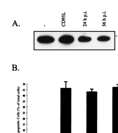
FIG. 6. CD95L expression in host cells is not responsible for SV-induced apoptosis. (A) CV-1 cells were infected with SV (MOI, 10), and FasL was detected by Western blot analysis at 24 and 36 h p.i. Lanes 1 and 2, preparations from uninfected controls (2) and from cells incubated with CD95L (at 12 h), respectively. (B) After infection with SV (MOI, 10), CV-1 cells were incubated with chimeric receptor decoy proteins consisting of the extracellular part of CD95 (aCD95L) fused to IgG-Fc. At 48 h p.i., the cells were analyzed by flow cytometry (propidium iodide staining). Cells with subgenomic DNA content were considered apoptotic. The bar labeled CV-1 indicates the results of the analysis of uninfected controls, and the bar labeled SV represents the results of the analysis of infected cells without chimeric receptor decoy proteins in the supernatant. In our setting concentrations of 5 to 10mg of aCD95L/ml in the supernatant blocked CD95L-induced apoptosis completely. The error bars rep-resent standard deviations.
on November 9, 2019 by guest
http://jvi.asm.org/
[image:4.612.326.531.382.613.2]replication. The twofold difference observed in HepG2 cells,
however, represents only one dilution step and thus may not be
significant (Table 1). A mere quantitation of SV progeny
viri-ons cannot provide information on the functionality, i.e.,
in-fectivity, of such particles. To investigate the functional
impor-tance of apoptosis induction for SV particle maturation, the
TCID50
of progeny virions was determined. Surprisingly, the
calculated ratio of TCID50
per HA unit (TCID50/HA) was
found to be independent of apoptosis induction in host cells
(Table 1). From these data we conclude that apoptotic death of
host cells is not necessary for efficient SV replication or
parti-cle maturation.
DISCUSSION
Mechanisms of virus-induced cell injury play an important
role in our understanding of the pathogenesis of viral
infec-tions. In this study we show that SV infection leads to apoptotic
cell death in all host cell types tested so far. The strong
cyto-pathic effect which can be observed soon after SV infection can
thus be attributed to the induction of a SV-triggered apoptotic
cell death program. Interestingly, the SV leader region at the
39
end of the viral RNA, which lies outside the protein coding
region, has recently been suggested to influence this process;
however, the underlying mechanisms remain to be determined
(18).
Caspases play a central role in the effector phase during
apoptotic cell death. To date, 10 caspases have been described
(11, 32). Still, it is not clear how many different caspases have
to be activated for the successful execution of an apoptosis
program (27, 47). Furthermore, little is known about which
individual caspases are activated during viral infections leading
to apoptosis in host cells. The involvement of CPP32/caspase-3
cleavage in virus-infected cells has been shown recently for
HIV (4), adenovirus (9), and hepatitis C virus (37). To further
understand the role that apoptosis plays in viral infections, it
seems to be crucial to define the virus-triggered steps of the
apoptosis signal transduction cascade. In parallel, this would
potentially open up new possibilities for therapeutic
manipu-lation of these processes. In this context, it has been shown for
HIV-1 that apoptosis inhibition by the broad-spectrum caspase
inhibitor z-VAD-fmk could result in deleterious consequences
for the infected host, such as enhanced viral replication or
stimulation of endogenous virus production in cells derived
from asymptomatic individuals (8).
In the present study we investigated the molecular
mecha-nisms as well as the consequences of SV-induced apoptosis for
virus propagation. Incubation of infected cells with the
broad-spectrum caspase inhibitor z-VAD-fmk prevented apoptosis,
indicating that caspases were involved in the cytopathic effect
of SV. Furthermore, as demonstrated by the processing of its
precursor, CPP32/caspase-3 was found to be activated upon SV
infection. CPP32/caspase-3 is thought to be a critical
execution-er protease, because it is activated by a multitude of apoptosis
stimuli and is able to cleave various cellular substrates (11, 32).
The most striking finding was the observation that, in
addi-tion to CPP32/caspase-3, FLICE/caspase-8 was also
proteolyti-cally processed to its active subunits. FLICE/caspase-8 is
con-sidered to be an important initiator caspase which is able to
activate other caspases, among them caspases 3, 4, 6, and 7
(30). At present, activation of FLICE/caspase-8 has been
dem-onstrated only during death receptor-mediated apoptosis, where
it is recruited and activated at the DISC level. It is conceivable
that activation of FLICE/caspase-8 during SV infection is
me-diated by receptor-dependent or -independent mechanisms.
Because CD95L-neutralizing decoy proteins failed to prevent
SV-induced apoptosis and CV-1 cells were not sensitive to
TNF-induced apoptosis, it is unlikely that CD95 or TNF-R1
was involved. However, the possibility cannot be excluded that
the TRAIL pathway participates in SV-induced cell death. The
cytokine TRAIL (also called Apo2L), which belongs to the TNF
family, has previously been reported to activate the caspase
cascade by a FADD-independent mechanism (35, 42). We will
therefore investigate the possible contribution of TRAIL as
soon as the reagents are available.
New insights into the mechanisms of apoptosis were recently
provided by the cloning of Apaf-1 (apoptosis-activating
fac-tor 1), the mammalian homolog of the ced-4 death gene
from Caenorhabditis elegans (55). At its N terminus, Apaf-1
has sequence similarities to the prodomain of certain caspases.
This region in Apaf-1 serves as a caspase recruitment domain
(CARD) by binding to and activating caspases that have
sim-ilar CARD motifs. Since FLICE/caspase-8 contains a CARD
motif, it is possible that FLICE/caspase-8 is activated upon
binding to Apaf-1. In such a scenario, FLICE/caspase-8
acti-vation would not require interaction with the DISC of TNF-R1
or CD95. In support of this assumption, it was recently found
that the chemotherapeutic agent betulinic acid triggers FLICE/
caspase-8 activation independently of the CD95 pathway and
probably of the TNF-R1 and TRAIL pathways (17). Thus,
future studies will address the question of whether FLICE/
caspase-8 is activated during SV infection by a death
receptor-dependent or -inreceptor-dependent mechanism. Taking the latter into
account, ongoing work has to carefully investigate the role of
FLICE/caspase-8 activation in the caspase death cascade of
SV-infected host cells.
We further investigated the role of apoptosis in SV
replica-tion. Looking at virus progeny release, comparable amounts of
virions were released from SV-infected cells incubated either
with or without the caspase inhibitor z-VAD-fmk. This
dem-onstrates that efficient SV replication does not depend on
apoptosis induction. Therefore, apoptosis inhibition did not
lead to dramatically enhanced viral replication, as was
demon-strated for HIV (1, 8, 38), or to growth limitation, as shown,
e.g., for Semliki Forest virus (40). Our results correspond to
recent observations made with reovirus-infected cells, where
blocking of apoptosis by bcl-2 did not change the virus yield,
suggesting that apoptosis induction is not a major determinant
of viral replication efficacy (36). Taken together, these studies
show that there is no common role for the apoptosis process
during different viral infections; rather it has to be determined
individually for different virus species.
Inhibition of apoptosis by the proto-oncogene bcl-2 during
influenza virus infection leads to an alteration in the
glycosyl-TABLE 1. Effect of caspase activation on virus progeny
release and infectivity
aCell type z-VADb HA units/5310 6cellsc
TCID50d TCIDHA unit50/
20 h 24 h 36 h
CV-1
2
160
640
2,560
640
1
1
320
640
1,280
640
1
HepG2
2
160
640
5,120
320
0.5
1
1,280
1,280
10,240
320
0.25
aResults are representative of three independent experiments.
bThe peptide inhibitor z-VAD-fmk (100mM) was added to the supernatants
after infection with SV (MOI, 10) and every 12 h thereafter.2, not present;1, present.
cDetermined at the indicated times p.i. with SV (MOI, 10).
dDetermined 24 h p.i. with SV (MOI, 10). Later time points p.i. were not
considered due to the short half-life of infective virions in the supernatant of infected cells (11).
on November 9, 2019 by guest
http://jvi.asm.org/
[image:5.612.54.292.92.165.2]ation pattern of hemagglutinin at the viral surface (34). This
hemagglutinin modification impaired infection activity of
prog-eny virions, indicating that the apoptotic death of host cells
is necessary for progeny maturation during influenza virus
infection. To look at the influence of apoptosis on SV progeny
maturation, we determined the infectivity of progeny in
rela-tion to the total number of progeny particles (TCID50
per HA
unit) from cells undergoing apoptosis or being incubated with
the apoptosis inhibitor z-VAD-fmk. As no differences in the
infectivity of SV progeny were found, we have to conclude that
neither SV replication nor maturation depends on apoptotic
death of target cells. Thus, from an evolutionary perspective
one could speculate that SV-triggered apoptosis might serve to
allow the organism to restrict virus progeny release by
elimi-nating infected cells and preventing the establishment of
per-sistent infections.
ACKNOWLEDGMENTS
Michael Bitzer and Florian Prinz contributed equally to this work.
This work was supported by grants from the Deutsche
Forschungs-gemeinschaft (BI 669/3-1), the fortu¨ne-program of the Medical Faculty
at Tu¨bingen (184) and the Bundesministerium fu¨r Bildung,
Wissen-schaft, Forschung und Technologie, Programm Gesundheitsforschung
2000.
REFERENCES
1. Antoni, B. A., P. Sabbatini, A. B. Rabson, and E. White. 1995. Inhibition of apoptosis in human immunodeficiency virus-infected cells enhances virus production and facilitates persistent infection. J. Virol. 69:2384–2392. 2. Aragane, Y., D. Kulms, D. Metze, G. Wilkes, B. Po¨ppelmann, T. A. Luger,
and T. Schwarz.1998. Ultraviolet light induces apoptosis via direct activation
of CD95 (Fas/APO-1) independently from its ligand CD95L. J. Cell Biol.
140:171–182.
3. Badley, A. D., J. A. McElhinny, P. J. Leibson, D. H. Lynch, M. R. Alderson,
and C. V. Paya.1996. Upregulation of Fas ligand expression by human
immunodeficiency virus in human macrophages mediates apoptosis of unin-fected T lymphocytes. J. Virol. 70:199–206.
4. Banki, K., E. Hutter, N. J. Gonchoroff, and A. Perl. 1998. Molecular ordering in HIV-induced apoptosis—oxidative stress, activation of caspases, and cell survival are regulated by transaldolase. J. Biol. Chem. 273:11944–11953. 5. Bitzer, M., U. Lauer, C. Baumann, M. Spiegel, M. Gregor, and W. J.
Neu-bert.1997. Sendai virus efficiently infects cells via the asialoglycoprotein
receptor and requires the presence of cleaved F0precursor proteins for this
alternative route of cell entry. J. Virol. 71:5481–5486.
6. Boldin, M. P., T. M. Goncharov, Y. V. Goltsev, and D. Wallach. 1996. Involvement of MACH, a novel MORT1/FADD-interacting protease, in Fas/APO-1- and TNF receptor-induced cell death. Cell 85:803–815. 7. Brunner, T., R. J. Mogil, D. LaFace, N. J. Yoo, A. Mahboubi, F. Echeverri,
S. J. Martin, W. R. Force, D. H. Lynch, C. F. Ware, and D. R. Green.1995.
Cell-autonomous Fas (CD95)/Fas-ligand interaction mediates activation-in-duced apoptosis in T-cell hybridomas. Nature 373:441–444.
8. Chinnaiyan, A. M., C. Woffendin, V. M. Dixit, and G. J. Nabel. 1997. The inhibition of pro-apoptotic ICE-like proteases enhances HIV replication. Nat. Med. 3:333–337.
9. Chiou, S. K., and E. White. 1998. Inhibition of ICE-like proteases inhibits apoptosis and increases virus production during Adenovirus infection. Vi-rology 244:108–118.
10. Chow, S. C., M. Weis, G. E. Kass, T. H. Holmstrom, J. E. Eriksson, and S.
Orrenius.1995. Involvement of multiple proteases during Fas-mediated
apoptosis in T lymphocytes. FEBS Lett. 364:134–138.
11. Cohen, G. M. 1997. Caspases: the executioners of apoptosis. Biochem. J. 326:1–16.
12. Collins, P. L., R. M. Chanock, and K. McIntosh. 1996. Parainfluenza viruses, p. 1205–1241. In B. N. Fields, D. M. Knipe, and P. M. E. Howley (ed.), Fields virology. Raven Publishers, Philadelphia, Pa.
13. Crowe, P. D., T. L. VanArsdale, B. N. Walter, K. M. Dahms, and C. F. Ware. 1994. Production of lymphotoxin (LT alpha) and a soluble dimeric form of its receptor using the baculovirus expression system. J. Immunol. Methods
168:79–89.
14. Darlington, R. W., A. Portner, and D. W. Kingsbury. 1970. Sendai virus replication: an ultrastructural comparison of productive and abortive infec-tions in avian cells. J. Gen. Virol. 9:169–177.
15. Enari, M., R. V. Talanian, W. W. Wong, and S. Nagata. 1996. Sequential activation of ICE-like and CPP32-like proteases during Fas-mediated apo-ptosis. Nature 380:723–726.
16. Fernandes-Alnemri, T., G. Litwack, and E. S. Alnemri. 1994. CPP32, a novel
human apoptotic protein with homology to Caenorhabditis elegans cell death protein Ced-3 and mammalian interleukin-1 beta-converting enzyme. J. Biol. Chem. 269:30761–30764.
17. Fulda, S., C. Friesen, M. Los, C. Scaffidi, W. Mier, M. Benedict, G. Nunez,
P. H. Krammer, M. E. Peter, and K. M. Debatin.1997. Betulinic acid triggers
CD95 (APO-1/Fas)- and p53-independent apoptosis via activation of caspases in neuroectodermal tumors. Cancer Res. 57:4956–4964. 18. Garcin, D., G. Taylor, K. Tanebayashi, R. Compans, and D. Kolakofsky.
1998. The short Sendai virus leader region controls induction of pro-grammed cell death. Virology 243:340–353.
19. Gavrieli, Y., Y. Sherman, and S. A. Ben Sasson. 1992. Identification of programmed cell death in situ via specific labeling of nuclear DNA fragmen-tation. J. Cell Biol. 119:493–501.
20. Hinshaw, V. S., C. W. Olsen, N. Dybdahl Sissoko, and D. Evans. 1994. Apoptosis: a mechanism of cell killing by influenza A and B viruses. J. Virol.
68:3667–3673.
21. Hoff, H. S., and R. O. Donis. 1997. Induction of apoptosis and cleavage of poly(ADP-ribose) polymerase by cytopathic bovine viral diarrhea virus in-fection. Virus Res. 49:101–113.
22. Jacobsen, M. D., M. Weil, and M. C. Raff. 1996. Role of Ced-3/ICE-family proteases in staurosporine-induced programmed cell death. J. Cell Biol. 133: 1041–1051.
23. Kischkel, F. C., S. Hellbardt, I. Behrmann, M. Germer, M. Pawlita, P. H.
Krammer, and M. E. Peter. 1995. Cytotoxicity-dependent APO-1 (Fas/
CD95)-associated proteins form a death-inducing signaling complex (DISC) with the receptor. EMBO J. 14:5579–5588.
24. Leyrer, S., M. Bitzer, U. Lauer, J. Kramer, W. J. Neubert, and R. Sedlmeier. 1998. Sendai virus-like particles devoid of HN protein infect cells via the human asialoglycoprotein receptor. J. Gen. Virol. 79:683–687.
25. Liang, X. H., S. Mungal, A. Ayscue, J. D. Meissner, P. Wodnicki, D.
Hock-enbery, S. Lockett, and B. Herman.1995. Bcl-2 protooncogene expression in
cervical carcinoma cell lines containing inactive p53. J. Cell Biochem. 57: 509–521.
26. Liu, X., C. N. Kim, J. Pohl, and X. Wang. 1996. Purification and character-ization of an interleukin-1beta-converting enzyme family protease that acti-vates cysteine protease P32 (CPP32). J. Biol. Chem. 271:13371–13376. 27. MacFarlane, M., K. Cain, X. M. Sun, E. S. Alnemri, and G. M. Cohen. 1997.
Processing/activation of at least four interleukin-1beta converting enzyme-like proteases occurs during the execution phase of apoptosis in human monocytic tumor cells. J. Cell Biol. 137:469–479.
28. Medema, J. P., C. Scaffidi, F. C. Kischkel, A. Shevchenko, M. Mann, P. H.
Krammer, and M. E. Peter.1997. FLICE is activated by association with the
CD95 death-inducing signaling complex (DISC). EMBO J. 16:2794–2804. 29. Muzio, M., A. M. Chinnaiyan, F. C. Kischkel, K. O’Rourke, A. Shevchenko,
J. Ni, C. Scaffidi, J. D. Bretz, M. Zhang, R. Gentz, M. Mann, P. H. Krammer,
M. E. Peter, and V. M. Dixit.1996. FLICE, a novel FADD-homologous
ICE/CED-3-like protease, is recruited to the CD95 (Fas/APO-1) death-inducing signaling complex. Cell 85:817–827.
30. Muzio, M., G. S. Salvesen, and V. M. Dixit. 1997. FLICE induced apoptosis in a cell-free system. Cleavage of caspase zymogens. J. Biol. Chem. 272: 2952–2956.
31. Nicholson, D. W., A. Ali, N. A. Thornberry, J. P. Vaillancourt, C. K. Ding, M.
Gallant, Y. Gareau, P. R. Griffin, M. Labelle, Y. A. Lazebnik, et al.1995.
Identification and inhibition of the ICE/CED-3 protease necessary for mam-malian apoptosis. Nature 376:37–43.
32. Nicholson, D. W., and N. A. Thornberry. 1997. Caspases: killer proteases. Trends Biochem. Sci. 22:299–306.
33. Nicoletti, I., G. Migliorati, M. C. Pagliacci, F. Grignani, and C. Riccardi. 1991. A rapid and simple method for measuring thymocyte apoptosis by propidium iodide staining and flow cytometry. J. Immunol. Methods 139: 271–280.
34. Olsen, C. W., J. C. Kehren, N. R. Dybdahl-Sissoko, and V. S. Hinshaw. 1996.
bcl-2 alters influenza virus yield, spread, and hemagglutinin glycosylation.
J. Virol. 70:663–666.
35. Pan, G., J. Ni, Y.-F. Wei, G.-L. Yu, R. Gentz, and V. M. Dixit. 1997. An antagonist decoy receptor and a death domain-containing receptor for TRAIL. Science 277:815–818.
36. Rodgers, S. E., E. S. Barton, S. M. Oberhaus, B. Pike, C. A. Gibson, K. L.
Tyler, and T. S. Dermody.1997. Reovirus-induced apoptosis of MDCK cells
is not linked to viral yield and is blocked by Bcl-2. J. Virol. 71:2540–2546. 37. Ruggieri, A., T. Harada, Y. Matsuura, and T. Miyamura. 1997. Sensitization
to Fas-mediated apoptosis by hepatitis C virus core protein. Virology 229:68– 76.
38. Sandstrom, P. A., D. Pardi, C. S. Goldsmith, D. Chengying, A. M. Diamond,
and T. M. Folks.1996. bcl-2 expression facilitates human immunodeficiency
virus type 1-mediated cytopathic effects during acute spreading infections. J. Virol. 70:4617–4622.
39. Sastry, K. J., M. C. Marin, P. N. Nehete, K. McConnell, A. K. el Naggar, and
T. J. McDonnell.1996. Expression of human immunodeficiency virus type I
tat results in down-regulation of bcl-2 and induction of apoptosis in hema-topoietic cells. Oncogene 13:487–493.
40. Scallan, M. F., T. E. Allsopp, and J. K. Fazakerley. 1997. bcl-2 acts early to
on November 9, 2019 by guest
http://jvi.asm.org/
restrict Semliki Forest virus replication and delays virus-induced pro-grammed cell death. J. Virol. 71:1583–1590.
41. Shen, Y., and T. E. Shenk. 1995. Viruses and apoptosis. Curr. Opin. Genet. Dev. 5:105–111.
42. Sheridan, J. P., S. A. Marsters, R. M. Pitti, A. Gurney, M. Skubatch, D.
Baldwin, L. Ramakrishnan, C. L. Gray, K. Baker, W. I. Wood, A. D.
God-dard, P. Godowski, and A. Ashkenazi.1997. Control of TRAIL-induced
apoptosis by a family of signaling and decoy receptors. Science 277:818–821. 43. Sigmund, M., H. Einberger, and W. J. Neubert. 1988. Simple method for rapid and highly sensitive detection of antiviral-antibodies in serum and cerebrospinal fluid of small laboratory animals. J. Virol. Methods 22:231– 238.
44. Slee, E. A., H. Zhu, S. C. Chow, M. MacFarlane, D. W. Nicholson, and G. M.
Cohen.1996. Benzyloxycarbonyl-Val-Ala-Asp (OMe) fluoromethylketone
(Z-VAD-FMK) inhibits apoptosis by blocking the processing of CPP32. Biochem. J. 315:21–24.
45. Steller, H. 1995. Mechanisms and genes of cellular suicide. Science 267: 1445–1449.
46. Strack, P. R., M. W. Frey, C. J. Rizzo, B. Cordova, H. J. George, R. Meade,
S. P. Ho, J. Corman, R. Tritch, and B. D. Korant.1996. Apoptosis mediated
by HIV protease is preceded by cleavage of Bcl-2. Proc. Natl. Acad. Sci. USA
93:9571–9576.
47. Takahashi, A., and W. C. Earnshaw. 1996. ICE-related proteases in apopto-sis. Curr. Opin. Genet. Dev. 6:50–55.
48. Takizawa, T., R. Fukuda, T. Miyawaki, K. Ohashi, and Y. Nakanishi. 1995. Activation of the apoptotic Fas antigen-encoding gene upon influenza virus
infection involving spontaneously produced beta-interferon. Virology 209: 288–296.
49. Teodoro, J. G., and P. E. Branton. 1997. Regulation of apoptosis by viral gene products. J. Virol. 71:1739–1746.
50. Tewari, M., L. T. Quan, K. O’Rourke, S. Desnoyers, Z. Zeng, D. R. Beidler,
G. G. Poirier, G. S. Salvesen, and V. M. Dixit.1995. Yama/CPP32 beta, a
mammalian homolog of CED-3, is a CrmA-inhibitable protease that cleaves the death substrate poly(ADP-ribose) polymerase. Cell 81:801–809. 51. Tropea, F., L. Troiano, D. Monti, E. Lovato, W. Malorni, G. Rainaldi, P.
Mattana, G. Viscomi, M. C. Ingletti, M. Portolani, C. Ceremelli, A.
Cossa-rizza, and C. Franceschi.1995. Sendai virus and herpes virus type 1 induce
apoptosis in human peripheral blood mononuclear cells. Exp. Cell Res. 218: 63–70.
52. Ubol, S., S. Park, I. Budihardjo, S. Desnoyers, M. H. Montrose, G. G.
Poirier, S. H. Kaufmann, and D. E. Griffin.1996. Temporal changes in
chromatin, intracellular calcium, and poly(ADP-ribose) polymerase during Sindbis virus-induced apoptosis of neuroblastoma cells. J. Virol. 70:2215– 2220.
53. Westendorp, M. O., R. Frank, C. Ochsenbauer, K. Stricker, J. Dhein, H.
Walczak, K. M. Debatin, and P. H. Krammer.1995. Sensitization of T cells
to CD95-mediated apoptosis by HIV-1 Tat and gp120. Nature 375:497–500. 54. Yuan, J., S. Shaham, S. Ledoux, H. M. Ellis, and H. R. Horvitz. 1993. The C. elegans cell death gene ced-3 encodes a protein similar to mammalian interleukin-1 beta-converting enzyme. Cell 75:641–652.
55. Zou, H., W. J. Henzel, X. Liu, A. Lutschg, and X. Wang. 1997. Apaf-1, a human protein homologous to C. elegans ced-4, participates in cytochrome c-dependent activation of caspase-3. Cell 90:405–413.
on November 9, 2019 by guest
http://jvi.asm.org/