CUTANEOUS MANIFESTATIONS OF SYSTEMIC
LUPUS ERYTHEMATOSUS
Dissertation submitted to
THE TAMILNADU DR.M.G.R MEDICAL UNIVERSITY
In partial fulfillment of the regulations
For the award of the degree of
M.D.BRANCH- XII A
DERMATOLOGY, VENEREOLOGY AND LEPROSY
MADRAS MEDICAL COLLEGE
CHENNAI, INDIA
.
CERTIFICATE
This is to certify that the dissertation entitled “
CUTANEOUS
MANIFESTATIONS OF SYSTEMIC LUPUS ERYTHEMATOSUS”
is a bonafide
original work of
Dr. D. NITHYA GAYATHRIDEVI
, in partial fulfillment of the
requirements for
M.D.BRANCH XIIA (DERMATOLOGY, VENEREOLOGY AND
LEPROSY)
examination of the Tamilnadu Dr. M.G.R. Medical University to be held in
March 2010.
.
DEAN
Madras Medical College Chennai-600 003 PROFESSOR and HOD
Department of Dermatology & Leprosy
DECLARATION
I,
DR. D. NITHYA GAYATHRIDEVI,
solemnly declare that dissertation titled,
“
CUTANEOUS
MANIFESTATIONS
OF
SYSTEMIC
LUPUS
ERYTHEMATOSUS”
is a bonafide work done by me at Department of Dermatology
and Leprosy, Madras Medical College, Chennai-3 during the period of August 2007 to
September 2009 under the supervision of my
Prof. DR.D.PRABHAVATHY, M.D, D.D,
Professor and HOD, The Department of Dermatology and Leprosy, Madras Medical
College, Chennai. The dissertation is submitted to Tamilnadu Dr. M.G.R. Medical
University, towards partial fulfillment of requirement for the award of
M.D. Degree
(Branch-XII A) in DERMATOLOGY, VENEREOLOGY AND LEPROSY
.
Place: Chennai
SPECIAL ACKNOWLEDGEMENT
My sincere thanks to
Prof. Dr.J.Mohana Sundaram
,
M.D., D.N.B., PhD
DEAN,
Madras Medical College
for allowing me to do this
ACKNOWLEDGEMENT
I am gratefully indebted to
Prof. Dr. D. PRABHAVATHY, M.D., D.D.,
Professor
and Head, Department of Dermatology and Leprosy for her invaluable guidance,
motivation and help throughout the study. I would like to express my sincere and
heartful gratitude to
Prof. Dr. N. KUMAR, M.D., D.V., DMRD,
Director in-charge,
Institute of Venereology.
I wish to thank Prof.
Dr. B.PARVEEN, M.D., D.D.,
Former Professor and Head,
Department of Dermatology and Leprosy for her constant support and motivation.
I wish to thank Prof. Dr
. V.S. DORAIRAJ, M.D., D.V., & Prof. Dr.
GAJENDRAN, M.D., D.V.,
Former Directors, Institute of Venereology for their
constant support and motivation.
I express my gratefulness to Prof.
Dr. V.SOMASUNDARAM, M.D., D.D.,
Professor and Head of Department of Occupational Dermatology and Contact Dermatitis
for his constant motivation and guidance. I thank
Prof. Dr.
V.THIRUNAVUKKARASU, M.D., D.D.,
Additional Professor, Department of
Occupational Dermatology and Contact Dermatitis for his benevolent help and support.
I am very grateful to
Prof.Dr.S.JAYAKUMAR M.D.,D.D.,
Additional Professor,
Department of Dermatology for his invaluable guidance and help. I sincerely thank
Prof.Dr.C.JANAKI,M.D.,D.D.,
Additional Professor, Department of Dermatology
(Mycology) for her priceless support.
Additional Professor (Leprosy), Department of Dermatology &
Prof. Dr.
JEYAKUMARI JEEVAN,M.D,D.D.,
Former Professor of Leprosy for their support.
I specially thank
Dr.A.HAMEEDULLAH,M.D.,D.D.,
Assistant Professor,
Department of OCD for his immense guidance and support.
I incline to thank
Dr.G.K.Tharini,M.D., Dr.N.Hema, M.D(D.V.L)., Dr.Samuel
Jayaraj Daniel, M.D (D.V.L)., Dr.S.Anupama, D.D.,
Assistant Professors, Department
of Dermatology for their kind support and encouragement.
I thank
Dr.S.Kumaravel,M.D.,D.D., Dr.J.Manjula,M.D.,D.N.B.,
and
Dr.Afthab
Jamila Wahab,M.D.,D.D.,
Assistant Professors, Department of Occupational
Dermatology and Contact Dermatitis for their support and help.
My sincere thanks to
Dr. V. Thirunavukkarasu, M.D.,D.V.,
Dr.K.Venkateswaran, M.D., D.V., Dr.S.Thilagavathy, M.D., D.V., Dr.P.Mohan,
M.D., D.V., Dr. S. Arunkumar, M.D., D.V., Dr. S. Kalaivani, M.D., D.V., Dr. S.
Prabhakar, M.D(D.V.L)., Dr. V.N.S.Ahamed sheriff M.D (D.V.L).,
Assistant
Professors, Institute of Venereology for their help and suggestions.
I am also thankful to
Dr. R. Priyavathani, M.D.,
Dr.V.Anandan,M.D(Derm).,D.C.H., D.N.B., & Dr.S.Thilagavathy, M.D., D.V
for
their guidance and support.
I am thankful to
Prof.Dr.R.Porkodi,M.D.,D.M.,
Professor and Head of
Department of Rheumatology,
Prof.J.Sasikala,M.D.,
Professor of Immunology and
Rheumatology, GGH, Chennai for their support and guidance.
I am thankful to Prof
.Dr.C.Balachandran,M.D.,
Professor and Head of
Department, Department of Skin & STD, Kasturba Medical College, Manipal,
Karnataka for his help and support.
I duly acknowledge the paramedical staff and my collegues for their help and
favour.
CONTENTS
Sl.No. Title Page
No.
1. INTRODUCTION 1
2. REVIEW OF LITERATURE 2
3. AIM OF THE STUDY 45
4.
MATERIALS AND METHODS
465. OBSERVATIONS AND RESULTS 50
6. DISCUSSION 61
7.
8.
SUMMARY AND CONCLUSIONS
ANNEXURES
REFERENCES
PROFORMA
MASTER CHART
KEY TO MASTER CHART
ABBREVIATIONS
INTRODUCTION
HIPPOCRATES, ROGERIUS, PARACELSUS, MANARDI, AMATUS LUSITANUS,
SENNERT etc. are some of the early renowned Physicians who have described "LUPUS"
which is derived from the Latin word which means "WOLF"1 depicting its nature of
destruction. The ulcerated skin lesions typical of this disease signify this feature as it "bites,
eats away and destroys".
Lupus erythematosus (LE) was identified only as a cutaneous disease, until a century
ago when emphasis was transferred from the integument to include visceral manifestations. At
the beginning of this century blood vessels and connective tissues distributed throughout the
body came to be implicated in the pathogenesis, which lead to the concept of "multisystem
malady”.2 In 1942, KLEMPERER et al 3 were struck by the many, morphological features that
were common to diseases as distinct as Lupus erythematosus, Scleroderma, Dermatomyositis,
Rheumatoid Arthritis, Acute Rheumatic Fever and Poly Arteritis Nodosa and classified them as
"Collagen disease" or "Collagenosis". The discovery of auto antibodies to various cellular
components of different tissue in these diseases has of late given place to the concept of
REVIEW OF LITERATURE
HISTORICAL BACKGROUND:
In 1826 RAYER1 described lupus erythematosus (LE) as "Flexus Sebaceui"; in 1828
BIETT4 described the condition as "Erythema centrifuge". In 1845 HEBRA1 described it as
"Seborrhea congestiva" in which the term "butterfly rash” was used for the first time. In 1851,
CAZENAVE5 introduced the term "lupus erythematoides" meaning red wolf, in order to
distinguish this disease from lupus vulgaris. KAPOSI4 differentiated LE into two forms namely
the discoid form and lupus erythematosus disseminatus.
KAPOSI4 was the first to describe the systemic manifestation of LE (1872). WILLIAM
OSLER1 in 1903 discussed the significance of systemic manifestations and its relationships to
the cutaneous lesions.
In 1925 DROCQ6 classified lupus erythematosus (LE) into 3 major categories. In 1934
O’LEARY6 classified LE into chronic discoid, sub acute disseminate and acute disseminate
types. The discovery “LE cell phenomenon" in 1948 by HARGRAVES et al1 paved way to the
subsequent discoveries of various autoantibodies. The finding of immunoglobulin deposition at
the DEJ of LE lesions by BURNHAM et al7 in 1963 further strengthened autoimmune etiology.
In 1971, COHEN and CANOSO8 proposed the American Rheumatic Association (ARA)
criteria for the classification of systemic lupus erythematosus (SLE), which following criticism
was revised in 1982 by TAR EM et al.9 In 1994 CASCIOLA ROSEN et al first demonstrated
autoantigens that are targeted in SLE. WORTH et al demonstrated presence of 30SA TNF
promoter polymorphism in SLE.
DEFINITION:
LE is a systemic autoimmune disorder associated with polyclonal B-cell activation,
spectrum ranging on one end from DLE to SLE on the other end. The term SLE has been used
in the past synonymously with LE to all patients suffering from this autoimmune disorder. But
in our discussion SLE will be used to refer only to the patient with systemic manifestations.
AETIOLOGY AND PATHOGENESIS:
The regular manifestations of specific DLE lesions in systemic lupus erythematosus,
common histological and laboratory abnormalities, immunoglobulin depositions in the
involved skin, the transition of discoid lupus erythematosus to systemic lupus erythematosus,
etc.10-13 have lead to the concept that DLE and SLE have common pathogenetic mechanisms in
which discoid lupus erythematosus (DLE) represents one polar expression with minimal
immunological alteration and the systemic lupus erythematosus on the other end with maximal
immunological alterations.11,12
The current concepts suggest that this is a multifactorial disorder in which there is
profound disturbance of immune mechanism provoked by constitutional and environmental
factors.
I. CONSTITUTIONAL FACTORS:
GENETIC PREDISPOSITION: A genetic role is suggested by the following
observations; 1)Familial incidence (5% in the first degree relatives) 14 , 2) Concordance rate in
identical twins is 65% , 3)The higher incidence of other autoimmune diseases in the patients
and among family members,16 4) Increased association of HLA B8, B7 and DR3 in idiopathic
type 17 and increased HLA DR6Y in drug induced lupus erythematosus, 5)The occurrence of
linkage / disequilibrium among individual alleles at neighboring loci in some which is referred
as “persistent haplotype” 6) Increased allele of TNF Alfa, IL-I, HSP 70-2 polymorphism and
AGE, SEX AND HORMONAL FACTORS:13 There is an increased oestrogen levels as there is an
abnormal oestrogen metabolism in patients with SLE, this leads to increased number of self
reactive lymphocytes and increased number of B cells which has high affinity of recognizing
self DNA. This observation has a bearing on the propensity for females to develop systemic
lupus erythematosus and the higher incidence between menarche and menopause and during
the 3rd trimester of pregnancy, when the circulating oestrogen levels are high.22 Women in
childbearing age have 15 times more preponderance for SLE than men.21 Female patients with
DLE have noted exacerbation of the lesions during the premenstrual and menstrual periods.13
Prolactin is immunogenic and is associated with high Anti DNA levels.
II. ENVIRONMENTAL FACTORS:
Environmental factors like viruses, drugs, UV light, trauma by interfering with the
immune mechanism can precipitate or exacerbate SLE.
VIRUSES: The demonstration of Paramyxovirus like cytoplasmic tubular structure in
glomerular capillaries23 and endothelial cells of dermal blood vessels by Ken Hashimoto et al24
suggested a possible viral aetiology. The pathogenesis behind proposed viral etiology include;
1) Virus infected cells while getting apoptised, express their Ro/SSA and related antigens
towards the surface25. 2) An alteration of class II proteins by viral antigens, may result in
preferential activation by altered self MHC class II reactive cells26. 3) By altering MHC class II
expression there is also a molecular mimicry by viruses and auto antibodies are produced27.
Other corroborative findings include precipitation or exacerbation of the disease following viral
infection, and increased titres of antibodies against various viruses like REO viral RNA,
exaggeration28,29.
DRUGS: A number of drugs have been incriminated either to produce or unmask LE. These
are listed in Table-1. Hydralazine induced SLE is dose dependent, dosage <50mg/day does not
Table -1
DRUGS INDUCING SLE LIKE SYNDROME
NO TYPE OF DRUGS NAME OF THE DRUG
1. Cardio vascular Procainamide, quinidine
2. Antimicrobial INH, penicillins, sulphonamides, griesofulvin, streptomycin, tetracyclins, nitrofurantoin
3. Antihypertensives Hydralazine, methyldopa, reserpine, atenolol, captopril, labetalol
4. Antithyroid Propylthiouracil
5. Psychotropic Chlorpromazine, lithium
6. Antiepileptic Phenytoin, sodium valproate, ethosuximide, carbamazepine, clobazam
7. Miscellaneous d-penicillamine, phenylbutazone, gold salts, allopurinol, PAS,ibuprofen,OCP’s
Possible mechanisms hypothesized are:30 1) Structural similarity of the drug to the
purine base of DNA, with subsequent cross-reactivity in the induction of antibody production
to DNA, 2) Interaction of drugs with nuclear antigens expressing a new determinant to evoke T
lymphocytes help B lymphocytes in producing antibodies, 3)Inhibition of T suppressor cell
activity, 4) By genetic predisposition through an immune response gene and through slow
acetylation, 5) Drugs may induce T-cell DNA hypomethylation, may cause increased auto
reactivity of lymphocytes, 6)increased keratinocyte apoptosis, expose intracellular peptides on
epidermal surface, enhances pro inflammatory cytokines like IFN alfa and TNF alfa30,31.
UV LIGHT: Incidence of photosensitivity among SLE patients ranges from 32 to 37%13, in
patients with DLE from 5 to 40%13 and in patients with sub acute cutaneous LE upto 50%11, 31. It
the degree of increased activity is related to the duration of sun exposure.13 Kesten proposed
that the local reaction to UV injury represents an isomorphic or koebner Phenomenon.25 DLE
lesions in patients with SLE have been explained to be the result of excessive damage induced
by sister chromatid inducing agents, the action of which is increased by exposure to near UV
light. Recent studies using monochromatic sources have confirmed that the photo reactivity lies
within the sunburn range (UVB). 13
Pathogenesis stated behind UV light includes; 1) UV light causes apoptosis of
keratinocytes, which in turn makes previously cryptic peptides available for immune
surveillance leading to self immunity and loss of tolerance, 2) UVB displaces intracellular
Ro/SSA, LA/SSB, Calreticulin to their cell surfaces, 3) UVB induces release of CCL-27, a
chemokine, activates autoreactive T Cells, IFN alfa and dendritic cells leading on to alteration
of DNA, 4) UV light affects immunoregulatory cells which normally help in suppressing
abnormal cutaneous inflammation27,28,16,.
OTHER FACTORS: Hemolytic anaemia with increased anti dsDNA antibodies has been
documented following the ingestion of sprouts, seeds and dietary supplements containing
L-canavanin. Heavy metals like cadmium, mercury, gold, silica, trichlorethylene has also been
associated with SLE. Lipogenic aromatic amines present in tobacco can induce SLE27,28. Lodin
reported DLE following trauma (Koebner phenomenon) that included chemical burns,
diathermy, scars of herpes zoster and exposure to X-rays.
III. IMMUNOLOGICAL FACTORS:
Complex interaction between various cells like B and T lymphocytes, dentritic cells,
complements defects, apoptotic abnormalities, receptor defects, and aberration in chemokine
T LYMPHOCYTE: It is postulated that in LE, varying degree of impairment of the
suppressor T lymphocytes occur, leading to defect in tolerance to self antigen and over activity
of B lymphocyte, followed by the production of antibodies to a wide variety of antigens,
preferably to nuclear proteins.CTL cell has role both in induction and expansion phases by its
increased number and surface DR antigens23,32.
B LYMPHOCYTE: B Cells are involved in expansion phase of pathogenesis. Production of
auto antibodies against nuclear antigens and immune complexes is the hallmark of SLE, which
may causes tissue damage by causing direct cell death, cellular activation, opsonization and
blocking of target molecule function. The excess B cell is hypothesized to result from;
1)Primary B cell defect, 2)excess helper T Cell function, 3)increased polyclonal B cell
activation and 4) genes for high responsiveness to certain auto antigens or antigens which
cross-react with auto antigens. RES is less efficient in clearing circulating immune complexes,
which accounts for the widespread tissue injury27,28,32,33.
DEFECTIVE APOPTOSIS: In SLE apoptosis of peripheral blood mononuclear cells is
increased and defective. Normally apoptotic cells remain intact, but in SLE apoptotic cells
dissolute and release nucleoprotein there by exposing the antigen. The above factor also adds
up to inherited defect of apoptotic body clearance.34
IDIOTYPE AND ANTI IDIOTYPE ANTIBODY: Idiotype is an antigenic determinant present
on the variable region of an antibody formation. Antibodies are produced against this idiotype.
So there is dysregulation of production of immunoglobulins bearing these idiotypes35.
COMPLEMENT DEFECTS:36 There is both inherited and acquired complement deficiency are
associated with disease. Normally complement binds to apoptotic cells which are then disposed
by house keeping macrophages but when complement proteins are deficient there is a defective
viral neutralization and linkage of gene foci controlling complement synthesis with those
controlling the immune response.36
ROLE OF DENDRITIC CELLS AND IFN ALFA: Immature dendritic cells normally perform
a watch dog role by capturing self antigen and keep self antigen in check. In SLE there is more
plasmacytoid maturation of DC’s via IFN stimulation. These mature DC’s unlike immature
DC’s present antigen to auto reactive T-cells, there by producing antibodies37.
TOLL LIKE RECEPTORS: Circutating DNA / Anti DNA complex trigger TLR signaling
which induces proliferation of auto reactive B-Cells, IFN secretion from DC’s30,31,18.
TNF: TNF alfa secreted in response to UVR induce HLA – DR expression, stimulate NF
Kappa which upregulates proinflammatory cytokines and adhesion molecules30,18.
Other corroborative finding include the following: Detection of circulating immune
complexes,36 the deposition of immunoglobulin and complement in different tissues like
kidney, skin etc.,10 the pattern of disease expressivity correlating with the detection of specific
autoantibodies, lupus like rash in graft versus host disease,11 lymphopenia,25 positive
intradermal test on patients with autologus WBC, marked depression or absence of skin
reactivity to all test antigens and finally, failure of sensitization to DNCB by majority of the
patients with SLE.
The immunological aberration increases, as the spectrum moves from DLE to SLE. The
possible explanation would be the presence of isolated cellmediated autoimmunity in DLE,
without producing immune complexes. But, in patients with SLE, antigen antibody complexes
may be the reason for multi organ involvement. However, it is the dichotomy in T suppressor
cells control over the B-lymphocyte that could best explain the polar expression, though the
Table-2
I. ANTIBODIES TO DNA AND HISTONES
A. ANTIBODY REACTIVE WITH DOUBLE STRANDED DNA ONLY (dsDNA)
B. AGAINST ds AND ss DNA – 60 TO 70% OF PATIENTS WITH SLE
C. AGAINST ss DNA ONLY
D. AGAINST HISTONES (H, AH2A, H2B, H3 AND H4) -DRUG
INDUCED LE (95-100%), SLE 30%
II ANTIBODIES TO NON-HISTONE NUCLEAR PROTEINS AND
RNA PROTEINS
Sm ANTIGEN – SLE IN 30 TO 40% - MARKER ANTIBODY
NUCLEAR RNP (N RNP) – SLE 30 TO 40%
SSA/RO ANTIGEN – SLE 30-40%
SSB/LA ANTIGEN – SLE 10-15%
PROLIFERATING CELL NUCLEAR ANTIGEN (PCNA) – SLE
5% - 10%
III KU – SLE/PM/SCLERODERMA – OVERLAP SYNDROME
Table-3
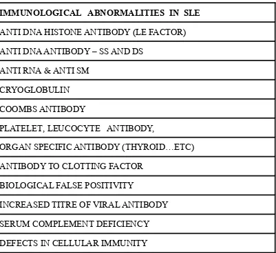
IMMUNOLOGICAL ABNORMALITIES IN SLE
ANTI DNA HISTONE ANTIBODY (LE FACTOR)
ANTI DNA ANTIBODY – SS AND DS
ANTI RNA & ANTI SM
CRYOGLOBULIN
COOMBS ANTIBODY
PLATELET, LEUCOCYTE ANTIBODY,
ORGAN SPECIFIC ANTIBODY (THYROID…ETC)
ANTIBODY TO CLOTTING FACTOR
BIOLOGICAL FALSE POSITIVITY
INCREASED TITRE OF VIRAL ANTIBODY
SERUM COMPLEMENT DEFICIENCY
DEFECTS IN CELLULAR IMMUNITY
CLASSIFICATION:
The classifications of LE by Brocq in 1925, O'Leary' in 1934, Urbach and Thomas in
1939 and Wilson and Jordon4 in 1950 can at best be considered as anecdotal importance, since
there were lacunae in the clinical and histopathological correlations. The exercise of
classification is further complicated by the fact that visceral involvement may or may not be
accompanied by skin changes. Based entirely upon "clinical and specific histopathological
findings” of LE, Gilliam et al11 classified cutaneous lesions of LE into,1) LE specific (or)
cutaneous Lupus Erythematosus (CLE) showing specific histopathological findings and 2) LE
Table -4
I. CUTANEOUS LUPUS ERYTHEMATOSUS (LE SPECIFIC SKIN LESIONS)
A. CHRONIC CUTANEOUS LUPUS ERYTHEMATOSUS : (CCLE) 1. LOCALISED DISCOID LUPUS ERYTHEMATOSUS (LDLE)
(LESIONS ARE CONFINED TO HEAD AND NECK) 2. GENERALISED OR DISSEMINATED DISCOID LUPUS
ERYTHEMATOSUS (DDLE) –(LESIONS ARE PRESENT ABOVE AND BELOW THE NECK)
3. HYPERTROPHIC OR VERRUCOUS DISCOID LUPUS ERYTHEMATOSUS
4. LUPUS ERYTHEMATOSUS PROFUNDUS (LUPUS ERYTHEMATOSUS PANNICULITIS)
B SUBACUTE CUTANEOUS LUPUS ERYTHEMATOSUS (SCLE) 1. PAPULOSQUAMOUS (PSORIASIFORM) SCLE
2. ANNULAR – POLYCYCLIC (OCCASIONALLY VESICULAR) SCLE C. ACUTE CUTANEOUS LUPUS ERYTHEMATOSUS (ACLE)
1. FACIAL (MALAR) ERYTHEMA
2. WIDESPREAD ERYTHEMA OF FACE. SCALP, NECK, UPPER CHEST, SHOULDERS, EXTENSOR SURFACES OF ARMS AND BACK OF HANDS
3. BULLOUS OR TEN LIKE ACLE
II. NON SPECIFIC BUT DISEASE RELATED SKIN LESIONS IN PATIENTS WITH LUPUS ERYTHEMATOSUS
A. VASCULAR LESIONS
1. TELANGIECTATIC LESIONS (45-65%) 2. DERMAL VASCULITIS (10-20%)
3. THROMBOPHLEBITIS (15-20%)
4. RAYNAUD’S PHENOMENON (15-20%) 5. LIVEDO RETICULARIS (10%)
6. CHRONIC ULCERS (2-8%)
7. RHEUMATOID NODULES (5-10%)
9. DEGOS LIKE DERMAL INFARCTS OR ATROPHIE BLANCHE (LESS THAN 5%)
B ALOPECIA (40-60%)
1. FRONTAL (“LUPUS HAIR”) 2. DIFFUSE (NON SCARRING)
C MUCOUS MEMBRANE LESIONS (7%) D PIGMENTARY ABNORMALITIES (10%)
E. SCLERODACTYLY (10%)
F CALCINOAIS CUTIS (RARE) G URTICARIA (7 – 14%)
H BULLOUS LESIONS (LESS THAN 5%)
This classification is again incomplete as it does not encompass other variants of
cutaneous LE namely, chilblain lupus,10 LE telangiectoides,10 LE profundus et hypertrophicus,10
LE tumidus,39 and LE/LP overlap syndrome.
On the other hand, Baeur and Orfanos preferred to use the general term "LE
syndrome" which comprised all types of LE disease and regarded the individual clinical picture
as LE subsets. In their classification, classical LE subsets included the chronic DLE, the
disseminated LE and systemic lupus erythematosus (SLE). Among the rare LE subsets few
were bullous LE, LE profundus, Mixed Connective Tissue Disease, Rowell syndrome, Drug
induced LE, Urticarial vasculitic LE, and ANA negative LE39 were recently recorded in the
literature.
CRITERIA FOR THE CLASSIFICATION OF SLE:
In 1964, Ropes40 proposed the following criteria;
1) A cutaneous eruption consistent with LE. 2) Renal involvement. 3) Serositis and 4)
The presence of 3 of the above mentioned 4 manifestations were considered for the
classification of SLE. The diagnosis of which however required confirmatory laboratory tests.
In 1971, American Rheumatism Association8 proposed the preliminary criteria for the
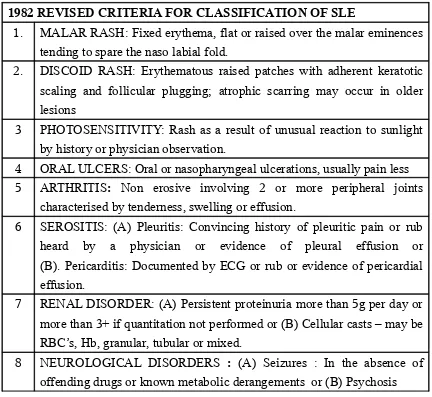
classification of SLE, which, after criticism was revised in 1982 by Tan Em et al9 (Table-5).
Among the 6 cutaneous criteria initially said, Malar flush, discoid rash, photosensitivity and
oral ulcers were retained and the Raynaud's phenomenon and alopecia were excluded. The
[image:23.612.49.481.295.692.2]revised criteria, however, was also subjected to criticism. (Table 5)
Table – 5
1982 REVISED CRITERIA FOR CLASSIFICATION OF SLE
1. MALAR RASH: Fixed erythema, flat or raised over the malar eminences tending to spare the naso labial fold.
2. DISCOID RASH: Erythematous raised patches with adherent keratotic scaling and follicular plugging; atrophic scarring may occur in older lesions
3 PHOTOSENSITIVITY: Rash as a result of unusual reaction to sunlight by history or physician observation.
4 ORAL ULCERS: Oral or nasopharyngeal ulcerations, usually pain less 5 ARTHRITIS: Non erosive involving 2 or more peripheral joints
characterised by tenderness, swelling or effusion.
6 SEROSITIS: (A) Pleuritis: Convincing history of pleuritic pain or rub heard by a physician or evidence of pleural effusion or (B). Pericarditis: Documented by ECG or rub or evidence of pericardial effusion.
7 RENAL DISORDER: (A) Persistent proteinuria more than 5g per day or more than 3+ if quantitation not performed or (B) Cellular casts – may be RBC’s, Hb, granular, tubular or mixed.
9 HEMATOLOGICAL DISORDER : (A) Hemolytic anemia with reticulocytosis (B) Leukopenia < 4000/cu mm on 2 or more occasions or (C) lymphopenia < 1500 / cu mm on 2 or more occasions or (D) Thrombocytopenia <1,00,000/cu mm in the absence of offending drugs 10 IMMUNOLOGICAL DISORDER : (A) Positive LE cell or (B) Anti
DNA antibody in abnormal titre or (C) presence of anti Sm anti body or (D) False positive VDRL for atleast 6 months and confirmed by TPI or FTA-ABS
11 Abnormal ANA by immunoflouresence in the absence of drugs
For the purpose of identifying in clinical studies a person shall be said to have SLE, is
any four or more of the 11 criteria are present serially or simultaneously, during any interval of
observation.
INCIDENCE:
SLE is not a rare disease, but neither is it a common one. The average annual incidence
of SLE has been estimated to be 27.5 per million population for white females and 75.4 per
million for black females.13 SCLE represents 9% of all cases of cutaneous LE12 and DLE
represents the commonest among the cutaneous LE.42,46 Other variants of cutaneous LE seems
to be extremely rare. 43,44,45,46
AGE INCIDENCE:
Average age of onset is between the 2nd and 5th decade,10,13 the peak incidence being
between 3rd and 4th decade in DLE,10,13 It occurs rarely in children47,48, on the other extreme,
DLE has also started at 83 years of age.10 In the Indian study by Pandh et al49 the majority of
SEX INCIDENCE:
Females are predominantly affected and the female to male ratio increases as the
spectrum moves from DLE to SLE. 10,13,25 In DLE the ratio ranges from 1.2:1 in the localised
type49 upto 3.8:1 in the disseminated type.50 70% of the patients are females in the SCLE type31
and the ratio in ACLE ranges from 9:110,13 upto 15:1 in the childbearing age group. 21
RACE:
SLE is three times more common in whites than blacks. Blacks with SLE more
commonly have Sm and RNP antibodies, DLE lesions, Serositis and internal organ damage.
SYMPTOMS:
Patients with DLE are often asymptomatic, but for the most common symptom cum
precipitating factor is photosensitivity. The early papular lesion of DLE may however be
slightly pruritic,13 and intolerable itching is a symptom peculiar to scalp involvement.51,52 Pain
on scratching in lesions of DLE has been explained by the carpet tack phenomenon. 53 Pain is
also present in some patients with LE/LP overlap syndrome46 and LEP. Other symptoms
include symptoms of associated cutaneous and systemic lesions. 10
DISTRIBUTION OF THE LESIONS:
The classical cutaneous LE lesions predominantly involve the face and other sun
exposed areas of the body.10,13 The DLE lesions may be solitary, multiple, localised or
disseminated.10,13 The localised type11,53,54 is confined to the head and neck and the sites
commonly affected are malar area, nose, forehead, ears, scalp and external auditory canals.
SCLE is usually widespread and commonly involves the shoulder, extensor surface of the
13,31ACLE may manifest as malar erythema of the face or as generalised morbilliform eruption.
13,54Hypertrophic lesions mainly manifest on the upper extremities. 45 LEP predominantly
involve the face, abdomen, buttocks, upper arm, thighs and breast. 10,56 LE/LP overlap syndrome
predominantly involve the acral areas palms, soles, face and less commonly the trunk.37,57
Chilblain lupus predominantly involve the digits, calves and heels.
MORPHOLOGY AND COURSE:
DISCOID LUPUS ERYTHEMATOSUS (DLE): 10-13 Starts as an erythematous papule or scaly
patch and on evolution is characterised by well demarcated erythematous scaly, disc shaped or
irregular plaque with follicular dilatation and keratotic plugging. When the plug comes out, the
prickly under surface resembles "Carpet tacks" or "Cats tongue", which is very characteristic of
DLE.52 There is an active infiltrated margin with peripheral hyperpigmentation. The lesions
have a tendency to spread peripherally and the central area heals with hyper, hypo or more
commonly depigmentation, telangiectasia, and atrophy. Scarring with depigmentation is a
characteristic feature of DLE and it persists indefinitely. Size varies and lesions may remain
active for months or years together. Scarring of the concha of the ear as a sign of DLE has been
stressed by Rebra58 and SAM Shuster. 57,58,59 DLE lesions that occur only on the head and (or)
neck are referred to as localized DLE whereas DLE occurring both above and below the neck
are referred to as generalized DLE. Various other types of localized DLE includes, patch, warty,
non itchy hyperkeratotic, papulonodular lesions, annular lesions, acneiform lesions, rosacea
like. Likewise, morphological types of disseminated DLE include annular variant, lupus
erythematosus gyratus repens, bullous lesions, linear lesions, arteritic lesions.
Oral mucosal involvement has been reported to occur in 3% to 25% of DLE patients,19,60
while other mucosa like conjuctival, nasal, vulval, perianal are rarely involved. The sites of
involved. The lesions of localised LE have a predilection for the vermillion border of the lip
which presents as cheilitis or as superficial ulcer or as crusting. While silvering or whitening of
the vermillion border of the lip is a pathognomonic sign of DLE,51 early lesions appear as,
superficial painless erythematous patches or plaques with telangiectasia and may exhibit
atrophy, erosion or ulceration. Chronic lesions typically show sharply marginated irregular
atrophic plaques with scalloped radiating white striae and telangiectasia. Nasal mucosa is
involved in 9% of patients. Erythema in vulva is seen in 5%. In the eye they produce velvety
edema and redness. Erythematous plaques occur on lower eyelid in 6% which may be
associated with scaring of conjunctiva and symblepharon60,61.
Rarer presentations include vesicles, bullae,10 DLE in nail causing nail dystrophy and
cutaneous horns.61 Scarring alopecia is the end result of scalp lesions. Rare complications
include calcinosis10,62 and neoplastic changes10,49 like basal cell epithelioma or squamous cell
carcinoma.
LE PROFUNDUS/LE PANNICULITIS/KAPOSI–IRGANG DISEASE: Kaposi in 1883 first
described the subcutaneous nodules in LE. In 1940, Irgang introduced the term LE profundus.63
Two third of LEP patients had DLE lesions. The lesions are usually non tender, firm,
subcutaneous nodules which are sharply defined, one to several centimeters in size. The
overlying skin may be normal but is frequently drawn inward 51 with saucerized depression.
Ulceration may occur spontaneously 51 while, atrophy, scarring and cosmetic deformity may
ensue as the nodules resolve. This condition occurs most often in the absence of associated
internal diseases. LEP presented as discrete breast nodules - lupus mastitis, that simulated
breast carcinoma on mammogram. 51
HYPERTROPHIC LE /VERRUCOUS LE: 45 It was first described by Behcet in 1942, as
lesions often resembled keratoacanthoma, prurigo nodularis, Lichen planus or fibrokeratoma.
The clinical course is marked by chronicity and absence of regression of lesions and resistance
to therapy.
LE HYPERTROPHICUS ET PROFUNDUS: 10 Described by Behcet, starts as a violaceous,
scaly, tender lesion. It rapidly enlarges and develops a warty hypertrophic surface with coarse
adherent scales that form a hard brown black tar like plaque with rolled border and central
crateriform atrophy. This name is ambiguous as the pathology does not reveal any LEP
features.
LE TUMIDUS: 39 Lesions are of violet red colour, the epidermis appears normal and there is
little or no follicular plugging. The patches are well defined, raised and soft.
LE TELANGIECTOIDES:10 Described by Radcliffe Crocker in 1888, is characterised by
persistent blotchy reticulate telangiectasia and heals with punctate atrophic scarring. Scaling is
absent.
CHILBLAIN LUPUS: 44 Described first by Jonathan Hutchinson in 1888, which is
characterised by lesions of purplish blue discoloured patches or plaques on toes, fingers and
face with little hyperkeratosis. The pulp of the fingers becomes atrophic but does not ulcerate.
Patients usually have Ro antibody positivity and abnormal peripheral circulation and rarely
cryofibriginogens or cryoagglutinins.
ROWELL’S SYNDROME: 10 Described by Rowell in 1963.This is unusual variant with
transitions between SCLE and EMF. 30 The syndrome was described in DLE but it also occurs
show bulla, necrosis and ulceration. When the syndrome occurs in DLE, the lupus band test is
positive in the discoid lesion and negative in EMF lesion. They characteristically have speckled
ANA pattern and anti La positivity.
LE/LP OVERLAP SYNDROME/RUBRIC LP: This refers to a condition in which the skin
lesions present clinical, histologic and/or immunopathologic characteristics that may be typical
for both or either of the disease at the same time. 46 It is usually not possible to assign a
diagnosis of LE or LP alone. The lesions are characterised by large circumscribed atrophic
patches and plaques with hypopigmentation and a livid red to violet colour, mild
hyperpigmentation at the borders, minimal silvery scaling, fine telangiectasia and occasionally
tending towards ulceration.37,46 Transient bullae and verrucous changes may occasionally
SUBACUTE CUTANEOUS LUPUS ERYTHEMATO SUS: (SCLE) 11,39,55 In 1977 Gilliam31
introduced the term SCLE to describe a distinct type of cutaneous LE, which had been
described in the past as superficial disseminated LE, subacute disseminated LE,11 pityriasiform
LE,11, maculo papular photosensitive LE,11 and annular vesicular erythema.11 Approximately
85% of all SCLE patients report of photosensitivity. It erupts as a small erythematous scaly
papule that evolves into a psoriasiform lesion or extends peripherally to form annular or
polycyclic figurate configuration. The lesions tend to involve large confluent areas in contrast
to the individual lesions that tend to remain separate and well circumscribed in DLE. Follicular
plugging is not a prominent feature. Greyish hypopigmentation is usually seen in the centre of
the annular lesions that are bordered by erythema and superficial scales. Vesiculo bullous
lesions are present sometimes. The lesions heal without scarring. Residual hypo or
depigmentation and telangiectasia may be present which resolve in months to years. Mucous
membrane ulcers occur in 39% of patients particularly in patients with systemic manifestations.
More than 50% have non scarring alopecia. Other associated cutaneous findings in
uncomplicated cases include facial telangiectasia, livedo reticularis, periungual telangiectasia,
vasculitis and cutaneous sclerosis. Most patients have mild systemic complaints and 50%
satisfy ARA criteria54,55.
ACUTE CUTANEOUS LUPUS ERYTHEMATOSUS : (ACLE)11,54,55 The classic malar
butterfly rash or the more extensive morbilliform eruptions occurs at sometime in 30 to 40% of
patients with SLE. ACLE is typically characterised by abrupt onset of confluent symmetrical
erythema and edema over malar eminences and last few hours or days, which frequently
coincides with the activity of disease. If it extends over bridge of nose it completed the body of
classical butterfly rash but sparing nasolabial fold. Lesions do not leave a scar. Vesiculation,
superficial ulceration and crusting develop if the edema is severe and healing with
after exposure to sunlight. The more widespread acute morbilliform or exanthematous eruption
may resemble drug eruption which is also referred as “photosensitive rash”
DRUG INDUCED LE:2 7,28: Skin lesions are less common.29 Drugs like dapsone, sulphonamides
and griseofulvin can produce chronic cutaneous LE. 10 In contrast to classical SLE, drug
induced SLE is uncommon in blacks, has HLA-DR4 association, CNS and renal system is
infrequently involved, low anti DNA antibodies, normal serum complement levels and more
frequent association with antihistone antibodies. ACLE, Polyserositis, hepatomegaly,
polyarthritis, lymphadenopathy and pulmonary infiltration do rarely occur.
BULLOUS LE:27,28 First description by Haradway in 1889.68 Bullous LE is apparently systemic
in most cases. 69 It may be the sole manifestation of LE. Typically the lesions have little, if any,
surrounding erythema, induration or inflammation. Usually erythema follows the rupture of the
vesicle with ulceration and possible infection and scarring. 69 Bullous skin lesions that can be
seen in LE patients can be specific or nonspecific. Specific bullous lesions can be seen with,
TEN like SCLE and ACLE, vesiculobullous annular SCLE, bullous DLE. LE non specific
bullous lesions included DA like cutaneous LE and Epidermolysis bullosa like cutaneous LE.
NEONATAL LE: LE in neonates may occur at birth, within few hours or upto 6 weeks. 52,71’72
This disorder is by transplacental passage of maternal antibodies. Cutaneous manifestations are
seen in 50% of cases. Most common finding is erythematous, scaly eruption confined mostly to
head and neck. Lesions over orbital skin called as Racoon sign or Owl eye. Rashes improve
within few months with residual dyspigmentation.72 Systemic involvement like congenital heart
block, hematological and hepatic manifestations are common. Neonates are Ro/SSA antibody
positive in which 52 KDa protein and 60 KDa protein of Ro antibody are responsible for CHD
CHILDHOOD LE:72,73 DLE is rare in children. The common age of occurrence is between 8
and 10 years of age. Lesions are the same as adults52 but they encounter severe disease and
often associated with hepatosplenomegaly and lymphadenopathy. Other types of CLE are also
rare.
SLE IN PREGNANCY:27,28 Fertility is not affected but there are more chances of recurrent
abortion and poor fetal outcome particularly when associated with APLS. Renal function
detoriates if it has already affected.
SLE IN ELDERLY:27,28 It is associated with HLA DR3, increased incidence of lung disease and
anti Ro and La antibody.
ANTIPHOSPHOLIPID ANTIBODY SYNDROME: This is a procoagulant syndrome. This in
SLE is associated with recurrent abortions, thrombosis, ulcer, livido reticularis, purpura,
ecchymosis, retinal changes. This occurs due to interruption in protective function of beta2
microglobulin against coagulation.
URTICARIAL VASUCLITIC LE: 39 This is probably an LE subset with sense of combination
of SCLE with leukocytoclastic vasculitis. Clinically there are patches of erythema as well as
persistent urticaria with some hyperpigmentation and purpura.
COMPLEMENT DEFICIENCY LE: 36 Deficiency of early components of complement
especially C2 is well known. 60% of the persons with C2 deficiency may be affected with SLE
or DLE. Striking feature is the presence of LE like rash involving sun exposed areas.
Laboratory abnormalities and systemic associations are minimal.
ANA NEGATIVE SLE: 39 Malar rash and annular type of lesions may be prominent.
Photosensitivity and oral ulcerations are common and the systemic involvement is usually less
URTICARIAL PLAQUE:51 The commonest manifestation. It is a reddish purple plaque,
relatively fixed in shape and time, showing no atrophy or scaling, and occurs usually on the
face. It occurs in both cutaneous and systemic LE. In the systemic form a more acute
violaceous, urticarial, papular lesion that look like hives except for their violaceous colour
occurs that lasts only for few days and disappears without scarring. Urticaria in SLE is usually
the cutaneous expression of the underlying circulating immune complex. Chronic urticaria may
be a presenting manifestation of LE but is very uncommon.
ALOPECIA (40 to 60%):11,54 Scarring alopecia is a sequlae of DLE. Diffuse non scarring
alopecia occurs during the acute toxic exacerbation of systemic disease. A receding frontal hair
line with broken hairs has been called "lupus hair” which is seen in 6% of patients.. 11,54 Other
causes of non scarring alopecia may include drugs, stress and telogen effluvium.
PHOTOSENSITIVITY REACTIONS:51 Photosensitivity reactions include atrophic scaling
plaques, fixed urticarial plaques, transient urticarial papules and a persistent violaceous flush
over the face and ‘V’ of the neck. The lesions may persist for hours, days or weeks.
Photosensitivity reactions may cause fatal exacerbation of the disease. Although, most
photosensitive patients react to the sunburn portion of the UV spectrum, but rarely to blue
green radiation emitted by fluorescent lamps54.
VASCULAR LESIONS (50-70%):11,54 Three types are commonly seen namely linear
telangiectasia involving the posterior nail fold and cuticle, erythematous polyangular macules
and lastly papular telangiectasia (45-65%). Discrete papular telangiectasia on the palms and
finger is characteristic sign of SLE.51 Discrete erythema with or without scaling is sometimes
observed over the interphalangeal and large joints and the periorbital tissues.51Centrifugal
RAYNAUD’S PHENOMENON: 51 (30%) May precede or follow LE lesions. Digital gangrene
and ulceration may be produced by severe Raynaud's phenomenon.
DERMAL AND SUBCUTANEOUS VASCULITIS: Dermal vasculitis may produce palpable
purpura occurring in the lower extremities, small dermal ischaemic infarcts with ulcerations,
peripheral gangrene, painful subcutaneous nodules, chronic and recurrent ulcers, especially in
the legs and forearms (2-8%) and livedo reticularis (10%).11Purpura can also occur secondary
to thrombocytopenia and steroid therapy. 10
ATROPHIE BLANCHE: The lesions closely resembling malignant atrophic papulosis.10 Small
splinter haemorrhages are rarely seen. 13
SUBCUTANEOOS NODULES (Rheumatoid nodules 5-10%):11,54 Usually non tender and
cartilagenous in consistency and may occur over the proximal inter phalangeal joints, elbow,
dorsum of the wrists, extensor surface of the extremities and occiput.
RECURRENT SUPERFICIAL AND DEEP THROMBOPHELBITIS: (5%-10%) May be an
early sign of SLE. 51
GENERALISED HYPERPIGMENTATION: This may be seen11,54 in 10%.
BULLOUS LESIONS:(< 5%) Including haemorrhagic bullae can occur in LE.11,54
MUCOUS MEMBRANE LESIONS: They are often histologically LE specific usually occur
during acute flares of the disease and painless.10,11,51 Palate is most commonly involved
followed by cracked edematous crusted lips. Mucosal haemorrhage, painful erosions, shallow
ulceration surrounded by erythema, gingivitis and palatal erythema may also occur as
nonspecific lesions. Nasal septal, vulva and perianal ulcers may rarely occur, especially during
SCLERODACTYLY:(10%11) Sclerodactyly and clubbing is rarely seen.10
OTHERS: Erythema multiforme may also be an expression of increased disease activity in
SLE.51 Calcinosis can develop in subcutaneous tissue, muscle and periarticular structures in
longstanding disease.11 Other rare manifestations include poikiloderma atrophicans vasculare,
erythromelalgia, pyoderma gangrenosum and panniculitis. Allergic reactions like allergic
dermatitis, rhinitis, asthma or food or drug allergies have a higher than normal incidence in
SLE. 13
OVERLAP SYNDROMES: Variable signs of more than one disease are shared in this
condition for example, Dermatomyositis and LE. 27 In some cases such a differentiation is not
seen and in such cases, a diagnosis of undifferentiated connective tissue disease seems
warranted. 74
MIXED CONNECTIVE TISSUE DISEASE: Was first described by Sharpe et al in 1971.74 The
most common presentations include Raynaud’s phenomenon, joint pain and cutaneous LE. 75
Among the cutaneous LE, the majority have chronic discoid LE, minority have SCLE, and
rarely have ACLE, LEP and others.75 Sharpe et al found lupus like rash, SCLE and DLE in upto
50% of cases. The defining feature of this disease is high titre of haemagglutinating antibody to
the ribonuclease sensitive component of extractable nuclear antigen (ENA). 51,76
SJOGREN SYNDROME: This syndrome also can have LE lesions and immunological
findings of LE syndrome. 30
SENEAR USHER SYNDROME:77 The exact categorization as a distinct subset of Pemphigus
erythematosus or LE or co-existence of Pemphigus erythematosus and LE is still under debate
though Chorzelski et al78 had concluded that in the majority of cases reported, there is
SYSTEMIC MANIFESTATIONS IN LUPUS ERYTHEMATOSUS: Various studies show
different percentage incidence of various organ involvement.10,11,13,21,33,47,79 Constitutional
symptoms include fever, myalgia, malaise, weakness, anorexia, weight loss etc.
MUSCULOSKELETAL SYSTEM:13,79 This is the commonest system to be involved. Arthritis
without erosion or deformity, migratory polyarthritis, joint effusions, persistent arthralgia with
stiffness, and aseptic necrosis of the bone occur. One of the hall mark of lupus joint symptom is
more symptom and less deformity. Myopathy and lupus foot are rare occurrence.
CARDIOVASCULAR SYSTEM:(50%)79 Pericarditis, pericardial effusion, myocarditis,
conduction defects, cardiac failure, heart murmurs, Libmann Sack endocarditis, hypertension,
cardiomegaly etc.
RESPIRATORY SYSTEM:(45%)79 Pleurisy, pleural effusion, pneumonia, pulmonary
vasculitis, etc.
RENAL SYSTEM:(53%)79 Hematuria, localised, diffuse and membranous glomerulonephritis,
CENTRAL NERVOUS SYSTEM:(26%)79 Peripheral neuritis, convulsions, psychosis,
hemiparesis, aphasia, cranial nerve palsies, subarachnoid haemorrhage, Guillain Barre
syndrome, chorea and others.
GASTROINTESTINAL SYSTEM:(53%)79 Nausea, vomiting, diarrhoea, ulcerative colitis,
dysphagia, abdominal pain, hepatomegaly(25%), splenomegaly(10%), jaundice etc.
LYMPHATIC SYSTEM: Generalised lymphadenopathy (59%).79
EYE INVOLVEMENT: 13 Cytoid bodies, exudation, haemorrhage, arteriolar narrowing,
conjunctivitis, episcleritis, papilloedema, optic atrophy etc.
BONE CHANGES: These include avascular necrosis, cysts and sclerosis.
INTER RELATIONSHIP AND IMPLICATION OF CUTANEOUS LE IN SLE76 :
Cutaneous LE can be acute, subacute or chronic and scarring or non scarring. DLE is on
the benign end of the LE spectrum and SLE is on the other pole. All variations occur between
the cutaneous and systemic form of the disease and the host immune response is critical in
disease expression. There is a 'Grey area' between DLE and SLE as defined by ARA criteria.54
In this area are those patients who have DLE with mild serological or clinical symptoms of
systemic disease. The chronic cutaneous lesion in DLE and SLE are identical clinically and
histologically. 51,80 DLE is the presenting manifestation in approximately 2 to 10% of the
patients with SLE and. approximately 15-20% of the patients with SLE4,10,13,42 eventually have
DLE lesions.10,13,54,55 Females developing DLE before the age of 40 years and those with HLA
B8 are more prone to develop SLE. 17 Patients with DDLE and DLE lesions below the neck,
have more frequent clinical and laboratory abnormality suggestive of SLE. 50,81,82 Patients with
to 50% of DLE patients intermittently exhibit abnormal lab findings such as leucopenia,
increased ESR, mild anemia etc. Nearly all patients with DLE and extra cutaneous disease have
positive ANA test.34
SLE patients with DLE lesions usually have a better overall prognosis than those
patients who do not have DLE and these patients rarely develop renal insufficiency. 47,54,70,82,83 It
is impossible to predict whether a DLE patient will eventually go in for SLE or not from the
morphological or clinical picture of DLE alone. 51
About 50% of the patients with LEP go in for SLE56. 84SCLE manifests in approximately
10% of the SLE patients and 50% of patients with SCLE satisfy the ARA criteria for SLE.
55ACLE is the commonest of the specific cutaneous LE to be the presenting manifestation of
SLE10 and 30 to 40% of SLE patients have ACLE at sometime of the disease.55 It reflects the
activity of the systemic disease. The presence of overlapping cutaneous features of DLE and
SCLE may point towards the presence of probable development of SLE. 85The prognosis in
DIFFERENTIAL DIAGNOSIS: 10,11,13,51
DLE needs to be differentiated from polymorphic light eruption, seborrheic dermatitis,
psoriasis, acne rosaceae, tinea faciei, granuloma teenei, leishmania recidivans, sarcoidosis and
others. Scarring alopecia due to DLE should be differentiated from lichen plano pilaris and
pseudopelade of Brocq. SCLE should be differentiated from psoriasis and psoriasiform
syphilide. ACLE should be differentiated from erysipelas, leprosy in reaction, Senear-Usher
syndrome, Sweet’s syndrome, drug eruption and others. Bullous LE should be differentiated
from eruption and others. Bullous LE should be differentiated from erythema multiforme,
pemphigus, pemphigoid and other bullous dermatosis. LEP should be differentiated from other
causes of panniculitis especially connective tissue pannicultis and Weber-Christian disease.
HISTOPATHOLOGY:
The histopathologic features of LE are well known and are usually considered to be
diagnostic. 10-13 But different clinical types cannot be reliably distinguished on histological
grounds alone.12 It is opined that most of the Pathologists had not tried to separate the different
LE subsets, the primary reason being, they considered the variations in the findings to be
related to the age of the lesion, the more aged showing more changes.
DLE lesions show hyperkeratosis, keratotic plugging (both follicular and
extrafollicular), epidermal atrophy, liquefaction degeneration of basal cells, squamatization of
basal cell layer,88 thickening of the basement membrane zone (BMZ), edema of the papillary
dermis, and lymphohistiocytic, mononuclear and macrophage infiltrate. Liquefaction
degeneration of the basal cell has been considered as the most important finding in the
diagnosis of cutaneous LE12,88 The lymphohistiocytic infiltrate is patchy and distributed around
rarely extending upto the subcutaneous fat. The infiltrate may extend into the appendageal
epi-thelium with vacuolation and liquefaction degeneration of the basal cells, which in the absence
of epidermal basal cell degeneration may be diagnostic.88 88 Civatte bodies (Dyskeratotic cells)
and incontinence of melanin into the upper dermis and increased production of melanin are
usually seen. 88 Dermal edema, fibrinoid deposits and focal dermal hemorrhage may be present.
Increased deposits of acid mucopolysaccharide also been observed in special stains. Excepting
for fibrinoid deposit, these changes are characteristically seen in DLE. Atypical keratinocytes
may be seen occasionally in chronic lesions of DLE.
In ACLE, the lesions show sparse dermal infiltrate, focal liquefaction degeneration of
basal cells and upper dermal edema. Epidermal necrosis may be noted in severe forms12. In
SCLE, the hydropic degeneration of the basal cells and edema of the dermis are more
pronounced than DLE lesions,12 but the hyperkeratosis is less and inflammatory infiltrate
independent and around the appendages is confined to the upper third of the dermis only.87 The
oedema may be severe enough to produce clefts and even vesicles between the epidermis and
dermis.12 Focal extravasation of erythrocytes and fibrinoid deposits may be seen. The presence
of hyperkeratosis, BMZ thickening, extensive follicular damage, dense leukocytoclastic
infil-trate and involvement of the deep dermis favoured the diagnosis of DLE. While minimal
changes of this type and sparing of the follicles and deep dermis favoured the diagnosis of
SCLE. Liquefaction degeneration was considered as one of the unimportant factor in the
diagnosis of SCLE. They concluded that irrespective of the age of the lesion, the two subtypes
can be distinguished in that the lesions of SCLE were superficial with slight follicular
involvement, while those of DLE were more extensive and deeper.
LEP shows septal and lobular panniculitis composed of lymphoid cells, plasma cells and
deposits, vascular changes like thrombosis, perivascular fibrosis and calcification are rarely
seen.12 The overlying epidermis and dermis may show DLE changes or changes of verrucous
type i.e. hyperkeratosis, hyperplasia and papillomatosis. 56
In LE/LP overlap syndrome, some may show findings suggestive of LP, some LE and
in some both. It appears that on histological grounds clear differentiation may be impossible
and only the subsequent evolution may make a diagnosis possible.90
In bullous LE, bulla is subepidermal.69 Mucosal LE shows parakeratosis or
hyperkeratosis, liquefaction degeneration of basal cell layer, degeneration of collagen and
ELECTRON MICROSCOPIC EXAMINATION:12
EMS of the specific cutaneous lesions of LE show marked vacuolation in the
cytoplasm of the basal cells, numerous greatly elongated narrow cytoplasmic projections in to
the dermis with surrounding basement membrane. The basal cells progress to disintegration
with necrosis of the cytoplasm, giving the impression that the basal cells are the primary site of
change in cutaneous LE. The colloid bodies appears as homogenous eosinophilic ovoid bodies,
of 20 nm in diameter filled with filaments of 6 to 8 nm in diameter. They are largely located in
the papillary dermis but sometimes are seen in the lower epidermis. They contain fibrin,
immunoglobulin and complement.90
CUTANEOUS IMMUNO FLOURESCENCE TEST (LUPUS BAND TEST-LBT):
In 1963, Burbham et al first reported the presence of immunoglobulin (Ig) at the
dermo-epidermal junction (DEJ) in patients with LE. 7 Cormane (1964) pointed out Ig and complement
(C) deposition at DEJ in clinically normal skin of patients with SLE.91 The test entails the
Direct Immunoflourescence (DIF) testing of involved and uninvolved sun exposed skin for the
purpose of diagnosis, and of uninvolved sun protected skin for the purpose of prognosis. For
this purpose skin biopsy specimen is transported in Micheli’s medium and incubated with
All three major immunoglobulin classes (Ig G, M, A) and a variety of complement
components may be present in the subepidermal deposits. The pattern is usually granular,
thready, stippled and continuous, but may be homogenous in a small percentage. Stippled band
occurs in uninvolved skin, thready band in new lesions and homogenous band in older lesion.
IgM and IgG are most frequently detected in the subepidermal deposit while properdin along
with fibrin is also deposited occasionally.26,27
When involved skin were tested, Ig and complement deposition are seen between 50%
to 90% of the specimens in DLE, between 50% to 94% in SLE and around 60% in SCLE. 92
When uninvolved sun exposed skin were tested, it was almost negative in DLE, and was
positive in 81% to 90% with SLE and 26% with SCLE. 92 When uninvolved sun protected skin
was tested, 50% to 83% of specimens were positive in SLE, 46% in SCLE and was negative in
DLE.92 The basement membrane phenomenon can be demonstrated in uninvolved skin in
patients with transitory type of DLE. The percentage of positive LBT in uninvolved, sun
protected skin is correlated with the severity of renal involvement. In addition, the deposits in
normal skin may be found in SLE patients who lack skin involvement altogether. 93
HISTOLOGICAL DIFFERENTIAL DIAGNOSIS:12
Lichen planus, polymorphous light eruption of the plaque type, lymphocytic lymphoma,
lymphocytoma cutis and lymphocytic infiltration of Jessner. Bullous LE should be
differentiated from EMF, dermatitis herpetiformis, bullous pemphigoid and other bullous
LABORATORY ABNORMALITIES OF LE:
The lab abnormalities increase as the spectrum moves from DLE to SLE. Before
investigations are planned they can be categorized as biochemical abnormalities, ANA pattern
and its titre, investigations pertaining to systemic involvement.
BIOCHEMICAL ABNORMALITIES:
Since ACLE is usually the scene of SLE the lab abnormalities parallel those of SLE.
Literature report shows leukopenia in 37%, thrombocytopenia in 21%, anaemia in 75%, raised
serum gamma globulin in 29%, LE cell was positive in 83%, VDRL shows false positivity in
25%, while rheumatoid factor and positive coomb’s test was seen in 37% and 15% respectively.
Cryoglobulins are most often present in patients with lupus nephritis, raynaud’s phenomenon,
gangrene while APL antibodies are seen in patients with repeated abortion, gangrene,
thrombosis anywhere, livedo reticularis. Complement levels are low in patients with lupus
nephritis 26,27,94,95.
In patients with SCLE leucopenia was seen in 19%, anaemia in 15% increased ESR in
59%, LE cell test positivity in 56%, increased gamma globulin in 30%, false positive VDRL in
30%, Rheumatoid factor positivity in 17%, ANA in 30% and Anti DNA in 57%94,95
Studies of abnormal lab values in DLE has shown leucopenia in 12.5%, anaemia in
30%, thrombocytopenia in 5%, increased ESR in 26%, increased serum globulin in 29%,
positive RF in 15%, false positive VDRL in 5%, positive LE cell phenomenon in 1.7%, positive
ANA in 35%, positive cryoglobulin in 2.5%. The diversity of results in all these spectrum is
that patients in whom the disease changes from DLE to SLE have persistent multiple abnormal
lab findings from the beginning. This is in contrast to cases of simple DLE in which most
abnormal lab findings, if present at all are transient more over, no single lab abnormality value
in a patient with DLE can tell whether there will be an conversion to SLE or not.
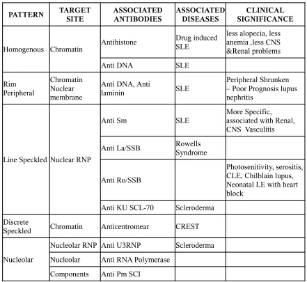
ANA PATTERN:
ANA are family of auto antibodies directed against contents of cell nucleus like dsDNA,
ENP (extractable nuclear proteins), Histone, nuclear RNA. ANA titre measures the pattern and
amount of auto antibodies. Substrates used are rat liver, mouse liver and human laryngeal cells
(Hep-2 cells). The pattern depends on the type of antibodies and substrate used. Hep-2 cells are
widely used now a days for its high specificity and sensitivity.26,27 Patient’s serum is incubated
with Hep-2 cells along with the fluorescence tagged auto antibody and viewed under
fluorescence microscope. Then the pattern is compared with standardized charts. Various other
quantitative methods as mentioned above are also used, which measures antibody in titres90,92.
Various types of ANA pattern (table 6) using Hep-2 cells are seen which includes
homogenous, fine speckled, discrete speckled, nucleolar pattern. Nucleolar pattern is further
divided into homogenous speckled and clumpy shows pattern of ANA, associated antibodies,
their target antigen and their significance.26,93,95 Homogenous ANA pattern is equivalent to LE
factor and it’s the most common pattern seen in SLE. Peripheral rim pattern is most commonly
associated with active disease, while peripheral shrunken pattern is most commonly seen in
patients with lupus nephritis.26,92,93 Peripheral shrunken pattern is seen 1-2 wks before disease
exacerbation and it is an indicator of poor prognosis. Presence of anti Sm antibody indicates
Table 6
PATTERN TARGET SITE ASSOCIATED ANTIBODIES ASSOCIATED DISEASES SIGNIFICANCECLINICAL
Homogenous Chromatin Antihistone
Drug induced SLE
less alopecia, less anemia ,less CNS &Renal problems
Anti DNA SLE
Rim Peripheral
Chromatin Nuclear membrane
Anti DNA, Anti
laminin SLE
Peripheral Shrunken – Poor Prognosis lupus nephritis
Line Speckled Nuclear RNP
Anti Sm SLE
More Specific, associated with Renal, CNS Vasculitis
Anti La/SSB Rowells Syndrome
Anti Ro/SSB
Photosenitivity, serositis, CLE, Chilblain lupus, Neonatal LE with heart block
Anti KU SCL-70 Scleroderma
Discrete
Speckled Chromatin Anticentromear CREST
Nucleolar
Nucleolar RNP Anti U3RNP Scleroderma
Nucleolar Anti RNA Polymerase
INVESTIGATIONS PERTAINING TO SYSTEMIC INVOLVEMENT:
As SLE involves almost all viscera, investigations are done depending upon the
symptoms. All base line investigations like LFT, RFT, urine routine, USG abdomen, CXR,
ECG is done. Expanded modes of investigations are done on individual needs which are shown
[image:47.612.95.521.245.569.2]in table 7.
Table 7
S.NO PATHOLOGY INVESTIGATION
1 CNS symptoms CT, MRI
2 Gangrene, ulcer APL levels
3 Livedo reticularis Cryoglobulins
4 Repeated abortions Cold agglutinins, APL
5 Cardiac ECHO
6 Respiratory CXR, HRCT
7 Lupus nephritis Renal biopsy, 24 hrs urine protein,C3, C4 levels
6 Lupus retinopathy Retinoscopy, angiography
7 GIT Endoscopy
8 Avascular bone necrosis MRI, lupus anticoagulant
All the above investigations are considered and treatment and follow up are made based