PREDICTORS AND IMPACT ON OUTCOME OF REFRACTORY STATUS EPILEPTICUS
DISSERTATION SUBMITTED FOR M.D DEGREE (PAEDIATRICS) BRANCH VII
MARCH 2010
INSTITUTE OF CHILD HEALTH AND
HOSPITAL FOR CHILDREN MADRAS MEDICAL COLLEGE
THE TAMILNADU Dr. M.G.R. MEDICAL UNIVERSITY
CHENNAI
CERTIFICATE
This is to certify that the dissertation titled, “Predictors and impact on the outcome of
refractory status epilepticus” submitted by Dr. R Pradeep Raj, to the Faculty of
Paediatrics, The Tamilnadu Dr. M.G.R Medical University, Chennai, in partial
fulfillment of the requirements for the award of M.D. Degree (Paediatrics) is a bonafide
research work carried out by him under our direct supervision and guidance, during the
academic year 2007-2010
Dr. J. Mohanasundaram Dr.Saradha Suresh,
M.D., Ph.D, DNB, M.D., Ph.D, F.R.C.P.(Glascow), Dean, Madras Medical College Director and Superintendent
Chennai 600003 Institute of Child Health and Hospital for Children Chennai-600008
Prof. Stephen Abraham Suresh Kumar, Dr. R. Kandasamy M.D.,D.M (Neuro)., M.D., D.C.H,
Chief, Department of Neurology Addl Professor of Paediatrics Institute of Child Health Institute of Child Health
DECLARATION
I, Dr. R..Pradeep Raj, solemnly declare that the dissertation titled “Predictors
and impact on outcome of refractory status epilepticus” has been prepared by me.
This is submitted to the Tamilnadu Dr. M.G.R. Medical University, Chennai in
partial fulfillment of the rules and regulations for the M.D. Degree Examination in
Pediatrics.
Place : Chennai Dr. R. Pradeep Raj
ACKNOWLEDGEMENT
I thank with deep sense of gratitude Prof. Saradha Suresh, M.D., Ph.D., F.R.C.P.
( Glascow), Professor of Paediatrics, Director and Superintendent of Institute of Child
Health and Hospital for Children, Egmore, Chennai for her kind support and guidance.
I thank my unit Chief Prof.R.Kandasamy,M.D.,D.C.H., and my unit Assistant
Professors Dr. C.V.Ravisekar,M.D.,D.C.H.,D.N.B., Dr. S. Lakshmi ,M.D., D.C.H.,
Dr.K. Kumarasamy,M.D.,D.C.H.,D.N.B., for their whole hearted support, guidance
and help rendered to this work.
I express my sincere thanks to Prof. Thilothammal., M.D.,D.M(Neuro)., for her
guidance and constant support in doing this work.
I express my sincere thanks to my guide Prof. Stephen Abraham Suresh
Kumar., M.D.,D.M(Neuro)., for his guidance, meticulous supervision and constant
support in doing this work.
I express my sincere thanks to Dr.Velusamy.,M.D.,D.M(Neuro), for his support
and guidance.
I express my sincere thanks to Dr. Luke Ravi ,M.D.,D.C.H, Registrar, Institute of
Child health and Hospital for Children, for his support and guidance.
I express my sincere thanks to the statisticians, Balaji Hospital, Chennai for their
help and guidance.
I would fail on my part if I forget to thank all my patients and their caretakers for
CONTENTS
PAGE NO.
1. INTRODUCTION 1
2. REVIEW OF LITERATURE 21
3. STUDY JUSTFICATION 30
4. AIM 31
4. MATERIALS AND METHODS 33
5. OBSERVATIONS 39
6. DISCUSSION 58
7. SUMMARY AND CONCLUSION 64
8. BIBLIOGRAPHY 66
INTRODUCTION
Status epilepticus (SE) is a frequent neurological emergency . The incidence
of SE has a bimodal distribution with peaks in children aged less than a year and
the elderly[1].Although conventional antiepileptic drugs (AED) can terminate SE
in most cases, a substantial minority of patients develop medically refractory status
epilepticus (RSE).
Status epilepticus is defined as a continuous convulsion lasting longer than
30 minutes or the occurrence of serial convulsions between which there is no return of
consciousness[2]. Mild degrees of pure hypoxia causes impaired
judgement,inattentiveness,motor incoordinationand at times,euphoria. With hypoxic
ischemia, consciousness is lost within seconds. If circulation is restored
within 3-5 minutes, full recovery may occur, but if hypoxic-ischemia lasts beyond
3-5 minutes some degree of permanent cerebral damage is the rule[3]. Status Epilepticus
is common in childhood, and the reported current mortality is in the
range of 4-6% [4].
The evolution of a prolonged seizure into Status Epilepticus is associated
with increased morbidity and mortality. Hypoxia is currently thought to be responsible
REFRACTORY STATUS EPILEPTICUS (RSE):
Although the entity of RSE is widely recognized and discussed, a standard
definition has not yet been evolved and is usually defined as seizure activity that
continues after first- and second-line therapy has failed.[6]Such patients are considered
to be in Refractory Status Epilepticus and escalation of therapy with administration of
barbiturate or non-barbiturate anesthetic agent is then recommended with the therapeutic
endpoint of achieving seizure control, electrical silence or both [7]. These patients
require management in pediatric intensive care unit, with continuous cardiorespiratory
and electro-encephalographic monitoring along with the aggressive therapy to control
seizures
The optimal management of such patients remains unclear and large, controlled
studies comparing the various agents are lacking. Generalized status epilepticus is
refractory to standard anticonvulsant therapy in at least 9% of patients and additional
intervention is required [8 ,9].
The significant morbidity and mortality in refractory status epileptics is due to the
nature of underlying illness and sustained seizure activity as well as the toxicity of
con-current treatment modalities. Many studies have come out which suggest that continuous
intravenous infusion of midazolam in safe and effective therapy for refractory status
epilepticus. On the other hand studies using other drugs e.g. Pentobarbital, propofol,
disadvantages due to their side effects.
Though refractory status epilepticus is a life threatening condition, there is no
standard drug or standard protocol for its treatment and also treatment varies from
institution to institution. Many drugs are being tried and there is no fixed consensus
arrived as which would be the best in children for early control of seizures and having
minimal side effects. Very few studies have been done in India and abroad on Refractory
Status Epilepticus, so, the data on the clinical profile and treatment modalities and their
effectiveness is also limited. Moreover, the drugs and the doses at which refractory
status epilepticus was controlled on western population may not be same in our children.
Important causes of status epilepticus [10]: The important causes can be broadly
divided into:
1. Acute Causes - CNS Infections (Meningitis / Meningo Encephalitis)
Febrile Convulsions
Vascular Episodes
Trauma
Metabolic Problems and
2. Static
Causes-Status Epilepticus can occur as a first manifestation of epilepsy or during the
course of epilepsy with or without an underlying neurological disorder. In known
epilepticus, drug default or sudden withdrawal of antiepileptics or undercurrent
infections and stress may predispose to status epilepticus.
3. Degenerative or Progressive Neurological Conditions
After a single episode of status epilepticus, subsequent episodes are more
common with risk of recurrence of 17% for a second episode and 5% for further attacks
[11]. In relation to premorbid state, the recurrence risk for status epilepticus for those
with prior neurological abnormalities is about 50% and in those who are neurologically
normal, it is 3% [11].
If status epilepticus could not be controlled by front-line anticonvulsants
(Diazepam, lorazepam, phenytoin and phenobarbitone) and seizures become refractory
then the following reasons should be reassessed systematically [12].
1. Adequacy of drug therapy.
2. Initiation of appropriate maintenance antiepileptic therapy
3. Failure to identify and treat the underlying causes e.g. Acutve progressive
cerebral disorders and cerebral infections.
4. Misdiagnosis of pseudo status as status epilepticus
PATHOPHYSIOLOGY OF STATUS EPILEPTICUS (SE):
SE refers to a condition in which there is a failure of the "normal" factors
to terminate a typical seizure. y-Aminobutyric acid (GABA) receptor-mediated
inhibition may be responsible for the normal termination of a seizure. In addition,
the activation of the N-methyl-D aspartate (NMDA) receptor by the excitatory
neurotransmitter glutamate may be required for the propagation of seizure activity.[13]
SE that is refractory to treatment may be the result of several processes and has been
attributed to a mechanistic shift from inadequate GABAergic inhibitory
receptor-mediated transmission to excessive NMDA excitatory receptor-receptor-mediated transmission.
[14] In experimental models, resistance to both benzodiazepines and barbiturates
develops during prolonged seizures and it has been hypothesized that prolonged seizure
activity alters the structure and/or function of GABAA receptors.[15] SE induced
neuronal death is morphologically necrotic and is initiated by excessive glutamate
release, which activates postsynaptic NMDA receptors and triggers receptor-mediated
calcium influx (excitotoxicity). This results in a cascade of events and cell death.[16]
The alterations in inhibitory and excitatory pathways have important implications
for the pharmacological management of SE. Another important aspect of self-sustaining
SE is the progressive, time-dependent development of pharmacoresistance. Currently
recommended agents acts primarily through the
GABA A receptor and have been shown to become less effective in SE of longer
than benzodiazepine receptor site, propofol acts at a site distinct from the
benzodiazepine and barbiturate binding sites, isoflurane acts by potentiation of
inhibitory postsynaptic GABA A receptor-mediated currents, although effects on
thalamo-cortical pathways also have been implicated.[17]
COMPLICATIONS OF STATUS AND REFRACTORY STATUS EPILEPTICUS:
Systemic changes and complications of status epilepticus and protean with the
involvement of all organ systems and these undoubtedly contribute to the ultimate
mortality seen in patients with Status Epilepticus and Refractory Status epilepticus. As
the duration of status epilepticus progresses, there is systemic alteration, with worsening
of general clinical state. The final stages are characterized by respiratory compromise,
hypotension, hypothermia and ongoing epileptiform paroxysms without motor
accompaniments.
The table given below lists the various complications [4]:
1. Interictal coma
2. Cumulative anoxia- cerebral and systemic
3. Cardiovascular complications – Tachycardia, Bradycardia, cardiac arrest,
hypertension, cardiac failure, shock.
4. Respiratory system failure- Apnoea, Cheyne-stokes breathing, aspiration,
cyanosis, respiratory acidosis.
5. Renal failure- Oliguria, uremia, acute tubular necrosis, rhabdomyolysis.
6. Autonomic disturbances – Hyperpyrexia, sweating, vomiting, hypersecretion,
airway obstruction.
7. Metabolic and Biochemical abnormalities – Acidosis (Metabolic, lactic acidosis),
hyper and hyponatremia, hyperkalemia, hypoglycemia, hepatic failure,
dehydration, acute pancreatitis.
8. Infections- pulmonary, bladder
9. Others – Disseminated intravascular coagulation, multiple organ dysfunction,
fractures, thrombophlebitis.
INVESTIGATIONS:
A battery of investigations will be required not only for identifying the underlying
etiology but also to monitor the alterations in the metabolic parameters during status
epilepticus. The investigations for cause of status are guided by thorough history and
clinical examination of the child. The following are the investigations, which are helpful
in the accurate diagnosis and management of the child.
1. Complete blood counts, Blood Sugar, electrolytes, Serum calcium,
phosphorus, magnesium levels
2. Mantoux test, resting gastric juice for acid fast bacilli toxicology screening.
4. Liver function tests, blood culture
5. Anticonvulsant levels, urine analysis and toxicology
6. Lumbar puncture and analysis of fluid for protein, sugar, Grams stain and
culture
7. Arterial blood gas analysis.
8. X-ray chest, CT scan brain, MRI brain
9. Continuous electro encephalographic monitoring.
INITIAL EVALUATION:
After performing a rapid but thorough cardiopulmonary assessment and
stabilizing the airway, breathing and circulation, a detailed physical and neurological
examination should be carried out to pick up the probable etiology and complications of
seizures.
Assess for evidence of trauma; signs of raised intracranial pressure like
papilloedema, bulging anterior frontenalle or focal neurological deficit; manifestations
of sepsis or meningitis. Look for Kussmaul breathing and dehydration suggestive of
metabolic acidosis or irregular respirations signifying brain stem dysfunction; evidence
of failure to thrive, a peculiar body odor or abnormal hair pigmentation that suggests an
inborn error of metabolism and constricted or dilated pupils suggesting a toxin or drug
as the cause of status epilepticus[2].
MANAGEMENT:
shown that there is progressive loss in the ability of antiepileptic drugs to control the
severe seizures of status epilepticus. This time dependant development of
pharmacoresistant status epilepticus is extremely important in the management of status
epilepticus [18].
The main goals of management of status epilepticus are to:
1. Support vital function
2. Terminate seizure activity as fast possible
3. Prevent recuurence of seizures
SUMMARY OF EMERGENCY MANAGEMENT OF STATUS EPILEPTICUS
Immediate
Airway - Protect airway – use 100% oxygen and endotracheal tube if
necessary
Breathing - Support and use muscle relaxant if necessary
Circulation - Maintain adequate perfusion and normal blood pressure- support if
necessary
Establish secure intravenous line.
Draw - Laboratory samples
Anticonvulsants
First Line - Diazepam ( 0.2 mg/kg) IV
(0-10 min) (or)
Lorazepam (0.1 mg/kg) IV given over 30-60 sec. (Repeat same dose
after 10 min. if seizures are not controlled).
Second Line- Phenytoin 20 mg/kg IV infusion
in 20 ml of Normal saline over 20 minutes
( Not to exceed 1 mg/kg/mt)
Seizures not controlled
Phenytoin 10mg/kg infusion half loading dose
Seizures are not controlled
Phenobarbitone loading dose 20 mg/kg slow IV infusion
(Intubate )
Seizures not controlled
MANAGEMENT OF REFRACTORY STATUS EPILEPTICUS
RSE requires more aggressive treatment and however, the optimal treatment has
not been defined. Patients should be treated in intensive care unit, as artificial
ventilation and hemodynamic support is required. These patients generally require
intravenous fluids and vasopressors to treat hypotension associated with high dose
intravenous use of anesthetic agents. In a third of adults in SE, arterial pH falls
below 7;[19] the main contribution to this change is lactic acidosis from skeletal
muscle,[20] which responds well to oxygen and control of convulsive activity.
Mild acidosis might be an anticonvulsant and neuroprotective. The usual
practice is to treat with bicarbonate if the patient is hypotensive and arterial pH if it
is < 7 due to metabolic acidosis. Control of hypothermia is neuroprotective.[21]
Pharmacological treatment
To date, no randomized controlled trials have been done for SE refractory to first-
and second-line therapy. The most experience exists with continuous infusion
(cIV) of pentobarbital, midazolam and propofol,[22]. The best comparative information
comes from the systematic review by Claassen and colleagues. No difference was found
in mortality among the groups treated with cIV propofol, cIV midazolam and cIV
pentobarbital. Mortality was related to patient's age and duration of SE rather than AED
choice. A recent retrospective study investigated the effect on RSE prognosis of various
coma-inducing pharmacologic options. Mortality and likelihood of the patient's
the three arms, barbiturates (pentobarbital and phenobarbital), propofol and midazolam.
This study did not find any evidence for mortality related to propofol infusion syndrome.
Traditionally, barbiturates such as pentobarbital or thiopental have been used to
terminate RSE, inducing coma and EEG suppression.[23],[24] However their
effectiveness has not been studied systematically. In a systematic review of 109 adult
patients with RSE who were treated with pentobarbital 8% experienced
acute failure; 12%, breakthrough seizures; 43%, withdrawal seizures within 48
hours; and 8%, refractory hypotension during the therapy. EEG burst suppression or
complete suppression has been achieved more frequently in episodes treated with
barbiturates. Pentobarbital use is also often accompanied by prolonged sedation and life
threatening infections. Episodes treated with barbiturates were associated with
significantly longer hospital stay for surviving patients compared with episodes in which
barbiturates were not used.
Superior pharmacokinetics and favorable adverse effect profile makes propofol the
drug of choice. The two main advantages of propofol are a rapid onset and short
duration of action. Propofol is a GABAA agonist that suppresses seizure activity
via GABA-mediated inhibition of neuronal firing. Other mechanisms of action
include inhibition of N-methyl-D aspartate receptor and modulation of calcium
influx through slow calcium ion channels. The safety of propofol was further
supported in recent retrospective series both in adults[22] and children. A
by Rossetti et al[40] in which 27 patients who failed to intravenous clonazepam
and phenytoin therapy were induced into burst suppression pattern on cEEG with
cIV propofol at a dose of 2.1 to 13 mg/kg/h for 1 to 9 days while continuing
clonazepam infusion. RSE was successfully treated with propofol in 21 (67%)
episodes. Seven deaths (23%) wee reported and none were attributable directly to
propofol use and no patient experienced propofol infusion syndrome. In pediatric
RSE also propofol has been shown to be a safe and effective drug, 14 (64%) of the
22 episodes could be adequately controlled. Two patients who were successfully
treated with propofol died and the death was related to the underlying etiology and
not to the use of propofol. However propofol may cause metabolic acidosis
and cardiovascular collapse with prolonged use in children and deaths have been
reported the propofol infusion syndrome.[26] Propofol should therefore
be used with caution in children, ideally for short time only and the infusion rate
should not exceed 67 ug/kg/min.[27] In a prospective study the quality of burst
suppression was unsatisfactory in most patients. The maintenance of continuous
burst suppression is difficult and vigilant titrating of dosage of propofol is
necessary under EEG monitoring.[25]
Midazolam is an effective, short acting benzodiazepine that when given as an
infusion has an efficacy in RSE, including at sub-anesthetic doses. It has the
advantages of rapid onset of activity and greater water solubility, avoiding the
benzodiazepines and barbiturates. Midazolam binds to GABAA receptors and
augments GABAergic transmission, there by imparting anticonvulsant and
sedative-hypnotic properties.[28] Duration of antiepileptic effects is minutes to
hours. The elimination half-life is 1.5 to 3.5 hours initially. With prolonged use,
there may be tolerance, tachyphylaxis and significant prolongation of half-life, up
to days.[29] After 24-48 h, the dose of the drug must often be increased severalfold
to maintain seizure control. Clinical experience with midazolam for RSE is limited.
The reported failed treatment with midazolam ranges between 14 to 18% . [29] , [30] .
In the series of Claassen and colleagues acute treatment failure occurred in 18% of
episodes, breakthrough seizures in 56%, post treatment seizures in 68% and ultimate
treatment failure in 18%. The authors suggest that titrating continuous intravenous
midazolam to burst suppression, more aggressive treatment with concurrent AED or a
longer period of initial treatment may reduce the high proportion of patients with RSE
who relapse after midazolam is discontinued. In this series only 24% had an immediate
and sustained response.
Other Pharmacological treatment:
High dose phenobarbital:
High dose of phenobarbital with serum levels of 100 to 200 ug/ml, has been found
effective and safe in the treatment of RSE in children. In another study a very high dose
phenobarbital at accumulated daily doses up to 80 mg/kg, with a resulting serum level of
children with RSE. In this study the adverse effects were milder compared with
thiopental infusion.
Ketamine:
Ketamine, a NMDA antagonist, has been proved useful in RSE[31] and it is also a
neuroprotective. However, because ketamine can raise intracranial pressure, the absence
of intracranial mass lesion should be confirmed by neuroimaging. The experience with
this agent in RSE is very limited.
Inhalational Anesthetics:
Inhalational anesthesia (IA) is an alternative approach to the treatment of RSE. Its
attractive feature include efficacy, rapid onset of action and the ability to titrate the doses
according to the effects demonstrated on the EEG.[32] Of the various agents, isoflurane
and desflurane are the two agents that have been administrated for RSE because of their
safety associated with long-term administration.[33] In a recent retrospective study,
seven patients with RSE were initiated to IAs (all patients to isoflurane and one patient
in addition to desfluratne) after 1 to 103 (mean,19) days. They received multiple AEDs
(mean 10, range 7 -15) in addition to IAs. Regardless of seizure type, isoflurane and
desflurane consistently stopped epileptic discharges with adequate, sustained
electrographic burst suppression within minutes of initiating IA therapy. Four patients
Newer AEDs:
The use of newer AEDS in the treatment of RSE has not been studied systematically. In
6 patients with RSE unresponsive to sequential trials of multiple agents, a suspension of
topiramate administered via nasogastric tube was effective in aborting RSE. Effective
dosages ranged from 300 to 1,600 mg/d.[34] RSE was terminated in three children with
topiramate loading, 5 mg/kg/day.[35] Seizure control has been achieved in patients with
RSE by administration of levetiracetam (500-3000 mg/day) by nasogastric route.[36]
Injectable levetiracetam formulation is available and the pharmacokinetics of
levetiracetam administered by IV infusion was comparable across all dose groups and
infusion rates and the pharmacokinetic profile was consistent with that for levetiracetam
administered orally.[37] Well designed studies are needed to determine the place of
newer AEDs as the use of drugs can avoid pharmacologic coma.
Target of treatment-burst suppression:
Experimental studies demonstrated maximal depression of cerebral metabolism with
barbiturates with burst suppression intervals of 30 seconds.[38] Burst suppression and
isoelectric background EEG have been shown to be accompanied by fewer recurrent
seizures than simply stopping seizures. There is uncertainty about the optimal extent of
EEG suppression in RSE. Several authors used different burst suppression intervals.
Kofke et al[39] used 15 to 30 seconds as burst suppression interval. Van Ness used 3 to
9 bursts per minute during pentobarbital treatment. Mirsattari and colleagues[33]
and suppression duration longer than 10 seconds as the goal of therapy. Where as Bleck
advocates a more aggressive approach using isoelectric EEGs. In a recent retrospective
study the outcome was independent of the extent of EEG burst suppression and probably
related to the underlying cause of RSE.
Maintenance therapy:
In parallel with emergency treatment attention must be given to maintenance AED
therapy to prevent recurrence of seizures. In patients known to have epilepsy, their usual
AEDs should be maintained and dose adjustments may be required depending on AED
levels. In patients presenting denovo the AEDs, phenytoin/fosphenytoin or valproate,
used to control the status can in principle be continued as oral maintenance therapy. In
others, unless relatively short-lived treatment is anticipated, the preference is to initiate
oral maintenance therapy, valproate or carbamazepine, starting immediately at standard
doses.[40] If additional medication is needed, the most appropriate AEDs are topiramate
and levetiracetam as these drugs can be started at high doses with a low risk of
REVIEW OF LITERATURE
A retrospective cohort ctudy was conducted by Stephen et al[42] in the
neurological intensive care unit (NICU) at Columbia-Presbyterian Medical Center
between January 1, 1994, and March 31, 1998 to determine the frequency, risk factors,
andimpact on outcome of RSE. Consecutive sample of 83 episodes of status epilepticus
in 74 patients were studied. In 57 episodes (69%), seizures occurred aftertreatment with
a benzodiazepine, and in 26 (31%), seizures occurredafter treatment with a second-line
anticonvulsant drug (usually phenytoin), fulfilling our criteria for RSE. Nonconvulsive
SE (P=.03) and focal motor seizures at onset (P= .04) were identified as independent
risk factors for RSE. Eleven (42%) of 26 patientswith RSE had seizures after receiving a
third-line agent (usually
phenobarbital). Although mortality was not increased (17% overall), RSE was associated
with prolonged hospital length of stay (P<.001) and more frequent functional
deterioration at discharge (P= .02). They concluded that refractory status epilepticus
occurs in approximately 30% of patients with SE and was associated with increased
hospital length of stay and functional disability. Nonconvulsive SE and focal motor
seizures at onset were risk factors for RSE.
M Holtkamp et al[43] retrospectively analysed all episodes of status epilepticus
(SE) treated between 1993 and 2002 on the neurological intensive care unit (NICU) of
the Charité-Universitätsmedizin Berlin. The predictive and prognostic features of RSE
up to identify the possible development of post-SE symptomatic epilepsy. A total of 83
episodes fulfilled their criteria of SE. Of these 43% were refractory to first line
anticonvulsants. The mean age of patients with SE was 53.3 (SD 19) years, with only
two patients younger than 18 years. Encephalitis was significantly more often the
primary cause in RSE (p<0.05), whereas low levels of antiepileptic drugs were
significantly more often associated with NRSE (p<0.001). Hyponatraemia within the
first 24 hours after onset of status activity was significantly more often associated with
RSE (p<0.05). In RSE, compared with NRSE, significantly longer duration of seizure
activity (p<0.001), more frequent recurrence of epileptic activity within the first 24
hours after the end of seizure activity (p<0.001), longer stay in the NICU and in hospital
(p<0.001 and p<0.01, respectively), and more frequent development of symptomatic
epilepsy (p<0.05) were seen. SE treated in the NICU was frequently refractory to first
line anticonvulsant drugs. Encephalitis was a predictor for RSE, which was associated
with markedly poor outcome, in particular, the development of post-SE symptomatic
epilepsy.
In a prospective study conducted by Ramon Rivera, Miguel Seginini et al [44], 24
children diagnosed with the diagnosis of status epilepticus were admitted and treated in
the Intensive Care Unit, National Children’s Hospital, Costa Rica with midazolam
infusion. Out of 24 patients, 10 were male and 14 were female. The mean age was 2.2
yrs (range 2 months to 12 yrs). Fourteen patients had history of epilepsy and were in
(n=4) and carbamazepine (n=4)). But at the moment of admission, the serum levels of
these drugs are not known. The other ten patients presented at the emergency room with
their first conclusive episode. Four patients had infection of central nervous system. Six
had idiopathic epilepsy. Majority of patients (n=18) had generalized tonic-clonic
seizures, followed by focal seizures (n=6) and focal with secondary generalization
(n=1). The mean time between admission to the emergency room and the establishment
of midazolam infusion was 0.75 hrs (range 0.25 to 2.5 hrs). Complete arrest of seizures
was achieved with midazolam therapy in all 24 patients. The mean infusion rate of
midazolam necessary to control the fits was 2.3 µg/kg/min. (range 1 to 18). They
observed that none of the patients experienced clinically important changes in blood
pressure or respiratory status while receiving midazolam as an intravenous infusion. In
addition, none of the patients required endotracheal intubation and assisted ventilation.
The metabolic parameters were within normal limits and all of them regained full
consciousness at a mean time 4.2 hrs (range 2 to 8.5 hrs) after discontinuation of the
midazolam infusion. They concluded that midazolam is an effective and safe therapeutic
approach for the management of pediatric patients with status epilepticus.
In another interesting study conducted by Roshan Lal Koul, Raj Ajithala G et al
[45] as in a tertiary referral centre in Sultanate of Oman, 20 children with status
epilepticus were admitted of which majority were males ( n=15) and the rest ( n=5) were
females. The mean age was 4.07 yrs ( range 2 months to 13 yrs). Eleven children had
epilepsy, three had acute purulent meningitis, three had acute meningo encephalitis and
the remaining had various vascular or degenerative lesions of the brain. Majority
presented with generalized tonic clonic seizures (n=13), four children had partial
seizures, myoclonic status in one child and Lennox-Gastaut status in two children.
Twelve patients had refractory status epilepticus. Complete arrest of seizures was
achieved with midazolam infusion in all but one child who had Batten’s disease. The
mean time to control seizures in refractory status epilepticus was 64.6 minutes (range
15-240 minutes) and in established status epilepticus 34.3 minutes (range 10-60
minutes). The mean time between start of midazolam infusion and total cessation of
seizures in all patients was 54 minutes. The mean infusion rate of midazolam required to
control the seizures completely was two µg /kg/min (range 1-5 µg/kg/min). Only two
children had transient fall in oxygen saturation (to 90%) as demonstrated by pulse
oxymetry however, none of the patients required mechanical ventilation and none
showed any abnormalities in electrolyte and glucose levels. So this study too suggested
that midazolam infusion is an effective and safe therapeutic approach for refractory
status epilepticus (RSE).
The meta-analysis conducted by Donald L. Gilbert, Peter S. Gartside [46] in 1999
was the first study that systematically analyzed the published literature on the treatment
of refractory generalized convulsive status epilepticus in children. One hundred eleven
Five drugs (midazolam, pentobarbital, thiopental, isoflurane and diazepam) were used in
patients who met inclusion criteria. They found that midazolam, pentobarbital,
thiopental and isoflurane were 100% efficacious, in an average of less than 1 hour after
starting treatment. The only treatment failures were seen in diazepam study in which
seizures continued in 14% of children. The efficacy of all treatments in symptomatic
cases was 92% versus 96% in idiopathic cases (p=0.42). The overall mortality in
children treated for refractory generalized convulsive status epilepticus was 16%.
Mortality in children with symptomatic etiologies was significantly greater than that in
idiopathic group (1 of 27; Fisher’s exact test; p=0.033). The overall mortality rate in
children treated with diazepam was 19%; with isoflurane 40%, with pentobarbital 17%
and with thiopental was 31%. But there were no deaths among children treated with
midazolam. The incidence of new neurological defects was 25% in the midazolam
studies. With the apparent effectiveness of midazolam in children with refractory
generalized convulsive status epilepticus, they suggested that midazolam should be
considered as first line therapy for treatment of refractory status epilepticus.
Minagawa K, Yanai s [47] reviewed 48 episodes of refractory status epilepticus in
children. The mean age of patients was 3.5 years (range 1 month to 18 years). Nine
children had epilepsy, one purulent meningitis, one encephalitis, one acute hypoxic
ischemic encephalopathy. The types of seizure were generalized tonic clonic seizures in
in 3 children. All patients were treated with midazolam infusion after a bolus dose. Forty
one of the 48 episodes of seizures were controlled within 30 minutes after initiation of
midazolam therapy. The mean infusion rate of midazolam required was 0.22 mg/kg/hr.
The duration of treatment was 4.1 days. None of the patients had serious changes in the
blood pressure or respiratory status attributable to the use of midazolam infusion. They
concluded that midazolam infusion is an effective and safe therapeutic approach for the
management of childhood status epilepticus.
Parent JM, Lowenstein DH in their study [48] comprising of four patients with
refractory generalized status epilepticus, who were monitored by electroencephalogram,
documented the cessation of seizure activity within minutes of the loading dose in all the
patients. No adverse effects occurred during midazolam treatment. Only one patient
required fluid boluses and pressure support for hypotension while the remainder of the
patients safely tolerated midazolam despite preexisting hemodynamic instability. All the
patients had recovered and maintained good seizure control.
In an Indian study done by Singhi S, Murthy A et al., at Chandigarh, they
compared the efficacy of continuous midazolam versus diazepam infusion in RSE [49].
It was an open-label, randomized control study at their Intensive Care Unit. The subjects
included 40 children, 2 to 12 years of age with refractory status epilepticus. Either
continuous midazolam (n=21) or diazepam infusion (n=19) in incremental doses was
administered for seizure control. The two groups were similar in age (mean ± SD = 4.9
system infection. RSE was controlled in 18 (86%) and 17 (89%) patients in the
midazolam and diazepam groups respectively (p=not significant). They found that the
median time to control seizures was 16 minutes in both the groups, but in the midazolam
group, seizures recurred in more children (57% versus 16% in diazepam group; p<
0.05). The maximum dose ( mean ±SD) of midazolam and diazepam required was
5.3±2.6 µg/kg/min and 0.04±0.02 mg/kg/min, respectively. About half of the patients
needed mechanical ventilation and 40% had hypotension in both groups but they found
that the mortality was higher in the midazolam group ( 38%) as compared to the
diazepam group ( 10.5 %, p< 0.01>0.05). They concluded that continuous midazolam
and diazepam infusions were equally effective for control of RSE. However, midazolam
was associated with more seizure recurrence and higher mortality in RSE predominantly
caused by central nervous system infections.
In a small retrospective study comprising of seven patients with refractory status
epilepticus done by Kumar A, Bleck TP [50], they found that midazolam infusions
terminated status epilepticus in all patients in less than 100 seconds as determined by
clinical observation ( three patients) or electroencephalographic monitory ( four
patients). But all the patients received mechanical ventilation before receiving
midazolam. They found that one patient developed mild hypotension. In this small study,
they concluded that midazolam appears to be an effective and safe alternative to high
dose barbiturate coma for the termination of status epilepticus when conventional agents
STUDY JUSTIFICATION
Identification of predictors for RSE is crucial for detection of patients at risk
early in the course of the disease.Despite its frequency, little is known about the
predictive and prognostic features of the critical condition of RSE.The clinical
characteristics of refractory status is poorly understood and therefore the current
management approaches are still unsatisfactory.Refractory status epilepticus (RSE)
is a condition in search of improved clinical characterisation and more efficient
treatment options. In contrast with status epilepticus (SE) in general, only a few
studies have been reported on the subgroup of refractory status and moreover there
AIMS AND OBJECTIVES
• To determine the predictors of refractory status epilepticus
MATERIALS AND METHODS
STUDY DESIGN:
Case – control study
STUDY PERIOD:
October 2007- September 2009
STUDY PLACE:
Institute of child health and hospital for children,Egmore.
STUDY POPULATION:
INCLUSION CRITERIA:
Continuous seizure activity lasting for more than 30 minutes or
intermittent seizure activity without recovery of consciousness in between the
episode in children aged between 1 month to 12 years.
EXCLUSION CRITERIA:
(1) Age < 1 month
(2) Children with seizures only at home.
(3) Children with head injury
(4) Children in case group of RSE in whom first line
anticonvulsants were not given sequentially before starting midazolam.
CASE DEFINITION:
CONTROL DEFINITION:
Cases with seizures responding to benzodiazepine and a second line
intravenous anticonvulsant drug ( phenytoin or phenobarbitone).
LIMITATION:
• Serum levels of anticonvulsants were not known during admission
• Electroencephalographic monitoring was not available CONSENT:
Institutional consent was obtained from the parents after explaining the
MANOEUVRE:
One hundred and eleven children between the age group of 1 month to
12 years admitted in our hospital with the diagnosis of refractory status epilepticus
were included in the study group and one hundred children between the age group
of 1 month and 12 years admitted in our hospital with status epilepticus were
included in control group in our study.
Status epilepticus was diagnosed when child had continuous seizure lasting
more than 30 minutes or several seizures occurring without regaining consciousness
between the seizure activity. These children were treated with 0.1
mg/kg of lorazepam 2 doses, 20mg/kg loading dose of phenytoin followed by 10
mg/kg half loading dose of phenytoin and 20 mg/kg loading dose of phenobarbitone at
predetermined time interval in succession after stabilizing the
airway, breathing and circulation. Intubation was done prior to phenobarbitone
administration. Refractory status epilepticus was the diagnosis if the child continued to
have seizure activity despite adequate doses of medications mentioned above.
A thorough history was obtained from the parents regarding name, age, sex,
area, duration of seizure prior to reaching hospital, type of seizure , presence or
absence of fever. Past history of seizure / status epilepticus and the anticonvulsant
drug and dose ( whether adequate ) and any history of drug default or any recent
change in anticonvulsant drug was enquired and noted down. Any history of drug
enquired. Presence of any neurological co morbidity and its type if present , was
enquired in detail.
Total time taken to control the seizure , number of drugs required , whether
ventilator support was required was noted down. The complications during status
epilepticus and the consciousness and neurological status at the end of status
epilepticus were recorded. Later the patients were followed up in the ward for any
recurrence of seizure , time taken for complete control of seizure. A thorough
clinical examination was carried out including monitoring of vitals, oxygen saturation
and systemic examination to find out the cause, if any, involving the
central nervous system, cardiovascular system, renal and respiratory system.
Complete blood count, liver function test,renal function test and chest X ray
were done in all patients. In order to exclude electrolyte and metabolic disturbances as a
cause or complication of seizures , blood samples were taken for
electrolytes, glucose and calcium estimation. Mantoux, resting gastric juice for
Acid fast bacilli, cerebrospinal fluid analysis ,cerebrospinal fluid lactate and
pyruvate were done if indicated. Computed tomography of brain was done in all
patients. Magnetic resonant imaging of brain was done in selected cases. Final
diagnosis of various underlying problems was made based on the clinical history,
physical examination and various investigation.
hospital stay, duration of ventilation required length of stay in hospital stay. The
neurological status of the child at the time of discharge was noted.
All statistical analysis (mean, median, standard deviation) was done using
SPSS version 11 for windows and data were statistically analysed, compared and
OBSERVATIONS
A total of 111 children in case group with refractory status epilepticus and
100 children in control group with non refractory status epilepticus were studied.
The age distribution is as follows:
TABLE – 1
Age distribution
Age Case group Control group
< 1 yr 27(24.3%) 21( 21%)
1 – 5 yrs 28(25.2%) 23(23%)
5 – 9 yrs 24( 21.6%) 29( 29%)
9 – 12 yrs 32( 28.8%) 27(27%)
The percentage of RSE cases was comparable in all the age groups.The number of
RSE cases in children <1 year was 27(24.3%) with an insignificant p value of 0.565 and
Odds ratio of 1.209 . This is in contrast to the previous studies which concluded <1 year
as a risk factor for RSE. The youngest child in the study group was 2 months and the
child with maximum age in study group was 12 yrs. The commonest age group in this
study was 9 – 12 yrs with 32 cases (28.8%).
TABLE- 2
SEX DISTRIBUTION
SEX CASES CONTROL TOTAL
MALE 61(55%) 57(57%) 118(55%)
FEMALE 50(45%) 43(43%) 93(45%)
The total number of male patients in the study was 118(55%), with 61 in the
RSE group and 57 in the control group. The total number of female patients in the
study was 93(45%), with 50 in the RSE group and 43 in the SE group. Not either
of the sex had a significant p value and so neither were found to be a risk factor
for RSE.
TABLE – 3
Duration of seizure prior to reaching hospital
Vast majority of the patients with RSE were brought to the hospital more
than 60 min after onset of seizure, with the exact number being 51 in RSE group
and 30 in NRSE group with a p value of 0.015 which is significant and an odds
ratio of 2.1 . The median duration of seizures before reaching the hospital was 40
minutes and standard deviation was 38.12 minutes. Minimum time of seizures
before reaching hospital was 10 minutes while the maximum time was 90 minutes.
Long duration of seizure prior to reaching hospital was found to be a risk factor for
RSE.
TYPE OF SEIZURE
Type of seizure Cases Control p value Odds ratio 95% confidence interval Focal to GTCS 26(23.4%) 11(11%)
0.018 2.475 1.152-5.319 GTCS 85(76.6%) 89(89%)
Total 111 100
Majority of the patients in both the groups had generalized tonic clonic
seizures numbering 85 in RSE group and 89 in NRSE group. However focal to
secondary generalization was more associated with RSE than NRSE with a Chi
square value of 5.615 and a significant p value of 0.018 and an odds ratio of 2.475
with 95% confidence interval being 1.152- 5.319. This result goes hand in hand
with the observation made by Stephan et al that focal onset of seizure was a major
risk factor for RSE.
TABLE-5
Past history of status epilepticus
Past H/O Status epilepticus
Cases Control
Yes 7(6.3%%) 6(6%)
No 104(93.7%) 94(94%)
7 patients among RSE group had previous history of status epilepticus as
against 6 in NRSE group. Among this 7 in RSE group 2 patients had primary
seizure disorder and 5 were cerebral palsy children with seizures. Among this 6 in
NRSE group 1 patient had primary seizure disorder and 5 were cerebral palsy
children with seizures. Past history of status epilepticus was not a risk factor for
RSE and had an insignificant p value.
TABLE-6
Drug withdrawal
Withdrawal Cases Control p value Odds ratio 95% confidence interval Yes 27(71%) 7(26%)
0.001 6.6 3.56-13.62
No 11(29%) 19(74%)
Total 38 26
38 patients in RSE group had seizure disorder out of which 5 had primary
seizure disorder, 20 were cerebral palsy children with secondary seizure disorder
and 3 were post meningitic/encephalitic sequelae with secondary seizure disorder.
7 patients in NRSE group had seizure disorder out of which 2 had primary
seizure disorder,4 were cerebral palsy children with secondary seizure disorder
and 1 was a post meningitic/encephalitic sequelae with secondary seizure disorder.
Drug withdrawal had a significant association with RSE with a p value of 0.001
TABLE-7
Inadequate drug dose
Dose inadequate
Case Control p value Odds ratio 95% confidence interval
Yes 18(47.3%) 5(19.2%)
0.031 3.78 1.82-7.42 No 20(52.7%) 21(80.8%)
Total 38 26
18 patients in RSE group had a history of drug withdrawal as against 5 in
NRSE group. Among the 18 with history of drug withdrawal in RSE group 2 had
primary seizure disorder,15 were cerebral palsy children with secondary seizure
disorder and 1 was a post meningitic/encephalitic sequelae with seizure disorder.
Among the 5 with history of drug withdrawal in NRSE group 1 had primary
seizure disorder,3 were cerebral palsy children with secondary seizure disorder
and 1 was a post meningitic/encephalitic sequelae with seizure disorder. History of
drug withdrawal was associated with RSE with a significant p value of 0.031 .
Table-8
Recent change in AED
Recent change
in AED Cases Control p value Odds ratio
95%
confidence interval Yes 6(15.7%) 3(11.5%)
Total 38 26
6 patients in RSE group had a history of recent change in AED as against 3
in NRSE group. Among the 6 with history of drug withdrawal in RSE group 2had
primary seizure disorder,4 were cerebral palsy children with secondary seizure
disorder and. Among the 3 with history of drug withdrawal in NRSE group 1 had
primary seizure disorder,2were cerebral palsy children with secondary seizure
disorder. History of recent change in AED was significantly associated with RSE
with a p value of 0.031 .
Table-9
Neonatal seizure among children with CP
NNS among children with CP
Case Control p value Odds ratio
95%confi dence interval Yes 19(63.3%) 3(20%)
0.003 6.9 3.92-13.12 No 11(36.67%) 12(80%)
Total 30 15
19 cerebral palsy patients out of 30 in the RSE group had a prior history of
prior history of neonatal seizure. History of neonatal seizure among cerebral palsy
children had a significant association with RSE with a p value 0.003
Table-10
Shock in E- Room
Shock Case Control
Present 111(100%) 85(85%)
Absent 0 15(15%)
Total 111 100
All the 111 patients in RSE group had shock that was managed in
Emergency Room with fluid boluses followed by inotropes if necessary. 85(85%)
patients in control group had shock.
Table -11
Recurrence of seizure after control of status epilepticus
Recurrence Case Control p value Odds ratio
95% confidence
interval Yes 20(18%) 7(7%)
0.017 2.92 1.178-7.239 No 91(82%) 93(93%)
Total 111 100
Recurrence of seizure after control of status epilepticus was seen in 20
odds ratio of 2.92
Table-12
Ventilator support
Ventilator support Cases Control
Yes 104(93.7%) 48(48%)
No 7(6.3%) 52(52%)
Total 111 100
104(93.75%) patients in RSE group required ventilator support. In the
remaining 7 patients phenobarbitone and midazolam infusion was started in
ward and these cases were not intubated.
Table -13
Duration of ventilator support
Duration of ventilator support
Case Control
<1 day 17(16.3%) 18(37.5%)
1-5 days 40(38.5%) 22(45.8%)
>5 days 47(45.2%) 8(16.7%)
Total 104 48
47 patients in RSE group required ventilator support for a longer
period as against 8 in NRSE group. Patients in RSE group required longer
Table -14
Complications during hospitalization
Complication Case Control
Pneumonitis 58(52.3%) 29(29%)
Refractory shock 10(9%) 3(3%)
UTI 9(8.1%) 10(10%)
Renal failure 2(1.8%) 0(0%)
Sepsis 4(3.6%) 1(1%)
Pneumonitis 58 (52.3%) was the common complication among children
with RSE. Refractory shock was seen in 10(9%) of RSE cases. UTI was
seen in 9(8.1%) of RSE cases. Even in NRSE group pneumonitis was the common
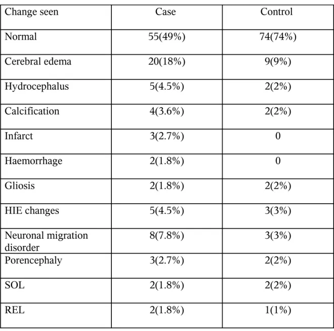
Table -15
CT changes
Change seen Case Control
Normal 55(49%) 74(74%)
Cerebral edema 20(18%) 9(9%)
Hydrocephalus 5(4.5%) 2(2%)
Calcification 4(3.6%) 2(2%)
Infarct 3(2.7%) 0
Haemorrhage 2(1.8%) 0
Gliosis 2(1.8%) 2(2%)
HIE changes 5(4.5%) 3(3%)
Neuronal migration disorder
8(7.8%) 3(3%)
Porencephaly 3(2.7%) 2(2%)
SOL 2(1.8%) 2(2%)
REL 2(1.8%) 1(1%)
Abnormal CT scan was reported in 56(51%) cases in RSE group as
against 26(26%) in NRSE group. The association between abnormal CT findings
and RSE is significant and has a p value of 0.001. CT scan was done in all the
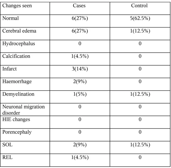
Table -16
MRI changes
Changes seen Cases Control
Normal 6(27%) 5(62.5%)
Cerebral edema 6(27%) 1(12.5%)
Hydrocephalus 0 0
Calcification 1(4.5%) 0
Infarct 3(14%) 0
Haemorrhage 2(9%) 0
Demyelination 1(5%) 1(12.5%)
Neuronal migration disorder
0 0
HIE changes 0 0
Porencephaly 0 0
SOL 2(9%) 1(12.5%)
REL 1(4.5%) 0
MRI was done only in selected cases. Out of the 22 cases in RSE
group MRI was abnormal in 16 cases and out of the 8 cases in control group
Table – 17
Etiology
Etiology Cases Control p value Odds ratio
95% confidence
interval CNS infection 35(31.5%) 15(15%) 0.005 2.61 1.323-5.148
Seizure disorder 15(13.5%) 20(20%) 0.206 0.625 0.301-1.3
CP 30(27%) 15(15%) 0.033 2.099 1.052-4.186
Febrile seizure 10(9.7%) 40(40%) 0.0731 1.2 0.501-2.8
CVA 5(4.6%) 0 0.032 -
-SOL 2(1.8%) 1(1%) 0.623 1.817 0.162-20.343 Hypertensive
encephalopathy
2(1.8%) 1(1%) 0.0623 1.817 0.162-20.343
Metabolic 1(0.9%) 0 0.341 -
-Neurodegenerativ e disorder
3(2.71%) 1(1%) 365 2.75 0.281-26.874
Toxins 3(2.7%) 0 0.098 -
-PES/PMS 5(4.6%) 7(7%) 0.434 0.621 0.192-2.041
CNS infection was the most common cause of RSE accounting for 31.5% of
cases in the RSE group with a p value of 0.005. However CNS infection was the
cause in only 15% of the cases in NRSE group. CNS infection was followed by CP
as the next common cause for RSE accounting for 27% of the cases and a
toxin ingestion had RSE. Febrile seizure was the most common cause for NRSE
accounting for 40% of cases in the control group.
Table-18
Precipitating factor
Precipitatin g factor
Case Control p value Odds ratio
95% confidence
interval Fever 54(48.6%) 49(49%) 0.959 0.986 0.572-1.693
Poor compliance
30(27%) 13(13%) 0.012 2.479 1.209-5.081
Lack of sleep
4(3.6%) 3(3%) 0.807 1.209 0.264-5.537
Stress 4(3.6%) 3(3%) 0.807 1.209 0.264-5.537
Fever was the precipitating cause for seizure in 48.6% of cases in RSE
group and 49% of cases in NRSE group. Poor drug compliance was the
precipitating factor in 27% of cases in RSE group and 13% of cases in NRSE
group with a significant association with RSE with a p value of 0.012 . Lack of
sleep and stress were the precipating factor in 3.6% and 3% of patients in RSE and
Table-19
Outcome
Outcome Case Control p value Odds ratio
95% confidence
interval Recovered
normally
36(32.4%) 59(59%) 0.87 0.33 0.172-0.782
Recovered with fresh weakness
33(29.7%) 18(18%) 0.045 1.9 1.01-4.012
Recovered with no fresh deficit
20(18%) 11(11%) 0.3 1.93 1.04-4.013
Mortality 25(22.5%) 12(12%) 0.038 2.13 1.23-4.62
32.4% of patients with RSE recovered without any weakness and 59% of
patients in NRSE group recovered without weakness. Fresh deficit was seen in
29.7% of cases in RSE group compared to 18% in NRSE group. Mortality was
Table-20
Risk factor for refractory status epilepticus as derived by Univariate Logistic
Regression analysis
Table- 21
Risk factors for refractory status epilepticus as derived by multiple logistic
regression analysis
Among the various risk factors which were analysed previously by univariate
analysis, focal onset of seizure (OR(95% C.I ) = 2.1234 (1.012-4.239)), drug
withdrawal ( OR(95% C.I ) = 8.5909 (1.574-46.88) ) , prolonged duration of seizure
prior to hospitalization ( OR(95% C.I) = 0.012( 1.701-6.54) ) , history of neonatal
seizure among children with CP( OR(95% C.I ) = 13.1429 (0.8687-60.2)), abnormal
neuroimaging ( OR(95% C.I ) = 4.3293 (2.0877-8.97) ), acute CNS infection ( OR(95%
C.I ) = 2.2368 ( 1.118-4.47) ), were found to be the independent risk factors f or
DISCUSSION
This case-control study was conducted to study the risk factors for refractory
status epilepticus and its impact on outcome of the patient.
Table-20
Comparison of age and sex distribution with other studies
Mean age Range M:F
Present study 5.91yrs 2mo-12yrs 1.06:1
Roshan la et al.,[45] 4.07yrs 2mo-13ytr 3:1
Ramon rivera et al.,[43] 2.2yrs 2mo-12yrs 1:1.4
John Igartua et al.,[51] 4.25yrs 17days-16yrs
-Minagawa K,at al.,[47] 3.5yrs 1mo-18yrs
-The mean age of children in our study was 5.91years, while in the study by
Roshan Lal et al was 4.07 years and2.2 years in study by Ramon Rivera et al. The
mean age was 3.5 years and 4.25 years in studies by Minagawa K et al.,and John
Igartua et al. Less than 1 year as a risk factor for RSE as made out in the study by
Col.M.K.Behera et al was not a risk factor in this present study.There was no
clustering of cases in any age group. So age was not a risk factor for refractory
status epilepticus. The ratio of male to female in the present study was 1.06:1
1:1.4. higher incidence in males (3 times more) was noticed in study by Roshan
Lal et al.
Table-21
Comparison of seizure type with other studies
Seizure type Present study Stephen et al[42] Roshan Lal et al[45] Ramon Rivera et al[44] John Igartua et al[51]
GTCS 76.6% 23% 65% 755 62.55
Partial to GTCS
23.4% 73% 20% 4.2% 25%
Though 76.6% of the patients had GTCS in the present study, focal to
secondary generalization was more associated with RSE with a p value of 0.018 in
univariate analysis and a p value of 0.042 in multiple logistic regression. A
similar observation has already been made in the study by Stephen et al.
6.3% of patients in the present study with RSE had prior history of status
epilepticus as against 6% in the NRSE group with an insignificant p value. No
previous studies are available to compare this as a risk factor.
Duration of seizure prior to hospitalization was >60 minutes in 46% of
patients in RSE group compared to 30% in NRSE group with a significant p value
of 0.012 in multiple logistic regression . Prolonged duration of seizure prior to
hospitalization was a risk factor for RSE , similar to the observation made by
Ramon Rivera et al ., where the mean time was 0.75 hours.
disorder. Drug withdrawal was a cause of RSE in 71% of the patients in RSE
group with a significant p value of 0.001in univariate analysis and a p value of
0,0130 in multiple logistic regression . Inadequate drug dose as a cause of RSE
was seen in 47.3% of patients in RSE group as against 19.2% in the NRSE group
and this as a risk factor for RSE had an insignificant p value of o.8106 in multiple
logistic rregression .
History of neonatal seizure among children with CP was observed in 63.3%
of RSE group compared to 20% in NRSE group with a significant p value of
0.0009 in multiple logistic regression. This result is similar to the observation made by
Yoko Ohtsuka et al where100% of refractory cases with CP had a history of neonatal
seizure.
Recurrence of seizures after control of status epilepticus was seen in 18% of
cases in RSE group as against 7% in NRSE group with a significant p value of
0.017. A similar result of recurrence of seizure in 57% of cases of RSE was
observed in a study conducted by Singhi S. et al.
Ventilatory support was required in 93.7% of patients in RSE group as
against 48% in NRSE group. 50% of patients with RSE required ventilator support
in a study by Shinghi S et al. Most of the patients requiring ventilation in RSE
group required it for longer duration.
followed by refractory shock (9%) and urinary tract infection (8.1%)
Abnormal neuroimaging (including CT and MRI) was seen in 63.03% of
patients in RSE group. CT alone was abnormal in 51% of patients in RSE group as
against 74% in NRSE group. Abnormal neuroimaging as a cause of RSE had a
significant p value of 0001 in multiple logistic regression analysis.
CNS infection (31.5%) was the most common etiology of RSE with a
significant p value of 0.0227 in multiple logistic regression analysis.This was followed
by CP(27%), however it had an insignificant p value of 0.3266 in multiple logistic
regression analysis. A similar observation has been made by M. Holtkamp et al., where
CNS infection had significant association with a p value of <0.05 .
Children with RSE had a significantly longer duration of hospital stay and
longer duration of stay in intensive care unit.
Fever was the precipitating factor in 48.6% of cases in RSE and 49% of
cases in NRSE group. Poor drug compliance was observed in 27% of cases in
RSE group as against 13% in NRSE group. Poor drug compliance had a
significant association with RSE with a p value of 0.012 .
32.4% of patients in RSE group recovered normally as against 59% in NRSE
group. 29.7% of patients in RSE group developed fresh weakness as against 18%
in NRSE group with a significant p value of 0.045 . There was mortality in 22.5%
of patients in RSE group as against 12% in NRSE group with a significant p value
SUMMARY AND CONCLUSION:
In this study the following factors were found to be significantly associated with
refractory status epilepticus
• Focal onset of seizures
• Withdrawal of antiepileptic drugs in patients with seizure disorder
• Past history of neonatal seizure among patients with cerebral palsy
• Prolonged duration of seizure prior to treatment
• Abnormal neuroimaging
• Acute CNS infection
Seizure recurrence after control of status epilepticus was more observed in
patients with refractory status epilepticus.
Ventilator support was required for more patients with refractory status epilepticus
and they required it for a longer period.
Pneumonitis was the most common complication seen in patients with refractory
status epilepticus followed by refractory shock.
Most of the patients with refractory status epilepticus recovered with weakness.
BIBLIOGRAPHY
1. Chin RF, Neville BG, Scott RC. A systematic review of the epidemiology of status epilepticus. Eur J Neurol 2004; 11: 800-10
2. Kleigman , Behrman , Jenson , Stanton : Nelson Textbook of Pediatrics; 18; 2473-75
3. Kasper , Braunwald , Fauci , Hauser , Longo , Jameson : Harrison’s principles of
Internal Medicine; 16 ; 1634-37
4. David JJ , Tasker RC : The management of acute epileptic seizures and status epilepticus. Recent advances in pediatrics 2001 ; 19 : 1-16
5. Hanhan VA ,Fiallos MR: Status Epilepticus. Ped clinic of North America2001 ; 48 : 683-94
6. Shorvon SD , Status epilepticus : Its clinical features and treatment in children and adults : Cambridge University Press : England ; 1994 p 201
7. Hanhan UA, Fiallos MR : Status Epilepticus. Ped Clinics of North America 2001
June ; 48 : 683-94
8. Bleck TP : Advances in the management of refractory status epilepticus. Crit Care Med 1993 ; 21(7) : 955-57
9. Hanley DF, Kross JF : Use of midazolam in the treatment of refractory status epilepticus. Clinical therapeutics 1998 Nov-Dec : 20 : 1093-105
10.Pratibha D. Singhi, Sunit Singhi : Management of Status Epilepticus. Current Concepts in Pediatric Critical Care 2000 : 147-54
11.Shinnar S, Maytal J, Krasnoff L et al : Recurrent status epilepticus in children. Anna Neurol 1992 ; 31 : 598 – 604
12.Duncan JS, Simon DS : Emergency treatment. Clinical Epilepsy 1995 ; 241-64
13.Rice AC, DeLorenzo RJ. NMDA receptor activation during status epilepticus is required for the development of epilepsy. Brain Res 1998;782:240-7.
1989;61:417-26.
15.Kapur J, Coulter DA, Experimental status epilepticus alters gamma-aminobutyric acid type A receptor function in CA1 pyramidal neurons. Ann Neurol
1995;38:893-900.
16.Fujikawa DG. Prolonged seizures and cellular injury: Understanding the connections. Epilepsy Behav 2005;7:S3-11.
17.Peduro VA, Concas A, Santoro G, Biggio G, Cessa GL. Biochemical and
electrophysiological evidence that propofol enhances GABAergic transmission in the rat brain. Anesthesiology 1991;75:1000-9.
18.Douglas A. Coulter, Robert J. Delorenzo et al : Jasper’s Basic Mechanism of the Epilepsis. Advance in Neurology : 1999; 79 : 725 – 33
19.Simon RP, Aminoff MJ. Clinical aspects of status epilepticus in an unselected population. Trans Am Neurol Assoc 1980;105:46-7.
20.Wasterlain CG. Mortality and morbidity from serial seizures: An experimental study. Epilepsia 1974;15:155-76
21.. Lungren J, Smith ML, Blennow G, Siesjo BK. Hyperthermia aggravates and hypothermia ameliorates epileptic brain damage. Exp Brain Res 1994;99:43-55.
22.Rossetti AO, Reichhart MD, Schaller MD, Despland PA, Bogousslavsky J. Propofol treatment of refractory status epilepticus: A study of 31 episodes. Epilepsia 2005;45:757-63.
23.Rashkin MC, Youngs C, Penovich P. Pentobarbital treatment of refractory status epilepticus. Neurology 1987;37:500-3.
24.Lowenstein DH, Aminoff MJ, Simon RP. Barbiturate anesthesia in the treatment of refractory status epilepticus. Neurology 1988;38:395-400.
25.Parviainen I, Uusaro A, Kalviainen R, Mervaala E, Ruokonen E. Propofol in the treatment of refractory status epilepticus. Intensive Care Med 2006;32:1075-9.
27.Cornfield DN, Tegtmeyer K, Nelson MD, Milla CE, Sweeney M. Continuous profol infusion in 142 critically ill children. Pediatrics 2002;110:1177-81.
28.Fountain NB, Adams RE. Midazolam treatment of acute and refractory status epilepticus. Clin Neuropharmacol 1999;22:261-7.
29.Naritoku DK, Sinha S. Prolongation of midazolam half-life after sustained infusion for status epilepticus. Neurology 2000;54:1366-8.
30.Crisp CB, Gannon R, Knauft F. Continuous infusion of midazolam hydrochloride to control status epilepticus. Clin Pharm 1988;7:322-4.
31.Sheth RD, Gidal BE. Refractory status epilepticus: Response to ketamine. Neurology 1998;51:1765-6.
32.Ropper AH, Kofke WA, Bromfield EB, Kennedy SK. Comparison of isoflurante, halothane and nitrous oxide in status epilepticus. Ann Neurol 1986;19:98-9.
33.Mirsattari SM, Sharpe MD, Young GB. Treatment of refractory status epilepticus with inhalational anesthetic agents isoflurane and desflurane. Arch Neurol
2004;61:1254-9.
34.Towne AR, Garnett LK, Waterhouse EJ, Morton LD, Delorenzo RJ. The use of topiramate in refractory status epilepticus. Neurology 2003;60:332-4.
35.Perry MS, Holt RJ, Sladky JT. Topiramate loading for refractory status epilepticus. Epilepsia 2006;47:1070-1.
36.Patel NC, Landan IR, Levin J, Szaflarski J, Wilner AN. The use of levetiracetam in refractory status epilepticus. Seizure 2006;15:137-41.
37.Ramael S, Daoust A, Otoul C, Toublanc N, Troenaru M, Lu ZS, et al .
Levetiracetam intravenous infusion: A randomized, placebo-controlled safety and pharmacokinetic study. Epilepsia 2006;47:1128-35.
38.Kassell NF, Hitchon PW, Gerk MK, Sokol MD, Hill TR. Alterations in cerebral blood flow, oxygen metabolism and electrical activity produced by high dose sodium thiopental. Neurosurgery 1980;7:598-603.
39.Kofke WA, Young RS, Davis P, Woelfel SK, Gray L, Johnson D, et al . Isoflurane