EFFECTIVENESS OF ISCHEMIC COMPRESSION TECHNIQUE
AND TAPING TECHNIQUE TO REDUCE PAIN IN
LEVATOR SCAPULAE TRIGGER POINT
RELATED NECK PAIN
A dissertation submitted in partial fulfillment of the requirement for the degree of
MASTER OF PHYSIOTHERAPY
(ELECTIVE – PHYSIOTHERAPY IN ORTHOPAEDICS)
To
The Tamil Nadu Dr. M.G.R. Medical University
Chennai – 600 032
April 2013
(Reg. No.27111021)
RVS COLLEGE OF PHYSIOTHERAPY
(Affiliated to the Tamil Nadu Dr. M.G.R Medical University, Chennai – 32)
SULUR, COIMBATORE – 641 402
CERTIFICATE
Certified that this is the bonafide work of Mr. K. ARUN of R. V. S. College
of Physiotherapy, Sulur, Coimbatore submitted in partial fulfillment of the
requirements for Master of Physiotherapy Degree course from the Tamil Nadu
Dr. M. G. R. Medical University under the Registration No: 27111021.
ADVISOR
Mrs. L.Vinola M.P.T
Professor,
RVS College of Physiotherapy,
Sulur, Coimbatore.
PRINCIPAL
Prof. Mrs. R. Nagarani, M. P. T., M. A., (Ph. D.),
RVS College of Physiotherapy,
Sulur, Coimbatore.
Place:
EFFECTIVENESS OF ISCHEMIC COMPRESSION TECHNIQUE
AND TAPING TECHNIQUE TO REDUCE PAIN IN
LEVATOR SCAPULAE TRIGGER POINT
RELATED NECK PAIN
INTERNAL EXAMINER:
EXTERNAL EXAMINER:
SUBMITTED IN PARTIAL FULFILLMENT OF THE REQUIREMENT FOR
THE DEGREE OF “MASTER OF PHYSIOTHERAPY”
AT
THE TAMIL NADU DR. M. G. R. MEDICAL UNIVERSITY,
CHENNAI
DECLARATION
I hereby declare and present my thesis work entitled “EFFECTIVENESS OF
ISCHEMIC COMPRESSION TECHNIQUE AND TAPING TECHNIQUE
TO REDUCE PAIN IN LEVATOR CAPULAE TRIGGER POINT
RELATED NECK PAIN”.
The outcome of original research work undertaken and carried out by me, under
the guidance of Mrs. L.Vinola M.P.T R.V.S. College of Physiotherapy, Sulur,
Coimbatore.
I also declare that the material of this project work has not formed in anyway the
basis for the award of any other degree previously from the Tamil Nadu Dr. M. G.
R. Medical University, Chennai.
Place: SIGNATURE
ACKNOWLEDGEMENT
First of all I want to acknowledge the almighty, who has given me the
required knowledge, wisdom, strength and opportunity to do this project
successfully.
I would like to express my gratitude to our principal Mrs. R. Nagarani, M.
P. T., M.A., (Ph.D), for providing me constant support and motivation in the form
of resources and inputs.
I owe my sincere thanks to Prof. Mrs. L.Vinola M.P.T , my guide for her
inspiration, assistance and support, from the inception of this research study to its
completion.
I am grateful to all patients who rendered their valuable co-operation during
my data collection.
I’m thankful to all my friends for their support. And last but not least
I would like to acknowledge my parents, my collegues for their unconditional
support and encouragement to pursue higher aspirations. Many people, directly or
indirectly, knowing or without knowing the importance of their contribution, helped
CONTENTS SERIAL NO CHAPTER PAGE NO I INTRODUCTION
1.1Need for the study
1.2Objective of the study
1.3Statement of the problem
1.4Hypothesis
1.5Operational definition
1 5 5 5 5 6
II REVIEW OF LITERATURE 7
III METHODOLOGY
3.1 Study design
3.2 Sampling design
3.3 sample population
3.4 Study setting
3.5 Study duration
3.6 Criteria for selection
3.7 Variables
3.8 Assessment tool
3.9 Measurement procedure
3.10. Treatment procedure
15 16 16 16 16 16 17 17 18 18 19
IV DATA ANALYSIS AND RESULTS
4.1 Data analysis of pain measurement
4.2 Data analysis of active range of motion of cervical rotation
4.3 Result
21
22
23
26
V CONCLUSION 27
VII ANNEXURE
7.1 Assessment Chart 31
7.2 Raw datas 34
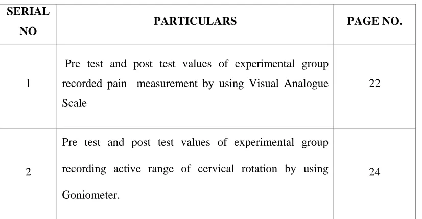
LIST OF TABLES
SERIAL
NO PARTICULARS PAGE NO.
1
Pre test and post test values of experimental group
recorded pain measurement by using Visual Analogue
Scale
22
2
Pre test and post test values of experimental group
recording active range of cervical rotation by using
Goniometer.
24
[image:8.612.116.529.90.305.2]LIST OF FIGURES
FIGURE NO CONTENT PAGE NO.
1 Graphical representation of Mean and Mean difference of Visual Analogue Scale
23
2
Graphical representation of Mean and Mean
difference of Active Range of Motion of cervical
rotation measured by goniometer.
ABSTRACT
OBJECTIVE: To find out the effectiveness of taping and ischemic compression
technique to reduce pain in levator scapulae trigger point related neck pain
DESIGN: This study is an experimental with pre test and post test evaluation.
PARTICIPATION: 20 subject aged 22-45 years neck pain patients are selected, the
client were treated by ischemic compression and taping techniques.
OUTCOME MEASURES: The outcome was measured using visual analogue scale ,
and active Range of cervical spine rotation measure by goniometer.
RESULTS: Statistical analysis done by using paired ‘t’ test showed that that there was
significant improvement in subject who underwent the treatment of taping and ischemic
compression technique.
CONCLUSION: Hence, it is concluded that the taping and ischemic compression
I. INTRODUCTION
Neck pain is the common problem in all over the countries. It occurs any age
group Individuals due to many causes like trauma, poor posture, pathological and
ergogenic problems.
Levator scapulae muscles trigger points also produce neck pain and
shoulder pain.Levator scapulae located on each side of the neck situated posteriorly. It
named for its action in“ Elevating” or “lifting” the scapulae. In Greek word , Levator
means “To lift”. Levatorscapulae muscle arise from posterior tubercle of transverse
process of first four cervical vertebrae. It is inserted into the medial border of superior
angle of scapulae. Levator scapulae muscle is blood supplied by dorsal scapular artery
and nerve supply is third, fourth cervical nerve and Dorsal scapular nerve(C5). Levator
muscles act along with trapezius to shrug the shoulder by its raising of the Scapula. If
the scapula fixed the muscle assist in cervical extension and used alone flex the neck
Laterally to one side.
Levator scapulae trigger point are frequently produce stiff neck because of
markedly limited neck movements. Myofascial trigger point in levator scapulae muscle
can be activated and perpetuated by Occupational activities such as typing, with the
head and neck turned to look towards one side of the type writer, making long telephone
calls ,and taking length with head turned toward someone sitting by one side. Vigorous
exercise that involved head turning such as playing tennis or swimming when out of
shape; The repetitive rotation of the head as in “spectator neck” using a cane or crutches
that are too long, Improper positioning, Flattend arch foot, Nutritional in adequacies
such as Folic acid, Vitamin B2, B6, B12,Vitamin C deficiency, Metabolic, Endocrine
infections. Others factor such as impaired sleep, Radiculopathy, prolonged immobility
also produce or activate Trigger points.
Trigger points are “A highly irritable localized spot of exquisite tenderness in
nodule in a taut band of skeletal muscle” (Travell,simon 1999).
Trigger points develop in the myofascia, mainly in the center of the muscle belly
where the motor end plate enters. However , secondary or satellite trigger point after
develop in a response to a primary trigger point. These satellite trigger point tend to
develop along the line of the stress. These line of stress may be built in at the time of
embryogenesis. The trigger point causes muscle to be sore, stiff, weak, and less flexible,
and may trigger sensory, motor and autonomic phenomena.
The Trigger points are classified as central or primary Trigger point, satellite or
secondary Trigger point, Attachment Trigger point, Diffuse Trigger point, Inactive or
latent Trigger point, Active Trigger point. The theories of Trigger point:
1. Motor end plate theory
2. Energy crisis theory
3. Radiculopathic theory
4. Polymodel theory (PMRS)
Levator scapulae is a postural muscle. Levator scapulae develops trigger point
tenderness in two locations. Central trigger points are at the where the muscle emerges
from beneath the anterior border of the upper trapezius and much more readily identified
secondary area where the muscle attaches to the superior angle of scapula. So the poor
posture is a powerful “activator and perpetuator” of Trigger points. The involved muscle
The Pain from trigger point activities in this muscle is mainly felt at the base of
the neck, but it also extended upwards towards the occiput; Outward to the back of the
shoulder and downwards along the inner border of the scapula.
The pain may also radiate interiorly around the chest wall along the course of the
fourth and fifth intercostal nerve when it may erroneously be diagnosed as being either
anginal or pleural or even more frequently as being due to intercostal nerve entrapment.
Trigger point examination by palpation of the muscle as it emerges from beneath
the trapezius at the angle of neck dislodges it most important central trigger point that
above the superior angle of the scapula often locates a second region of marked
tenderness.
During examination, it is only possible to palpate taught band that lie close to the
surface in superficially placed muscles. And if a palpable band is snapped by drawing
examining finger sharply across it at a trigger point site in a manner similar to that
employed when plucking a violin strings it is possible to evoke a transient contraction of
the muscle fibers. This local twitch response (LTR) may be either visible or felt under
the examine finger. And in same cases it is both seen and felt.
Levator scapulae Trigger points are differentially diagnosed with the condition
like scapulo costal syndrome, zygophyseal pain and bursitis.
Levator scapulae trigger point related neck pain is treated conservatively using
Non steroidal anti inflammatory drugs, analgesics, Muscle relaxants, Non manual
methods like saline injection and Dry needling and manual methods like Myofascial
massage, Ischemic compression technique or Manual inhibition technique, Taping,
Stretching, Positional release technique, Muscle energy technique, and Strengthening
exercises.
Ischemic compression ,technique help to reduce the Trigger points. Trigger
points can be deactivated by temporarily occluding their blood supply and causing a
reactive hyperemia (Increase blood supply): effectively flusing out the muscle of
inflammatory exudates and pain metabolites, breaking down scar tissue, and reducing
muscle tone. The muscle is nourished by the extra – flow through of blood, nerve ending
are desensitized and scar tissue is broken down so that the muscle fiber can move better.
In this technique pressure is progressively over the trigger point area or Nodule
or taut band in the muscle. The pressure is maintained until the tension is released. The
pressure is applied by the therapist thumb, finger pad, knuckles and elbow. The pressure
is applied 60 seconds maximum but mostly the desired effect is achieved in 10 – 20 sec.
repeated for 3or 4 times. Perhaps, moving to another part of the muscle, if the treated
area felt looser or softer to touch. stretching, and active exercises are beneficial after the
ischemic compression of the technique.
Taping will encourage correct the posture of the upper back and neck. It is
currently used by therapist to change muscle tone, more Lymphatic fluids, Correct the
posture, Tape can also be used facilitate weakened or lengthened muscles. It can be
positioned along the direction of the muscle fiber and pulled together or shorten the
tissue. Tape enhance our proprioceptive awareness of the muscles and increase muscle
1.1 NEED FOR THE STUDY
Levator scapulae trigger point related neck pain occurs mainly in poor posture
and overuse injuries of the muscles. Central trigger point in the levator scapulae at the
angle of neck is palpated. The lower trigger point tenderness by electrical stimulates
which produce pain refer to the neck and back of the shoulder and inter scapular region.
Many treatment have been a recommended various degree of success. But still
there is a need to find out the effective conservative treatment for levator scapulae
trigger point neck pain.
So the need for the study is to find out the effectiveness of ischemic
compression and taping technique to reduce pain in levator scapulae trigger point
related neck pain.
1.2. OBJECTIVE OF THE STUDY
To find out the effectiveness of ischemic compression and taping technique to
reduce pain in levator scapulae trigger point related neck pain
1.3.STATEMENT OF THE PROBLEM
The effectiveness of ischemic compression and taping technique to reduce pain
in levator scapulae trigger point related neck pain
1.4 HYPOTHESIS
Null hypothesis
There is no significant difference in pain and Range of motion following taping
and ischemic compression technique among levator scapulae trigger point related neck
Alternative hypothesis
There is significant difference in pain and Range of motion following taping
and ischemic compression technique among levator scapulae trigger point related neck
pain subjects.
1.5.OPERATIONAL DEFINITIONS
Trigger point
A trigger point is a hyper irritable spot associated with a taut band of a skeletal
muscle that is painful on compression or skeletal, and that can give rise to a typical
reffered pain pattern as well as autonomic phenomenon (Simon et. al. 1999)
Ischemic compression
Ischemic compression technique, pressure is applied slowly and progressively
over the trigger point as the tension in the trigger point and its taut band sub sides. The
pressure is maintained until the tenderness (or) tension is released.
Taping
Therapeutic taping techniques are techniques that utilize adhesive strapping tape
as a component of the management of the patients with musculoskeletal conditions.
(compact oxford dictionary 2009)
Tape can be used clinically to reduce strain on damaged tissue provide support
II.
REVIEW OF LITERATURE
SECTIONS
Section A: Studies on effect of ischemic compression technique for levator
scapulae trigger point related neck pain patients.
Section B: Studies on effect of taping technique of levator scapulae trigger point
related neck pain patients.
Section C: Studies on Reliability and validity of Visual Analog Scale (VAS) in
measuring pain
Section D: Studies on Reliability and validity of Goniometer in measuring Range
of Motion (ROM)
Section A
Studies on Effectiveness of Ischemic Compression Technique for Levator scapulae
Trigger point Related Neck pain Patients
Auguliera and Martin D.P et al. (2010) In their case study used a 27 years old
female patient effectiveness of ischemic compression for deliberate the blockage of
blood in trigger point area to increase local blood flow this result so that application of
ischemic compression is effective in presents of myofascial trigger point in neck.
Gemmell H. et al. (2007) did a study to determine immediate effect of ischemic
compression, trigger point technique and ultra sound on pain. This study was
Randomized control trial study in which two treatment group with 15 subjects
Penas et al. (2006) did a pilot study to compare the effect of ischemic compression
with transverse friction massage for myofascial in neck pain. In their study they include
40 subjects participated. This study concluded ischemic compression technique and
transverse massage technique were equally effective in reducing pain and tenderness of
neck muscles.
Bosch – Morell et al. (2006) The immediate effect of ischemic compression as a
trigger point therapy in a case of a patient with neck pain. The application of ischemic
compression is a safe and effective method to successfully treat elicited myofascial
trigger point. The purpose method is deliberate the blockage of blood in trigger point
area in order to increase blood flow. This washes away the waste products supplies
necessary oxygen and helps the affected tissue heal. The physical examination is
revealed of neck pain and stiffness in the neck muscle.
Fernandez - de - las - penas et al. (2004) A first systemic review analysing the
effectiveness of manual therapies in the management of trigger points found that few
studies had analysed manual interventions for trigger points follow up studies have
found that ischemic compression technique is effective in reducing pain sensitivity on
latent and active trigger point as well as pain elicited by active trigger point in patient
with neck pain we have recently shown that neuromuscular approaches are also effective
for reducing pain sensitivity in latent trigger point.
Nowicki et al. (2000) This technique of ischemic compression followed by stretching
provided the best and most effective decrease in trigger point.
Travell, Simon et al. (1999) Ischemic compression technique is non-invasive and
The virture of this technique is that is painless and imposes no additional strain on
attachment trigger point and there by avoids and aggravate them.
Hanten et al. (1997) Reported that stretching techniques reduce the intensity of
reffered pain and reduced the sensitivity of the trigger points treated. Many other authors
recommended that stretching alone is not enough but it is helpful as an adjunct to
ischemic compression.
Travell and Simon et al. (1983)In this ischemic compression technique, pressure is
applied slowly and progressively over the trigger point as the tension in the trigger point
and its taut band subsides pressure is maintained until the tenderness (or) tension is
released. This is followed by stretching the muscle.
Sola et al. (1980) This “stiff neck” muscle, when involved consistently limits neck
rotation due to pain on movements. If trigger points are active enough, they refer severe
Section B
Studies on effect of taping technique of levator scapulae trigger point related neck
pain patients.
John Gibbons et al. (2012) Neck pain can be caused by a number of factors
including muscle strain, ligament sprain, arthritis or a pinched nerve. Neck pain most
successfully treated by taping technique include cervical strain and cervical myofascial
pain. Myofascial pain in the neck can develop after trauma or within the medical
conditions such as physical stress, depression or insomnia. Tape alleviates pain by
providing support and increased circulation around the affected area allowing for rapid
recovery.
Thelen et al. (2008) Taping has become a widely used rehabilitation modality for the
prevention and treatment of musculoskeletal conditions. It alleviates pain and
improving healing in soft tissue.
Niddam et al. (2007) The Taping technique claims four effects :to normalize muscular
functions, to increase lymphatic and vascular flow, to diminish pain and aid in
correction of possible articular mal alignments. This taping technique is frequently
applied for pathologies in musculoskeletal system, especially in the field of sport
injuries.
Jaraczewska and Long et al. (2006) Their study concluded that tape provides
proprioception feedback to achieve postural alignment and joint position. Muscle
facilitation is another hypothesized benefit of application of tape.
Hammer et al. (2006) It is proposed that applying taping from the muscle origin to
insertion will produce a concentric pull on the fascia, stimulating increased muscle
contraction. To facilitate an eccentric or diminished contraction believed to occurs from
an eccentric pull on underlying fascia, application of tape from insertion to origin is
Julie Webe et al. (2005) Tape provide gentle tissue sheering all the day before
providing beneficial analgesic effect on the area in addition the tape gives as feedback to
prevent painful and improper positioning. Tape can also used to facilitate muscle, inhibit
a muscle or provides structural support to the joint.
Leon Chaitow et al. (2003) Tape can provide an analgesic effect by stimulation of
receptor in the skin as well as helping the client by providing feedback when the client
goes into a poor posture or incorrect posture.
Reimaan and Lephart et al. (2002) It is proposed that cutaneous mechanoreceptors
are stimulated by the stretch upon tape application which conveys information regarding
joint movement and position.
Lewit et al. (2000) In their case study Levator scapulae trigger point is treated by the
application of taping to the computer workers. The taping application is very effective to
Section C
Studies on Reliability and Validity of Visual Analog Scale (VAS) in measuring
pain.
Williamson and Hoggart et al. (2005) The Visual Anologue Scale while uses a 10 cm
blank line. The patent asked to record their pain level on the line where one end is
indicative of ‘ no pain’ and other is indicative of the ‘worst imaginable pain’. This scale
need to be delivered in a written format and consistency in its delivery to be being in
either a horizontal (or) a vertical line is necessary.
Anna Maria Carlsson et al. (2008) The Visual Analog Scale is simple and frequently
used method for of variation in intensity of pain. In clinical practice the percentage of
pain relief, assessed by Visual Analog Scale, is often considered as a measure of
efficacy of treatment. However as illustrated in the present study the validity of Visual
Analog Scale estimates performed by patients with chronic pain may be unsatisfactory.
Two type of Visual Analog Scale, an absolute and a comparative scale. Where
comparative with respective factor influencing the reliability and validity of pain
estimate.
Mark et al. (2003) The Visual Analog Scale also demonstrated present pain intensity
and change in pain intensity in association with performance status, diagnosis, setting,
psychological distress and global quality of life. Visual Analog Scale also analysed with
umerical ratings scale, box scale, or verbal rating scales. Visual Analog Scale is one of
the easiest method to access pain intensity.
Lisa Janice et al (2002) Validity is generally seen as the most important consideration
and requires little to no training or equipment however it has several limitation in
clinical use. It is likely that this represents a significant reduction of pain but we cannot
determine the percentile using Visual Analog Scale score. The FACES scale is an
adaptation of Visual Analog Scale for children.
John Gallagher et al. (2000) Realiability of Visual Analog Scale for acute pain
measurement as assed by the ICC appears to be high. Ninety percent of pain rating were
reproducible with in 9 mm. These data that the Visual Analog Scale is sufficiently
reliable to be used to assess acute pain.
Section D
Studies on Reliability and validity of Goniometer in measuring Range of Motion
(ROM)
Richard L. Gajdoslk et al. (2008) clinical measurement of range of motion is a
fundamental evaluation procedure with ubiquitous application in physical therapy
objective measurement of range of motion and correct interpretation of the measurement
result can have a substantial impact on the development of the scientific basis of
therapeutic intervention. The purpose of this article is to review the related literature on
the reliability and validity of goniometric measurement of the area.
Poussa M.S et al. (2005) according to the cervical Range of Motion (ROM) Reliability
measurement via the goniometer and excellent inter examinar Reliability for most of the
movement evaluated.
Bevilaqua – grossi et al. (2003) Range of Motion (ROM) evaluation has been widely
used to quantity musculoskeletal deficits, besides serving as a basics for evaluating
efficacy of therapeutic intervention. One of the most common musculoskeletal
Chen et al. (2000) who conducted a review to compare studies on cervical spine Range
motion with different measurement equipment. They reported that in most of the studies
III. METHODOLOGY
Subjects N=20
Patients with Levator scapulae Trigger Point related neck pain
Study design
Pre –test and post test, single group
design
Pre test
Ischemic compression therapy
and taping technique
Post test after 2 weeks
Statistical Analysis-Paired ‘t’
3.1. STUDY DESIGN
¾ The study design consists of experimental group with pre test and post test
evaluation.
3.2 SAMPLE DESIGN
¾ Sample is selected by using consecutive sampling techniques.
3.3. SAMPLE POPULATION
¾ The sample population include 20 patients.
3.4 STUDYSETTING
¾ Kumar Ortho hospital, Nagercoil
3.5 STUDY DURATION
¾ 6 months
3.6 CRITERIA FOR SELECTION OF SUBJECTS
3.6.1 Inclusion criteria
¾ Age : 20 - 40
¾ Gender : Male and Female
¾ Active and palpable myofascial trigger point on one side of the neck of levator
3.6.2. Exclusion criteria
¾ More than 45 years
¾ Clinical evidence of myelopathy or Radiculopathy
¾ Fracture or dislocation of cervical vertebrae
¾ Recent Neck and Shoulder Surgery patient
¾ Nonco – operated patients
¾ Mentally retarded patient
¾ Hyper sensitivity patient
¾ Tumours of neck and shoulder
¾ Skin infection like dermatitis
¾ Loss of sensation
¾ Open wounds in levator scapulae area
3.7 VARIABLES
3.7.1. Independent variables
¾ Ischemic compression technique
¾ Taping technique
3.7.2 Dependent variables
¾ Pain
¾ Range of Motion(ROM)
3.8 ASSESSMENT TOOL
¾Visual Analog Scale (VAS)
3.9 MEASUREMENT PROCEDURE
3.9.1 Visual analogue scale
Visual anolog scale is used to measure the severity of pain response that
patients experience immediately after the completion of treatment.
0 1 2 3 4 5 6 7 8 9 10
(No pain) (severe pain)
It consists of 10 cm horizontal line labled as no pain (0) and severe pain(10).
The patients correspond to the severity of pain patient’s experience.
3.9.2. GONIOMETER
Patient position – Sitting position
Axis – Vertex of the skull
Movable arm – tip of the nose
Stable arm – Imaginary connect the patients to acromian process
Procedure : Asking to turned the head of the patient activity and the range of motion
3.10 Tre 3.10 first knu and seco proc
to th
be d
0. TREATM
eatment pro
0.1 Ischemic
Patient p
t for palpati
uckle compre
then gradua
Figur
The isch
ond small am
cedure in ord
he position o
done.
MENT PRO
cedure:
c compressi
position is su
ing the trigg
ession applie
ally progress
re – 1 shows
hemic compr
mount of talc
der to reduce
of comfort, f
CEDURE
ion techniqu
upine lying o
ger point. A
ed to that ar
s deeper into
s the techniq
ression will
cum powder
e the noxiou
following w
ue
or sitting po
After locating
rea. The pre
the tissue, b
ques of isch
be maintain
r will be appl
us skin frictio
which unilater
osition. The t
g the trigger
essure will b
but it not hur
hemic compr
ned for 5 sec
lied over the
on. After thi
ral stretchin
therapist rela
r point, a fi
be gentle at t
rt patient.
ression ther
onds and rel
e trigger poin
is method w
g of levator
ax the musc
irm digital o
the beginnin
rapy
leased for
2-nt, before th
will be brough
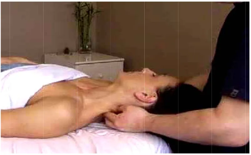
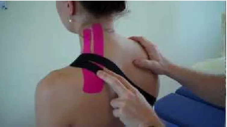
[image:28.612.118.519.247.495.2]3.10.2. Taping technique
¾ First clean skin with alcohol and lay down non-sticky tape (typically DonJolly
fix tape) perpendicular to the levator scapulae muscle or remove the hair of the
part.
¾ Relax the levator scapulae muscle and palpate tender points.
¾ lay one end of the tape at the lateral aspect of the clavicle near the acromion
clavicular joint.
¾ Pull the tape towards the medial aspect of the scapulae, pull tight to obtain
wrinkles in the skin.
¾ Apply end of tape at the medial border of the scapulae.
4.1. Des Pair Whe DATA scriptive ana
red ‘t’ test
ere,
S
=
∑
IV. D
ANALYSIS
The data co
lytical study
d – Differen
– Me
n – Total nu
s – Standard
(
2d
n
n 1
−
−
∑
∑
DATA ANA
S llected fromy was done b
nce between
ean of differ
umber of sub
d deviation
)
2d
n
ALYSIS A
m 20 subjects
by using Pair
pre test and
rence betwee
bjects
AND RESU
s were evalua
red ‘t’ test.
d post test va
en pre test an
ULT
ated statistic
alues
nd post test v cally.
4.2 DATA ANALYSIS AND INTERPRETATION
4.2.1 Data analysis of pain measurement
The tables -1 shows difference between the pre test and post test values of
experimental group recorded pain measurement by using Visual Analogue Scale (VAS)
Visual Analogue Scale
Measurement Mean Mean
difference
Standard
deviation
‘p’ value
Pre test
Post test
6.8
4.25
2.55 1 11.40
The ‘t’ test value for pre test and post test datas of experimental group was
11.40 and the table value was 2.861 at 0.005 level of significance. This shows that there
exist a significance difference between pre test and post test valued of experimental
Figure: 1 Graphical representation of Mean and Mean difference of Visual Analogue
Scale
6.8
4.25
2.55
0 1 2 3 4 5 6 7 8
Pre test Post test Mean difference
Means
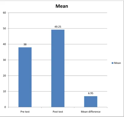
4.2.2. Data analysis of Active Range of Motion of cervical rotation
The tables showing difference between the pre test and post test values
of experimental group recording active range of cervical rotation by using Goniometer.
Goniometer
Measurement Mean Mean
difference
Standard
deviation
Paired ‘t’ test value
Pre test
Post test
38
49.25
11.25 6.95 7.23
The ‘t’ test value for pre test and post test datas of experimental group
was 7.23 and the table value was 2.861 at 0.05 level of significance. This shows that
there exist a significance difference between pre test and post test valued of
Figure: 2 Graphical representation of Mean and Mean difference of Active Range of
Motion of cervical pain measured by goniometer.
38
49.25
6.95
0 10 20 30 40 50 60
Pre test Post test Mean difference
Mean
4.3
RESULTS
The number of twenty(20) subjects were selected based on the selection
criteria and underwent pre test assessment by visual Analog Scale and goniometer
to measure pain and active Range of motion of cervical rotation respectively. The
subject were treated with ischemic compression and taping technique for two
weeks. After the intervention the post test measures were measured.
In the statistical analysis, the calculated t value for the Visual Analog Scale
was 11.40 which is greater than the table value 2.861 at 0.005 level. The active
Range of Motion measurement the calculated t value is 7.23 which is greater than
the table value 2.861 at 0.005 level of significance. Hence, the calculated t value for
pain and Active Range of Motion is more than the table value, the above values
shows there is significant difference in both Pain and Active Range of Motion after
V. CONCLUSION
An experimental study was conducted to investigate the effectiveness of
Ischemic compression and taping technique to reduce pain in levator scapulae
related neck pain patients.
20 patients were selected in this study consecutive manner. The levator
scapulae trigger point pain assessed by Visual Analogue Scale. The limited Range
of Motion of cervical pain assessed by goniometer.
The statistical result shows that the ischemic compression and taping
technique is effective for the reduction of pain in levator scapulae trigger point
related neck pain patients
5.1 Limitations
This study was very short term and therefore to make it more valid long term is
necessary.
Since the study has been done with smaller number of subjects further studies
should be conducted the large group of population.
5.2 Recommendation
• Number of subjects may be increased,
• More research in both interventions with consistent outcome measures,
• Study can be done with different variables.
VI.
BIBLIOGRAPHY
1) Simons DG, Travell J, Simons LS. (1999) Myofascial Pain and Dysfunction: The Trigger Point Manual. 2nd ed. Volume 1. Baltimore, MD: Williams & Wilkins.
2) Burnstock G. (2002) Structural and chemical organization of the autonomic neuroeffector system. In: Bolis CL, Licinio J, Govoni S, editors. Handbook of the Autonomic Nervous System in Health and Disease. New York: Marcel Dekker.
3) Macefield VG,Wallin BG.(1995) Modulation of muscle sympathetic activity during spontaneous and artificial ventilation and apnœa in humans. J Auton Nerv Syst.
4) Fernández-de-las-Peñas C, 2005). Manual therapies in the myofascial trigger point treatment: A systematic review. J Bodywork Mov Ther.
5) Fryer G, Hodgson L (2005) The effect of manual pressure release on myofascial trigger points in the upper trapezius muscle. J Bodywork Mov Ther.
6) Fernández-de-las-Peñas C, Alonso-Blanco (2006) The immediate effect of ischemic compression technique and transverse friction massage on tenderness of active and latent myofascial trigger points: A pilot study. J Bodywork Mov Ther.
7) Gemmell H, Miller P, Nordstrom H. (2008) Immediate effect of ischæmic compression and trigger point pressure release on neck pain and upper trapezius trigger points: A randomized controlled trial. Clin Chiropr.
8) Ibáñez-García J, Alburquerque-Sendín F, Rodríguez-Blanco C, et al. (2009)Changes in masseter muscle trigger points following straincounter/strain or neuro-muscular technique. J Bodywork Mov Ther.
9) Fernández-de-las-Peñas C, Fernández-Car-nero J, Galán-del-Río F, (2004) Are myofascial trigger points responsible of restricted range of motion? A clinical study (abstract) J Musculosk Pain.
muscle involving post-isometric relaxation or strain/counterstrain. J Bodywork Mov Ther. ;10:197–205.
11)Vernon H, Schneider M. (2009) Chiropractic management of myofascial trigger points and myofascial pain syndrome: A systematic review of the literature. J Manipulative Physiol Ther.
12)Shah JP, Phillips TM, Danoff JV, Gerber LH. (2005) An in vitro micro-analytical technique for measuring the local biochemical milieu of human skeletal muscle. J Appl Physiol.
13)Howing JL, Gross A, Gasner D, et al. (2001) A critical appraisal of review articles on the effectiveness of conservative treatment for neck pain. Spine.
14)Jette DU, Jette AM.( 1997) Professional uncertainty and treatment choices by physical therapists. Arch Phys Med Rehabil.
15)Triano JJ. (2001) Biomechanics of spinal manipulative therapy. Spine J.
16)Fernández-de-las-Penas C, (2005) Validity of the lateral gliding test as a tool for the diagnosis of inter-vertebral dysfunctions in the lower cervical spine. J Manipulative Physiol Ther.
17)Fernández-de-las-Peñas C, Downey C, (2005) Immediate changes in radiographically determined lateral flexion range of motion following a single cervical HVLA manipulation in patients presenting with mechanical neck pain: A case series. Int J Osteopath Med.
18)Martínez et al. (2006) Immediate effects on neck pain and active range of motion a"er a single cervical high-velocity low-amplitude manipulation in subjects presenting with mechanical neck pain: A randomized controlled trial. J Manipulative Physiol Ther. 19)Vernon H, Humphreys K, Hagino C. (2007) Chronic mechanical neck pain in adults
treated by manual therapy: A systematic review of change scores in randomized clinical trials. J Manipulative Physiol Ther.
21)Gerwin RD, Dommerholt D, Shah JP. (2004) An expansion of Simons' integrated hypothesis of trigger point formation.Curr Pain Head Reported.
22)McPartland JM, Simons DG.( 2006) Myofascial trigger points: Translating molecular theory into manual therapy. J Man Manipulative Ther.
23)Simons DG. (2001)Do endplate noise and spikes arise from normal motor endplates? Am J Phys Med Rehabil.
24)Couppé C, Midttun A (2001) Spontaneous needle electromyographic activity in myofascial trigger points in the infraspinatus muscle: A blinded assessment. J Musculosk Pain.
25)Simons DG, Hong CZ (2002)Endplate potentials are common to midfiber myofascial trigger points. Am J Phys Med Rehabil
VII ANNEXURE
Annexure- I
ASSESSMENT CHART
Physical therapy assessment chart
Name
Age
Gender
Occupation
Chief complaints
Medical history
• Past
• Present
Family history
Social history
Associated problems
On observation
• Body Built
• Posture
• Attitude of limbs
• Muscle wasting
• Edema
• Gait
• Deformity
On palpation
• Tenderness
• Swelling
• Muscle tightness
• Warmth
Pain assessment
• Side
• Site
• Duration
• Nature
• Aggravating factor
• Relieving factor
• Other if any
On examination
• Vital signs
• Motor Assessment
o Range Of Motion
o End Feels
o Manual Muscle Testing
o Joint Positions
• Sensory Assessment
o Superficial Sensations
o Combined
• Reflexes
o Superficial
o Deep
o Clonuses
• Dermatomes and Myotomes
• Limb Length Discrepancies
• Special Tests
• Functional Assessments
• Gait Assessments
Investigations
Clinical Impression
Differential Diagnosis
Final Diagnosis
Goals
• Short Term Goals
• Long Term Goals
Treatment Plan
• Electrotherapy Modalities
• Manipulations
• Exercise Therapy
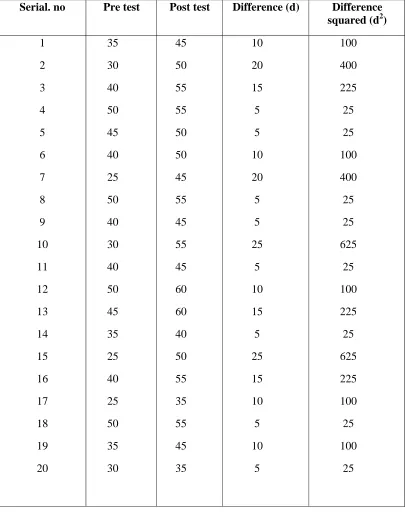
ANNEXURE – II
[image:43.612.123.532.147.607.2]RAW DATAS
Table: 7.2 Pre and post-test Visual Analog Scale values of Pain
Serial .No Pre test Post test Difference (d)
Difference squared (d2)
Table: 7.2 Pre and post-test Goniometer Range of Motion values of Pain
Serial. no Pre test Post test Difference (d) Difference squared (d2)
ANNEXURE- III
PATIENT CONSENT FORM
I ………aged……yrs,
voluntarily consent to participate in the research named “Effectiveness of
ischemic compression technique and taping technique to reduce pain in
levator scapulae trigger point related neck pain”
The researcher has explained me the treatment approach in brief, risk of
participation and has answered all the questions pertaining to the study to my
satisfaction.
Signature of Subject Signature of Researcher
Figure -2 shows levator scapulae trigger point
[image:46.612.124.501.483.694.2]