COMPARATIVE EVALUATION OF MECHANICAL
PROPERTIES OF BASE METAL ALLOYS AND
COMMERCIALLY PURE TITANIUM WITH EFFECT OF
LASER SURFACE TREATMENT - AN IN VITRO STUDY
Dissertation Submitted to
THE TAMILNADU Dr. M.G.R. MEDICAL UNIVERSITY
In partial fulfillment for the Degree of
MASTER OF DENTAL SURGERY
BRANCH I
PROSTHODONTICS AND CROWN & BRIDGE
ACKNOWLEDGEMENT
This dissertation is the result of work with immense support from many
people and it is a pleasure now that I have the opportunity to express my
gratitude to all of them.
I would be failing in my duty if I do not adequately convey my heartfelt
gratitude and my sincere thanks to my Head of the Department, Professor
Dr. N.S. Azhagarasan, M.D.S., Department of Prosthodontics and Crown & Bridge, Ragas Dental College and Hospital, Chennai, for his exceptional
guidance, tremendous encouragement, well-timed suggestions and heartfelt
support throughout my postgraduate programme which has never failed to
drive the best out of me. I would like to profoundly thank him for giving an
ultimate sculpt to this study. I will remember his help for ages.
I wish to express my gratitude to Dr. S. Ramachandran, M.D.S., Principal, Ragas Dental College and Hospital, Chennai, for his
encouragement throughout my postgraduate course. I also thank him for
permitting me to make use of the amenities in the institution.
I would like to express my real sense of respect, gratitude and thanks
to my Professor Dr. K. Chitra Shankar M.D.S., for her guidance, constant support, back up and valuable criticism extended to me during the period of
my study. The timely help and encouragement rendered by her had been
I would like to immensely thank Dr. R. Hariharan, M.D.S., Reader, for the valuable guidance and encouragement rendered by him. This
dissertation has been the fertile outcome of his massive endurance, support,
proficient guidance and counsel.
I would also like to thank Dr. K. Madhusudan, M.D.S.,
Dr.S.Jayakrishnakumar, M.D.S., Dr.Manoj Rajan, M.D.S., Dr.Saket
Miglani, M.D.S., Dr. M. Saravana Kumar, M.D.S., Dr. Sabarinathan. M.D.S., Dr. Vallabh Mahadevan, M.D.S and Dr. Divya Krishnan, M.D.S., for their valuable suggestions and help given throughout my study.
I thank Mr.Gurusami, M.E., Department of Mechanical Engineering,
Anna University, Chennai for his support and constant encouragement.
My sincere thanks to Mr.Parthasarathy, Metallurgist, and to
Mr.Jeyaraj, M.E., MET MECH LAB, Chennai for permitting me to do testing of samples and to Mr.Prabhakaran, Laser specialist, Srirangam for helping
me in laser surface treatment of samples.
I thank Mrs.Deepa for helping me with the statistical analysis for the
study,
It would not be justifiable on my part if I do not acknowledge the help
of my fellow colleagues, seniors and juniors and my friends for their
Last but not the least, even though words wouldn’t do much justice , I
would like to specially thank my father Dr. Gopal Periyasamy Pillai, my
mother Mrs.Manjula, my fiancée Dr.Vishali, my brother Mr.Arun, my sister Mrs.Renuka for their blessings and love.
CONTENTS
S.NO TITLE PAGE.NO
1.
INTRODUCTION 1
2.
REVIEW OF LITERATURE 9
3.
MATERIALS AND METHODS 18
4.
RESULTS 35
5.
DISCUSSION 56
6.
CONCLUSION 65
7.
SUMMARY 73
LIST OF TABLES
S.NO. TITLE PAGE
No.
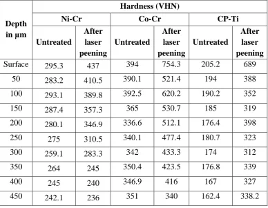
1. Hardness depth profile of Ni-Cr, Co-Cr and CP-Ti samples before (Control groups I, II & III) and after laser peening (Test groups I, II & III)
38
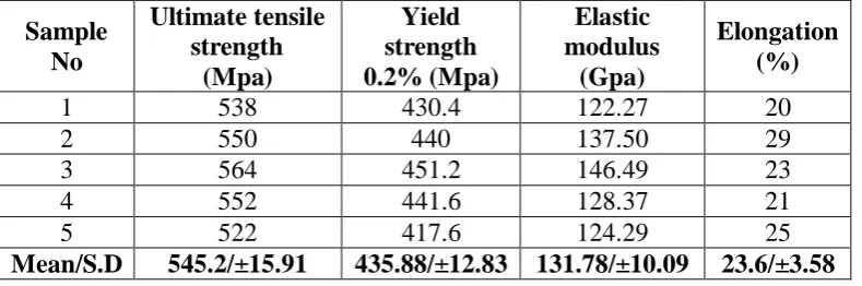
2. Basic values and mean values of Ultimate tensile strength, Yield strength, Modulus of elasticity and Percentage of elongation of Ni-Cr samples before laser peening (Control group I)
39
3. Basic values and mean values of Ultimate tensile strength, Yield strength, Modulus of elasticity and Percentage of elongation of Co-Cr samples before laser peening (Control group II)
39
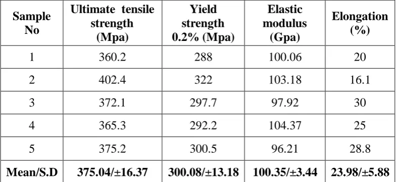
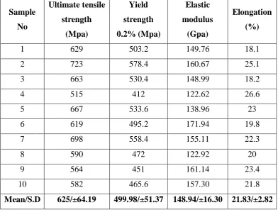
4. Basic values and mean values of Ultimate tensile strength, Yield strength, Modulus of elasticity and Percentage of elongation of CP-Ti samples before laser peening (Control group III)
40
5. Basic values and mean values of Ultimate tensile strength, Yield strength, Modulus of elasticity and Percentage of elongation of Ni-Cr samples after laser peening (Test group I)
41
6. Basic values and mean values of Ultimate tensile strength, Yield strength, Modulus of elasticity and Percentage of elongation of Co-Cr samples after laser peening (Test group II)
42
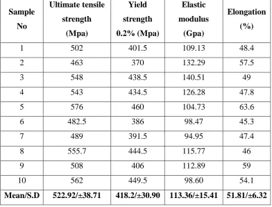
7. Basic values and mean values of Ultimate tensile strength, Yield strength, Modulus of elasticity and Percentage of elongation of CP-Ti samples after laser peening (Test group III)
43
8. Comparison of Surface Hardness of Ni-Cr, Co-Cr and CP-Ti samples before (Control groups I, II & III) and after laser peening (Test groups I, II & III)
44
9. Comparison of means of Ultimate tensile strength, Yield strength, Modulus of elasticity and Percentage of elongation of Ni-Cr samples before (Control group I) and after laser peening (Test group I)
10. Comparison of means of Ultimate tensile strength, Yield strength, Modulus of elasticity and Percentage of elongation of Co-Cr samples before (Control group II) and after laser peening (Test group II)
46
11. Comparison of means of Ultimate tensile strength, Yield strength, Modulus of elasticity and Percentage of elongation of CP-Ti samples before (Control group III) and after laser peening (Test group III)
47
12. Comparison of means of Ultimate tensile strength of Ni-Cr, Co-Cr and CP-Ti samples before laser peening (Control groups I,II & III)
48
13. Comparison of means of Yield strength of Ni-Cr, Co-Cr
and CP-Ti samples before laser peening (Control groups I, II & III)
49
14. Comparison of means of Modulus of elasticity of Ni-Cr, Co-Cr and CP-Ti samples before laser peening (Control groups I, II & III)
50
15. Comparison of means of Percentage of elongation of Ni-Cr, Co-Cr and CP-Ti samples before laser peening (Control groups I, II & III)
51
16. Comparison of means of Ultimate tensile strength of Ni-Cr,
Co-Cr and CP-Ti samples after laser peening (Test groups I, II & III)
52
17. Comparison of means of Yield strength of Ni-Cr, Co-Cr and CP-Ti samples after laser peening (Test groups I, II & III)
53
18. Comparison of means of Modulus of elasticity of Ni-Cr,
Co-Cr and CP-Ti samples after laser peening (Test groups I, II & III)
54
19. Comparison of means of Percentage of elongation of Ni-Cr,
Co-Cr and CP-Ti samples after laser peening (Test groups I, II & III)
LIST OF GRAPHS
GRAPH
NO.
TITLE
1. Hardness depth profile of Ni-Cr samples before (Control group I) and
after laser peening (Test group I)
2. Hardness depth profile of Co-Cr samples before (Control group II) and
after laser peening (Test group II)
3. Hardness depth profile of CP-Ti samples before (Control group III)
and after laser peening (Test group III)
4. Basic values and Comparison of means of Ultimate Tensile Strength of
untreated Ni-Cr, Co-Cr and CP-Ti samples (Control groups I, II & III)
5. Basic values and Comparison of means of Yield Strength of untreated
Ni-Cr, Co-Cr and CP-Ti samples (Control groups I, II & III)
6. Basic values and Comparison of Modulus of Elasticity of untreated
Ni-Cr, Co-Cr and CP-Ti samples (Control groups I, II & III)
7. Basic values and Comparison of Percentage of Elongation of untreated
Ni-Cr, Co-Cr and CP-Ti samples (Control groups I, II & III)
8. Basic values and Comparison of Ultimate Tensile Strength of Ni-Cr,
Co-Cr and CP-Ti samples after laser peening (Test groups I, II & III)
9. Basic values and Comparison of Yield strength of Ni-Cr, Co-Cr and
10. Basic values and Comparison of Modulus of Elasticity of Ni-Cr,
Co-Cr and CP-Ti samples after laser peening (Test groups I, II & III)
11. Basic values and Comparison of Percentage of Elongation of Ni-Cr,
Co-Cr and CP-Ti samples after laser peening (Test groups I, II & III)
12. Comparison of means of Ultimate Tensile Strength of Ni-Cr, Co-Cr
and CP-Ti samples before (Control groups I, II & III) and after laser
peening (Test groups I, II & III)
13. Comparison of means of Yield Strength of Ni-Cr, Co-Cr and CP-Ti
samples before (Control groups I, II & III) and after laser peening
(Test groups I, II & III)
14. Comparison of means of Modulus of Elasticity of Ni-Cr, Co-Cr and
CP-Ti samples before (Control groups I, II & III) and after laser
peening (Test groups I, II & III)
15. Comparison of means of Percentage of Elongation of Ni-Cr, Co-Cr and
CP-Ti samples before (Control groups I, II & III) and after laser
ANNEXURE
LIST OF FIGURES
Fig No. TITLE
Fig: 1 Nickel-Chromium alloy ingots
Fig: 2 Cobalt-Chromium alloy ingots
Fig: 3 Commercially pure titanium (Grade 2) ingots
Fig: 4 Auto-polymerizing acrylic resin
Fig: 5 Sprue wax
Fig: 6 Silicone casting ring
Fig: 7 Titanium metal casting ring
Fig: 8 Surfactant spray
Fig: 9(a) Phosphate bonded investment material -Powder
Fig: 9(b) Phosphate bonded investment material- Liquid
Fig: 10(a) Magnesium oxide based investment material-Powder
Fig: 10(b) Magnesium oxide based investment material-Liquid
Fig: 11 Aluminum oxide powder
Fig: 13 Tungsten carbide burs
Fig: 14 Silicon carbide rubber points
Fig: 15 Bristle brushes
Fig: 16 Alloy polishing paste
Fig: 17 Dental occlusal film
Fig: 18 Vacuum mixer
Fig: 19 Burnout furnace for Ni-Cr and Co-Cr groups
Fig: 20 Burnout furnace for CP-Ti group
Fig: 21 Induction casting machine
Fig: 22 Argon arc melting pressure casting machine
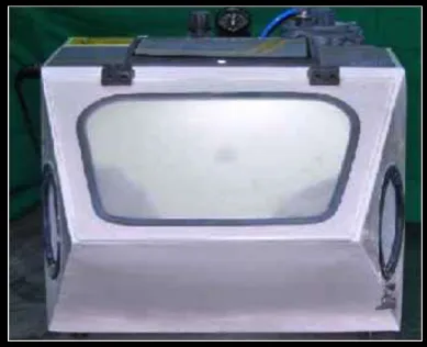
Fig: 23 Sandblaster
Fig: 24 Alloy grinder & polisher
Fig: 25 Dental X-ray unit
Fig: 26 Metallurgical microscope
Fig: 27 Optical layout of metallurgical microscope
Fig: 28 Micro Vickers Hardness Tester
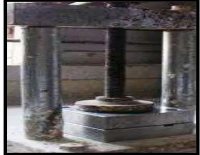
Fig: 29 Tensometer
Fig: 30 Schematic representation of Tensometer
Fig: 31 Nd-YAG laser machine
Fig: 32 Schematic representation of Nd-YAG laser machine
Fig: 33(a & b) Stainless steel metal die
Fig: 34 Dimensions of stainless steel die
Fig: 35 Dimensions of cast test samples
Fig: 36 Filling of slots of die with auto-polymerizing acrylic resin
Fig: 37 Bench pressing of patterns in stainless steel die
Fig: 38 (a & b) Separation of acrylic patterns from metal die
Fig: 39 Finished acrylic patterns
Fig: 40 Sprue attached to the acrylic patterns
Fig: 41 Positioning of silicone casting ring
Fig: 42 Patterns sprayed with surfactant
Fig: 43 Investing the pattern for Ni-Cr / Co-Cr group with silicone ring
Fig: 44 Set investment mold
Fig: 46 Mold placed in burnout furnace (Ni-Cr and Co-Cr groups)
Fig: 47 Mold placed in burnout furnace (CP-Ti groups)
Fig: 48 Mold placed in induction casting machine
Fig: 49 Bench cooling of mold after casting
Fig: 50 CP-Ti casting done in Argon arc melting casting machine
Fig: 51 CP-Ti Ingot heated under argon atmosphere
Fig: 52 Divesting of castings
Fig: 53 Sandblasting the sample
Fig: 54 Sprue cut using carborundum disc
Fig: 55 Trimming of sample with tungsten carbide bur
Fig: 56 Finishing of sample using silicone carbide rubber points
Fig: 57 Polishing the sample
Fig: 58 Finished Ni-Cr samples
Fig: 59 Finished Co-Cr samples
Fig: 60 Finished CP-Ti samples
Fig: 61 Radiographic inspection of Ni-Cr samples for defects
Fig: 63 Radiographic inspection of CP-Ti samples for defects
Discarded sample due to defect (A)
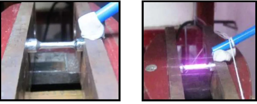
Fig: 64 Sample mounted on a customised jig
Fig: 65 Argon gas shielding during laser treatment
Fig: 66 Prepared sample for microscopic examination
Fig: 67 Microscopic examination of sample
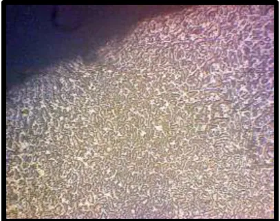
Fig: 68 Metallurgical microscopic image of untreated Ni-Cr sample
Fig: 69 Metallurgical microscopic image of laser treated Ni-Cr sample
Fig: 70 Metallurgical microscopic image of untreated Co-Cr sample
Fig: 71 Metallurgical microscopic image of laser treated Co-Cr sample
Fig: 72 Metallurgical microscopic image of untreated CP-Ti sample
Fig: 73 Metallurgical microscopic image of laser treated CP-Ti sample
Fig: 74 Measurement of Vickers microhardness
Fig: 75 Control sample engaged in the tensometer
Fig: 76 Fractured control samples
Fig: 77 Test sample engaged in the tensometer
ABSTRACT
Background: Prosthodontic restorations require metals or alloys with good
mechanical properties. CP-Ti has good biocompatibility but is weaker
mechanically in comparison to Ni-Cr and Co-Cr alloys. An attempt was made
in this study to improve the mechanical properties of CP-Ti using laser
peening.
Materials and methods: 51 dumbbell shaped cast samples were fabricated
with Ni-Cr (n=17), Co-Cr (n=17) and CP-Ti (n=17). 6 cast samples from each
group served as Control group while 11 cast samples were subjected to laser
peening (Test group). Cross section of 1 Control and test group sample each of
Ni-Cr, Co-Cr and CP-Ti were subjected to microscopic examination following
which microhardness was evaluated. Tensile testing of 5 control group and 10
test group samples each for Ni-Cr, Co-Cr and CP-Ti were conducted using
Tensometer to evaluate mechanical properties.
Results:Upon laser peening surface hardness increased for Ni-Cr, Co-Cr and
CP-Ti samples. Ultimate Tensile Strength and Yield Strength of CP-Ti
increased whereas these values reduced for Ni-Cr and Co-Cr samples.
Modulus of elasticity increased for Co-Cr and CP-Ti while it reduced for
Ni-Cr. Percentage of Elongation reduced for CP-Ti samples.
Conclusion:Laser peening for CP-Ti resulted in improvement of mechanical
properties.
Keywords: CP-Ti, Mechanical properties, Laser peening, Nd:YAG laser
1
INTRODUCTION
Dental casting alloys play a prominent role in the fabrication of fixed,
removable and implant prostheses. The use of alloys provides physical,
mechanical and biologic properties that are required for successful, long-term
prosthodontic restorations.46,53,54,55 The significant improvement in the
properties of casting alloys ensures their role as the principal material for years
to come since no other material has the combination of strength, modulus of
elasticity, wear resistance and biologic compatibility that a material must have
for long term survival in the mouth.55
The American Dental Association (ADA) divides casting alloys into
three groups on the basis of wt % composition as high-noble, noble and
base-metal alloys.13,54,55 The ADA revised the classification system (Mar 2003) for
alloys for fixed prosthodontics with regard to the use of titanium and titanium
alloys in dentistry. The Council classified casting alloys as high-noble,
titanium, noble and base-metal alloys in the revised classification system
because of the excellent biocompatibility of titanium.1
The cast restorations must be made of an alloy that meets certain
minimum requirements for strength, stability, castability, corrosion and tarnish
resistance, burnishability, polishability and biocompatibility for the long term
success.44 Metal-ceramic alloys have additional requirements that include
higher melting temperature, thermal compatibility with ceramics, oxide
2
tensile strength, yield strength, flexural strength, torsional strength, fatigue
strength, impact strength, modulus of elasticity, percentage elongation,
malleability, fracture toughness and hardness are important for good clinical
performance of dental alloys.2,15 Among these properties, the ultimate tensile
strength, yield strength, modulus of elasticity and percentage elongation are
the most important.5,15,16,18,31,34,37,41,43,45,46,51,54,55,57,58
Ultimate tensile strength is the maximum stress that a material can
withstand while being stretched or pulled before necking.2,15 It indicates the
ability of a metal structure to endure the applied forces.15,18 Tensile strength is
necessary for the prosthesis to withstand occlusal forces within the oral
cavity.36 Yield strength is the amount of stress required to produce a
pre-established amount of permanent strain (i.e., change in length) of the
alloy.2,15
The modulus of elasticity (elastic modulus) is a measure of the
stiffness or rigidity of an alloy.54 The elastic modulus for prosthodontic alloys
needs to be high so that the prosthesis can resist flexure, especially in
metal-ceramic restorations where any flexure will cause fracture of the porcelain. It
is also important for removable partial dentures so that the major connectors
have adequate rigidity to prevent flexure during placement and function and
will therefore transmit occlusal forces more efficiently to the remaining teeth
or other tissues.54 Resistance to flexure is also beneficial because clasps can be
3
The percentage of elongation is the measure of ductility of the
material.5,15 Ductility is the degree to which a material can be permanently
deformed by a tensile force without undergoing fracture or rupture. It is
requiredto improve marginal fit of a restoration by burnishing.7 The hardness
of the alloy must be enough to resist occlusal forces but not wear opposing
teeth.15,55
The high-noble alloys are generally favourable for manipulation and
clinical service because of high ductility, adequate yield strength and ultimate
tensile strength, excellent castability, excellent porcelain bonding, favourable
esthetics, good biocompatibility, excellent corrosion and tarnish resistance,
not technique sensitive, good burnishability and easy to adjust and finish, but
none of these alloys have a high elastic modulus value.54,55 In single units and
short-span fixed partial dentures, gold-based alloy systems possess ample
elastic modulus to prevent this flexure. However, in long span fixed partial
dentures, elastic modulus is not sufficient to prevent clinically problematic
levels of flexure.54 The other disadvantages are high cost, poor sag resistance
in case of long span fixed partial dentures, low hardness and high density. 2,15
Noble alloys with lower content of noble elements and inclusion of
Cu, Ag and Ga overcome the drawback of low elastic moduli of high noble
alloys, thereby increasing the flexural resistance.2,15 Also, they have better sag
resistance than high-noble alloys.46,54,55 Whereas, the corrosion resistance of
the noble alloys is variable; it depends on the microstructure and the presence
4
it has the drawback of higher costs when compared to base metal
alloys.2,15,46,54,55
The need for better physical and mechanical properties and affordable
costs led to the development of base metal alloys.2,8,15,54,55 The base-metal
alloys like Nickel-chromium (Ni-Cr) and Cobalt-chromium (Co-Cr) share high
physical properties, and these alloys have higher modulus of elasticity6,8,46,47
and superior mechanical properties when compared to high noble and noble
alloys. They have low density8 and excellent sag resistance making them
suitable for long span fixed partial dentures.2,15,46,54,55 They are much lower in
cost6,8,53 and have excellent porcelain bonding.2
These alloys also have several disadvantages namely higher corrosion
in acidic environments,22,53 risk of patient allergy,22,53 and difficult in
soldering 2 when compared to high-noble and noble alloys. Furthermore, their
liquidus temperatures are the highest among all prosthodontic alloys, making
them harder to cast, finish and polish and ensure appropriate marginal fit of
restoration.2,8,14 From the standpoint of porcelain application, these alloys all
form heavy, dark oxide layers that are more difficult to esthetically manage
than those formed by alloys in the noble and high-noble alloy groups.55 The
compatibility of coefficients of thermal expansion between Co-Cr alloys and
porcelains may also be problematic.45
The base metal alloys, such as Ni-Cr and Co-Cr have been used for the
fabrication of fixed and removable prosthesis, but there are concerns about
5
In recent years CP-Ti and its alloys have become an alternative to
gold and base metal alloys due to their excellent biocompatibility, good
corrosion resistance, low density, high mechanical sag resistance and
relatively low cost.4,7,9,11,19,21,22,53,54,55 These alloys can be used for all-metal
and metal-ceramic prostheses, as well as for implants and removable partial
denture frameworks.7,9,18,25 According to the American Society for Testing
and Materials (ASTM), there are four unalloyed grades of CP-Ti (Grades 1-4),
based on the concentration of oxygen and iron.
The mechanical strength of pure titanium increases due to the inclusion
of impurities during casting.10 However, this strength might not be sufficient
for multi-unit fixed partial dentures and metal frameworks of removable
partial dentures18,26,30,32,33,34,40,57 and are reported to possess low wear
resistance.23,26 Moreover, titanium has a high melting point(1668° C), and a
special casting machine with arc-melting capability and an argon atmosphere
is typically used, along with a compatible casting investment, to ensure
acceptable castability which makes them expensive and technique
sensitive.12,19,20,24,31,59 This high melting point is accompanied by a relatively
low thermal expansion coefficient, necessitating special low-expansion dental
porcelains for bonding to titanium.25,61
Considering the properties of all the casting alloys, none of them
satisfy all the physical, mechanical and biologic requirements for the
fabrication of prosthodontic restorations.2,54,55 Various methods like
6
have been reported in literature for high-noble, noble and base metal alloys for
enhancing their physical and mechanical properties. Recently, laser peening
method has been employed for improving mechanical properties of
commercially pure titanium and has given good results.43,57 Laser peening
(laser surface treatment) is an innovative surface enhancement process used to
improve fatigue life. This process creates residual compressive stresses deep
into the surfaces. These compressive surface stresses inhibit the initiation and
propagation of fatigue cracks. When laser surface treatment is applied to cast
titanium metal frameworks, it is expected that the titanium framework will
have high mechanical strength to withstand mastication stresses.57
In the literature, comparative evaluation of the various mechanical
properties of Ni-Cr, Co-Cr and CP-Ti has been reported.6,18,38,39,45,46,49,54,55,57,58
The evaluations have been done on both untreated and surface treated alloy
specimens.12,47,60 However, literature regarding the effect of laser peening on
the mechanical properties of Ni-Cr, Co-Cr and CP-Ti alloys is recently
emerging and sparse.43,57
Considering the significance of this new technology in potentially
improving the mechanical properties and its relevance to clinical situations,
the present in-vitro study was aimed to comparatively evaluate the mechanical
properties of base metal alloys and commercially pure titanium with effect of
laser surface treatment. Also added to the aim of the study were the following
7
1. To evaluate the hardness of Ni-Cr, Co-Cr and CP-Ti test samples at
various depths before and after laser peening.
2. To evaluate the ultimate tensile strength, yield strength, modulus of
elasticity and percentage of elongation of Ni-Cr before laser peening.
3. To evaluate the ultimate tensile strength, yield strength, modulus of
elasticity and percentage of elongation of Co-Cr before laser peening.
4. To evaluate the ultimate tensile strength, yield strength, modulus of
elasticity and percentage of elongation of CP-Ti before laser peening.
5. To evaluate the ultimate tensile strength, yield strength, modulus of
elasticity and percentage of elongation of Ni-Cr after laser peening.
6. To evaluate the ultimate tensile strength, yield strength, modulus of
elasticity and percentage of elongation of Co-Cr after laser peening.
7. To evaluate the ultimate tensile strength, yield strength, modulus of
elasticity and percentage elongation of CP-Ti after laser peening.
8. To comparatively evaluate the surface hardness of Ni-Cr, Co-Cr and
CP-Ti before and after laser peening.
9. To comparatively evaluate the ultimate tensile strength, yield strength,
modulus of elasticity and percentage of elongation of Ni-Cr before and
after laser peening.
10.To comparatively evaluate the ultimate tensile strength, yield strength,
modulus of elasticity and percentage of elongation of Co-Cr before and
8
11.To comparatively evaluate the ultimate tensile strength, yield strength,
modulus of elasticity and percentage of elongation of CP-Ti before and
after laser peening.
12.To comparatively evaluate the ultimate tensile strength, yield strength,
modulus of elasticity and percentage of elongation of Ni-Cr, Co-Cr and
CP-Ti before laser peening.
13.To comparatively evaluate the ultimate tensile strength, yield strength,
modulus of elasticity and percentage of elongation of Ni-Cr, Co-Cr and
9
REVIEW OF LITERATURE
Civjan S et al (1972)12 stated that the main problems with the base
metal alloys are their excessive hardness and inadequate elongation. They
studied the effects of heat treatment on the mechanical properties of two
nickel-chromium-based casting alloys and concluded that both alloys showed
rapid response to heat treatment. Both alloys had similar mechanical and heat
treatment characteristics and in their softened state, they lost up to 50% of
their as-cast hardness and had elongations of 25 to 45%.
Baran GR et al (1983)5 in their article on metallurgy of Ni-Cr alloys
for fixed prosthodontics has mentioned that the important mechanical
properties necessary for dental casting alloys are hardness, yield strength,
tensile strength and percentage elongation.
Preston DJ (1984)44 in his article on metal-ceramic restoration has
stated that alloy selection should consider physical properties, mechanical
properties, compatibility with a given porcelain, ease of manipulation and
cost. Among the mechanical properties he has considered that the yield
strength and modulus of elasticity are the most important.
Asgar K et al (1985)3 conducted a study on influence of different
casting machines on castability of crown and bridge alloys. The alloys
included one base metal alloy, two high-fusing noble metal alloys and one
10
among casting machines and alloys and that the type of casting machine had a
stronger effect on castability.
Hensten-Pettersen A (1992)22 in his article on side-effects of casting
alloys has stated that the incidence of side-effects in prosthodontics was about
l:400. About 27% of the reactions were related to base-metal alloys containing
cobalt, chromium, and nickel and to noble based palladium alloys. They also
inferred from various studies that all dental casting alloys had potential for
eliciting adverse reaction in hypersensitive patients except unalloyed titanium
which was considered most biocompatible among all other casting alloys.
Berg E et al (1995)7conducted a study on the mechanical properties of
laser welded cast and wrought titanium base and compared them with those of
a brazed Type 4 gold casting alloy. Ultimate tensile strength, 0.2 % yield
strength and percentage elongation were recorded for joined and unjoined
bars. The strength of the laser welded titanium equalled that of the brazed gold
alloy, suggesting that dental restorations made of cast and wrought titanium
would satisfy ordinary clinical requirements.
Wang R et al (1996)52 in their review article on titanium for
prosthodontic applications have mentioned that the light weight of titanium
and its strength-to-weight ratio, high ductility and low thermal conductivity
would permit design modifications in titanium restorations and removable
11
titanium raw material and its favourable microhardness make them attractive
for dental prostheses.
Chai T et al (1998)10 studied the mechanical properties of laser
welded cast CP-Ti under different laser welding conditions to find the optimal
parameters in terms of duration and voltage (energy level). They found that
the voltage was the only significant factor that influenced the ultimate tensile
strength and 0.2% yield strength of the joint, whereas the duration was not a
significant factor. They concluded that the laser-welded joint achieved
superior mechanical properties.
Wataha JC et al (2000)53in their review article on biocompatibility of
dental casting alloys stated that the single most relevant property of a casting
alloy to its biologic safety is its corrosion. Several elements such as nickel and
cobalt have relatively high potential to cause allergy. To minimize biologic
risks, dentists should select alloys that have the lowest release of elements by
using high-noble or noble alloys with single-phase microstructures. They
concluded that selection of an alloy should be made on a case-by-case basis
using corrosion and biologic data from dental manufacturers.
Zinelis S et al (2000)62 conducted a study to evaluate the effect of
pressure of helium, argon, krypton, and xenon on the porosity, microstructure,
and mechanical properties of commercially pure titanium (Cp-Ti) castings.
Groups A, C, E, and G were cast under a pressure of 1 atm, and groups B, D,
12
respectively. The VHN of the as-received cp Ti was significantly greater than
all the cast groups tested. Groups cast under He showed the highest VHN,
yield strength, and fracture stress. They concluded that the porosity and
mechanical properties of cp Ti castings are dependent on the gas type and
pressure, whereas the microstructure remains unaffected.
Cecconi TB et al (2002)9 have stated that titanium is the most
biocompatible metal available for dental castings. However, they expressed
concern about the castability of titanium used on a daily basis. They conducted
a study on radiographic evaluation of titanium partial denture frameworks to
ascertain whether these castings were technically acceptable for clinical use.
Three hundred Grade II titanium removable partial denture frameworks were
cast and were evaluated radiographically and 97% were rated acceptable for
clinical use and concluded favourably for use of titanium castings on a
daily basis.
Liu J et al (2002)35 examined the joint strength of titanium laser
welding using several levels of laser output energy. They observed that the
penetration depth by laser was different among the parent metals because the
rate of laser beam absorption, thermal conductivity and melting point are
different in each metal. Base metals have a greater rate of laser beam
absorption and lower thermal conductivity compared to the noble metals. They
found that titanium in particular has a very low thermal conductivity and high
rate of laser beam absorption which makes it easy for the laser to penetrate
13
Wataha JC et al (2002)54 in their article on alloys for prosthodontic
restorations have stated that several properties of alloys are critical to the
clinical performance of restorations which includes the grain size and
structure, mechanical properties such as ultimate tensile strength, yield
strength, modulus of elasticity and hardness, porcelain bonding properties
such as coefficient of thermal expansion and oxide color and biological
properties such as corrosion resistance.
Lin Chai-Wei et al (2004)34 conducted a study to compare the
castability and mechanical properties of CP-Ti, Ti-6Al-7Nb and a newly
developed Ti-15Mo-1Bi in regard to their as-cast state. Experimental results
indicated that the hardened layer thicknesses of three materials were similar.
The bulk hardness of CP-Ti was much lower than the other two alloys which
displayed similar hardness. The bending modulus of Ti-15Mo-1Bi was
significantly lower than CP-Ti and Ti-6Al-7Nb. Tensile test also indicated that
the Ti-15Mo-1Bi and Ti-6Al-7Nb alloys have far higher strengths and lower
elongations than CP-Ti.
Wataha JC et al (2004)55in their article on casting alloys have stated
that the the mechanical properties of the casting alloy must be considered
important and customized for a particular clinical situation whereas, cost and
color are the least important factors in selecting a material for a successful
14
Eliopoulos D et al (2005)18conducted a study to evaluate the effect of
the type of the investment material on the thickness of the contamination zone
and on the mechanical properties such as modulus of elasticity, yield strength,
elongation, and hardness of CP-Ti castings. From the results of this study they
concluded that the extent of the contamination zone as well as the yield
strength and percentage elongation of the CP-Ti castings were significantly
affected by the type of the investment material.
Rocha SS et al (2006)47 evaluated the effect of different heat
treatments on the Vickers hardness of CP-Ti and Ti-6Al-4V cast alloys. They
concluded that heat treatments enhanced the hardness of both CP-Ti and
Ti-6Al-4V alloy which may be due to the martensitic transformation and
micro-structural alteration.
Watanabe I et al (2006)56 conducted a study to investigate the effect
of surface preparation on the Nd:YAG laser penetration into cast titanium and
gold alloy. They found that increasing the voltage or pulse duration and
decreasing the spot diameter generally increases the laser penetration into the
alloys. Other factors affected the laser penetration are thermal conductivity
(Tc) and the rate of laser beam absorption (Ba) of the alloys. Lower the Tc and
the greater Ba of the alloy, the deeper the laser can penetrate into the alloys.
Oliveira PC et al (2007)41 conducted a study on the influence of the
final temperature of investment heating on the tensile strength and Vickers
15
significant differences in tensile strength and hardness was found only
between the materials used and not between the temperatures of the
investment mold used for casting for both the materials.
Roach M (2007)45 reviewed the base metal alloys used for dental
restorations and implants and concluded that Ni-Cr alloys have superior
properties for use in porcelain-fused-to-ceramic (PFM) applications. He also
observed that the Co-Cr alloys are more corrosion-resistant than the Ni-Cr
alloys and have physical properties similar to that of the Ni-Cr alloys. Cast
Titanium and its alloys have physical properties which is comparable to that of
other base metal alloys. Also titanium and its alloys have excellent
biocompatibility and corrosion resistant.
Roberts W et al (2009)46 in their review article on metal-ceramic
alloys in dentistry stated that the increased use of metal-ceramic restorations
was the result of their proven history of clinical performance, acceptable
esthetics, and satisfactory physical properties. Good clinical performance has
been attested to by longitudinal studies that reported that up to 88.7% of
metal-ceramic crowns and 80.2% of metal-ceramic fixed partial dentures were
still in function after 10 years. He was also in opinion that although
considerable function may be borne by the ceramic portion of a metal-ceramic
restoration, the success of the entire prosthesis depends largely on the physical
16
Watanabe I et al (2009)57 conducted a study to investigate the effect
of laser surface treatment on the mechanical properties of cast titanium. The
cast titanium specimens were laser-treated on the surface using a dental
Nd:YAG laser machine at 240V and 300V. After laser treatment, tensile
testing was conducted to obtain the tensile strength, percent elongation and
modulus of elasticity. Results showed that the laser-treated titanium specimens
with 300V showed a tensile strength equivalent to the Co–Cr alloy indicating
that the laser treatment significantly improved the mechanical properties of
cast titanium and they concluded that laser treatment on cast titanium surfaces
could produce reliable titanium metal framework for prosthesis.
Machha S et al (2011)36 conducted a study on microstructure,
mechanical performance and corrosion properties of base metal solder joints.
Mechanical properties of base metal alloys joined by gas oxygen soldering and
laser fusion were compared to a one-piece casting. Mechanical properties
evaluated were tensile strength, percentage of elongation and hardness of the
solder joint. Results showed that tensile strength of one-piece casting was
higher than laser fused and gas oxygen torch soldering joints. Properties of gas
oxygen soldered joints were inferior when compared to laser fused joints in
both mechanical performance and corrosive properties. They concluded that
the laser fused joints have properties between those of one-piece casting and
the gas oxygen torch soldering.
Ucar Yurdanur et al (2011)51 conducted a study to compare the
17
important mechanical properties necessary for long term performance were
0.1% and 0.2% yield strength, ultimate tensile strength, elastic modulus and
percentage elongation.
Bauer J et al (2012)6 conducted a study to evaluate the tensile
strength, elongation, microhardness, microstructure and fracture pattern of
various metal ceramic alloys cast under different casting conditions. Two
Ni-Cr alloys, Co-Cr and Pd-Ag were used. Results showed that Ni-Cr-Mo
alloy had the highest elongation and lowest Vickers microhardness. Regarding
tensile strength, Ni-Cr-Be alloy had the highest ultimate tensile strength. They
concluded that the composition of the alloys, as well as the casting methods
significantly influenced the properties evaluated.
Poulon-Quintin A et al (2012)43 conducted a study to investigate the
effect of laser surface treatment of cast titanium alloy on microstructure and
mechanical properties. After the cast surfaces of each specimen were laser
treated using a dental Nd:YAG laser machine at 240V and 300 V with and
without argon gas shielding, tensile testing and microstructure analysis were
conducted. The results of tensile testing and Vickers hardness depth profiling
showed that laser treatment improved the mechanical properties. The X-ray
diffraction analysis indicated that the beta phase formation was clearly
noticeable after laser surface treatment. They concluded that laser treatment on
cast titanium surfaces showed significant enhancement of mechanical
18
MATERIALS AND METHODS
This study was conducted to comparatively evaluate the mechanical
properties of base metal alloys and commercially pure titanium with effect of
laser surface treatment. The mechanical properties evaluated were hardness,
ultimate tensile strength, yield strength, modulus of elasticity and percentage
of elongation for Ni-Cr, Co-Cr and CP-Ti cast test samples.
The following materials and equipments were used for the study:
Materials used:
1. Nickel-chromium alloy ingots (Bellabond plus,Bego,Germany) (Fig.1)
2. Cobalt-chromium alloy ingots (Wirobond C,Bego,Germany) (Fig.2)
3. Commercially pure titanium(Grade2) ingots(Bio-Ti,Orotig,Italy)(Fig.3)
4. Auto-polymerizing acrylic resin (DPI, India) (Fig.4)
5. Sprue wax (Wachsdraht, Renfert, Germany) (Fig.5)
6. Silicone casting ring (Sili ring, Delta labs, Chennai, India) (Fig.6)
7. Titanium metal casting ring (Dent-care labs, Kerala, India) (Fig.7)
19
9. Phosphate bonded investment material - Powder (Bellasun, Bego,
Germany) (Fig.9a) and Liquid (Begosol, Bego, Germany) (Fig.9b)
10.Magnesium oxide based investment material - Powder (Titec Polvere,
Orotig, Italy) (Fig.10a) and Liquid (Titec liquido, Orotig, Italy)
(Fig.10b)
11.Aluminum oxide powder, 110 μm (Delta labs, Chennai,India) (Fig.11)
12.Carborundum discs (Dentorium, New York, U.S.A.) (Fig.12)
13.Tungsten carbide burs (Sunshine burs, Germany) (Fig.13)
14.Silicon carbide rubber points (Dentsply, Germany) (Fig.14)
15. Bristle brush (Dentsply, Germany) (Fig.15)
16.Alloy polisher (Universal polishing paste,Ivoclar,Switzerland)(Fig.16)
17.Dental occlusal flim (Insight, Kodak, India) (Fig.17)
Equipments employed:
1. Vacuum mixer (Vacuret mini, Reitel, Germany) (Fig.18)
2. Burnout furnace (Technico, TechnicoLaboratory, India) (Fig.19)
3. Burnout furnace (Silfradent, Italy) ( Fig.20)
20
5. Argon arc melting pressure casting machine (Titec 205 M, Orotig,
Italy) (Fig.22)
6. Sandblaster (Delta labs, Chennai, India) (Fig.23)
7. Alloy grinder and polisher (Cutty, Germany) (Fig.24)
8. Dental x-ray unit (Confident, India) (Fig.25)
9. Metallurgical microscope (De-wintor trinocular, Germany) (Fig.26)
10.Micro Vickers Hardness Tester (Wilson Wolpert, Germany) (Fig.28)
11.Tensometer (Associated Scientific Engg. Works, India) (Fig.29)
12.Nd-YAG Laser machine (Lee lasers, U.S.A) (Fig.31)
Description of equipments used:
1. METALLURGICAL MICROSCOPE:
Metallurgical microscope (De-wintor inverted trinocular, Germany)
(Fig.26) is the optical microscope, differing from other microscopes in the
method of the specimen illumination (Fig.27). Metallurgical microscope was
used to observe the micro-structural features of the base metal alloys such as
the grains, grain boundaries and dendritic patterns. Since metals are opaque
substances they must be illuminated by frontal lighting, therefore the source of
21
reflector, installed in the tube. The image quality and its resolving power are
mainly determined by the quality of the objective. The image obtained is
magnified by eyepiece in x6, x8 or x10.
2. MICRO VICKERS HARDNESS TESTER:
Vickers hardness tester (Wilson Wolpert – Germany) (Fig.28) is an
instrument used to evaluate the microhardness of the metals. The specimens,
which were used to test the microhardness, were cleaned and polished at the
entire surface. It has a flat table for positioning the specimen for testing. The
Vickers indenter tip is used to create indentation for checking the hardness of
the sample. The indenter tip is pyramidal in shape and produces diamond
shaped indentation. The parameters such as testing load and load distribution
time can be programmed in the computer connected to the Vickers hardness
tester. The hardness was calculated using the following formula, VHN=C×P/L,
P-Applied load in Kgs, L-Length of diagonal, C-Constant for each indenter based on
the angle.
3. TENSOMETER:
A tensometer (Associated Scientific Engg. Works, India) (Fig.29) is a
device used to determine a material's response to varying strains, called loads.
Tensometer devices consist of two grips that hold a section of test
22
compression force, called a load, to the test piece. Tensometer instruments can
create the force through the use of either a screw or a hydraulic arm, which are
powered by mechanical or electrical means. It is used to evaluate the Young's
modulus (how much it stretches under stress) of a material and other tensile
properties of materials, such as ultimate tensile strength, yield strength,
modulus of elasticity and percentage elongation. The strain measurements are
most commonly measured with an extensometer consisting of electronic
sensors connected to a data collection device (often a computer) and software
to manipulate and output the data.
4. Nd:YAG LASER MACHINE:
Nd:YAG laser machine (Lee lasers, U.S.A) (Fig.31) is widely used in
material processing such as laser metal welding, drilling, engraving and in
laser surface treatment (laser peening) of metal. The components of Nd:YAG
laser machine are (a) Pump source, (b) Nd:YAG laser crystal and (c) Optics of
the Nd:YAG laser. (Fig.32) Flash tubes of krypton flash lamps are used as a
pump source to activate the laser crystal. The Nd:YAG laser crystal is
approximately 10 cm long and has a diameter of 8 mm. On activation by the
pump source, it emits red coloured laser. The laser beam is reflected,
23
METHODOLOGY:
The methodology of this study has been divided into the following stages:
I. Fabrication of Stainless steel die
II. Preparation of Ni-Cr, Co-Cr and CP-Ti cast test samples
a. Preparation of the acrylic patterns for casting
b. Sprue attachment of acrylic patterns
c. Investing the acrylic patterns
d. Burnout of acrylic patterns
e. Casting procedures
f. Divesting and finishing of cast samples
g. Grouping of samples
III. Laser surface treatment of test group samples
IV. Microscopic examination of control and test group samples
V. Hardness depth profile of control and test group samples
VI. Tensile testing of control group samples
24
I. FABRICATION OF STAINLESS STEEL DIE: (Fig.33-35)
A die was prepared for the fabrication of base metal alloy test
specimens. The die was made with stainless steel. The die was a two-piece
unit. The upper and lower parts were made with stainless steel. Two rivets
were present at the corners of the lower part (Fig.33a) and corresponding holes
were present in the upper counterpart (Fig.33b), which fits onto the lower part
precisely. A U-shaped slot was given in the side of lower compartment to
facilitate separation of the two compartments. Three equal dimensions shaped
slots were made according to ISO specification 6871 (Fig.34). The completed
mold space dimension were such that three specimens could be obtained at a
stretch with the following dimensions: Total length of 42 mm, 8.5 mm length
and 6 mm diameter at the gripper surface, 18 mm length and 3 mm diameter at
the functional gauge area (Fig.35).
II. PREPARATION OF Ni-Cr, Co-Cr AND CP-Ti CAST TEST
SAMPLES: (Fig.36-63)
(a) Preparation of the acrylic patterns for casting:(Fig.36-39)
The patterns were prepared in auto-polymerizing acrylic resin to avoid
distortion. A total of 51 acrylic patterns were prepared according to ISO
Sp.6871 dimensions and were used for fabricating seventeen (n=17) castings
25
A thin coat of separating medium was applied all over the components
of the metal die on all sides. The slots of the metal die on both upper and
lower compartments were filled with auto-polymerizing acrylic resin using
sprinkle-on technique (Fig.36). The upper compartment of the metal die was
kept into the rivets of the lower compartment and bench pressed (Fig.37).
Excess acrylic outside the slots was removed and the material was allowed to
set. After poured acrylic came to a plastic stage the metal die was separated
(Fig.38a&b) and thus acrylic patterns were obtained after fine trimming and
finishing (Fig.39).
(b) Sprue attachment of acrylic patterns: (Fig.40)
For the Ni-Cr, Co-Cr and CP-Ti groups, 17 acrylic patterns each were
attached individually to a preformed round wax sprue of 5 mm diameter. The
sprue was attached to the base of acrylic pattern at the centre of the gripper
surface of dumb bell on each sides which was bent into U-shape and then
attached to the base of the crucible former as suggested by ADA Sp.No.38
(Fig.40).
(c) Investing the acrylic patterns: (Fig.41-45)
For the Ni-Cr and Co-Cr groups, Silicone casting ring was positioned
over the base of the crucible former (Fig.41). The acrylic patterns were
26
tension and to improve wettability (Fig.42). The acrylic patterns for Ni-Cr and
Co-Cr groups seventeen (n=17) each were invested using phosphate bonded
investment material (Bellasun, Bego,Germany). A 6 mm distance was
provided between the acrylic patterns and top of the silicone casting ring
(Siliring, Delta, India). As per the manufacture's recommendation, 160 gms of
phosphate bonded investment powder is mixed with 30ml of colloidal silica
and 8ml of distilled water. Initially the investment was hand mixed thoroughly
for 15 seconds and then vacuum mixed for 60 seconds in a mixing unit.
Vibrator was used during pouring of the investment material into the casting
ring over the patterns to avoid formation of air bubbles (Fig.43). The
investment material was allowed to set for 30 minutes after which the silicone
casting ring and crucible former was separated from the set invested mould
(Fig.44).
For the CP-Ti group, titanium metal casting ring was lined with an
cellulose liner having a thickness of 1 mm that was short of the ring at either
ends by 3mm. The acrylic patterns for CP-Ti group seventeen (n=17) were
invested using magnesium oxide based investment material (Fig.45) following
the manufacturer’s recommendation.
(d) Burnout of acrylic patterns: (Fig.46-47)
After 20 minutes of bench cooling, the set investment molds for the
27
Technico Laboratory, India) (Fig.46). Burn out of the acrylic patterns was
done using a programmed preheating technique. The investment mold was
kept into the furnace at room temperature and was heated continuously up to
the temperature of 950o C at the rate of 8oC/min. The investment mold was
kept in such a way in the furnace so that the crucible end was in contact with
the floor of the furnace for the escape of molten acrylic resin. It was
maintained at that final holding temperature of 950 o C for 30 mins and casting
was done at that same temperature.
For the CP-Ti groups, the molds were placed in burnout furnace (Logic
heat, Silfradent, Italy) (Fig.47) at room temperature and was heated up to the
temperature of 150o C at the rate of 4oC/min. It was then maintained at that
temperature for 60 mins. After the holding time the temperature was gradually
raised to 300 o C and held at that temperature for 60 mins. It was then heated
to maximum temperature of 920o C and maintained for 30 mins. The mold was
then gradually cooled to 450 o C and maintained at that final temperature for
30 mins and casting was done at 450 o C.
The investment molds were kept in such a way in the furnace so that
the crucible end was in contact with the floor of the furnace for the escape of
molten acrylic resin. The investment mold was reversed later near the end of
28
the entrapped gases and allow oxygen contact to ensure complete burnout of
the acrylic patterns.
(e) Casting procedures: (Fig.48-51)
The molds for Ni-Cr and Co-Cr groups were transferred from the
burnout furnace to the induction casting machine (Fornax Genu, Germany)
(Fig.48) casting procedure was performed quickly to prevent heat loss from
the mold resulting in the thermal contraction of the mold. Ni-Cr alloy ingots
(Bellabond plus, Bego, Germany) and Co-Cr alloy ingots (Wirobond C, Bego,
Germany) were used to cast the molds of their respective groups. The alloy
ingots were heated sufficiently till they turned into the molten state. The
crucible was then released and the centrifugal force ensured the completion of
the casting procedure. Following casting, the hot investment molds were left
for bench cooling at room temperature (Fig.49).
For the CP-Ti groups, casting were done in argon arc melting pressure
casting machine (Titec F 205M, Orotig, Italy) (Fig.50). The CP-Ti Grade-2
ingots (Bio-ti, Orotig, Italy) were heated sufficiently into the molten state with
tungsten electrode under argon atmosphere (Fig.51) and then the grip lifter
was switched on and the argon flow pressure of 4.1 bars ensured the
29
(f) Divesting and finishing of cast samples: (Fig.52-63)
After cooling, the investment molds were cleaved along its long axis
and the casting was left free using sledge hammer (Fig.52). After this the
adherent investment was removed from the casting by sandblasting with 110
microns alumina (Delta labs, India) at 80 psi pressure (Fig.53). The sprue was
cut and removed with the help of a thin carborundum disc (Dentorium, U.S.A)
(Fig.54). Fine trimming was done with tungsten carbide burs (Sunshine
burs,Germany) mounted on alloy grinder (Fig.55) and finishing done with
silicon carbide rubber points (Dentsply,Germany) (Fig.56). Polishing of the
sample was done with alloy polisher (Universal polishing paste, Ivoclar,
Switzerland) using bristle brush (Bison brushe, Dentsply, Germany) (Fig.57).
All the samples were finished in a similar manner. Thus seventeen
(n=17) cast samples each for Ni-Cr, Co-Cr and CP-Ti were obtained (Fig.58,
59&60). Every sample was subjected to visual inspection for voids, defects or
porosity. It was also radiographically examined with the help of dental X-ray
unit (Confident, India) using occlusal films (Insight, Kodak, India) for the
presence of porosity or defects (Fig.61,62&63). The samples with defects
30
(g) Grouping of test samples
A total of seventeen (n=17) cast samples each for Ni-Cr, Co-Cr and
CP-Ti were obtained after casting. Six (n=6) untreated samples of Ni-Cr,
Co-Cr and CP-Ti each were grouped as Control groups I, II and III
respectively. The remaining eleven (n=11) samples of Ni-Cr, Co-Cr and CP-Ti
each were grouped as Test groups I, II and III respectively. The test group
samples were subjected to laser surface treatment (laser peening).
III. LASER SURFACE TREATMENT OF TEST GROUP SAMPLES:
(Fig.64-65)
11 samples each from test group I (Ni-Cr), test group II (Co-Cr) and
test group III (CP-Ti) were subjected to laser surface treatment in order to
determine the effects of laser surface treatment on the mechanical properties
of base metal alloys. Laser surface treatment was performed using a dental
Nd:YAG laser welding machine (Lee lasers, Q-switched, U.S.A) using the
following parameters for all the three groups: spot diameter of 1.2 mm, pulse
duration of 10 ms, frequency of 1.1 Khz and fluence value of 450 J/Cm2. Each
sample was mounted on a customised jig which allowed the sample to be
rotated manually in slow rpm in order to treat the sample uniformly on the
functional gauge area (Fig.64). The laser beam was detected and observed
using infra - red camera and used to position the laser on top of the sample’s
31
shielding was applied from one nozzle set at a 45o angle above the treatment
area of the sample (Fig.65). Laser single pulses were applied perpendicular to
the long axis of each sample linearly in order to avoid misshaping of the
straight gauge due to the induction of compressive stresses by the laser. After
uniform laser treatment on the functional gauge area of the sample, the sample
was then allowed to cool to room temperature and stored in clean container to
keep it ready for tensile testing. The same was repeated for all the 11 test
samples from each group. One sample treated with laser surface treatment was
observed with metallurgical microscope and subjected to Vickers micro
hardness testing. The remaining 10 samples were subjected to tensile testing.
IV. MICROSCOPIC EXAMINATION OF CONTROL AND TEST
GROUP SAMPLES: (Fig.66-73)
Cross section of one (n=1) untreated Control group samples of Ni-Cr,
Co-Cr and CP-Ti samples (Control groups I, II and III) and cross section of
one (n=1) Test group samples of Ni-Cr, Co-Cr and CP-Ti samples after laser
peening (Test groups I, II and III) respectively was prepared for microscopic
examination. These samples were prepared by embedding in bakelite and
ground to a smooth surface using silicon carbide paper upto 320 grit. Final
polishing was completed with alumina paste, followed by chemical etching in
a mixture of water, nitric acid and hydrofluoric acid (85:10:5 in volume). The
32
using metallurgical microscope (Fig.67). Metallurgical microscopic images of
these representative samples were captured by using metallurgical microscope
(Fig.68-73)
V. HARDNESS DEPTH PROFILE OF CONTROL AND TEST GROUP
SAMPLES: (Fig.74)
Hardness testing was done in order to determine the Vickers
microhardness of as-cast samples and samples subjected to laser surface
treatment. Cross section of one (n=1) untreated Control group samples of
Ni-Cr, Co-Cr and CP-Ti (Control groups I, II and III) and cross section of one
(n=1) Test group samples of Ni-Cr, Co-Cr and CP-Ti after laser peening
(Test groups I, II and III) respectively was subjected to Vickers microhardness
measurements with 0.5 kg load using a microhardness tester (Wilson Wolpert
– Germany) (Fig.74). A total of ten measurements started from the cast surface
to 450 µm in depth with 50 µm increments were noted. The results were then
tabulated.
VI. TENSILE TESTING OF CONTROL GROUP SAMPLES: (Fig.75-76)
Tensile testing was done in order to evaluate the mechanical properties
such as ultimate tensile strength, yield strength, modulus of elasticity and
33
5 untreated samples (n=5) of Ni-Cr, Co-Cr and CP-Ti samples (Control
groups I, II and III) respectively were subjected to tensile testing.
Each control sample was placed in the custom made securing device
that can be engaged in the tensometer (Associated Scientific Engineering
Works, New Delhi, India) (Fig.75). An incremental tensile load of 0.1 KN per
minute was applied with cross head speed of 0.5mm per minute. Load was
applied until the sample was fractured (Fig.76). The strain measurements were
measured with an extensometer consisting of electronic sensors connected to a
computer and software to manipulate and output the data. The values of
ultimate tensile strength, yield strength, modulus of elasticity and percentage
of elongation were displayed and recorded for each sample. The results were
tabulated. The same was repeated for all the 15 control samples (n=5 Ni-Cr,
n=5 Co-Cr & n=5 CP-Ti).
VII. TENSILE TESTING OF TEST GROUP SAMPLES: (Fig.77-78)
Ten (n=10) each of Ni-Cr, Co-Cr and CP-Ti laser treated samples
(Test groups I, II and III respectively) were subjected to tensile testing.
The tensile testing of test group sample (Fig.77-78) was done in a
similar manner as done previously for the control group sample. The same was
1
17 cast samples obtained with Co-Cr alloy
Divestment, sandblasting, trimming and finishing of the metal specimens done Burn-out of acrylic patterns done with burn-out furnace
17 acrylic patterns were grouped for Ni-Cr Casting
METHODOLOGY – OVERVIEW
Acrylic patterns invested with phosphate bonded investment material
Stainless steel die made according to ISO specification 6871
51 Acrylic patterns made with auto-polymerizing acrylic resin
17 acrylicpatterns were grouped for Co-Cr Casting
17 acrylic patterns were grouped for CP-Ti Casting
Acrylic patterns invested with magnesium based investment material Acrylic patterns invested
with phosphate bonded investment material
Casting done with centrifugal induction casting machine using bellabond plus (Ni-Cr) alloy ingots.
Casting done with centrifugal induction casting machine using wirobond C (Co-Cr) alloy ingots.
Casting done with argon-arc melting argon pressure casting machine using bio-ti (CP-Ti) ingots.
17 cast samples obtained with Ni-Cr alloy
17 cast samples obtained with CP-Ti 1 sample tested for hardness & microscopic examination 11 samples subjected to laser peening (Test group I)
10 samples subjected to tensile testing for UTS, YS, MOE & PE 6 Untreated samples (Control group I) 5 samples subjected to tensile testing for UTS, YS, MOE & PE
1 sample tested for hardness & microscopic examination 1 sample tested for hardness & microscopic examination 11 samples subjected to laser peening (Test group III)
10 samples subjected to tensile testing for UTS, YS, MOE & PE 6 Untreated samples (Control group III) 5 samples subjected to tensile testing for UTS, YS, MOE & PE
1 sample tested for hardness & microscopic examination 1 sample tested for hardness & microscopic examination 11 samples subjected to laser peening (Test group II)
10 samples subjected to tensile testing for UTS, YS, MOE & PE 6 Untreated samples (Control group II) 5 samples subjected to tensile testing for UTS, YS, MOE & PE
1 sample tested for hardness & microscopic examination
Results tabulated and subjected to statistical analysis
MATERIALS
Fig: 1 Ni-Cr alloy ingots Fig: 2 Co-Cr alloy ingots
Fig: 3 CP-Ti (Grade 2) ingots Fig: 4 Auto-polymerizing acrylic resin
Fig: 7 Titanium metal casting ring Fig: 8 Surfactant spray
Fig: 9 Phosphate bonded investment material – (a) Powder & (b) Liquid
Fig: 11 Aluminum oxide powder Fig: 12 Carborundum disc
Fig: 13 Tungsten carbide burs Fig: 14 Silicon carbide rubber points
EQUIPMENTS
Fig: 18 Vacuum mixer Fig: 19 Burnout furnace
(For Ni-Cr & Co-Cr groups)
Fig: 20 Burnout furnace for CP-Ti Fig: 21 Induction casting machine
[image:54.595.79.265.129.300.2] [image:54.595.313.508.569.727.2]
Fig: 24 Alloy grinder & polisher Fig: 25 Dental x-ray unit
Fig: 26 Metallurgical microscope Fig: 27 Optical layout of Metallurgical microscope