0022-538X/96/$04.0010
Copyrightq1996, American Society for Microbiology
Susceptibility of Human Bone Marrow Cells and Hematopoietic
Cell Lines to Coxsackievirus B3 Infection
TYTTI VUORINEN,1* RAIJA VAINIONPA¨ A¨ ,1RIITTA VANHARANTA,2ANDTIMO HYYPIA¨1
Department of Virology, University of Turku,1and Central Laboratory, Department of Hematology,
Turku University Central Hospital,2FIN-20520 Turku, Finland
Received 17 April 1996/Accepted 28 August 1996
Viremia is commonly observed in association with enterovirus infections, and during this phase viruses can be transmitted to secondary target organs in the body. It is not known, however, whether blood cells play a role in the pathogenesis of enterovirus infection supporting virus replication. Our earlier work (T. Vuorinen, R. Vainionpa¨a¨, H. Kettinen, and T. Hyypia¨, Blood 84:823–829, 1994) demonstrated that coxsackievirus B3 is able to replicate in representatives of B- and T-cell lines but not in a monocytic cell line or peripheral blood mononuclear cells, indicating that virus replication may depend on the differentiation and maturation stages of the cells. Therefore, we have broaden our studies and analyzed the susceptibility of granulocyte-macrophage CFU and hematopoietic cell lines with various differentiation and maturation stages to coxsackievirus B3 infection. Virus replication was detected in B- and T-cell lines with no direct correlation to the maturation stage. Granulocyte-macrophage CFU were also able to support virus multiplication.
Coxsackie B viruses (CBVs) are members of theEnterovirus
genus in the picornavirus family. CBVs are important human pathogens and cause diseases varying from mild respiratory illness to severe meningoencephalitis and myocarditis (8). Pri-mary replication of enteroviruses is thought to take place in the tissues of the nasopharynx and alimentary tract, from which the virus spreads to secondary target organs (e.g., the heart and central nervous system). In general, enteroviruses cause acute infection, but prolonged and persistent infections have also been reported in vivo and in vitro (2, 3, 15, 25, 26). An exten-sive viremia has been detected in mice infected with CBV3 (28), and enteroviruses have also been isolated from blood specimens collected from patients during acute infection (4, 24). The finding that the yield of enteroviruses from the leu-kocyte fraction may exceed that from plasma (24) suggests that enteroviruses may be capable of replicating in blood cells.
In general, the knowledge about the effects of enteroviruses on the human immune system is rather incomplete. Poliovirus infection in immune cells has been examined in a relatively detailed manner. The virus replicates in human peripheral blood leukocyte cultures (6, 7), and the production of infec-tious virus is enhanced by phytohemagglutinin stimulation (31). Poliovirus is also known to infect hematopoietic cell lines (14, 21) and to persist in both lymphoid and monocytic cell lines (5, 25).
CBVs are also able to replicate and persist in lymphoid cell lines (16), but the ability of CBV3 to infect human peripheral blood leukocytes differs from that of poliovirus. Although CBV3 has been reported to replicate in peripheral blood mononuclear cell cultures (11), we were unable to demonstrate CBV3 production in in vitro-infected peripheral blood mono-nuclear cells from several individuals (29). On the other hand, extensive CBV3 replication occurred in representatives of B (Raji)- and T (Molt-4)-cell lines and viral antigens were also detected in infected bone marrow aspirate cultures, suggesting that the maturation and differentiation stages of the immune cells may affect the permissiveness for CBV3 (29). To obtain a more comprehensive picture of the role of maturation and
differentiation stages on the susceptibility of blood cells to CBV3 infection we examined virus replication in granulocyte-macrophage CFU (CFU-GM) and in monocytic, granulocytic, B-, and T-cell lines with different maturation stages.
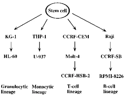
The cell lineages and maturation stages of the cell lines are summarized in Fig. 1. Hematopoietic cell lines representing different maturation stages (18, 19, 30) of granulocytic (KG-1 and HL-60), monocytic (THP-1 and U-937), T-cell (CCRF-CEM, Molt-4, and CCRF-HSB-2) and B-cell (Raji, CCRF-SB, and RPMI 8226) lineages were obtained from the American Type Culture Collection. CCRF-HSB-2 and KG-1 cell lines were main-tained in Dulbecco’s modified Eagle medium (IDMEM), and the other cell lines were maintained in RPMI 1640 medium,
supple-mented with 10% fetal bovine serum and 50mg of gentamicin per
ml. Cells (106cells per ml; 0.53106CFU-GM cells per ml) were
inoculated with the Nancy strain of CBV3 at a multiplicity of infection of 1 in serum-free Hanks’ balanced salt solution. After 1 h of incubation, the cells were washed three times in order to remove the unadsorbed virus.
Bone marrow mononuclear cells were obtained with the COBE 2991 model I Blood Cell Processor from Ficoll-Paque
[image:1.612.334.536.554.707.2]* Corresponding author. Phone: 358-2-3337461. Fax: 358-2-2513303.
FIG. 1. Maturation and differentiation stages of hematopoietic cell lines (18, 19), modified from the study by Okada et al. (21).
9018
on November 9, 2019 by guest
http://jvi.asm.org/
(Pharmacia)-separated heparinized bone marrow collected from autologous transplantation patients (disease free at the time of collection). The in vitro colonies of hematopoietic progenitors were cultured by the methyl cellulose method (23) with the modification described by Guilbert and Iscove (9).
Briefly, 23105mononuclear bone marrow cells per ml were
mixed with culture medium containing 1% methyl cellulose, 20% fetal bovine serum (HyClone), 1% delipidated and
deion-ized bovine serum albumin (Sigma Cell Culture), 1024M
mer-captoethanol, and 0.5 mg of iron-saturated human transferrin (Behringwerke) in IDMEM per ml and added to plastic petri dishes. The growth of granulocyte-macrophage precursors was
FIG. 2. Production of infectious CBV3 in human CFU-GM and hematopoietic cell lines infected at a multiplicity of infection of 1. The means and standard deviations (error bars) of dilutions (log10) are shown. Open circles are values obtained from the supernatants, while closed circles represent the cells.
on November 9, 2019 by guest
http://jvi.asm.org/
stimulated with 10% conditioned medium from the human bladder carcinoma cell line 5637 (30). The plates were
incu-bated at 378C in humidified atmosphere with 5% CO2.
CFU-GM were counted on the 14th day, picked under a light mi-croscope, washed, and suspended in the culture medium (with-out methyl cellulose).
The amount of infectious virus in the cells and supernatants
was determined by a 50% tissue culture infective dose assay on
confluent monolayers of LLC-Mk2 cells. Cells and
[image:3.612.101.515.67.607.2]superna-tants for infectivity titration were collected 1, 7, 24, 48, 78, and 96 h postinfection (p.i.), except for CFU-GM, which were harvested 1, 4, 24, and 48 h p.i. CBV3 was able to replicate in the CFU-GM (Fig. 2; Table 1), and a small amount of virus was released into the culture medium. A slight increase was also
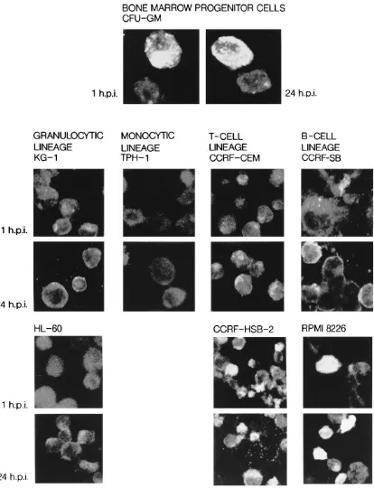
FIG. 3. Immunofluorescence staining of CBV3 proteins in human CFU-GM and hematopoietic cell lines with different maturation and differentiation stages examined by laser-scanning microscopy. Results after 1 and 24 h p.i. are shown.
on November 9, 2019 by guest
http://jvi.asm.org/
observed in virus titers determined from the CCRF-HSB-2 cells and culture medium. Because interpretation of the sus-ceptibility of the other cells to the CBV3 infection on the basis of infectivity titration was difficult because of the high back-ground level originating from the virus inoculum, immunolog-ical and molecular methods were used for further analysis of the presence and synthesis of viral components in the cells.
Immunofluorescent staining was performed in order to de-termine the presence of CBV3 antigens in infected CFU-GM and hematopoietic cell lines after the adsorption and at 24 h p.i. For confocal microscopy, the CBV3-infected CFU-GM were fixed in 4% formalin at room temperature for 15 min, incubated with the CBV3 antiserum (diluted 1:30), prepared by immunizing rabbits with highly purified virus (29), and then stained with fluorescein isothiocyanate-conjugated goat anti-rabbit immunoglobulins (diluted 1:150; Cappel). The fluores-cence was examined with a laser scanning confocal microscope (CCM; EMBL) at the Department of Medical Physics and Chemistry, University of Turku.
Intensive fluorescence in CFU-GM was seen already after the adsorption, whereas no viral antigens were detected in the monocytic cell line THP-1 (Fig. 3). CBV3 antigens were found in the KG-1 and HL-60 cells of granulocytic lineage, with an increase in the intensity during the first 24 h p.i. possibly due to reorganization of the antigen-positive clusters. The B- and T-cell lines were also positive after the adsorption, and no increase in the intensity of the fluorescence could be detected above this background.
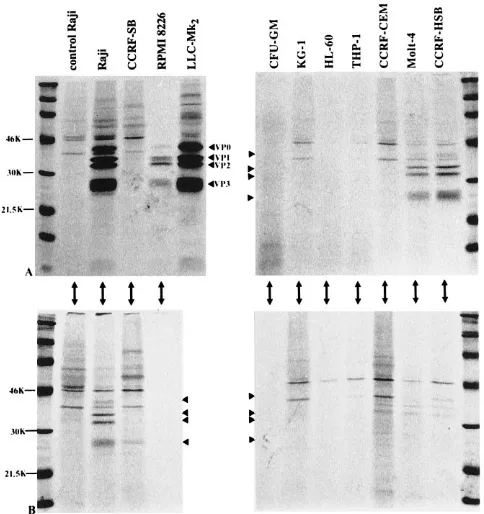
Synthesis of viral capsid polypeptides was analyzed by im-munoprecipitation after metabolic labeling between 4 and 22 and 73 and 91 h p.i. The CFU-GM, the hematopoietic cell lines
(106 cells), and the LLC-Mk
2cells were infected at a
multi-plicity of infection of 5. At 3 and 72 h p.i., the cells were pre-incubated in methionine-free medium for 1 h and labeled
for 18 h with [35S]methionine (50mCi/ml; Amersham).
Immu-noprecipitation was done with rabbit polyclonal antiserum against purified CBV3 and protein A-Sepharose beads as de-scribed previously (29). The precipitated proteins were ana-lyzed in 12.5% polyacrylamide gels, and the dried gels were exposed to X-ray films.
No virus-specific protein synthesis was detected in human CFU-GM or in the granulocytic (KG-1 and HL-60) and mono-cytic (THP-1) cell lines, whereas virus-specific proteins were
immunoprecipitated with the CBV3 antiserum from the CCRF-HSB cells at 22 h p.i. (Fig. 4). In the other T-cell line (CCFR-CEM) no viral polypeptides were detected until 91 h p.i. Synthesis of viral proteins was also seen in the RPMI 8226 and CCRF-SB cells; the signals in the CCRF-SB cells were weak at 22 h p.i., and only a faint, evidently nonspecific band comigrating with VP3 was detected at 91 h p.i.
The proportional amount of CBV3 RNA in the infected cells was determined by spot hybridization as described earlier (28) with the following modifications: the CBV3 cDNA probe
(13) was labeled with [32P]dCTP by using a nick translation kit
(Bethesda Research Laboratories) to a specific activity of 23
107cpm/mg. The prehybridization solution contained 50%
for-mamide, 1 M NaCl, 53Denhardt’s solution, 9% dextran sulfate,
100mg of denatured herring sperm DNA per ml, 1% sodium
dodecyl sulfate (SDS), and 50 mM HEPES
(N-2-hydroxyeth-ylpiperazine-N9-2-ethanesulfonic acid [pH 7.3]). After
hybrid-ization at 428C overnight, the membrane was washed three
times for 5 min each at room temperature in 23SSC (13SSC
is 0.15 M NaCl plus 0.015 M sodium citrate) containing 0.1%
SDS and three times for 30 min each at 428C in 0.13 SSC
containing 0.1% SDS and exposed to an X-ray film. Hybrid-ization signals were also analyzed with a PhosphorImager (GS-250 Molecular Imager; Bio-Rad).
No viral RNA was detected in the granulocytic and mono-cytic cell lines (Fig. 5). Infected CCRF-CEM cells appeared to be positive only at 96 h p.i., while CCRF-HSB-2 cells sup-ported detectable viral RNA synthesis during the first 24 h. A clear increase in the amount of CBV3 RNA was seen in the RPMI 8226 cells, while the CBV3 RNA synthesis in the CCRF-SB cells was less extensive.
[image:4.612.57.299.92.263.2]When different methods were used for the detection of virus replication in the cells and cell lines (Table 1), there were difficulties in the interpretation of the results obtained by in-fectivity titration and immunofluorescence due to the high
[image:4.612.316.558.441.698.2]FIG. 4. Synthesis of viral proteins detected by immunoprecipitation with rabbit antiserum against purified CBV3 at 22 (A) and 91 (B) h p.i. Virus polypeptides VP0, VP1, VP2, and VP3 are indicated by arrowheads.
TABLE 1. Summary of the susceptibility of CFU-GM and hematopoietic cell lines to CBV3 infection by different methods
Cell Virus
production
Virus protein synthesis
Virus minus-strand RNA synthesis
CFU-GM 1 2 1
KG-1 6 2 2
HL-60 2 2 2
THP-1 2 2 2
U-937 2a 2a 2
Raji 1 1 1
CCRF-SB 6 6 1
RPMI 8226 1 1 1
CCRF-CEM 6 1 1
Molt-4 1a 1 1
CCRF-HSB-2 1 1 1
a
Data from reference 23.
on November 9, 2019 by guest
http://jvi.asm.org/
background originating from the inoculum virus. Both immu-noprecipitation and nucleic acid hybridization suffered, to some extent, from the lack of sensitivity. Therefore, the results were confirmed by a sensitive reverse transcription (RT)-PCR for the detection of genomic (positive) and complementary (negative) RNA strands by protocols described earlier (1, 12). The oligonucleotide primers for the RT-PCR were from the
conserved parts of the 59-untranslated and the VP2
capsid-protein coding regions of the enterovirus genome (1, 12). Ex-traction of RNA from the cells was performed by using the Ultraspec (Biotecx) RNA isolation system with slight
modifi-cations. In brief, 200ml of chloroform was added to the
Ultra-spec mixture containing the cells; the mixture was incubated on
ice for 5 min and centrifuged 12,000 3gat 48C for 15 min.
RNA was precipitated by an equal volume of isopropanol and
0.04 mg of glycogen at 2208C overnight and centrifuged
12,0003gat 48C for 20 min, and the precipitates were washed
twice with 75% ethanol. Finally, the pellets were dried at room
temperature, dissolved in RNase-free water, incubated at 558C
for 15 min, and stored at2708C until used in the RT-PCR.
In principle, negative strands should represent de novo syn-thesis, thus being the most informative indication for virus replication. Uninfected control cells representing each cell line were also analyzed by RT-PCR, but no specific signals were detected (data not shown). Some background, obviously caused by the presence of negative strands in the virus inoculum, was occasionally observed (Fig. 6). Therefore, the samples col-lected late during the infection were compared with those collected immediately after inoculation. This approach clearly confirmed that both B- and T-cell lines are susceptible to CBV3 infection while monocytic and granulocytic cell lines are not (Fig. 6; Table 1). The cells of monocytic and granulocytic lineage did not support CBV3 replication as already shown by infectivity titration, immunoprecipitation, and analysis of viral RNA synthesis by spot hybridization. Viral antigens detected by immunofluorescence in the KG-1 and HL-60 cells could originate from the virus inoculum ingested into the cells by phagocytosis. It appears that negative-strand RT-PCR offers the best alternative for the detection of low levels of entero-virus replication.
Our previous work showed that CBV3 replicates in human hematopoietic cell lines with B (Raji)- and T (Molt-4)-cell characteristics (29). In the present study the permissiveness of the more mature T-cell (CCRF-HSB-2) and B-cell (RPMI 8226) lines for CBV3 was greater than that of the more im-mature cell lines. One explanation for these differences could be that the virus receptors are expressed on the cell surfaces in a variable manner depending on the maturation stage of the cells. Recently, the decay-accelerating factor (CD55) has been
FIG. 5. (A) Detection of CBV3 RNA by spot hybridization in hematopoietic cell lines after different incubation periods p.i. C, uninfected control cells. (B) Intensity of32P signals in the spot hybridization assay in the hematopoietic cell lines determined by PhosphorImaging analysis. The intensities of the32P signals of the uninfected control cells have been deducted from the intensity of infected cells.
FIG. 6. (A) Detection of viral RNA synthesis by negative-strand RT-PCR in infected hematopoietic cell lines of granulocytic, monocytic, B-cell, and T-cell lineages after adsorption and at 2 days p.i. Lanes: MW, molecular weight marker; 1, Raji; 2, CCRF-SB; 3, RPMI (B-cell lines); 4, KG-1; 5, HL-60 (granulocytic cell lines); 6, THP-1 (monocytic cell line); 7, CCRF-CEM; 8, Molt-4; 9, CCFR-HSB (T-cell lines). (B) Negative- and positive-strand RT-PCR from infected CFU-GM. Lanes: MW, molecular weight marker; A, negative control (uninfected LLC-Mk2cells); C, positive control (LLC-Mk2cells infected with CBV3). The 41/52primer pair (1) was used for negative-strand RT-PCR, and 52/41primer pair used for positive-strand RT-PCR. Arrows indicate the CBV3-specific am-plification products.
on November 9, 2019 by guest
http://jvi.asm.org/
[image:5.612.66.286.62.459.2] [image:5.612.321.548.393.633.2]reported to be a cellular receptor for CBV3 (27). It is ex-pressed on blood cells, and expression is increased during the cell maturation (20, 22). However, when our earlier results of the CBV3 infection in Raji and Molt-4 cells are also consid-ered (29) this cannot be directly explained by the maturation of the cells, because virus replication in these cell lines, repre-senting earlier maturation stages, is more effective. Replication of measles virus in peripheral blood mononuclear cells is de-pendent on the cellular activation stage (10); thus, an alterna-tive explanation could be the differences in the metabolic ac-tivity of the cell lines.
An increase in the levels of both complementary and geno-mic RNA could be detected in the CFU-GM, indicating vi-rus replication (Fig. 6). CBV3 antigen-positive cells were also detected in CFU-GM, and the infectivity titration indicated some virus release. Because we were unable to detect synthesis of CBV3 proteins by immunoprecipitation, it is possible that CBV3 replicates only in a certain subpopulation of the CFU-GM and the low number of the infected cells does not allow sufficient labeling.
The cell tropism of CBV3 is different from that of poliovirus, which can infect hematopoietic cell lines with monocytic and granulocytic characteristics (21). The most likely explanation is the diverse expression of virus receptors, since the poliovirus receptor, a member of the immunoglobulin superfamily, is expressed on human peripheral mononuclear phagocytes (17), although intracellular factors may also play a role. Further studies characterizing the presence of enterovirus receptors in different human hematopoietic cell lines, and transfection ex-periments using viral RNA to determine the level of the re-striction in the replication cycle would provide important in-formation about the pathogenesis of enterovirus infections.
We thank Marita Maaronen and Marjut Sarjovaara for technical assistance, Reinhard Kandolf for providing us with the CBV3 cDNA probe, Tapani Hovi for helpful discussions, and Yolanda Bullen for revising the language of the manuscript.
The study was supported by grants from the Academy of Finland, the Sigrid Juselius Foundation, and the Turku University Foundation.
REFERENCES
1.Arola, A., J. Santti, O. Ruuskanen, P. Halonen, and T. Hyypia¨.1996. Iden-tification of enteroviruses in clinical specimens by competitive PCR followed genetic typing using sequence analysis. J. Clin. Microbiol.34:313–318. 2.Borzakian, S., T. Couderc, Y. Barbier, G. Attal, I. Pelletier, and G. F.
Colbere.1992. Persistent poliovirus infection: establishment and mainte-nance involve distinct mechanisms. Virology186:398–408.
3.Cunningham, L., N. E. Bowles, R. J. Lane, V. Dubowitz, and L. C. Archard. 1990. Persistence of enteroviral RNA in chronic fatigue syndrome is associ-ated with the abnormal production of equal amounts of positive and negative strands of enteroviral RNA. J. Gen. Virol.71:1399–1402.
4.Dagan, R., J. A. Jenista, S. L. Prather, K. R. Powell, and M. A. Menegus. 1985. Viremia in hospitalized children with enterovirus infections. J. Pediatr. 106:397–401.
5.Fagraeus, A., M. Bo¨ttinger, L. Heller, and E. Norrby.1981. Replication of poliovirus and measles virus in cultures of human lymphoblastoid and of Burkitt lymphoma cell lines. Arch. Virol.69:229–237.
6.Freistadt, M. S., H. B. Fleit, and E. Wimmer.1993. Poliovirus receptor on human blood cells: a possible extraneural site of poliovirus replication. Vi-rology195:798–803.
7.Gresser, I., and C. Chany.1963. Multiplication of poliovirus type 1 in prep-arations of human leukocytes and its inhibition by interferon. J. Immunol. 92:889–895.
8.Grist, N. R., E. J. Bell, and F. Assaad.1978. Enteroviruses in human disease. Prog. Med. Virol.24:114–157.
9.Guilbert, L. J., and N. N. Iscove.1976. Partial replacement of serum selenite,
transferrin, albumin and lecithin in haemopoietic cell cultures. Nature (Lon-don)263:594–595.
10. Helin, E., A. A. Salmi, R. Vanharanta-Hiltunen, and R. Vainionpa¨a¨.Measles virus infection in cells of the myeloid lineage. Submitted for publication. 11. Henke, A., C. Mohr, H. Sprenger, C. Graebner, A. Stelzner, M. Nain, and D.
Gemsa.1992. Coxsackievirus B3-induced production of tumor necrosis fac-tor-alpha, IL-1 beta, and IL-6 in human monocytes. J. Immunol.148:2270– 2277.
12. Hyypia¨, T., P. Auvinen, and M. Maaronen.1989. Polymerase chain reaction for human picornaviruses. J. Gen. Virol.70:3261–3268.
13. Kandolf, R., D. Ameis, P. Hirschner, A. Canu, and P. H. Hofschneider.1987. In situ detection of enteroviral genomes in myocardial cells by nucleic acid hybridization: an approach to the diagnosis of viral heart disease. Proc. Natl. Acad. Sci. USA84:6272–6276.
14. Kitamura, Y., M. Masuda, and H. Yoshikura.1985. Effect of myelocytic maturation of HL60 cells on replication of influenza and polioviruses. Vi-rology141:299–301.
15. Klingel, K., C. Hohenadl, A. Canu, M. Albrecht, M. Seemann, G. Mall, and R. Kandolf.1992. Ongoing enterovirus-induced myocarditis is associated with persistent heart muscle infection: quantitative analysis of virus replica-tion, tissue damage, and inflammation. Proc. Natl. Acad. Sci. USA89:314– 318.
16. Matteucci, D., M. Paglianti, A. M. Giangregorio, M. R. Capobianchi, F. Dianzani, and M. Bendinelli.1985. Group B coxsackieviruses readily estab-lish persistent infections in human lymphoid cell lines. J. Virol.56:651–654. 17. Mendelsohn, C., E. Wimmer, and V. R. Racaniello.1989. Cellular receptor for poliovirus: molecular cloning, nucleotide sequence and expression of a new member of the immunoglobulin superfamily. Cell47:855–865. 18. Minowada, J.1982. Membrane and other phenotypes of leukemia cells.In
Proceedings of 13th International Cancer Congress, p. 215–227. Alan R. Liss, New York.
19. Minowada, J., E. Tatsumi, K. Sagawa, M. S. Lok, T. Sugimoto, K. Minato, L. Zgoda, L. Prestine, L. Kover, and D. Gould.1984. A scheme for human hematopoietic differentiation based on the marker profiles of cultured and fresh leukemia-lymphomas: the results of a workshop study, p. 519–527.In
A. Bernard, L. Boumsell, C. Dausset, C. Milstein, and S. F. Schlossman (ed.), Leucocyte typing: human leucocyte differentiation antigens detected by monoclonal antibodies. Springer-Verlag, Berlin.
20. Nicholson-Weller, A., and C. E. Wang.1994. Structure and function of decay accelerating factor CD55. J. Lab. Clin. Med.123:485–491.
21. Okada, Y., G. Toda, H. Oka, A. Nomoto, and H. Yoshikura.1987. Poliovirus infection of established human blood cell lines: relationship between the differentiation stage and susceptibility or cell killing. Virology156:238–245. 22. Okuda, K., A. Kanamaru, E. Ueda, T. Kitani, N. Okada, H. Okada, E. Kakishita, and K. Nagai.1990. Expression of decay-accelerating factor on hematopoietic progenitors and their progeny cells grown in cultures with fractionated bone marrow cells from normal individuals and patients with paroxysmal nocturnal hemoglobinuria. Exp. Hematol.18:1132–1136. 23. Pike, B. L., and W. A. Robinson.1970. Human bone marrow colony growth
in agargel. J. Cell. Physiol.76:77–84.
24. Prather, S. L., R. Dagan, J. A. Jenista, and M. A. Menegus.1984. The isolation of enteroviruses from blood: a comparison of four processing meth-ods. J. Med. Virol.14:221–227.
25. Roivainen, M., and T. Hovi.1989. Replication of poliovirus in human mono-nuclear phagocyte cell lines is dependent on the stage of cell differentiation. J. Med. Virol.27:91–94.
26. Sato, S., R. Tsutsumi, A. Burke, G. Carlson, V. Porro, Y. Seko, K. Okumura, R. Kawana, and R. Virmani.1994. Persistence of replicating coxsackievirus B3 in the athymic murine heart is associated with development of myocar-ditis lesions. J. Gen. Virol.75:2911–2924.
27. Shafren, D. R., R. C. Bates, M. V. Agrez, R. L. Herd, G. F. Burns, and R. D. Barry.1995. Coxsackieviruses B1, B3, and B5 use decay accelerating factor as a receptor for cell attachment. J. Virol.69:3873–3877.
28. Vuorinen, T., M. Kallajoki, T. Hyypia¨, and R. Vainionpa¨a¨.1989. Coxsack-ievirus B3-induced acute pancreatitis: analysis of histopathological and viral parameters in a mouse model. Br. J. Exp. Pathol.70:395–403.
29. Vuorinen, T., R. Vainionpa¨a¨, H. Kettinen, and T. Hyypia¨.1994. Coxsack-ievirus B3 infection in human leukocytes and lymphoid cell lines. Blood 84:823–829.
30. Welte, K., E. Platzer, L. Lu, J. L. Gabrilove, E. Levi, R. Mertelsmann, and M. A. S. Moore.1985. Purification and biochemical characterization of hu-man pluripotent hematopoietic colony-stimulating factor. Proc. Natl. Acad. Sci. USA82:1526–1530.
31. Willems, F. T. C., J. L. Melnick, and W. E. Rawls.1969. Replication of poliovirus in phytohemagglutinin-stimulated human lymphocytes. J. Virol.3: 451–457.