ASSESSING RV AND LV FUNCTION IN PATIENTS WITH
Dissertation submitted to
THE TAMIL NADU DR. M.G.R. MEDICAL UNIVERSITY
In partial fulfillment of the r
BRANCH II
MADRAS MEDICAL COLLEGE
RAJIV GANDHI GOVERNMENT GENERAL HOSPITAL
THE TAMIL NADU DR. M.G.R. MEDICAL UNIVERSITY
ASSESSING RV AND LV FUNCTION IN PATIENTS WITH
CORPULMONALE
Dissertation submitted to
THE TAMIL NADU DR. M.G.R. MEDICAL UNIVERSITY
In partial fulfillment of the requirements for the award of the degree of
D.M. CARDIOLOGY
BRANCH II – CARDIOLOGY
MADRAS MEDICAL COLLEGE &
RAJIV GANDHI GOVERNMENT GENERAL HOSPITAL
CHENNAI - 600 003
THE TAMIL NADU DR. M.G.R. MEDICAL UNIVERSITY
CHENNAI, INDIA
AUGUST 2013
ASSESSING RV AND LV FUNCTION IN PATIENTS WITH
THE TAMIL NADU DR. M.G.R. MEDICAL UNIVERSITY
equirements for the award of the degree of
RAJIV GANDHI GOVERNMENT GENERAL HOSPITAL
Dean,
Rajiv Gandhi Government
General Hospital &
Madras Medical College
Chennai - 600 003.
DECLARATION
I,
Dr.N.VISWANATHAN
, solemnly declare that this
dissertation
entitled,
“
NEWER
ECHOCARDIOGRAPHIC
PARAMETERS IN ASSESSING RV AND LV FUNCTION IN
PATIENTS WITH CORPULMONALE
” is a bonafide work done by
me at the department of Cardiology, Madras Medical College and
Government General Hospital during the period 2010 – 2013 under the
guidance and supervision of the Professor and Head of the department
of Cardiology of Madras Medical College and Government General
Hospital, Professor V.E.Dhandapani M.D.D.M. This dissertation is
submitted to The Tamil Nadu Dr.M.G.R Medical University, towards
partial fulfillment of requirement for the award of
D.M. Degree
(Branch-II) in Cardiology
.
Place:
SIGNATURE OF THE CANDIDATEDate
Professor and Head of Department
Department of Cardiology
A great many people made this work possible. I thank
Prof.
V.KANAGASABAI, M.D
., Dean for allowing me to conduct this
study.
My warmest respects and sincere gratitude to our beloved Prof
V.E.Dhandapani,
Professor and Head of the Department of
Cardiology, Government General Hospital, Chennai who was the
driving force behind this study. But for his constant guidance this study
would not have been possible.
I am indebted to
Prof. M.S.Ravi, Prof K.Meenakshi,
Prof.
D.Muthukumar,
Prof.
N.Swaminathan
and
Prof.
G.Ravishankar
without whom, much of this work would not have been
possible.
I acknowledge
Dr.S.Venkatesan
for the many useful comments
he made during this project.
In addition, I am grateful to Dr. G.Gnanavelu, Dr. G.Palanisamy,
Dr.Murthy, Dr. G. Prathap kumar, Dr. C. Elangovan, Dr. Rajasekar
Ramesh, Dr.S.Murugan, and Dr .G. Manohar, for tracing all those
waveforms and guidance.
I also thank all my patients for their kind cooperation.
CONTENTS
PAGE NO 1.
INTRODUCTION 6
2.
AIMS AND OBJECTIVES 10
3.
REVIEW OF LITERATURE 11
4.
MATERIALS AND METHODS 33
5.
RESULTS AND DATA ANALYSIS 36 6.
DISCUSSION♣ 55
7.
CONCLUSION 58
8.
LIMITATION OF STUDY 59
9.
APPENDIX 60
a. Bibliography
b. Abbreviations and acronyms
c. Proforma d. Master chart
INTRODUCTION
In India still pulmonary tuberculosis is prevalent and stays as one
of the leading cause of death. The cause of the death in most of the
terminally ill pulmonary tuberculosis patient is corpulmonale and
related cardiac problem. With the increase in incidence of retro viral
infection and diabetes the prevalence of multi drug resistant pulmonary
tuberculosis is high .This leads to increase in the chances for the
corpulmonale.
In the rural areas poverty and malnutrition lead to increased
incidence and prevalence of tuberculosis. In the urban areas exposure to
the automobile gases and dust particles predisposes patients to
pulmonary diseases and COPD in later life.
COPD in the later stages lead to right ventricular dilatation and
corpulmonale. The development and implementation of the national
programs for the detection and treatment of the tuberculosis is trying to
treat the disease in the early stage itself to prevent such corpulmonale in
future. In this study we are going to find easy solution to diagnose the
lung disease related cardiac problem in the early stage itself without
In corpulmonale patients right ventricular systolic dysfunction is
recognized to occur and it has been proved in many studies. But the
corpulmonale patients are symptomatic not only because of the lung
pathology and right heart failure, but also due to left ventricular
dysfunction in the form of left ventricular systolic and left ventricular
diastolic dysfunction .The dilated right ventricle pushes the
interventricular septum towards left ventricle and interferes with left
ventricular filling, which lead onto diastolic dysfunction of left
ventricle. The additional metabolic factors like hypoxia, hypercapnea
and acidosis lead on to direct myocardial depressant action and produce
myocardial dysfunction in both left and right ventricle.
So picking up left ventricular systolic and left ventricular diastolic
dysfunction in patients with corpulmonale using simple
echocardiographic parameters helps us to better assessment and
understanding of the patients. So treatment can be planned accordingly.
Corpulmonale is an end stage disease of the heart due to many
pulmonary diseases like pulmonary tuberculosis, COPD, interstitial lung
disease, occupational diseases and others. The treatment for
corpulmonale needs extra medication and diet restrictions. We are going
In corpulmonale patients the echo window is bit difficult to view
.so assessment of right ventricular systolic, left ventricular systolic and
left ventricular diastolic dysfunction in patients with corpulmonale
using simple parameters like mitral annular plane systolic excursion
(MAPSE),mitral annular systolic velocity(MASV), tricuspid annular
plane systolic excursion(TAPSE) ,tricuspid annular systolic
velocity(TASV),and e propagation velocity makes echocardiography
more complete and easy.
These parameters are studied individually in many studies and
results correlate well with left and right ventricular dysfunctions. In the
out patients department the usage of these simple parameters help the
physicians to assess the patients quickly also. we are going to compare
these parameters with one another and going to prove their validity.
So in this study we are going to study the prevalence of right
ventricular systolic, left ventricular systolic and diastolic dysfunction as
well as the utility of these simple and newer echocardiographic
AIMS & OBJECTIVES
1. To study the utility of newer echocardiographic parameters like
mitral annular plane systolic excursion, mitral annular systolic
velocity, tricuspid annular plane systolic excursion, tricuspid
annular systolic velocity, and ‘e’ propagation velocity in
identifying right ventricular systolic ,left ventricular systolic and
diastolic dysfunction in patients with corpulmonale
2. To study the prevalence of right ventricular systolic, left
ventricular systolic and left ventricular diastolic dysfunction in
REVIEW OF LITERATURE
Cor – Heart (Latin) and Pulmonale – of the Lungs (New Latin)
Pulmonary Heart disease
Chronic obstructive pulmonary disease (COPD), definition of
GOLD is preventable and treatable disease with some significant extra
lung pathology. Cor pulmonale was defined by the WHO in 1963 as
“hypertrophy of the right ventricle resulting from diseases involving the
physiology and or anatomy of the lungs, except when these pulmonary
alterations are the result of diseases that primarily affect the left side of
the heart, example as in congenital cardiac disease.
COPD is one of the major causes of morbidity worldwide. World
Bank data tells that it is expected to move from 4th and 12th most
frequent cause of mortality in 2000, to 3rd and 5th leading cause of
mortality in 2020. COPD is associated with more effects on heart.
Cardiovascular disease is responsible for 50% of all hospital admission
and nearly one third of deaths. In more advanced disease heart problem
is responsible for 20%–25% of all mortality in COPD. COPD affects
lung blood vessels, right ventricle and left ventricle leading to
development of pulmonary hypertension, cor pulmonale, right
Echocardiography is a rapid, noninvasive and exact method to evaluate
the right ventricular systolic function and filling pressure, tricuspid
regurgitation, left ventricular function and functions of valve. Many
studies have proved that echocardiographically arrived estimates of
pulmonary arterial pressure co-relate closely with pressures measured
by right heart catheter
The Right Ventricle in Pulmonary Hypertension.
The right ventricle is exposed to pressure overload by chronic
corpulmonale from any cause. The initial adaptive response of right
ventricular hypertrophy is followed later in life by right ventricular
contractile dysfunction. Right ventricular dilatation follows to allow
compensatory preload and maintain stroke volume in spite of reduced
fractional shortening.
As contractility weakens, clinical evidence of right ventricular
failure occurs, characterized by rise in filling pressures, diastolic
dysfunction, and decreasing cardiac output, which is compounded by
tricuspid regurgitation due to dilatation of right ventricular annulus and
poor leaflet cooptation. The increased volume and pressure overload of
the right ventricle also produce left ventricular diastolic dysfunction.
Right ventricular function is very important determinant of survival in
patients with pulmonary hypertension.
Clinical Presentation
Pulmonary hypertension in hypoxic lung diseases is common but
usually mild to moderate in severity. Breathlessness is the most frequent
symptom, but may not be helpful because breathlessness is so prevalent
in this patient population.
A change in breathlessness or the development of extra symptoms
such as angina, dizziness, syncope, and pedal edema may suggest
further evaluation. physical examination findings common in idiopathic
pulmonary hypertension such as a right ventricular heave, loud
pulmonary component of the S2, tricuspid regurgitant murmur, and RV
S4 may also be masked by the presence of parenchymal pulmonary
Severe pulmonary hypertension may lead to ascites and
peripheral edema. Few patients with severe COPD may develop pedal
edema in the absence of RV failure, the cause of which is hypercapnia,
suggesting that an elevated carbon dioxide partial pressure may be
responsible for sodium retention. Hypoxemia itself may cause renal
vasoconstriction, thus reducing urinary sodium excretion and lead to
pedal edema.
In corpulmonale patients the right ventricular systolic and
diastolic dysfunction recognized to occur. This concept is proved in
many studies. Presence of pulmonary hypertension interferes with the
right ventricular systolic and diastolic function. The dilatation of the
right ventricle interferes with diastolic filling of left ventricle. This also
leads to decreased left ventricular ejection fraction and finally systolic
function of left ventricle is also affected.
In the 1996, Italian study on echocardiographic Doppler
evaluation of left ventricular impairment in chronic corpulmonale
concluded that left ventricular filling is compromised due to shift of
interventricular septum towards left ventricle. This also concluded that
severity of pulmonary hypertension also correlates with the severity of
left ventricular diastolic dysfunction. It was published in 1996 June
Right ventricular volume overload causes flat interventricular
septum and the curvature is lost. Also in corpulmonale patients greater
negative intrathoracic pressure is needed for the lungs to get inflated.
This leads to increase in left ventricular after load. This increase in left
ventricular after load also impairs left ventricular systolic function.
Hypoxia interferes with intracellular calcium transport.
So the left ventricular relaxation during diastole is greatly
affected. In an Indian study published in Indian journal, the left
ventricular function was assessed in three categories of patients.
Category 1 (35 patients) patients had COPD only. Category ii (21
patients) is COPD with corpulmonale but without ventricular failure
and right ventricular dysfunction and category iii (14patients) is COPD
with corpulmonale with right ventricular dysfunction
The left ventricular ejection fraction was assessed in category iii
(14patients) i.e. COPD with corpulmonale with right ventricular
dysfunction. This was only on an average 45.6% in this category, but
68.5% and 70.8% in category ii and category I patients respectively.
This clearly implies that right ventricular dysfunction leads to left
ventricular dysfunction soon. Hypercapnea and acidosis of
action on myocardium. The left ventricular function further leads to
deterioration of corpulmonale due to metabolic and mechanical factors.
The left ventricular failure even if subclinical leads to delay in expected
improvement in overt right ventricular failure in corpulmonale patients.
Also biventricular failure in corpulmonale patients leads to kind of
refractory heart failure.
Assessment of right ventricular systolic function using TAPSE
(tricuspid annular plane systolic excursion) is validated in many studies.
In one study published in European heart journal 2007 June, right
ventricular systolic function was studied using TAPSE. This correlated
well with the right ventricular systolic function. Also TAPSE decreased
as the severity of the pulmonary hypertension increased.
In another TAIWAN study published in 2007 February, right
ventricular systolic function was studied in scleroderma patients. In this
study TAPSE and right ventricular systolic function were calculated
separately and compared. It showed excellent correlation between
TAPSE and right ventricular ejection fraction. TAPSE of less than
19.6mm was associated with right ventricular ejection fraction of less
In Indian heart journal 2009 January, TAPSE in normal subjects
and in patients with right ventricular dysfunction was studied. This was
correlated with fractional shortening and annular plane systolic velocity.
The value of less than 17mm TAPSE was identified in patients with
right ventricular dysfunction.
American journal of respiratory critical care medicine November
2006 published prognostic value of TAPSE in patients with established
pulmonary hypertension. The value of < 18mm was associated with
significantly increased mortality. This study concluded with the result of
TAPSE being the powerful indicator of right ventricular systolic
function.
Another important marker of right ventricular systolic function is
TASV (tricuspid annular systolic velocity).This is calculated by tissue
Doppler method. Sample volume is placed in the lateral tricuspid
annulus and systolic velocity is calculated. The normal value in person
with intact right ventricular systolic function is >10 cm/sec.
In one study conducted in Pittsburgh medical centre USA,
TAPSE was compared with TASV. In this study TAPSE correlated well
normal person. From this study we can infer that TASV is as good as
TAPSE and can be measured simultaneously and easily.
In another Swiss study published in 2005 the tricuspid annulus
systolic velocity of lateral annulus> 12cm/sec identified normal systolic
function with right ventricular ejection fraction of >55%. TASV
<9cm/sec in patients with right ventricular dysfunction was able to
identify the problem correctly and right ventricular ejection fraction in
this patients were between 30-55%.
MAPSE (mitral annular plane systolic excursion)
For assessment of left ventricular systolic function, MAPSE
(mitral annular plane systolic excursion) is used. It is the measure of
longitudinal systolic function of the left ventricle. It is calculated by the
1. Two dimensional Apical four chamber is viewed.
2. M-mode line is placed on the lateral mitral annulus
3. Distance between the end diastole and peak systole is measured.
MAPSE (mitral annular plane systolic excursion) can be
calculated in both medial and lateral mitral annulus and average can also
be taken. The normal value for MAPSE (mitral annular plane systolic
excursion) is more than 10 mm. This is very important tool and can be
very sensitive than left ventricular ejection fraction in identifying the
left ventricular function.
A study published in European heart journal 2011 September
compared 62 heart failure patients with preserved ejection fraction
patients with vo2 max 18.6±5 ml/min/kg with normal persons. MAPSE
(mitral annular plane systolic excursion) in these patients at rest was
10.9±2.1. But in normal persons it was 12.1±2.2. With exercise the
MAPSE in patient’s was12±2.2. But in normal persons it rose to
16.2±2.7. So this study concluded that MAPSE (mitral annular plane
systolic excursion) is a highly reliable and important tool in patients of
heart failure with normal left ventricular ejection fraction.
In the journal of cardiovascular magnetic resonance the cut off
males of age 35-50 years MAPSE is 12.8 ±7.3mm. For females of same
age it is 15.7±7.1mm.
For males of age 35-50 years TAPSE is 19.3 mm. For females of
same age it is 20.4mm. In patients with decreased ejection fraction,
MAPSE was less than 6 mm.
This study concluded that MAPSE has got accurate relationship
with left ventricular ejection fraction.
In the American journal of echocardiography September 2012 a
study of correlation of MAPSE with LVEF has been published. In this
study MAPSE was calculated by untrained person and LVEF was
calculated by experienced echocardiographer.
In patients with normal ejection fraction MAPSE >11mmfor
females and >13mm for males were found .In patients with decreased
ejection fraction MAPSE was <6mm. This had concluded that MAPSE
has got accurate relationship between with left ventricular ejection
fraction. many studies with MAPSE showed that the longitudinal left
ventricular systolic function correlate well with other parameters for left
MASV (mitral annular systolic velocity)
MASV (mitral annular systolic velocity) is measured by tissue
Doppler method. The sample volume is placed in the lateral mitral
annulus and the systolic velocity is calculated in c.m/sec.
In the journal of cardiovascular ultrasound 2010 march the
MASV (mitral annular systolic velocity) is compared with left
ventricular ejection fraction. Here the cutoff value was taken as 6.8
c.m/sec. For the patients with left ventricular systolic dysfunction the
value is less than 6.8cm/sec. In this study the left ventricular ejection
In another study published in 2012 cardiovascular ultrasound
journal MASV (mitral annular systolic velocity) was calculated in
hypertensive patients with diastolic dysfunction. This showed decreased
MASV (mitral annular systolic velocity) which is a marker of
longitudinal left ventricular systolic function. In this study control group
population without hypertension showed normal MASV (mitral annular
systolic velocity).
Mitral ‘e’ propagation velocity
To assess the left ventricular diastolic dysfunction the mitral e
propagation velocity is one of the parameters. It is calculated by
1. To obtain two dimensional echo apical four chamber view and
apply color Doppler at the mitral valve.
2. Then the M-mode line is placed on the line of color flow between
mitral valve and left ventricular apex.
3. The early diastolic E flow is recorded and the Nyquist limit is
adjusted as though no aliasing occur.
4. The velocity in the early phase of diastole i.e. the ‘e’ slope
velocity is recorded.
The normal value for ‘e’ propagation velocity is >50cm/sec.
The values of ‘e’ propagation velocity more than 50cm/sec
indicates that there is good diastolic flow from LA to LV and also
indicate intact diastolic function of the ventricle. The value of ‘e’
propagation velocity less than 50 cm/sec indicates left ventricular
There is also correlation between ‘E’ peak velocity and Vp
propagation velocity. The ratio of E/Vp more than 2.5 indicates raised
pulmonary capillary wedge pressure more than 15 mmHg.
In a study conducted in Poland in 2006 by Krystian Wite et al,
Propagation velocity was calculated in patients with known diastolic
dysfunction. The patients with diastolic dysfunction are categorized into
three groups. The third group comprised of Grade 3 diastolic
dysfunction. These patients had more decreased propagation velocity
In European respiratory journal 2008, LV diastolic dysfunction
was studied in idiopathic pulmonary fibrosis patients. In this study the
mitral inflow reversal was noted in all the patients and diastolic function
was assessed by ‘e’ propagation velocity was 46±13 cm/sec which was
higher in normal individuals of about 83±21 cm/sec.
In Chest journal 2003, Cystic fibrosis patients are studied for RV
and LV function. In the 40 patients studied all of them showed RV
systolic dysfunction and LV diastolic dysfunction. But LV systolic
function was not significantly decreased in these groups.
In 2006 Serbia Cardiovascular clinics of the Clinical centre NIS,
50 patients with chronic cor pulmonale were studied for LV diastolic
function and the results were compared with 56 patients of COPD
without heart failure.
In conclusion, that the LV diastolic dysfunction was due to the
progression of the right heart failure due to cor pulmonale and it
indicates advanced disease.
In an Indian study conducted in Rajasthan in 2009, COPD
patients were completely studied for Right and Left ventricular
functions using Echocardiography. Among them, 2/3 of the patients had
pulmonary hypertension and tricuspid regurgitation. The LV systolic
dysfunction was seen in <10% of the cases and left ventricular diastolic
also concluded the fact that echocardiography is extremely useful in
early detection of cardiac complication in COPD patients.
In Turkish study 30 patients of Sarcoidosis were studied for
cardiac involvement by echocardiography method. Among them,
sixteen patients showed LV diastolic dysfunction even if there is no
clinical proof of it. It was in the form of reversal of E/A ratio. This
figure of more than 50% of LV diastolic dysfunction was very
significant when compared to previous studies.
In our study, Cor pulmonale patients are studied by newer
echocardiographic methods, unlike the previous studies. This enables us
to detect the abnormality easily and accurately.
Traditionally, Cor pulmonale patients are defined by increase in
bulk of the right ventricle due to lung pathology. The lung pathology in
our group varied from chronic bronchitis, emphysema, old pulmonary
tuberculosis, previous lung surgery and bronchiectesis.
Interstitial Lung Disease
Diffuse parenchymal lung diseases (DPLDs) have also been
linked with cor pulmonale. DPLDs are a heterogeneous group of
diseases with similar clinical, x-ray, and physiological manifestations.
tissue, and other supporting structures. Occupational and environmental
exposures can lead to DPLD, although frequently the cause is Not clear.
The prevalence of pulmonary hypertension in patients with DPLDs
varies widely.
As in COPD, investigating method and criteria contribute to the
variability between studies, with prevalence tending to be higher where
echocardiography was used. Studies of persons undergoing evaluation
for lung transplantation have clearly demonstrated a higher prevalence.
Destruction and obliteration of the blood vessels secondary to loss of
lung parenchyma and fibrosis plays a role. Studies of IPF have reported
loss of blood vessels in areas of honeycomb lung and decrease in the
mean capillary surface area.
Vessel compression may lead to in situ thrombosis, fibrous
organization of blood vessels, and luminal obliteration. Abnormal
connections between the pulmonary and systemic circulation have also
been identified in patients with IPF.
Cor pulmonale is right ventricular enlargement due to high
resistance or pulmonary hypertension. Chronic increase in pulmonary
Pulmonary tuberculosis with corpulmonale
In Indian subcontinent still pulmonary tuberculosis is prevalent
and stays as one of the leading cause of corpulmonale. With the increase
in incidence of HIV infection and diabetes the prevalence of drug
resistant pulmonary tuberculosis is high .This leads to increase in the
chances for the corpulmonale .Immunodeficient states makes patient
susceptible to chronic infection and so the incidence of infection to
other people also increases.
In the rural areas poverty and malnutrition lead to increased
incidence of the tuberculosis. In the urban areas exposure to the
automobile gases and dust particles predisposes patients to pulmonary
diseases. Silicosis which is a occupational disease leads to pulmonary
tuberculosis. In tuberculosis the development of the corpulmonale is due
to decrease of lung elasticity, parenchymal pathology, alteration in the
diffusion capacity, obstruction of the bronchial tree due to brochospasm
and destruction of the lung tissue.
The development of right ventricular enlargement takes many
years to develop in corpulmonale patients. Smoking which is a risk
factor for the development of the COPD is also a risk factor for the
smokers. Presence of coronary artery disease in the COPD patients lead
on to left ventricular systolic and diastolic dysfunction.
DYSPNEA IN COPD
The grade of dyspnea in COPD increases in patients with
corpulmonale if coronary artery disease co exists. The development of
the corpulmonale lead to the state of irreversible disease process and
patients worsen day by day if the precipitating lung pathology is not
relieved. Ascites in corpulmonale lead to elevation of the diaphragm and
worsening of the dyspnea. Pleural effusion caused by the right heart
failure still increase the grade of dyspnea.
Most of the previous studies are done in particular subset of
patients with similar lung pathology and with only old
echocardiographic parameters. Some parameters like RV tei index, RV
ejection fraction and LV pulmonary flow reversal are not only time
consuming and also difficult to view and interpret in already sick cor
pulmonale patients.
So there is need for easier and simpler parameters like MAPSE,
MASV, TASV, TAPSE and ‘e’ propagation velocity are very much in
MATERIALS AND METHODS
All patients of corpulmonale referred for the study of the heart by
echocardiography are taken for the study and included. The patients
were selected irrespective of the lung pathology that caused
corpulmonale. Both male and female patients are selected for the study.
There was no age limit in our study population. Patients with
acute corpulmonale were not included because of the acute symptoms.
Relevant history, clinical examination, X ray chest, E.C.G was
taken for all the patients. Pulse oximetry, pulse rate, systolic and
diastolic blood pressure and respiratory rate were taken.
All the patients underwent detailed echocardiographic study.
Echocardiogram was done using Philips HD7XE Echocardiographic
machine. The following echocardiographic parameters were done.
Mitral annular plane systolic excursion (MAPSE), Mitral annular
systolic velocity(MASV), Tricuspid annular plane systolic
excursion(TAPSE), Tricuspid annular systolic velocity(TASV),and ‘e’
propagation velocity and E/A ratio were taken. TAPSE <17mm,
MAPSE < 10mm, TASV < 10cm/s, MASV <7 cm/s and ‘e’ propagation
American Society of Echocardiography recommendations. Pulmonary
hypertension was graded as mild, moderate and severe based on the
tricuspid regurgitation pressure gradient on the echocardiography.
INCLUSION CRITERIA:
All patients of Corpulmonale evaluated in Department of
Cardiology, Rajiv Gandhi Government General, Madras Medical
College, Chennai
EXCLUSION CRITERIA
1. Patients with past history of CABG
2. Patients with PTCA.
3. Patients with valvular heart disease.
4. Patients with hepatic dysfunction.
6. Unwilling to give consent.
7. Patients with coronary artery disease
8. Patients with systemic hypertension
RESULTS AND DATA ANALYSIS
40 patients of established cor pulmonale were
selected for the study. Among them 23 were males and 17 were females.
All are having right ventricular enlargement due to lung pathology.
Among them 30 patients have COPD, 8 patients have old pulmonary
tuberculosis and 2 patients have interstitial lung disease. The COPD
comprises of 75 % of study population; 20% of cases are old pulmonary
tuberculosis and 5% of cases are interstitial lung disease patients.
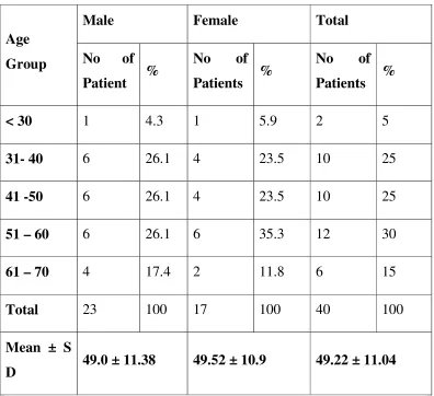
Age of the patients ranged from 29 years to 70 years with the
mean value of age is 49.5 years. Among female patients age range from
29 years to 68 years with the mean age of 48.5 years. For the male
Table 1: Age and Sex distribution of Study Population
Age Group
Male Female Total
No of Patient %
No of Patients %
No of Patients %
< 30 1 4.3 1 5.9 2 5
31- 40 6 26.1 4 23.5 10 25
41 -50 6 26.1 4 23.5 10 25
51 – 60 6 26.1 6 35.3 12 30
61 – 70 4 17.4 2 11.8 6 15
Total 23 100 17 100 40 100
Mean ± S
D 49.0 ± 11.38 49.52 ± 10.9 49.22 ± 11.04
Among the male patients corpulmonale is more common in the
age group between 30-60 years of age .corpulmonale is less common
before 30 years of age in both males and females. In female gender high
prevalence was noted between 50-60 years of age. In the age group of
60-70 years corpulmonale is slightly higher in the females when
Among these 40 patients 18 patients had mild pulmonary
hypertension, 8 patients had moderate pulmonary hypertension and14
patients had severe pulmonary hypertension. These comprises of 45% of
patients with mild pulmonary hypertension, 20% of patients with
moderate pulmonary hypertension and 35% of patients with severe
[image:35.612.99.529.297.537.2]pulmonary hypertension.
Table 2: Severity of Pulmonary Hypertension
Pulmonary Hypertension
Male Female Total
No of
Patient %
No of
Patients %
No of
Patients %
Mild 9 39.13 9 52.94 18 45
Moderate 5 21.74 3 17.64 8 20
Severe 9 39.13 5 29.42 14 35
Total 23 100 17 100 40 100
The prevalence of mild pulmonary hypertension is more in
females. The severe pulmonary hypertension prevalence is more in the
males. This severe pulmonary hypertension being more common in
males is probably due to increased smoking in the males. In the study
population also males are more commonly affected than the females
TAPSE of < 17mm was taken as having right ventricular systolic
dysfunction in our study. 38 out of 40 patients had TAPSE of <17 MM.
so prevalence of TAPSE calculated right ventricular systolic
dysfunction is 95 % in our study. 5 % of patients were not having right
ventricular systolic dysfunction when TAPSE was taken as the
echocardiographic parameter. TASV of < 10 cm/sec was taken as
having right ventricular systolic dysfunction in our study. 36 out of 40
patients had TASV of <10 cm/sec. so prevalence of TASV calculated
right ventricular systolic dysfunction is 90 % in our study. 10 % of
patients were not having right ventricular systolic dysfunction when
MAPSE (mitral annular plane systolic exertion)
MAPSE is considered to represent left ventricular systolic
dysfunction when it is <10mm in our study. MAPSE was < 10mm in 8
out of 40 patients. So prevalence of MAPSE calculated left ventricular
systolic dysfunction is 20 % in our study. 80 % of patients were not
having left ventricular systolic dysfunction when MAPSE was taken as
MASV (mitral annular systolic velocity)
MASV of < 7 cm/sec was taken as having left ventricular systolic
dysfunction in our study. 5 out of 40 patients had MASV of <7cm/sec.
so prevalence of MASV calculated left ventricular systolic dysfunction
is 12.5 % in our study. 87.5 % of patients were not having left
ventricular systolic dysfunction when MASV was taken as the echo
‘e’ propagation velocity
‘e’ propagation velocity of <50 cm/sec is taken as having left
ventricular diastolic dysfunction in our study. 18 out of 40 patients had
‘e’ propagation velocity of <50 cm/sec. so prevalence of e propagation
velocity calculated left ventricular diastolic dysfunction is 45 % in our
study. 55 % of patients were not having left ventricular diastolic
dysfunction when e propagation velocity was taken as the echo
parameter.
When E/A ratio is taken as echo parameter, to calculate left
ventricular diastolic dysfunction in our study, 22 out of 40 patients had
left ventricular diastolic dysfunction. So prevalence of E/A ratio
calculated left ventricular diastolic dysfunction is 55 % in our study. 45
% of patients were not having left ventricular diastolic dysfunction
when E/A ratio were taken as the echo parameter.
An average of about half of the study population had left
Mild pulmonary hypertension is more prevalent in the study patients 20%
35%
43%
Mild pulmonary hypertension is more prevalent in the study patients 45%
20%
Severity of PHT
Mild
Moderat e
57%
Sex Distribution
Male
Female Mild pulmonary hypertension is more prevalent in the study patients
Male
Male are more affected than the females.
Prevalence of RV systolic
TAPSE and TASV are more reliable
well with the right ventricular systolic dysfunction. Their sensitivity and
specificity in identifying the problem was very high. They can detect
even subtle problem which other parameters cannot do
simple parameters are proved useful in the acute set up also like right
ventricular infarction in various studies.
38 36 0 5 10 15 20 25 30 35 40
RV systolic dysfunction
Male are more affected than the females.
Prevalence of RV systolic dysfunction using TAPSE and TASV
TAPSE and TASV are more reliable parameters. Both of them correlate
well with the right ventricular systolic dysfunction. Their sensitivity and
in identifying the problem was very high. They can detect
which other parameters cannot do reliably. These
simple parameters are proved useful in the acute set up also like right
in various studies. 2 36
4
RV systolic dysfunction No RV systolic dysfunction
dysfunction using TAPSE and TASV
of them correlate
well with the right ventricular systolic dysfunction. Their sensitivity and
in identifying the problem was very high. They can detect
reliably. These
simple parameters are proved useful in the acute set up also like right TAPSE
Prevalence of LV systolic dysfunction
Assessment of LV sy
important for the evaluation of the dyspnea. sudden deterioration in the
dyspnea is due to the progressive worsening of the systolic
In our study patients with
prevalence of the LV systolic dysfunction. 0 5 10 15 20 25 30 35
LV Systolic Dysfunction
8
of LV systolic dysfunction
Assessment of LV systolic dysfunction in corpulmonale is very
evaluation of the dyspnea. sudden deterioration in the
the progressive worsening of the systolic dysfunction.
our study patients with severe pulmonary hypertension have more
LV systolic dysfunction. LV Systolic Dysfunction No LV Systolic
dysfunction
32
5
35 MAPSE
MASV
in corpulmonale is very
evaluation of the dyspnea. sudden deterioration in the
dysfunction.
hypertension have more MAPSE
Comparison of ‘e’ PV and E/A ration in detecting LV DD
compared to e propagation
diastolic dysfunction is
Comparing RV and LV systolic dysfunction in cor pulmonale 55%
'e' Propagation Velocity
0 5 10 15 20 25 e PV
PV and E/A ration in detecting LV DD
compared to e propagation velocity, E/A ratio calculated left ventricular
diastolic dysfunction is more.
Comparing RV and LV systolic dysfunction in cor pulmonale 45%
'e' Propagation Velocity
< 45 cm/sec
> 45 cm/sec
E/A
DD
NO DD PV and E/A ration in detecting LV DD. When
E/A ratio calculated left ventricular < 45 cm/sec
> 45 cm/sec
DD
TAPSE assess longitudinal right ventricular systolic dysfunction and its
calculation detects many patients with early systolic dysfunction .
left ventricular systolic dysfunction which occur very late in the course
of the disease is not that common when compared to right ventricular
systolic dysfunction. These two parameters are derived
echocardiography. 38 8 0 5 10 15 20 25 30 35 40 Systolic Dysfunction
TAPSE assess longitudinal right ventricular systolic dysfunction and its
calculation detects many patients with early systolic dysfunction .
left ventricular systolic dysfunction which occur very late in the course
of the disease is not that common when compared to right ventricular
systolic dysfunction. These two parameters are derived from m
2 32
Systolic Dysfunction No Systolic Dysfunction
TAPSE
MAPSE
TAPSE assess longitudinal right ventricular systolic dysfunction and its
calculation detects many patients with early systolic dysfunction .But
left ventricular systolic dysfunction which occur very late in the course
of the disease is not that common when compared to right ventricular
from m-mode TAPSE
Tissue Doppler derived
the true state of the systolic functions of the right and left ventricle
respectively. These results almost similar to results derived by other
parameters. 36 0 5 10 15 20 25 30 35 40 Systolic Dysfunction
Tissue Doppler derived parameters like TASV and MASV also reflects
the true state of the systolic functions of the right and left ventricle
results almost similar to results derived by other 4
5
35
Systolic Dysfunction No Systolic Dysfunction
parameters like TASV and MASV also reflects
the true state of the systolic functions of the right and left ventricle
results almost similar to results derived by other TASV
In patients with chronic corpulmonale both systolic
dysfunction of the left
patients had both systolic
They are more symptomatic than the patients with only diastolic
dysfunction.
The patients without even left
almost half of the study population. They are better
dyspnea and symptoms. 50%
Incidence of LV Systolic and Diastolic
In patients with chronic corpulmonale both systolic and diastolic
of the left ventricle co exist .In our study 20% of the
both systolic and diastolic dysfunction of the left ventricle.
They are more symptomatic than the patients with only diastolic
even left ventricular diastolic dysfunction make up
almost half of the study population. They are better in the
dyspnea and symptoms.
30%
20%
Incidence of LV Systolic and Diastolic
dysfunction
DD alone
Both SD & DD
No Dysfunction
and diastolic
In our study 20% of the
and diastolic dysfunction of the left ventricle.
They are more symptomatic than the patients with only diastolic
ventricular diastolic dysfunction make up
grade of
Incidence of LV Systolic and Diastolic
DD alone
Both SD & DD
Comparison of LV and RV dysfunction
In chronic cor pulmonale
dysfunction, Left ventricular dysfunction is rare. But some form of left
ventricular diastolic dysfunction co exist in almost half of the cor
pulmonale cases. Left ventricular diastolic dysfunction is common
malfunction than the systolic dysfunction. 0 5 10 15 20 25 30 35 40 Dysfunction 38 18
on of LV and RV dysfunction
In chronic cor pulmonale when compared to Right ventricular systolic
dysfunction, Left ventricular dysfunction is rare. But some form of left
ventricular diastolic dysfunction co exist in almost half of the cor
pulmonale cases. Left ventricular diastolic dysfunction is common
unction than the systolic dysfunction.
Dysfunction No Dysfunction
2 22
8
32
when compared to Right ventricular systolic
dysfunction, Left ventricular dysfunction is rare. But some form of left
ventricular diastolic dysfunction co exist in almost half of the cor
pulmonale cases. Left ventricular diastolic dysfunction is common RV Systolic
LV Diastolic
In our study, most common cause for cor pulmonale is COPD followed
by old pulmonary tuberculosis. Interstitial lung diseases constitute about
DISCUSSION
This study was undertaken to establish the role of simple and
useful parameters like TAPSE, TASV, MAPSE, MASV and e
propagation velocity in patients with corpulmonale. Also in this study
we aimed to study the prevalence of left ventricular systolic and
diastolic dysfunction in corpulmonale cases.
TAPSE is a simple method when compared to RVEF and RV TEI
index for studying right ventricular systolic dysfunction .It reliably
detects right ventricular systolic dysfunction and the value decreased as
the severity of systolic dysfunction worsen. Right ventricular systolic
dysfunction was very much in patients with severe pulmonary
hypertension when compared to mild pulmonary hypertension.
TASV is another newer parameter which utilizes tissue Doppler
to assess the right ventricular systolic dysfunction. The value of
<10cm/sec was taken as the cut off value .It correlated well with the
TAPSE in identifying the right ventricular systolic dysfunction. It is
well known that diseases like chronic bronchitis, pulmonary
tuberculosis, emphysema, bronchial asthma, interstitial lung diseases
pulmonary tuberculosis and interstitial lung diseases were the diseases
that caused the corpulmonale.
In tuberculosis cause for the development of the corpulmonale
include decrease in the lung elasticity, parenchymal disease, and
decrease in the diffusion capacity of the lung, obstruction of the
bronchioles due to brochospasm and destruction of the lung tissue.
Development of the corpulmonale leads to terminal stage of the
disease process and patient may go for congestive cardiac failure.
Biventricular failure in pulmonary tuberculosis patients can be easily
detected by echocardiography method using simple parameters like
MAPSE,TAPSE,MASV and TASV.
MAPSE and MASV which is used to study the left ventricular
function in our study are well accepted echo parameters. In future these
parameters may replace the conventional methods. They can be
compared with many other parameters for the similar purposes.
The concept that the left ventricular dysfunction which was
believed not to occur with corpulmonale patients is now disproved. In
our study almost half of the patients had left ventricular diastolic
dysfunction and 10-20 % of cases had left ventricular systolic
echocardiographic evaluation for the complete understanding about the
symptoms.
In corpulmonale cases sudden worsening of symptoms may be
due to the development of left ventricular systolic dysfunction. So
physician must be watchful about this problem.
It is also clear from the study that the above said echo parameters
are non inferior and can be used in routine echocardiographic evaluation
of cardiac patients. When there is doubt regarding the right and left
ventricular function, various parameters can be compared and doubts
LIMITATIONS OF THE STUDY
1. The study does not include the acute corpulmonale patients
2. The COPD patients without obvious right ventricular and right
atrial enlargement are not included in the study. So prevalence of
the right ventricular and left ventricular dysfunction in those
patients needs further evaluation
3. Right ventricular diastolic dysfunction which may be a earlier
CONCLUSION
1. Left ventricular diastolic dysfunction occurred in about 50% of
the corpulmonale patients.
2. Left ventricular systolic dysfunction was observed in 10-20%
percent in the study which is higher than the published literature.
3. MAPSE and MASV are simple and useful parameter to assess
the left ventricular systolic dysfunction.
4. TAPSE and TASV are simple and useful index in corpulmonale
patients to assess right ventricular systolic dysfunction.
5. It is found that atleast some amount of right ventricular systolic
dysfunction was detected when the newer echocardiographic
MASTER CHART
S. NO Patient No. Sex Age PHT Severity TRPG TAPSE TASV MAPSE MASV e PV E/A DD Diagnosis
1 Balamurugan M 36 Mild 30 15 8 11 10 52 0.8 1 PT
2 Neela F 40 Mild 32 15 9 11 8 70 0.7 1 PT
3 Kumaraguru M 61 Severe 64 16 8 11 7 43 0.8 1 COPD
4 Muralidharan M 56 Severe 87 12 9 8 7 39 1.1 2 COPD
5 Savithri F 45 Moderate 44 14 8 9 5.2 40 1.2 2 PT
6 Ismail M 48 Moderate 52 14 9 12 9.5 38 0.9 1 COPD
7 Hemavathy F 59 Severe 67 13 5.5 13.5 10 34 0.9 1 COPD
8 Kumaran M 43 Mild 33 16 8 13 10 68 1.1 0 COPD
9 Arumugam M 64 Severe 69 13 6.1 6.8 5.5 39 1.1 2 COPD
10 Chithra F 49 Mild 33 14 8 11 10 52 1.2 0 PT
11 Ramakrishnan M 51 Mild 35 16 9 11 8 51 1.1 0 COPD
12 Selvi F 58 Severe 72 13 9 11 8 40 0.8 1 COPD
13 Gowrishankar M 44 Moderate 40 15 8 11 8 45 0.9 1 COPD
14 Anandhan M 46 Severe 77 12 8 10 7 33 1.1 2 COPD
15 Shanmugam M 39 Mild 31 16 9 11 10 84 1.2 0 COPD
16 Annakili F 40 Severe 65 14 7 8 8 43 0.9 1 COPD
17 Dhandapani M 33 Mild 34 16 9 12 8 56 1.2 0 PT
18 Bhuvana F 35 Mild 35 20 15 11 9 96 1.3 0 COPD
20 Kanagavalli F 60 Moderate 53 16 7 10 7.5 46 1.1 0 COPD
21 Palaniappan M 44 Mild 32 15 8 11 10 52 1.1 0 COPD
22 Catherine F 49 Mild 33 16 9 11 8 70 1.1 0 COPD
23 Govindaraj M 38 Severe 64 16 8 10 7 43 0.8 1 PT
24 Ponniammal F 54 Severe 80 14 9 8 7 39 1.1 2 COPD
25 Kandasamy M 70 Moderate 44 13 8 10 6 40 1.2 2 COPD
26 Murugaselvan M 56 Moderate 52 16 9 12 9 38 0.9 1 COPD
27 Manoharan M 69 Severe 63 15 5.5 13 10 34 0.9 1 PT
28 Pushpammal F 64 Mild 31 16 8 13 10 68 1.1 0 COPD
29 Murugan M 59 Severe 64 15 6.1 6 5.1 39 1.1 2 COPD
30 Malathy F 44 Mild 33 16 8 11 10 52 1.1 0 COPD
31 Kannagi F 38 Mild 35 15 9 12 8 51 1.1 0 COPD
32 Sasikumar M 39 Severe 70 12 9 9 8 40 0.8 1 PT
33 Fathima F 56 Moderate 48 15 8 12 8 45 1.2 0 COPD
34 Abdul Khadar M 53 Severe 74 12 7 10 6.5 33 1.1 2 COPD
35 Valli F 54 Mild 30 16 9 11 10 84 1.2 0 COPD
36 Krishna Veni F 29 Severe 64 13 6 9 8 43 0.9 1 PT
37 Radha F 68 Mild 34 16 9 10 8 56 1.2 0 COPD
38 Dhayalan M 29 Mild 33 28 15 11 9 96 1.3 0 COPD
39 Joseph M 40 Mild 32 15 13 10 8 64 1.1 0 COPD
CONTENTS
PAGE NO 1.
INTRODUCTION 6
2.
AIMS AND OBJECTIVES 10
3.
REVIEW OF LITERATURE 11
4.
MATERIALS AND METHODS 33
5.
RESULTS AND DATA ANALYSIS 36 6.
DISCUSSION♣ 55
7.
CONCLUSION 58
8.
LIMITATION OF STUDY 59
9.
APPENDIX 60
a. Bibliography
b. Abbreviations and acronyms
c. Proforma d. Master chart
BIBLIOGRAPHY
1. Murray CJ, Lopez AD. Evidence based health policy-lessons
from the Global Burden of disease Study. Science 1996;
274:740-3.
2. World Health Report. Geneva: World Health Organization; 2000.
Available from: http://www.who.int/whr/2000/en/statistics.htm.
[Last accessed on 2004 Jan].
3. Anthonisen N, Connett JE, Kiley JP, Altose MD, Bailey WC, et
al. Effects of Smoking Intervention and the Use of an Inhaled
Anticholinergic Bronchodilator on the Rate of Decline of FEV1.
JAMA 1994;272:1497-1505.
4. Sin DD, Anthonisen NR, Soriano JB, Agusti AG. Mortality in
COPD: Role of comorbidities. Eur Respir J 2006;28:1245-57.
5. Daniels LB, Krummen DE, Blanchard DG. Echocardiography in
6. Yock PG, Popp RL. Noninvasive estimation of right ventricular
systolic pressure by Doppler ultrasound in patients with tricuspid
regurgitation. Circulation 1984;70:657-62.
7. Tramarin R, Torbicki A, Marchandise B, Laaban JP, Morpurgo
M. Doppler echocardiographic evaluation of pulmonary artery
pressure in chronic Gupta, et al. : Echocardiographic evaluation
of heart in COPD patientobstructive pulmonary disease. A
European multicentre study. Eur Heart J 1991;12:103-1.
8. Currie PJ, Seward JB, Chan KL, Fyfe DA, Hagler DJ, Mair DD,
et al Continuous wave Doppler estimation of right ventricular
pressure: A simultaneous Doppler-catheterization study in 127
patients. J Am Coll Cardiol 1985;6:750-6.
9. Chan KL, Currie PJ, Seward JB, Hagler DJ, Mair DD, Tajik AJ.
Comparison of three Doppler ultrasound methods in the
prediction of pulmonary artery pressure. J Am Coll Cardiol
1987;9:549-54.
10. Bredikis AJ, Liebson PR. The echocardiogram in COPD:
11. Rappaport E. Cor pulmonale. In, Murray JJ, Nadel JA, Mason
RM, Boushey H (eds). Textbook of respiratory medicine, 4th
Edition. Philadelphia, W.B. Saunders 2000;1631-48.
12. Chemla D, Castelain V, Humbert M, Simonneau JLHG,
Lecarpentier Y, Hervé P. New Formula for Predicting Mean
Pulmonary Artery Pressure using ystolic Pulmonary Artery
Pressure: Chest 2004;126;1313-17.
13. Braunwald’s Heart Disease. 9th Edition. By Libby P, Bonow RO,
Zipes DP, Mann DL. Philadelphia: Saunders 2008. p. 251.
14. Weitzenblum E, Hirth C, Ducolone A, Mirhom R,
Rasaholinjanahary J, Ehrhart M. Prognostic value of pulmonary
artery pressure In chronic COPD: Thorax 1981;36:752-8.
15. Weitzenblum E, Sautegeau A, Ehrhart M, Mammosser M, Hirth
C, Roegel E. long term course of pulmonary artery pressure In
chronic COPD. Am Rev Respir Dis 1984;130:993-8.
16. Burrows B, Kettel LJ, Niden AH, Rabinowitz M, Diener CF.
Patterns of cardiovascular dysfunction in COPD. N Engl J Med
17. Fishman AP. State of the art: Chronic cor pulmonale. Am Rev
Respir Dis 1976;114:775-94.
18. Pietra G. Pathology of the pulmonary vasculature and heart. In;
Cherniack N, editor. COPD. Philadelphia: WB Saunders; 1996. p.
21-6.
19. Thabut G, Dauriat G, Stern JB, Logeart D, Lévy A,
Marrash-Chahla R, et al : Pulmonary haemodynamics in advanced COPD
candidates for lung volume reduction surgery or lung
transplantation. Chest 2005;127:1531-6.
20. Weitzenblum E, Sautegeau A, Ehrhart M, Mammosser M,
Pelletier A. Long-term oxygen therapy can reverse the
progression of pulmonary hypertension in patients with chronic
obstructive pulmonary disease. Am Rev Respir
Dis 985;131:493-8.
21. Kessler R, Faller M, Weitzenblum E, Chaouat A, Aykut A,
Ducolone ́A, et al . “Natural history” of pulmonary hypertension
in a series of 131 patients with chronic obstructive pulmonary
22. Oswald-Mammosser M, Weitzenblum E, Quoix E, Moser G,
Chaouat A, Charpentier C, et al. Prognostic factors in COPD
patients receiving long-term oxygen therapy. Chest
1995;107:1193-8.
23. Higham MA, Dawson D, Joshi J, Nihoyannopoulos P, Morrell
NW. Utility of echocardiography in assessment of pulmonary
hypertension secondary to COPD. Eur Respir J 2001;17:350-5.
24. Chaouat A, Bugnet AS, Kadaoui N, Schott R, Enache I, Ducoloné
A, et al. Severe pulmonary hypertension and chronic obstructive
pulmonary disease. Am J Respire Care Med 2005;172:189-94.
25. Thabut G, Dauriat G, Stern JB, Logeart D, Lévy A,
Marrash-Chahla R, et al. Pulmonary Haemodynamics in advanced COPD
candidate for lung volume eduction surgery or lung
transplantation. Chest 2005;127:1531-6.
26. Springhouse. Respiratory disorders. In, Springhouse (ed).
Professional Guide to Diseases.9 th Edition.Philadelphia,
27. Rigolin VH, Robiolio PA, Wilson JS, Harrison JK, Bashore TM.
The forgotten chamber: The importance of the right ventricle.
Cathet Cardiovasc Diagn 1995;35:18-28.
28. Fishman AP. State of the art: Chronic Cor Pulmonale. Am Rev
Respire Dis 1976;114:775-94.
29. Macnee W. Pathophysiology of cor pulmonale in chronic
obstructive pulmonary disease. Part One. Am J Respire Crit Care
Med 1994;150:833-52.
30. Murphy ML, Adamson J, Hutcheson F. Left ventricular
hypertrophy in patients with chronic bronchitis and emphysema.
Ann Intern Med1974;81:307-13.
31. Fluck DC, Chandrasekar RG, Gardner FV. Left ventricular
hypertrophy in chronic bronchitis. Br Heart J 1966;28:92-7.
32. Robotham JL, Lixfeld W, Holland L, MacGregor D, Bryan AC,
Rabson J. Effects of respiration on cardiac performance. J Appl
Physiol 1978;44:703-9.
33. Render, ML, Weinstein, AS, Blaustein, AS Left ventricular
dysfunction in deteriorating patients with chronic obstructive
34. Vizza CD, Lynch JP, Ochoa LL, Richardson G, Trulock EP.
Right and left ventricular dysfunction in patients with severe
pulmonary disease. Chest 1998;113;576-83.
35. Jardin F, Gueret P, Prost JF, Farcot JC, Ozier Y, Bourdarias JP.
Two-dimensional echocardiographic assessment of left
ventricular function in chronic obstructive pulmonary disease.
Am Rev Respir Dis 1984;129:135-42.
36. Louridas G, Patakas D, Stavropoulos C. Left ventricular function
in patients with chronic obstructive pulmonary disease.
Cardiology 1981;67:73-80.
37. Funk GC, Lang I, Schenk P, Valipour A, Hartl S, Burghuber OC.
Left Ventricular Diastolic Dysfunction in Patients With COPD in
the Presence and Absence of Elevated Pulmonary Arterial
Pressure: Chest 2008;133:1354-9.
38. Poddar AK, Chakraborti BN, Ghosh JL, Nandy S, Hazra S.
Assessment of left ventricular function in patients of COPD :Ind J
ABBREVIATIONS AND ACRONYMS
RV - Right Ventricle
LV - Left Ventricle
MAPSE - Mitral Annular Plane Systolic Excursion
TAPSE - Tricuspid Annular Plane Systolic Excursion
MASV - Mitral Annular Systolic Velocity
TASV - Tricuspid Annular Systolic Velocity
LVEDD - Left Ventricle End Diastolic Dimension
LV ESD - Left Ventricle End Systolic Dimension
EF - Ejection Fraction
RVEDD - Right Ventricle End Diastolic Dimension
RVESD - Right Ventricle End Systolic Dimension
PROFORMA
Name:
Age:
Sex:
Address:
CD No. :
SYMPTOMS:
Chest pain:
SOB Class:
Palpitations
Right heart failure symptoms-
Risk Factors
h/o pul.TB Diabetes Mellitus
Smoking Family History
Past History:
Treatment History
Physical Examination
1. General Examination
2. Vital Signs
B.P Pulse
Respiration JVP Height cm Waveform
3. Systemic Examination
CVS
Inspection / Palpation
Apex Parasternal Heave
Palpable Sounds Thrills
Auscultation
S1 S2 Murmurs
Other System
RS: PA: CNS:
ECG:
CHEST X-RAY PA VIEW
ECHOCARDIOGRAPHIC ASSESSMENT OF corpulmonale
Name: Age: Sex:
Echocardiographic parameters:
M-mode:
LV: EDD- ESD- EF- FS-
MAPSE
RV: RVFWT RVEDD TAPSE
Mitral valve:
Aortic valve: LVOT diameter:
Tricuspid valve:
Pulmonary valve:
RVOT: MPA:
2 D and DOPPLER HEMODYNAMIC ASSESSMENT:
Mitral inflow:
E-wave: Peak velocity- DT-
A-wave: Peak velocity- TVI-
E/A ratio:
TISSUE DOPPLER
Mitral annular: E’ velocity- A’ velocity-
E/E’ ratio: MASV TASV
E PROPOGATION VELOCITY
IVRT:
IVCT:
Tricuspid inflow:
E-wave: Peak velocity- DT-
PATIENT CONSENT FORM
STUDY TITLE:
“NEWER ECHOCARDIOGRAPHIC PARAMETERS IN ASSESSING RV AND LV
FUNCTION IN PATIENTS WITH CORPULMONALE”
Patient may check () these boxes.
PARTICIPANT NAME: DATE:
AGE: SEX: I.P.NO. :
The details of the study have been provided to me in writing and explained to me in
my own language.
I confirm that I have understood the purpose of the above study. I have the
opportunity to ask the question and all my questions and doubts have been
answered to my complete satisfaction.
I understand that my participation in the study is voluntary and that I am free to
withdraw at any time without giving any reason, without my legal rights being
affected.
I understand that investigator, the institution, regulatory authorities and the ethical
committee will not need my permission to look at my health records both in respect
to the current study and any further research that may be conducted in relation to it,
even if I withdraw from the study. I understand that my identity will not be revealed
in any information released to third parties or published, unless as required under
the law. I agree not to restrict the use of any data or results that arise from this
study.
I hereby consent to undergo complete physical examination, and diagnostic tests
including hematological, biochemical, radiological and urine examinations
I have been given an information sheet giving details of the study.
I hereby consent to participate in the above study
Information to Participants
Title: NEWER ECHOCARDIOGRAPHIC PARAMETERS IN ASSESSING RV
AND LV FUNCTION IN PATIENTS WITH CORPULMONALE
Principal Investigator: Dr.N.VISWANATHAN
Co-Investigator (if any):
Name of Participant:
Site: RGGGH& MMC, Chennai
You are invited to take part in this research/ study/procedures/tests. The information in this document
is meant to help you decide whether or not to take part. Please feel free to ask if you have any queries
or concerns.
What is the purpose of research?
In patients with COPD not only RV dysfunction but LV diastolic and systolic dysfunction has
been reported.Newer echocardiographic parameters like tissue Doppler are useful in picking
early RV & LV dysfunction. The current study aims at using newer echocardiographic
parameters in assessing RV&LV function in patients with corpulmonale. we have obtained
permission from the institutional ethics committee.
The study design
It is a Retrospective cross-sectional study.
Study Procedures
The study involves evaluation of echocardiography newer echocardiographic parameters like
TAPSE, MAPSE, MASV and propagation velocity in assessing RV and LV function in patients
with corpulmonale. The results of the research may provide benefits to the society in terms of
Confidentiality of the information obtained from you
You have the right to confidentiality regarding the privacy of your medical information (personal
details, results of physical examinations, investigations, and your medical history). By signing this
document, you will be allowing the research team investigators, other study personnel, sponsors,
Institutional Ethics Committee and any person or agency required by law like the Drug Controller
General of India to view your data, if required.
The information from this study, if published in scientific journals or presented at scientific meetings,
will not reveal your identity.
How will your decision to not participate in the study affect you?
Your decision not to participate in this research study will not affect your medical care or your
relationship with the investigator or the institution. You will be taken care of and you will not lose
any benefits to which you are entitled.
Can you decide to stop participating in the study once you start?
The participation in this research is purely voluntary and you have the right to withdraw from this
study at any time