0022-538X/97/$04.00
1
0
Copyright © 1997, American Society for Microbiology
Structure-Function Analysis of the gE-gI Complex of Feline
Herpesvirus: Mapping of gI Domains Required for
gE-gI Interaction, Intracellular Transport,
and Cell-to-Cell Spread
J. D. F. MIJNES, B. C. H. LUTTERS, A. C. VLOT, E.
VANANKEN,
M. C. HORZINEK, P. J. M. ROTTIER,
ANDR. J.
DEGROOT*
Virology Unit, Department of Infectious Diseases and Immunology,
Veterinary Faculty, Utrecht University, 3584 CL Utrecht,
The Netherlands
Received 23 June 1997/Accepted 12 August 1997
Alphaherpesvirus glycoproteins gE and gI form a noncovalently associated hetero-oligomeric complex, which
is involved in cell-to-cell spread. In the absence of gI, feline herpesvirus (FHV) gE is transport incompetent and
fully retained in the endoplasmic reticulum. Here, we assess the effect of progressive C-terminal truncations
of FHV gI on the biosynthesis, intracellular transport, and function of the gE-gI complex. The truncated gI
proteins were coexpressed with gE in the vaccinia virus-based vTF7-3 expression system. The results were
corroborated and extended by studying FHV recombinants expressing truncated gI derivatives. The following
conclusions can be drawn. (i) Deletion of the cytoplasmic tail, the transmembrane region plus the C-terminal
half of the ectodomain of gI, does not affect intracellular transport of gE. Apparently, the N-terminal 166
residues of gI constitute a domain involved in gE-gI interaction. (ii) A region mediating stable association with
gE is located within the N-terminal 93 residues of gI. (iii) The cytoplasmic domain of gI is not essential for
gE-gI-mediated cell-to-cell transmission of FHV, as judged from plaque morphology. Deletion of the
cytoplas-mic tail of gI reduced plaque size by only 35%. (iv) Recombinants expressing the N-terminal 166 residues of
gI display a small-plaque phenotype but produce larger plaques than recombinants with a disrupted gI gene.
Thus, a complex consisting of gE and the N-terminal half of the gI ectodomain may retain residual biological
activity. The implications of these findings for gE-gI interaction and function are discussed.
The
Alphaherpesvirinae
subfamily, a group of large
envel-oped DNA viruses, comprises several important pathogens of
mammals and birds (for a review, see reference 35). Among
these are herpes simplex virus and varicella-zoster virus
affect-ing humans, pseudorabies virus affectaffect-ing swine, bovine
herpes-virus 1 affecting cattle, and feline herpesherpes-virus (FHV), the
caus-ative agent of feline rhinotracheitis (for a review, see reference
33). The viral genome is linear, 130 to 150 kb in length, and
contains more than 75 genes, a number of which encode
gly-coproteins. Several glycoprotein species have been shown to be
essential for the infection of cultured cells. For example, gB,
gH, and gL are involved in entry and mediate pH-independent
fusion of the viral envelope with the plasma membrane of the
target cell (for a review, see reference 39).
Membrane glycoproteins gE and gI are commonly regarded
as “dispensable,” as their genes can be deleted from the viral
genome with little or no effect on replication in vitro (3, 12, 28,
32, 40, 45). However, they do play an important role in vivo.
Mutant viruses lacking gE and/or gI are attenuated and, both
in the natural and in heterologous hosts, produce smaller
pri-mary lesions, cause milder clinical signs, and exhibit decreased
neurovirulence compared to the wild-type virus (8, 9, 12, 13,
16, 23, 24, 31, 34, 40, 41, 43, 45).
gE and gI form a noncovalently associated
hetero-oligo-meric complex (22, 30, 43, 44, 47, 49). Although its precise
function is unknown, there is ample evidence that gE-gI
pro-motes cell-to-cell transmission. Typically, viruses deficient in
either glycoprotein display a small-plaque phenotype (3, 12, 21,
28, 30, 32, 40, 48) and are impaired in virus-induced cell-cell
fusion (3, 11–13, 48). The mechanism of cell-to-cell spread is
by no means understood at the molecular level, but this mode
of infection differs in several respects from entry via the
extra-cellular route. It apparently entails the transfer of virus across
cell junctions in a manner resistant to neutralizing antibodies
(12). Moreover, viruses deficient in either gE or gI bind to and
enter the target cell with an efficiency equal to and with
kinet-ics similar to those of the parental wild-type virus (3, 12, 48).
We have recently identified the genes for gE and gI of FHV
and characterized their expression products both in infected
cells and in the vaccinia virus vTF7-3 expression system (30).
FHV gE and gI show all the characteristics of class I
mem-brane proteins. In accordance with findings made for other
alphaherpesviruses (43, 44, 47), the FHV proteins become
N-glycosylated and oligomerize shortly after synthesis in the
endoplasmic reticulum (ER) (30). The resulting gE-gI complex
is then transported through the Golgi apparatus to the plasma
membrane, concomitantly acquiring extensive
posttransla-tional modifications, including O-glycosylation. In the absence
of gE, gI is also transported to the plasma membrane, albeit
inefficiently. Transport of FHV gE, however, is dependent on
the presence of gI. gE, when expressed in the absence of gI, is
fully retained in the ER. Similarly, in cells infected with a
gI-deficient recombinant FHV only the endoglycosidase
H-sensitive 83-kDa ER species is produced and maturation of gE
does not occur (30). Here, we have studied gE-gI interaction in
further detail. By C-terminal deletion mutagenesis of gI, we
show that the N-terminal half of the gI ectodomain is sufficient
* Corresponding author. Phone: 31-30-2532460. Fax: 31-30-2536723.
E-mail: [email protected].
8397
on November 9, 2019 by guest
http://jvi.asm.org/
to induce gE maturation, thus defining a gI domain involved in
gE-gI complex formation. The effect of C-terminal deletions in
gI on cell-to-cell transmission is discussed.
MATERIALS AND METHODS
Cells, viruses, antisera, and plasmids.Cells were maintained in Dulbecco’s modified Eagle’s medium (Gibco BRL, Life Technologies, Inc.) supplemented
with 10% fetal calf serum and 100 IU of penicillin and 100mg of streptomycin
per ml (DMEM–10% FCS). FHV strain B927 (19) was obtained from D. A. Harbour and propagated in Crandell feline kidney (CRFK) cells (American Type Culture Collection). Recombinant vaccinia virus vTF7-3, expressing the bacteriophage T7 RNA polymerase (18), was obtained from B. Moss and prop-agated in RK-13 cells. Transient expression experiments were performed in
OST7-1 cells (15). The monospecific rabbit antisera against FHV gE (Ra-agE)
and gI (Ra-agI), the polyclonal cat antiserum against FHV (Cat-aFHV), and
plasmids pBS-gE and pBS-gI have all been described previously (30).
Recombinant DNA techniques.Recombinant DNA techniques were per-formed according to the methods of Sambrook et al. (37) and Ausubel (2). Sequence analysis was performed with the T7 sequencing kit (Pharmacia Bio-tech). PCR was performed as described previously (36) with the thermostable
DNA polymerase ofThermus aquaticus(Promega) according to the instructions
of the manufacturer.
Deletion mutagenesis of pBS-gI.Plasmid pBS-gI was constructed by inserting
the PCR-amplified FHV gI gene intoEcoRV-digested pBluescript SK2
(pBS-SK2) downstream of the T7 promoter. pBS-gIDM and -DB were made by cutting
pBS-gI with eitherMluI (position 497) orBamHI (position 926), respectively,
and withXbaI at a site downstream of the gI gene within the polylinker region
of pBS-SK2. The 39recessive ends were filled in by using the large fragment of
DNA polymerase I and then joined by blunt-end ligation. pBS-gIDXb was
cre-ated by directly ligating theXbaI site at position 1122 of the gI gene to theXbaI
site in the polylinker region. pBS-gIDC was made by ligating theClaI site at
position 635 of the gI gene to theClaI site in the polylinker region. pBS-gIDN
and pBS-gIDP were constructed by cutting pBS-gI with eitherNdeI (position
601) orPmlI (position 277), respectively, and withSmaI at a site in the polylinker
region. Prior to ligation, the 39recessive end generated byNdeI was made blunt
ended by using the large fragment of DNA polymerase I. pBS-gIDXh was
constructed by fusing the bluntedXhoI site at position 206 to the bluntedBamHI
site in the polylinker region. Finally, pBS-gI119 and pBS-gI152 were constructed
by amplifying nucleotides29 through 357 and29 through 456 of the gI gene,
respectively, by PCR with oligonucleotide primers 370 and 547 or 617 (Table 1).
The PCR products were cloned intoEcoRV-digested pBS-SK2. As a result of
the deletions and the nucleotide changes made, termination codons were created immediately downstream of each of the truncated gI genes. A sequence analysis of the relevant regions of each construct confirmed that no inadvertent changes had been introduced during cloning or during PCR amplification.
Transfection of vTF7-3-infected cells and metabolic labeling.Subconfluent monolayers of OST7-1 cells grown in 35-mm-diameter dishes were washed once with DMEM and infected with vaccinia virus vTF7-3 at a multiplicity of infection of 3 in DMEM at 37°C. One hour postinfection (p.i.), the cells were washed with DMEM and transfected with plasmid DNA as follows. A transfection mixture
consisting of 2 to 5mg of plasmid DNA, 500 ml of DMEM, and 10ml of
Lipofectin (Gibco BRL, Life Technologies, Inc.) was added to the monolayers.
After a 5-min incubation at room temperature, 500ml of DMEM was added and
incubation was continued at 37°C. At 2 h p.i., the incubation temperature was lowered to 32°C. From 4 to 5 h p.i., the cells were incubated with 1 ml of minimum essential medium with Earle’s salts, lacking cysteine and methionine
(Gibco BRL, Life Technologies, Inc.). Then, 100mCi of Redivue PRO-MIX
[35S] cell labeling mixture (35S[Met1Cys]; Amersham) was added to the culture
medium and the incubation was continued for 1 h. The cells were harvested either immediately or after a 2-h chase with DMEM–10% FCS containing 5 mM
(each)L-methionine andL-cysteine.
Metabolic labeling of FHV-infected cells.Subconfluent monolayers of CRFK cells in 35-mm-diameter dishes were washed once with DMEM and infected with either wild-type FHV strain B927 or recombinant FHV at a multiplicity of
50ml of a 10% (wt/vol) suspension of formalin-fixedStaphylococcus aureuscells
(Pansorbin; Calbiochem) in Detergent Mix. After a 30-min incubation at 4°C, they were washed three times with radioimmunoprecipitation assay (RIPA) buffer (10 mM Tris-Cl [pH 7.4], 150 mM NaCl, 0.1% SDS, 1% sodium deoxy-cholate, 1% Nonidet P-40). Treatment of immunoprecipitated proteins with endoglycosidase H (Endo-H; Boehringer Mannheim) was performed according to the method of Machamer et al. (26). Finally, the proteins were taken up in 30
ml of Laemmli sample buffer containing 5%b-mercaptoethanol, heated for 5 min
at 95°C, and analyzed in SDS-polyacrylamide gel electrophoresis (PAGE) gels.
Construction of FHV recombinants.FHVDgI-LZ has been described previ-ously (30). In this mutant, the gI gene has been disrupted by replacing nucleo-tides 203 to 923 with an expression cassette consisting of the LacZ gene under the control of the encephalomyocarditis virus (EMCV) internal ribosomal-entry
site (IRES). To restore the gI locus of FHVDgI-LZ or to introduce a gI gene
truncated either at nucleotide position 498 (gIDM) or 927 (gIDB), we
con-structed the transfer vectors pUS1, pUS1-gIDM, and pUS1-gIDB. pUS1 contains
a 7-kbEcoRV-BamHI fragment spanning the genes for gD, gI, gE, US8.5, and
US9 and approximately 2 kb of the terminal repeat sequence (38, 46).
pUS1-gIDM was constructed by replacing the sequences between theMluI site
in the gI gene and theXmaI site upstream of the gE gene by anMluI-Xma
I-digested PCR product which had been generated by amplifying the gE-gI inter-genic region with oligonucleotide primers 699 and 700 (Table 1). This product
contained a newly createdMluI site at its 59end immediately followed by the
authentic termination codon of the gI gene. pUS1-gIDB was constructed
simi-larly, i.e., by replacing the sequences between theBamHI site in the gI gene and
theXmaI site with aBamHI-XmaI-digested PCR product which had been
gen-erated with oligonucleotide primers 698 and 700 (Table 1). A sequence analysis
of the relevant regions of pUS1-gIDM and pUS1-gIDB showed that these
plas-mids had the desired structure and that no inadvertent mutations had been introduced during the cloning procedures.
To generate recombinant viruses FHV-gIrev, FHV-gIDM, and FHV-gIDB, 106
CRFK cells, seeded in 35-mm-diameter dishes, were subjected to cotransfection
with approximately650 ng of FHVDgI-LZ DNA and 1 to 2mg of transfer vector
DNA. The tissue culture supernatants were harvested 7 days after transfection, and plaque assays were performed. Recombinant viruses that had lost the EMCV IRES-LacZ expression cassette were identified by in situ staining of
plaques with X-Gal (5-bromo-4-chloro-3-indolyl-b-D-galactopyranoside) as a
substrate and were plaque purified three times prior to the preparation of virus stocks. In each case, proper insertion of the genes was confirmed by Southern blot analysis.
Plaque assays and immunohistochemistry.Confluent monolayers of CRFK cells grown in 35-mm-diameter dishes were washed with DMEM and infected with 50 to 100 PFU of either wild-type FHV B927 or recombinant FHV at 37°C. At 1 h p.i., the cells were washed three times with PBS and a solid-phase overlay of 2 ml of DMEM–10% FCS containing 1.5% agar was applied. Incubation was continued at 37°C. At 72 h p.i., the cells were fixed with 1 ml of 9% paraformal-dehyde for 1 h at room temperature. Subsequently, the overlay was removed and the cells were washed with PBS–10 mM glycine. The cells were permeabilized
with 1% Triton X-100 and then incubated with Cat-aFHV, diluted 1:500 in
PBS–0.5% Tween 80–5% FCS. Bound antibodies were detected by peroxidase staining with horseradish peroxidase-conjugated goat anti-cat immunoglobulin G (Cappel), diluted 1:500, as a conjugate and 3-amino-9-ethylcarbazole as a sub-strate. To determine the average plaque size, 25 randomly chosen plaques were
measured along thexandyaxes at a 20-fold magnification. The average plaque
size in millimeters squared was then calculated from the mean radius (r) by using
the termpr2.
RESULTS
A gI derivative truncated at residue 166 induces
intracellu-lar transport of gE.
To identify domains in gI required for the
formation and intracellular transport of the gE-gI
hetero-oli-gomer, we constructed a set of expression plasmids encoding gI
derivatives with progressive C-terminal deletions (Fig. 1). The
on November 9, 2019 by guest
http://jvi.asm.org/
[image:2.596.58.301.88.165.2]truncated gI genes were tested for proper expression in the
vaccinia virus-based vTF7-3 system (18). vTF7-3-infected
OST7-1 cells were transfected with plasmid DNA at 1 h p.i.
and labeled with
35S[Met
1
Cys] from 5 to 6 h p.i. Cell lysates
were subjected to immunoprecipitation with an antiserum
di-rected against residues 20 to 36 of gI (Ra-
a
gI), and the
immu-noprecipitates were analyzed in SDS–15% PAGE gels. In the
case of gI
D
Xb, -
D
B, and -
D
P single products of the anticipated
sizes of 65, 58, and 7.5 kDa, respectively, were found (Fig. 2a).
The detection of the 6-kDa gI
D
Xh protein was somewhat
un-expected. pBS-gI
D
Xh encodes a protein of this size, but this
product contains no Met or Cys residues other than the
initi-ator Met at its N terminus. It would thus appear that the
gI
D
Xh protein seen in Fig. 2a still retained its signal peptide.
Perhaps, because of its small size, gI
D
Xh becomes trapped in
the translocon and is not released into the lumen of the ER
(10). Expression of gI
D
M and gI-152 yielded four products
each. Treatment with Endo-H revealed that these represent
differentially glycosylated protein species (Fig. 2b and data not
shown). Potential N-glycosylation sites are present in gI at
amino acid positions 44, 65, and 117. Apparently, the 17- and
15-kDa products of gI
D
M and gI-152, respectively, represent
the nonglycosylated protein backbones, whereas the
Endo-H-sensitive larger products carry either one, two, or three
N-linked glycans. As illustrated for gI
D
M (Fig. 2b), the gI
deriv-atives did not acquire Endo-H resistance following a 2-h chase
and, like the membrane-anchored intact gI, were not efficiently
transported from the ER in the absence of gE.
Each of the truncated gI proteins was coexpressed with gE.
Metabolic labeling was performed for 1 h, followed by a 2-h
chase to allow for complex formation and intracellular
trans-port. RIPA was performed now with a gE-specific rabbit
anti-FIG. 1. Construction of a set of expression plasmids encoding gI derivatives with progressive C-terminal deletions. The upper panel shows a schematic represen-tation of the gI gene as cloned in plasmid pBS-gI. The gene is represented by an open box. The hatched regions indicate the nucleotide sequences coding for the
N-terminal signal peptide and the transmembrane region. Restriction sites are abbreviated as follows: Xh,XhoI; P,PmlI, M,MluI; N,NdeI; C,ClaI; B,BamHI; Xb,
XbaI. The bacteriophage T7 RNA polymerase promoter is represented by a black box. The arrow indicates the direction of transcription. The lower panel shows the
structures of the various gI deletion constructs and the sizes of the encoded polypeptides, which include the predicted cleavable N-terminal signal sequence. Also summarized are the effects of the gI mutations on the binding to and the maturation of gE. aa, amino acids.
FIG. 2. Biosynthesis of C-terminally truncated gI derivatives as studied by heterologous expression. (a) Monolayers of OST7-1 cells were infected with vaccinia virus
vTF7-3 and transfected with plasmid DNA encoding either full-length gI (gI) or the truncated gI derivatives, gIDXb, -DB, -DM, -152, -DP, and -DXh. Cells were
metabolically labeled with35S[Met1Cys] from 5 to 6 h p.i., and cell lysates were subjected to RIPA with the gI-specific antiserum, Ra-agI. (b) Biochemical analysis
of gIDM. vTF7-3-infected OST7-1 cells were mock transfected or transfected with pBS-gIDM and were metabolically labeled as described above. The cells were
harvested either immediately after labeling (pulse) or after a 2-h chase (chase). gIDM, immunoprecipitated with Ra-agI, was either treated with Endo-H (1) or mock
treated (2). Note that the Endo-H-deglycosylated 18-kDa form of gIDM still carriesN-acetylglucosamine moieties and thus migrates slightly slower than the
unglycosylated 17-kDa form. The proteins were separated in SDS–15% PAGE gels. Molecular sizes are in kilodaltons (K).
on November 9, 2019 by guest
http://jvi.asm.org/
[image:3.596.148.469.493.674.2]serum (Ra-
a
gE), and the precipitated proteins were analyzed
in SDS–7.5% PAGE gels. Maturation of gE, i.e., the
conver-sion of the sensitive 83-kDa species into the
Endo-H-resistant 95-kDa form, was interpreted to indicate the
forma-tion of a gE-gI hetero-oligomer and its subsequent transport
from the ER to the Golgi apparatus (Fig. 3; summarized in Fig.
1) (30). The deletion of the C-terminal 11 residues of gI, as in
gI
D
Xb, the complete cytoplasmic tail, as in gI
D
B, or even the
transmembrane region plus up to one-third of the ectodomain,
as in gI
D
C and gI
D
N (data not shown), did not affect
intracel-lular transport of gE. In fact, the N-terminal 166 amino acid
residues of gI, represented by gI
D
M, proved to be sufficient to
induce gE maturation (Fig. 3). However, maturation of gE no
longer occurred in the presence of gI derivatives truncated at
residue 152 (gI-152; Fig. 3) or at positions closer to the N
terminus.
gI
D
P, a gI derivative truncated at amino acid residue 93,
binds to gE but does not induce its release from the ER.
One
obvious explanation for the failure of gI-152 and the smaller gI
derivatives to induce gE maturation was that these truncated
proteins were no longer able to bind to gE. However, it was
also possible that binding to gE did occur but that the resulting
hetero-oligomers were recognized as misfolded by the ER
quality control system and therefore were retained in the ER
(14). Indeed, in a RIPA performed on lysates of cells
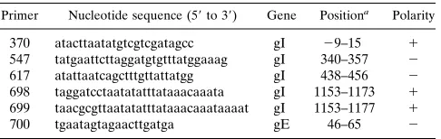
coex-pressing gI-152 and gE, a 28-kDa protein was coprecipitated
with the 83-kDa gE species (Fig. 4a, left lane). This product
corresponded to the fully glycosylated gI-152 species. Analysis
of the samples in SDS–15% PAGE gels revealed
coprecipita-tion also of the three smaller gI-152 species (data not shown).
In the 7.5% PAGE gels these products were not resolved but
rather migrated in the dye front. Consistent with these
find-ings, in RIPAs with the gI-specific antiserum, the 83-kDa gE
species was coprecipitated with gI-152 as well as with gI-119
and gI
D
P. However, gE was not coprecipitated with gI
D
Xh
(Fig. 1 and 4a). gI
D
P contains cysteine residues at positions 79
and 91. Theoretically, the coprecipitation of gE could have
resulted from illegitimate intermolecular disulfide bonding.
However, gE was also coprecipitated with gI
D
PC
0, a gI
D
P
derivative in which both Cys residues had been replaced by Ser
residues through PCR mutagenesis (Fig. 4a, right lane).
Fi-nally, to exclude the possibility that the coprecipitation merely
use of FHV
D
gI-LZ, an FHV strain B927 recombinant in which
residues 203 through 923 of the gI gene had been replaced by
an EMCV IRES-LacZ expression cassette (Fig. 5a) (30). The
disrupted gI locus of FHV
D
gI-LZ was restored by homologous
recombination with transfer vectors carrying either the intact
gI gene or truncated genes encoding gI
D
B or gI
D
M. The
genomic structures of the resulting FHV recombinant viruses,
FHV-gI
rev
, FHV-gI
D
B and FHV-gI
D
M, are depicted
schemat-ically in Fig. 5a.
To test for expression of the gI mutants and for gE
matu-ration, CRFK cells were infected and metabolic labeling was
performed with
35S[Met
1
Cys] from 7 to 8 h p.i.. Cells were
[image:4.596.322.551.462.611.2]harvested either immediately or after a 2-h chase, and cell
lysates were subjected to RIPA with the gI- or gE-specific
antisera. Cells infected with FHV
D
gI-LZ were taken along as
a negative control. As shown in Fig. 5b, the reintroduced gI
genes were properly expressed and in all cases products of the
anticipated size were found (for comparison, see Fig. 2a). In
cells expressing gI
D
M, each of the three N-glycosylated species
was detected, but the biglycosylated form was most abundant.
Maturation of gE occurred in cells infected with FHV-gI
rev
,
-gI
D
B, and -gI
D
M, but not in cells infected with FHV
D
gI-LZ
(Fig. 5c). In cells infected with FHV-gI
rev
or -gI
D
B, the
im-mature gE species was converted into the 95-kDa im-mature form.
FIG. 3. Coexpression of gE and C-terminally truncated gI derivatives in vTF7-3-infected cells. vTF7-3-infected OST7-1 cells were transfected with plas-mid pBS-gE and each of the pBS-gI derivatives listed in Fig. 1. The cells were metabolically labeled from 5 to 6 h p.i., followed by a 2-h chase. Cell lysates were
subjected to RIPA with the gE-specific rabbit antiserum, Ra-agE. Samples were
analyzed in SDS–7.5% PAGE gels. Only the results obtained for gI and gIDXb,
-DB, -DM, -152, -DP, and -DXh are shown. Arrowheads indicate the immature
83-kDa ER form (igE) and the Endo-H-resistant, mature 95-kDa gE species
(mgE). Molecular sizes are in kilodaltons.
FIG. 4. Coprecipitation of gE and C-terminally truncated gI derivatives. (a)
gE was coexpressed with gI-152, gI-119, gIDP, gIDXh, and gIDPC0in
vTF7-3-infected OST7-1 cells. The cells were metabolically labeled from 5 to 6 h p.i., followed by a 2-h chase. The cell lysates were subjected to RIPA with either
Ra-agE (agE) or Ra-agI (agI). The samples were analyzed in an SDS–7.5%
PAGE gel. Arrowheads indicate the immature, 83-kDa ER form of gE and the triglycosylated species of gI-152, migrating immediately above the dye front. The leftmost lane is an overexposure of the adjacent lane to show the coprecipitation
of gI-152 with gE in a RIPA with Ra-agE. The products of 59 and 55 kDa
represent vaccinia virus products nonspecifically binding to the immunosorbent (30). (b) vTF7-3-infected cells, transfected with either pBS-gE or pBS-gI-152, were metabolically labeled from 5 to 6 h p.i. Cells were lysed in 0.5 times the volume used in standard assays, and the lysates were mixed 1:1 and subsequently
subjected to RIPA with Ra-agE and Ra-agI. The samples were analyzed in an
SDS–10% PAGE gel. Molecular sizes are in kilodaltons.
on November 9, 2019 by guest
http://jvi.asm.org/
Mature gE, immunoprecipitated from lysates of FHV-gI
D
M-infected cells, produced a more diffuse band in SDS–7.5%
PAGE gels (Fig. 5c). Apparently, the gE-gI
D
M complex is
transport competent, but gE acquires more extensive
post-translational modifications.
Effect of C-terminal gI deletions on plaque size.
FHV
D
gI-LZ exhibits a small-plaque phenotype (30). To determine
wheth-er this defect was restored in the revwheth-ertant FHV-gI
rev
and to
study the plaque morphologies of FHV-gI
D
M and -gI
D
B,
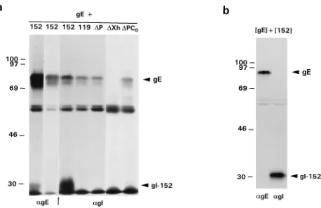
plaque assays were performed in CRFK cells. Plaques were
visualized immunohistochemically with an FHV-specific feline
hyperimmune serum (Fig. 6a). The plaque sizes (in millimeters
squared) were measured and compared to those of wild-type
FHV strain B927 and recombinant FHV
D
gI-LZ (Fig. 6b).
FHV-gI
rev
produced plaques indistinguishable from those of
wild-type strain B927. FHV-gI
D
B produced plaques much
larger than those of FHV
D
gI-LZ, but about 35% smaller than
those of wild-type FHV. In contrast, FHV-gI
D
M produced
small plaques, which were only about 25% the size of those
of wild-type FHV. On average, however, the plaques of
FHVgI
D
M were larger than those of FHV
D
gI-LZ.
DISCUSSION
We have previously reported that, in the absence of gI, FHV
gE is transport incompetent and fully retained in the ER (30).
Here, we have exploited this feature to study gE-gI interaction
in more detail and have assessed the effects of C-terminal
deletions in gI on the biosynthesis, intracellular transport, and
function of the gE-gI complex. Results, obtained by
vTF7-3-driven expression of cloned genes, were corroborated and
ex-tended by studying FHV recombinants expressing truncated gI
derivatives. Our data suggest that the N terminus of gI
repre-sents the business end of the molecule.
Comparative amino acid sequence analysis revealed that
most of the sequence conservation among alphaherpesvirus gI
proteins is in the N-terminal half of the ectodomain (1, 27). We
now show that this region of gI comprises a domain intimately
involved in gE-gI interaction. This conclusion is based upon
the observation that FHV gI derivative gI
D
M, which is
trun-cated at residue 166, can assemble into a transport-competent
complex with gE. Protein folding and oligomerization in the
ER are facilitated and controlled by foldases and molecular
chaperones. The latter proteins associate transiently with
fold-ing and assembly intermediates and, more permanently, with
misfolded products or incomplete subunits of oligomeric
com-plexes. In general, only properly folded proteins and fully
as-sembled oligomers are allowed to proceed to the Golgi
com-plex and beyond (14, 20, 42). Apparently, the interaction with
gI
D
M is sufficient to release gE from its chaperone(s); i.e., the
C-terminal half of the gI ectodomain, the transmembrane
an-chor, and the cytoplasmic tail are not required for this process.
How the dissociation of gE is brought about is not known.
gI
D
M may directly compete with chaperones for gE binding.
Alternatively, the interaction with gI
D
M may cause an
essen-tial conformational change in gE.
Complex formation per se is not sufficient to trigger
intra-cellular transport. The gI-152 product, which is only 14
resi-dues shorter than gI
D
M, and even gI
D
P, which is truncated at
residue 93, still associate with gE, as demonstrated by
coim-munoprecipitation. However, the resulting complexes are
re-tained in the ER. Our findings confirm and extend
observa-tions by Basu et al. (5); on the basis of deletion and linker
insertion mutagenesis of herpes simplex virus gI, these authors
suggested that a region comprising residues 43 to 192
(corre-sponding to residues 38 to 233 of FHV gI) complexes with gE.
Under the assumption that the overall protein structure of gI
has been conserved during alphaherpesvirus divergence, our
findings would suggest that a region mediating stable
associa-tion with gE is located between residues 38 and 93. It is not
known which regions of gE bind to gI. For HSV gE, a region
comprising residues 183 to 288 (corresponding to residues 153
to 255 of FHV gE) has been implicated in the gE-gI interaction
(4). It remains to be determined whether this part of gE, which
is highly conserved (1), binds to the N-terminal domain of gI.
Truncation of gI at residue 166 does not interfere with
com-plex formation and subsequent intracellular transport, but it
does have an effect on the posttranslational modification of gE.
In cells expressing gI
D
M, a mature gE species of 95 to 100 kDa
was found instead of the typical 95-kDa form (Fig. 3 and 5c).
FIG. 5. Biochemical analysis of FHV recombinants expressing C-terminally truncated gI derivatives. (a) Schematic representation of the gI loci in the unique short
regions of FHV-gIDM, FHV-gIDB, FHV-gIrev, and FHVDgI-LZ. The genes for gD, gI, gE, andb-galactosidase (LacZ) are depicted as open boxes. The hatched box
represents the IRES of EMCV. Arrows indicate the direction of transcription. X and B indicateXhoI andBamHI restriction sites, respectively. (b) Expression of
truncated gI proteins in FHV-infected cells. Monolayers of CRFK cells were infected with either FHV strain B927 (WT), FHV-gIrev(rev), FHV-gIDM (DM),
FHV-gIDB (DB), or FHVDgI-LZ (DgI). The cells were metabolically labeled from 7 to 8 h p.i. and lysed as described in Materials and Methods. To dissociate gE-gI
complexes, SDS was added to the lysates to a final concentration of 1.5% and the lysates were incubated for 5 min at 95°C prior to RIPA with Ra-agI. The samples
were analyzed in an SDS–15% PAGE gel. Arrowheads indicate the mono-, bi-, and tri-N-glycosylated forms of gIDM. The 80- to 100-kDa smears observed in lanes
WT andrevrepresent mature gI species. Previously, we failed to detect mature gI in FHV-infected cells (30). However, Ra-agI does recognize mature gI; because of
the heterogenous posttranslational modifications, the detection of this product is obscured in SDS–7.5% PAGE gels. (c) Maturation of gE in FHV-infected cells expressing truncated gI proteins. CRFK cells were infected and metabolically labeled as described above. After the labeling, the cells were harvested either immediately
(pulse) or after a 2-h chase (chase). Cell lysates were subjected to RIPA with Ra-agE. The samples were analyzed in an SDS–7.5% PAGE gel. Arrowheads indicate
the immature (igE) and mature (mgE) forms of gE. Molecular sizes are in kilodaltons.
on November 9, 2019 by guest
http://jvi.asm.org/
Apparently gE, when in a complex with gI
D
M, acquires
addi-tional posttranslaaddi-tional modifications. Experiments in which
gE was treated with peptide:
N
-glycosidase F indicate that the
increase in apparent molecular weight is caused by a more
elaborate decoration of glycans (29). Presumably, the
N-glycans involved are inaccessible to further processing in the
authentic gE-gI complex. These findings are not
unprece-dented. Aberrant posttranslational processing has also been
seen for varicella-zoster virus gE and gI when they are
ex-pressed separately (47) and for FHV gI produced in the
ab-sence of gE (30).
[image:6.596.80.505.76.618.2]Several lines of evidence suggest that gE-gI is critically
in-volved in cell-to-cell transmission (3, 11–13, 39, 48). Mutant
viruses deficient in gE and/or gI exhibit a small-plaque
pheno-type, yet are reportedly similar to the wild type with regard to
kinetics of entry, growth rate, and yield of extracellular virus (3,
12, 28, 29, 48). Recombinant FHV
D
gI-LZ, in which the gI gene
has been disrupted by the insertion of an EMCV IRES-LacZ
expression cassette, produces plaques only 15% the size of
those of the parental virus. In revertant FHV-gI
rev
, plaque
morphology was fully restored to that of wild-type FHV.
Plaque morphology was also restored in an FHV
D
gI-LZ
de-rivative which had been complemented allotopically by
in-serting a functional gI gene into the thymidine kinase locus
(29). These observations provide formal evidence that the
small-plaque phenotype of FHV
D
gI-LZ is caused by the
dis-ruption of the gI gene and not by “nearest neighbor effects,”
such as down-regulation of gD expression (46). Moreover,
these findings provided a basis to test the effects of C-terminal
deletions in gI on cell-to-cell transmission as measured by
plaque size. Like the gI proteins of other alphaherpesviruses,
FIG. 6. Plaque morphology of FHV recombinants expressing C-terminally truncated gI species. (a) Monolayers of CRFK cells, grown in 35-mm-diameter dishes were infected with 50 to 100 PFU. One hour p.i., a solid-phase overlay was applied, and incubation was continued for 72 h at 37°C. The monolayers were paraformaldehyde fixed, the overlay was removed, and the plaques were visual-ized immunohistochemically. Magnification, 2.3. (b) Quantification of plaque size. For each recombinant, 25 randomly chosen plaques were measured along
thexandyaxes at a 20-fold magnification to determine the mean diameter. The
average plaque size in millimeters squared was then calculated from the mean
radius (r) by using the termpr2. The histogram shows the plaque sizes of the
FHV mutants relative to that of parental FHV strain B927 (WT). Standard deviations are indicated by bars. The experiment was performed three times in two feline cell lines (CRFK and IRC5 cells), yielding essentially identical results. The results of a typical experiment are presented.
on November 9, 2019 by guest
http://jvi.asm.org/
FHV gI possesses a relatively large cytoplasmic tail, 73 residues
in length. However, this domain can be deleted—as in
FHV-gI
D
B—without much effect on plaque size. We therefore
con-clude that the cytoplasmic tail of gI is not essential for
gE-gI-mediated cell-to-cell spread, at least not under our in vitro
conditions. In contrast, the deletion of the C-terminal 218
residues, as in gI
D
M, resulted in a dramatic decrease in plaque
size. The most obvious explanation for these findings is that gI
sequences downstream of residue 166, for example, the
trans-membrane region, are required for efficient cell-to-cell spread.
Theoretically, the loss in function may also have resulted from
the altered posttranslational modifications of gE. It is of note,
however, that the plaques produced by FHV-gI
D
M were
con-sistently larger than those produced by gI-deficient FHV
D
gI-LZ. Interestingly, this difference was even more pronounced
when gI
D
M was expressed allotopically. From these results, it
would appear that the gE-gI
D
M complex retains residual
bio-logical activity and, thus, that membrane anchoring of gI is not
essential for function. These findings and their implications
will be described in more detail elsewhere (29).
The gE-gI complex is a virulence factor and as such plays an
important role in alphaherpesvirus pathogenesis. Although for
some alphaherpesviruses gE-gI may be involved in immune
evasion by acting as an Fc receptor (4–7, 17, 22, 25), its main
function appears to be to promote cell-to-cell spread. The
molecular mechanism of this process remains to be resolved.
As has been speculated (3, 12), gE-gI may be involved in
interactions at the plasma membrane and, by binding to a
specific ligand in the plasma membrane of adjacent cells, may
facilitate viral transmission via the contact route. Further
stud-ies on the biosynthesis of gE-gI may provide additional clues as
to its function and may perhaps even lead to the development
of new antiherpetic drugs.
ACKNOWLEDGMENTS
We thank D. Harbour and B. Moss for providing the virus stocks of
FHV strain B927 and vTF7-3, respectively, and Antoine de Vries for
stimulating discussions and for critical reading of the manuscript.
J. D. F. Mijnes was supported by Rho
ˆne Me
´rieux, Lyon, France. The
research of R. J. de Groot was made possible by a fellowship of the
Royal Netherlands Academy for Sciences and Arts.
REFERENCES
1.Audonnet, J.-C., J. Winslow, G. Allen, and E. Paoletti.1990. Equine herpes-virus type 1 unique short fragment encodes glycoproteins with homology to
herpes simplex virus type 1 gD, gI and gE. J. Gen. Virol.71:2969–2978.
2.Ausubel, F. M.1989. Current protocols in molecular biology. Greene Pub-lishing Associates and Wiley-Interscience, New York, N.Y.
3.Balan, P., N. Davis-Poynter, S. Bell, H. Atkinson, H. Browne, and T. Minson.
1994. An analysis of the in vitro and in vivo phenotypes of mutants of herpes simplex virus type 1 lacking glycoproteins gG, gE, gI or the putative gJ. J.
Gen. Virol.75:1245–1258.
4.Basu, S., G. Dubin, M. Basu, V. Nguyen, and H. M. Friedman.1995. Char-acterization of regions of herpes simplex virus type 1 glycoprotein E involved in binding the Fc domain of monomeric IgG and in forming a complex with
glycoprotein I. J. Immunol.154:260–267.
5.Basu, S., G. Dubin, T. Nagashunmugam, M. Basu, L. T. Goldstein, L. Wang, B. Weeks, and H. M. Friedman.1997. Mapping regions of herpes simplex virus type 1 glycoprotein I required for formation of the viral Fc receptor for
monomeric IgG. J. Immunol.158:209–215.
6.Baucke, R. B., and P. G. Spear.1979. Membrane proteins specified by herpes simplex viruses. V. Identification of an Fc-binding glycoprotein. J. Virol.
32:779–789.
7.Bell, S., M. Cranage, L. Borysiewicz, and T. Minson.1990. Induction of immunoglobulin G Fc receptors by recombinant vaccinia viruses expressing
glycoproteins E and I of herpes simplex virus type 1. J. Virol.64:2181–2186.
8.Card, J. P., M. E. Whealy, A. K. Robbins, and L. W. Enquist.1992. Pseu-dorabies virus envelope glycoprotein gI influences both neurotropism and
virulence during infection of the rat visual system. J. Virol.66:3032–3041.
9.Card, J. P., M. E. Whealy, A. K. Robbins, R. Y. Moore, and L. W. Enquist.
1991. Two alpha-herpesvirus strains are transported differentially in the
rodent visual system. Neuron6:957–969.
10.Crowley, K. S., S. Liao, V. E. Worrell, G. D. Reinhardt, and A. E. Johnson.
1994. Secretory proteins move through the endoplasmic reticulum
mem-brane via an aqueous, gated pore. Cell78:461–471.
11.Davis-Poynter, N., S. Bell, T. Minson, and H. Browne.1994. Analysis of the contributions of herpes simplex virus type 1 membrane proteins to the
induction of cell-cell fusion. J. Virol.68:7586–7590.
12.Dingwell, K. S., C. R. Brunetti, R. L. Hendricks, Q. Tang, M. Tang, A. J. Rainbow, and D. C. Johnson.1994. Herpes simplex virus glycoproteins E and I facilitate cell-to-cell spread in vivo and across junctions of cultured cells.
J. Virol.68:834–845.
13.Dingwell, K. S., L. C. Doering, and D. C. Johnson.1995. Glycoproteins E and
I facilitate neuron-to-neuron spread of herpes simplex virus. J. Virol.69:
7087–7098.
14.Doms, R. W., R. A. Lamb, J. K. Rose, and A. Helenius.1993. Folding and
assembly of viral membrane proteins. Virology193:545–562.
15.Elroy-Stein, O., and B. Moss.1990. Cytoplasmic expression system based on constitutive synthesis of bacteriophage T7 RNA polymerase in mammalian
cells. Proc. Natl. Acad. Sci. USA87:6743–6747.
16.Enquist, L. W., J. Dubin, M. E. Whealy, and J. P. Card.1994. Complemen-tation analysis of pseudorabies virus gE and gI mutants in retinal ganglion
cell neurotropism. J. Virol.68:5275–5279.
17.Frank, I., and H. M. Friedman.1989. A novel function of the herpes simplex virus type 1 Fc receptor: participation in bipolar bridging of antiviral
immu-noglobulin G. J. Virol.63:4479–4488.
18.Fuerst, T. R., E. G. Niles, F. W. Studier, and B. Moss.1986. Eukaryotic transient-expression system based on recombinant vaccinia virus that syn-thesizes bacteriophage T7 RNA polymerase. Proc. Natl. Acad. Sci. USA
83:8122–8126.
19.Gaskell, R. M., and R. C. Povey.1979. The dose response of cats to exper-imental infection with feline viral rhinotracheitis virus. J. Comp. Pathol.
89:179–191.
20.Hurtley, S. M., and A. Helenius.1989. Protein oligomerization in the
endo-plasmic reticulum. Annu. Rev. Cell Biol.5:277–307.
21.Jacobs, L., H. J. Rziha, T. G. Kimman, A. L. J. Gielkens, and J. T. Van Oirschot.1993. Deleting valine-125 and cysteine-126 in glycoprotein gI of pseudorabies virus strain NIA-3 decreases plaque size and reduces virulence
in mice. Arch. Virol.131:251–264.
22.Johnson, D. C., and V. Feenstra. 1987. Identification of a novel herpes simplex virus type 1-induced glycoprotein which complexes with gE and
binds immunoglobulin. J. Virol.61:2208–2216.
23.Kritas, S. K., M. B. Pensaert, and T. C. Mettenleiter.1994. Invasion and spread of single glycoprotein deleted mutants of Aujeszki’s disease virus (ADV) in the trigeminal nervous pathway of pigs after intranasal
inocula-tion. Vet. Microbiol.40:323–334.
24.Kritas, S. K., M. B. Pensaert, and T. C. Mettenleiter.1994. Role of envelope glycoproteins gI, gp63 and gIII in the invasion and spread of Aujeszky’s disease virus in the olfactory nervous pathway of the pig. J. Gen. Virol.
75:2319–2327.
25.Litwin, V., W. Jackson, and C. Grose.1992. Receptor properties of two varicella-zoster virus glycoproteins, gpI and gpIV, homologous to herpes
simplex virus gE and gI. J. Virol.66:3643–3651.
26.Machamer, C. E., S. A. Mentone, J. K. Rose, and M. G. Farquhar.1990. The E1 glycoprotein of an avian coronavirus is targeted to the cis Golgi complex.
Proc. Natl. Acad. Sci. USA87:6944–6948.
27.McGeoch, D. J.1990. Evolutionary relationships of virion glycoprotein genes
in the S regions of alphaherpesvirus genomes. J. Gen. Virol.71:2361–2367.
28.Mettenleiter, T. C., C. Schreurs, F. Zuckermann, and T. Ben-Porat.1987. Role of pseudorabies virus glycoprotein gI in virus release from infected
cells. J. Virol.61:2764–2769.
29.Mijnes, J. D. F., and R. J. De Groot.Unpublished observations. 30.Mijnes, J. D. F., L. M. van der Horst, E. van Anken, M. C. Horzinek, P. J. M.
Rottier, and R. J. de Groot.1996. Biosynthesis of glycoproteins E and I of feline herpesvirus: gE-gI interaction is required for intracellular transport.
J. Virol.70:5466–5475.
31.Mulder, W. A., L. Jacobs, J. Priem, G. L. Kok, F. Wagenaar, T. G. Kimman, and J. M. Pol.1994. Glycoprotein gE-negative pseudorabies virus has a reduced capability to infect second- and third-order neurons of the olfactory and trigeminal routes in the porcine central nervous system. J. Gen. Virol.
75:3095–3106.
32.Neidhardt, H., C. H. Schro¨der, and H. C. Kaerner.1987. Herpes simplex virus type 1 glycoprotein E is not indispensable for viral infectivity. J. Virol.
61:600–603.
33.Povey, R. C.1979. A review of feline viral rhinotracheitis (feline herpesvirus
1 infection). Comp. Immunol. Microbiol. Infect. Dis.2:373–387.
34.Rajcani, J., U. Herget, and H. C. Kaerner.1990. Spread of herpes simplex virus (HSV) strains SC16, ANG, ANGpath and its glyC minus and glyE
minus mutants in DBA-2 mice. Acta Virol.34:305–320.
35.Roizman, B.1996. Herpesviridae, p. 2221–2230.InB. N. Fields, D. M. Knipe, and P. M. Howley (ed.), Fields virology, 3rd ed, vol. 2. Lippincott-Raven, Philadelphia, Pa.
36.Saiki, R. K., D. H. Gelfand, S. Stoffel, S. J. Scharf, R. Higuchi, G. T. Horn, K. B. Mullis, and H. A. Erlich.1988. Primer-directed enzymatic
on November 9, 2019 by guest
http://jvi.asm.org/
Burg, A. Moerman, A. L. J. Gielkens, and J. T. van Oirschot.1994. A glycoprotein E deletion mutant of bovine herpesvirus 1 is avirulent in calves.
J. Gen. Virol.75:2311–2318.
42.Wei, J., and L. M. Hendershot.1996. Protein folding and assembly in the
endoplasmic reticulum. Exper. Suppl. (Basel)77:41–55.
43.Whealy, M. E., J. P. Card, A. K. Robbins, J. R. Dubin, H. J. Rziha, and L. W.
48.Zsak, L., F. Zuckermann, N. Sugg, and T. Ben-Porat.1992. Glycoprotein gI of pseudorabies virus promotes cell fusion and virus spread via direct
cell-to-cell transmission. J. Virol.66:2316–2325.
49.Zuckermann, F. A., T. C. Mettenleiter, C. Schreurs, N. Sugg, and T. Ben-Porat.1988. Complex between glycoproteins gI and gp63 of pseudorabies
virus: its effect on virus replication. J. Virol.62:4622–4626.