Copyright © 1993, AmericanSocietyforMicrobiology
Assembly,
Processing, and
Infectivity
of Human
Immunodeficiency
Virus Type 1 Gag Mutants
CHIN-TIEN WANGANDERICBARKLIS*
VollumInstitute for Advanced Biomedical Research and DepartmentofMicrobiology andImmunology,
OregonHealthSciences University, Portland, Oregon 97201 Received 11November1992/Accepted16April1993
We studied the effects of gag mutations on human immunodeficiency virus type 1 (HIV-1) assembly, processing, and infectivity by using areplication-defective HIV expression system. HIV mutants were screened for infectivity by transductionof a selectable marker and were examinedfor
assembly
by monitoringparticle release from transfected cells. Gag protein processing and reverse transcriptase activities of mutant particles were also assayed. Surprisingly,most Gagprotein mutants were assembled and processed.The twoexceptions tothis rule wereamyristylation-minus mutant, and one gagmatrix domainmutantwhichexpressed proteins that weretrapped intracellularly.Interestingly,
amutant with a 56-amino-acid deletion within the HIV gag capsid domain still could assembleandprocess virusparticles, exhibitedawild-type retrovirus particledensity,
and had
wild-type
reversetranscriptaseactivity.Indeed,althoughmostHIV-1gag mutants were noninfectious or poorly infectious, they produced apparently normal particles which possessed significant reverse tran-scriptase activities. These resultsstrongly
support the notion thatthe HIV-1 Gag proteins are functionally involved inpostassembly,postprocessing stages of virus infectivity.Assembly of human immunodeficiency virus(HIV) parti-cles occurs at theplasma membranes ofinfected cells (41) andresults in theincorporation of several viral components. They are the viral RNA genome, core (Gag) structural proteins, envelope (Env) glycoproteins, and virion-associ-ated enzymes encoded by the viral pol gene, including protease, reverse transcriptase (RT), RNase H, and inte-grase (6, 36). In addition to the characteristic retroviral
gag-pol-env genome (47), HIV type 1 (HIV-1) encodes several novelproteins. One small accessoryprotein, Vpu, is involved in theassembly process by assisting virus particle
budding, although Vpu itself isnotpackagedinto virions(7, 19, 43,44). However, in the absence of other viral products,
Gag polyproteins stillcan assemble as avirionlike particle, suggestingthat gagis theonlyviral generequiredforvirus
assembly (10,39). The HIVGagpolyprotein issynthesized as a precursorPr55, which is modified cotranslationally by methionine cleavage and the attachment of a myristic acid to the N-terminal glycine (45, 48). Myristylation is necessary for membrane association and virion formation (5, 13, 34).
Duringorafterbudding, Pr55 is cleaved by the viral protease intop17(matrix; MA), p24 (capsid; CA), p7 (nucleocapsid;
NC), and p6 (17, 25, 29, 32). p17 is myristylated and membraneassociated;p24 is themajor capsid structure;p7, containing two zinc finger motifs, is anucleic acid-binding
protein (1, 2, 11, 40); and the p6 domain, located at the carboxyl end of Pr55, may play a functional role in the processof virusbudding (10, 12).
Despite intensive efforts, the mechanism of HIV virus assembly is still unclear. One model, on the basis of previous studies, isthatmyristylated Gag precursors are targeted to the plasma membrane where they self-assemble into parti-cles. Envproteinsmight be incorporated by binding to Gag proteins at the plasma membrane; Gag-Pol fusion proteins would be incorporated into virions by virture of their
N-ter-*Corresponding author.
minal gag determinants; and viral RNA might be
encapsi-datedby interaction withGagproteinsatthe RNApackaging
signal(Psi)locatedaround the initiation sequence ofGag(16, 27).While Gagproteins playacentral role in the processof retroviralassembly, they also have been implicatedin other functions. Studies have shown that mutations of murine leukemia virus (MLV) gag canblock early stages of infec-tion (8),andHIVgagmutantscaninterfere withreplication
ofwild-type (wt)virus(46).This evidence suggests thatGag
proteins also may affect the process ofreversetranscription,
nuclear transport, orintegration (4, 37,38).
To investigate the mechanism of HIV assembly and the
potential functions of Gag proteins in other phases ofthe virusreplication cycle,wehaveadaptedageneticapproach. HIV gag mutations were created by deletion or linker insertion and were subcloned into a replication-defective HIV proviral genome (HIVgpt), which carries the
drug-resistant gpt gene(30)in theenvregion(33). Cotransfections
ofwt or mutantHIVgptconstructswithanenvelope expres-sionplasmidpermittedus toanalyzehow each gag mutation affected virusassemblyandinfectivity.TwelveHIV mutants werescreenedforinfectivity, assembly,processing,and RT
activity. Surprisingly, results showed that most mutants were assembled and released as particles. However, most mutants either were noninfectious or poorly infectious,
although they had significant RT activities. Interestingly, a mutantwith a56-amino-acid deletion in thecapsid domain wasstill assembled andprocessed. Indeed, theonlymutants incapable ofparticleassemblywere a myristylation mutant and a linker insertion mutant in the central portion of the matrix domain. Immunofluorescence studies showed that Gag proteins ofthis insertion mutant localized to the
peri-nuclear area, indicating that the matrix domain may be involved in transport or assembly of
Pr55sag.
Overall, our resultsstrongly support the notion that HIV Gagproteinsare functionally involved in postassembly and postprocessing stages ofviralinfectivity.4264
on November 9, 2019 by guest
http://jvi.asm.org/
MATERIALS AND METHODS
Cell culture, transfections, infections, and
infectivity
assays. HeLaand COS7 cells were maintained in Dulbecco's mod-ified Eagle's medium supplemented with 10% heat-inacti-vated fetal calfserum (GIBCO). Confluent COS7 cellswere split 1:10 onto 10-cm dishes 24 h before transfections. Fifteen micrograms ofplasmid DNAs of gag mutants or wt HIVgptwas transfected onto COS7 cells by calcium phos-phate precipitation (14). At 48 to 72 h after transfection, supernatants of COS7 cells were collected and filtered through a0.45-,um-pore-sizefilter and frozen at-80°C.
Forinfections, 15pzg of plasmid DNAs of each mutant was
cotransfected with 10,ug of plasmid DNA of the MLV
amphotropic env expression plasmid SV-A-MLV-env into COS7 cells. Two to three days later, cell supernatants of COS7 cellswere used toinfect HeLa cells, which had been split andgrown to10% confluence at the time ofinfection. Adsorption of virus was allowed to proceed at
37°C
in the presence of 4,ug of Polybrene per ml. Three days after infection, cells were trypsinized and split 1:8 onto 10-cm dishes containing selection medium. The selection medium was made of Dulbecco's modified Eagle's medium supple-mented with 10% heat-inactivated fetal calf serum, penicillin plus streptomycin (GIBCO), 50,ug
of xanthine per ml, 3,ug
ofhypoxanthine per ml, 4,ugof thymidine per ml, 10,ug
of glycine per ml, and 150,g of glutamine plus 25,ug
of mycophenolicacid (GIBCO) per ml. Cells were refed every 3 to 4 days with selection medium until colonies of drug-resistant cells formed. The number of colonies was con-verted into titer (infectious units per milliliter). The infectiv-ity of each mutant was determined by the ratio of its titer versus the titer of HIVgpt in parallel experiments.Recombinant plasmids. The parent DNA in this study is pHXB2 (36). Plasmids HIVgpt and SV-A-MLV-env were generously provided by D. Littman (33). Construction and sequencingfollowed the protocols described in Maniatis et al. (28). The methods for engineering linker insertion mu-tants weredescribed previously (15). Sequences in mutated regions are shown in Fig. 1.
Protein analysis. Supernatants of transfected COS7 cells were collected and filtered through a
0.45-,um-pore-size
filter. The filtered supernatants were centrifuged through 2 ml of 20% sucrose in TSE (10 mM Tris hydrochloride, 100 mM NaCl, 1 mM EDTA, 0.1 mM phenylmethylsulfonyl fluoride) at 4°C for 45 min at 274,000 x g (SW41 rotor at 40,000 rpm). Pelletswere suspended in 100
VI
of IPB (20 mM Tris hydrochloride [pH7.51,
150 mM NaCl, 1 mM EDTA, 0.1% sodium dodecyl sulfate [SDS], 0.5% sodium deoxy-cholate, 1% Triton X-100,0.02%sodium azide) plus 0.1 mMphenylmethylsulfonyl
fluoride. Cells were washed twice with 10 ml ofice-cold phosphate-buffered saline (PBS) and werecollected in 1 ml of PBS for 10-cm plates. Cells then were pelleted andresuspended in 1 ml of IPB plus 0.1 mMphenylmethylsulfonyl fluoride followed by microcentrifuga-tion toremove debris. Lysate was homogenized and saved. Supernatant and cell samples were prepared for loading by adding an equalvolume of 2x sample buffer (12.5 mM Tris
hydrochloride [pH 6.8], 2% SDS, 20% glycerol, 0.25% bromphenol blue) and 5% ,B-mercaptoethanol and boiling for 4to 5 min.Samples were subjected to SDS-polyacrylamide gel electrophoresis (PAGE) (21) and were electroblotted onto anitrocellulose filter. Procedures for immunodetection ofnitrocellulose-bound proteins were described previously (18). Mouse anti-p24 monoclonal antibody (Epitope Inc., Beaverton, Oreg.) was diluted at 1:20,000 as primary
anti-A
PSI
IV
mr Cia Acc Kndll PvuIldad Nsi-Pst
790 831960 1085 1147 1251-1418 1508
V00 I I II
ado
A1415 ApaBg9 PRO 2000 2010 2096
I"
IF
,
I
pInaDi cp4(CapsC) 15lnudloap)f
B
Psi ---39 bpdeletion--- AG AUG GGT GCG AGA
M G A R
Myr- AUGGCUGCG AGAGCGUCAGUAUUAAGC GGG GGA GAAUUAGAU
MGAA R A S V L S G G E L D
Cla GGA GAAUUA GAUCGAUGUCGACAUCGAUGGGAA AAAAUUOGG
G E L D R C R H R WE K R
AccI ACA UCA GAA GGC UGU AGC GGG AUCCCG AGA CAA AUA CUG GGA
T S E GC S G IP R Q I L G
HindIll GAC ACC AAG GAA GCUOGGAUCCGAGCUUUAGAC AAG AUA GAG
T K E A R I R A L D K I E
PvuII CAA GCA GCA GCA GAUOGUIGACGAUCU GAC ACA GGA CAC AGC
Q A A A D E R R S D T G H S
NsiPst UCACCU AGA ACUUUAAAU GCA/GAA UGG GAU AGA GUG CAU CCA
S P R T L N A E W D R V H P
Spe AUA GCA GGA ACU ACU AGU UCU AGU ACC CUU CAG GAA CAA AUA
I A G T TS S S T L QE Q I
A 14-15 UAU UUCAAUUAUGGCAAAGAAGGGCACACAGCCAGA AAUUGC
CYF N OY G K E K H T A R N C UAUUGG AAAUAUGGA AAG GAA GGA CAC CAA AUG AAA GAU UGU
OYW KCYG K E G H Q M K D C Apa AGA AAU UGC AGGGCCCCC CCUCGA GGGGGGGCCCCUAGG AAA
R N C R A PP E G G A P R K
Bglll AAU UUU UUA GGG AAGAUCCGU CGA CGG AUC UGGCCUUCCUAC
N F L G K IE T D I WP S Y
PR- AGACAGUAUGAU CCGUCGACG GAU CAG AUA CUCAUA GAAAUC
R Q Y D P S T D Q I L I E I
FIG. 1. HIV gag mutations. (A) Deletion, site-directed, and linker insertion mutations were generated at the designated
nucle-otide locations in the HIV-1 gag gene and introduced into the HIVgpt construct for analysis. HIVgpt, described by Page et al. (33), contains a simian virus 40
on
and the drug resistancegpt
gene in place of env coding sequences. The tat and rev genes remain intact, and expression of HIVgpt inCOS7cells results in release of noninfectious virus particles. As shown, four mutations were cre-ated within the matrix domain by linker insertion at restriction sites ofClaI-831,AccI-960,HindIII-1085, andPvuII-1147.
In addition, a Myr- version of HIVgpt was generated from a mutant kindly provided by L. Ratner (5). Two capsid mutations were generated, a mutation with a deletion fromNsiI-1251
to PstI-1418 (dl.NsiPst) and a mutant with a linker insertion atSpeI-1508.
Within the nucleocap-sid domain, there were two linker-generated mutations, ApaI-2010 and BglII-2096. A third nucleocapsid mutation, A14-15, was a site-specific mutation at the zinc finger motifs and was a gift from R. Young (1). In addition, there were two mutations outside the gag coding region, including a protease mutation with a linker insertion at Bcll-2429 (PRO) and a packaging signal (Psi [PSI]) deletion mutation, also from R. Young (1). (B) Viral RNA sequences and encoded protein sequences of mutatedHIVgag
regions. In the case of the Psi mutant, 39 bp between the first splice donor site and the gag initiation codon were deleted, while 56 amino acids were deleted in mutant dl.NsiPst. For all other mutants, inserted or changed amino acids are designated by boldface type.body. The secondary antibody was a goat anti-mouse immu-noglobulin G-alkaline phosphatase conjugate. HIV Gag pro-teins immunodetected on filters were quantitated by scanning densitometry with a Bio-Rad model 620 video densitometer on reflectance mode. For immunofluores-cence, protocols were as described elsewhere (18), with anti-p24 antibody at a 1:1,000 dilution as primary antibody and rhodamine-conjugated rabbit anti-mouse antibody at a 1:100 dilution as secondary antibody.
on November 9, 2019 by guest
http://jvi.asm.org/
RT assay. Supernatants of transfected COS7 cells were collected and pelleted as described above. Pellets were suspended in lx PBS and were incubated for 2 h at 37°C with 2x cocktail mix [100 mM Tris buffer(pH 8.3), 40 mM dithiothreitol, 1.2 mM MnCl2, 120 mM NaCl, 0.1% Nonidet P-40, 20 mMdTTP, 10 mg of
oligo(dT)1218
per ml, 20 mg of poly(A) per ml, and 20 ,Ci of Amersham [a-32P]dTTP]. Samples were precipitated with 10% trichloroacetic acid after the 2-h incubation. Theprecipitates then were washed three times with 10%trichloroacetic acid, followed by liquid scintillation counting.Sucrose
density
gradientfractionation. Linear sucrose gra-dients (20 to 50%) in TSE were poured into SW50.1 polyal-lomer tubes and allowed to sit at 4°C for 1 to 2 h. Cell supematants from transfectedCOS7 cells were spun through a 20% sucrose cushion at 4°C for 45 min at 274,000 x g. Pellets were resuspended in lx PBS and were carefully layered on top of the gradient. The gradients were centri-fuged in anSW50.1 rotor overnight at 50,000 rpm(300,000x g) at 4°C. Fractions of 0.4 ml were collected from top to bottom. Each fraction was measured for density and ana-lyzed for Gag proteins byimmunoblot.RESULTS
Infectivity
of HIV gag mutants. In order to define the functional domains of thegag gene in the processes of HIV-1 virus assembly and infectivity, a series of gagmutations was generated and introduced into a replication-defective HIV vector, HIVgpt. As described previously (33), HIVgpt con-tains a deletion of the HIV-1 env coding region and an insertion of a fragment from pSV2gpt (30). This insertion fragment contains the simian virus 40 origin of replication and early promoter driving expression of the selectable bacterial gpt gene. The two regulatory protein coding re-gions for tat and rev that are essential forvirus replication are intact, and transient expression of HIVgpt in COS7 cells results in assembly and release ofnoninfectious HIV virus particles. When HIVgpt is cotransfected into COS7 cells with a retrovirus envelope protein expressionplasmid, virus particles are produced which can transducethe gpt gene in a single round of infection(33).As described in Materials and Methods (Fig. 1), we constructed HIV mutations by deletion orlinkerinsertion in the gag orpol gene of HXB2 (36). In addition, we were kindly provided with three previously created mutants(1, 5). All 12 mutations wereintroduced into the HIVgpt backbone, yielding the constructs illustrated in Fig. 1. As shown, the Psi- construct contains a packaging signal deletion
(nucle-otides 748 to 786 [1]) which inhibits viral RNA packaging. Themyristylation-minus (Myr-) mutant construct possesses a second codon glycine-to-alanine mutation which reduces membrane association and blocks infectivity (5). The ClaI,
AccI, HindIII, and PvuII mutations were linker insertions into the HIV-1 matrix coding region atnucleotides 831, 960, 1085, and 1147, respectively (Fig. 1B). Within the capsid domain, we generated a 56-amino-acid deletion (dl.NsiPst) and a two-codon linker insertion at nucleotide 1508 (SpeI). Further towards the Gag protein carboxy terminus areApaI, a linker insertion in NC, and A14-15, a mutant which contains four individual mutations in the HIV-1 gag Cys-His motif, blocking RNAencapsidation (1). The final two muta-tions wereBglII, located near the HIV-1gag-pol frameshift region, and a linker insertion at the nucleotide 2429BclIsite, in the protease (PR) coding region.
For infectivity analysis, each mutant was cotransfected
intoCOS7 cells with the MLVamphotropicenvexpression plasmid SV-A-MLV-env (33). At 48 or 72 h after transfec-tion, virus-containing supernatants were used to infect re-cipient HeLa cells; infections and selections for drug-resis-tantcolonies were performed as described in Materials and Methods. In this regard, it should be noted that because cotransfectionswerenotwith an HIV env expression plas-mid, potential gagmutationsaffecting HIV env-gag interac-tions.could not be detected. Nevertheless, this system permitted analysis of gag mutants which perturbed other phases of the virus life cycle.With ourprotocol, the infec-tivity of each mutant was determined by the ratio of its titer versus the titer of wt HIVgpt in parallel experiments. De-pending on the date of transfection, the wt HIVgpt construct yieldedtiterswhich varied from 263 to 2,080 CFU for virus collected at 48 h and varied from 3,428 to 4,916 for virus collected at72 h. However, mostof our gagmutantviruses were 20-fold less infectious than comparable wt stocks (Table 1). As shown in Table 1, several mutants (Myr-, AccI, NsiPst, SpeI, A14-15, PR-) appeared noninfectious. Some mutants, such as Psi-, HindIII, ApaI and BglII,
retained low levels ofinfectivity. The PvuII mutant, within the gag MAdomain, had levels ofinfectivity approximately
10% that of wt. Even higher levelsofinfectivity, averaging over 50% of wt levels, were demonstrated by the ClaI mutant, also within the MAdomain.
On the basis ofpreviouswork, someof the results ofour infection assays were expected. For instance, the
Myr-mutation, which has been reported to block virusassembly
(5, 34), was noninfectious. Similarly, RNA encapsidation
mutants mapping to the packaging signal region (Psi-) and the nucleocapsid RNAbindingmotif also werepoorly infec-tious. The other NCdomain mutant,ApaI, which contained a six-amino-acid insertion between the two HIV Cys-His motifs, also had a low titer, as did our insertion within the protease region (PR-). However, infectious virus particles were produced on cotransfection of PR- with the noninfec-tious dl.NsiPst mutant, suggesting that NsiPst Gag-Pol pro-teins couldprovide a functional protease whenincorporated
into protease-minus particles. Also infectious were viruses produced by constructs with mutationsjust after the matrix myristylation signal(ClaI) andjust before the MA-CA cleav-age site (PvuII). These results suggested that, unlike other Gagproteinregions, theClaImutation (andtolesserextent the PvuII mutation) defines an area where variation is tolerated.
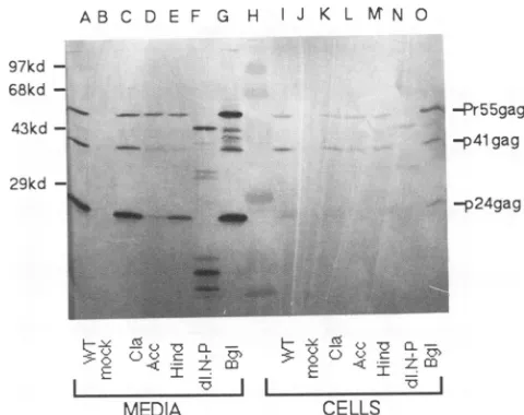
Expression and assembly of HIV gag mutants. To assay Gagprotein expression and to examine the effects of muta-tions onvirus assembly, immunoblottingwas performed to detect virus-associated Gag antigen in the medium versus inside the cells. At 48 h after transfection ofCOS7cells,cell supernatants and cell samples were prepared and subjected
toSDS-PAGE followed byelectroblottingonto a nitrocellu-lose membranes as described in Materials and Methods. HIV Gag proteins then were immunodetected with anti-p249ag monoclonal antibody. As illustrated for several mu-tants in Fig. 2, the mature Gag product p24gag, and incom-pletely processedproteinsPr55gag and p41gagwere detected in medium supernatants (lanes A to G). In cell samples, precursor
Pr55fag
and p41gag bandswerevisible (lanes I to 0), while p249agwasvariably detected(lanesIand0).In the case ofdl.NsiPst(Fig.2, lanes F andN)abandat49kDa and bands at 34 to 36 and 16to20kDawereobserved, consistent with a deletion of 56 amino acids. As shown, some mutant proteins were released efficientlyfrom cells, whereas otherson November 9, 2019 by guest
http://jvi.asm.org/
TABLE 1. InfectivityofHIV gag mutants
Construct' Titer HIVgpt titer" %Infectivity'
HIVgpt 1,681 1,681 100
Psi- 8 263 3.0
Myr- 0 263 0
ClaI 72 544 13.2
3,234 4,916 65.8
3,687 4,916 75.0
AccI 0 880 0
0 1,660 0
HindIll 2 1,392 0.1
183 3,428 5.3
PvUII 66 544 12.1
174 4,916 3.5
508 4,916 10.3
d1.NsiPst 0 544 0
0 660 0
SpeI 0 2,080 0
0 2,080 0
ApaI 2 984 0.2
251 3,428 7.3
A14-15 0 660 0
0 660 0
BglII 0 1,392 0
33 3,428 1.0
PR- 0 1,392 0
0 660 0
d1.NsiPst + PR- 3 660 0.5
'Each construct was cotransfected withSV-MLV-A-env into COS7 cells. In mostcases,2days later,cellsupernatantswereused toinfectHeLacells. In two experiments (those with HIVgpt titers of 3,428 and 4,916), cell supernatantswerecollected after 3daysrather than 2days. Infectionsand selections formycophenolic acid-resistant colonies wereperfornmed as de-scribed inMaterialsand Methods.
bThe value for the HIVgpt titer was an average of 10 independent
experiments.The averagesforHIVgpttiters from3- and 2-day collections were 4,172 and 1,058 + 611, respectively. Duplicate experiments were performedwithdifferent DNAs,atdifferenttimes,orboth.
cInfectivities for eachmutant weredetermined by the ratio of its titer versusthe wtHIVgpttiter(middle column)in aparallelexperiment.
werepresentin medium supematants atnoticeably reduced levels(seeAccI,Fig. 2, lanesD andL).
Becausetheseexperiments involved collection of medium
supernatantvirusproteins whichwere pelleted in a45-min
spin through a 20% sucrose cushion, we believe that they representvirus-associatedproteins. Based on centrifugation
clearing rate estimates, the minimum size of a pelletable
particle would be 165S, and >90% ofour medium HIVgpt
Gag protein was recoverable by this method (data not
shown). Nevertheless, wewere surprised tofind that even
dl.NsiPst, the56-amino-acid deletion protein, wasreleased as ahigh-molecular-weightcomplex, and thus we examined
particlesreleased from COS7 cells inmoredetail. To do so, medium supernatant proteins were fractionated by sucrose
density gradient centrifugation, assayed by immunoblotting,
AB C D E F G H J K L M N 0
97kd
-68kd -43kd -.
29d 29kd
-1w1,
-.-- -Pr55gag -p41gag
-p24gag
E E MIz
MEDIA CELLS
FIG. 2. Expression and release ofHIVGag proteins. COS7 cells were transfected with the designated plasmids. After48 to 72 h, supernatants and cells were collected and prepared for protein analysis asdescribed in Materials andMethods. Supernatant sam-ples (lanesA toG, corresponding to50% ofthe totalsample) and cell samples (lanes I to 0, corresponding to 5% of the total cell sample)were fractionatedby SDS-PAGE andwereelectroblotted
onto anitrocellulosefilter.HIVGag proteinsweredetectedwitha mouseanti-p24 monoclonal antibodyat a1:2,000 dilution, followed by a secondary alkaline phosphatase-conjugated goat anti-mouse antibodyat a1:1,500 dilution and detection of alkaline phosphatase activity. Molecular size markers (lane H)areindicatedontheleft, andHIVGagproteinsPrSS, p41, and p24areshownonthe right. Lanes: AandI,wt;B andJ, mock; CandK,ClaI;D andL, AccI; EandM,HindIII; FandN,NsiPst; G and 0,BglII.
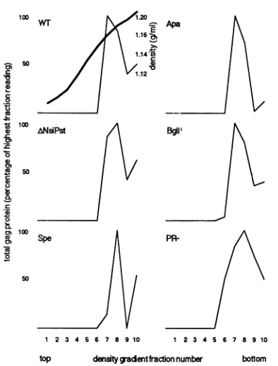
and quantitated by densitometry. Because ClaI and PvuII mutants wereinfectious (Table 1), Psi- and A14-15mutants had been examined previously (1), and limited release of AccIandHindIIImutantproteins prohibited theiranalysis, we focused our examination onwt, dl.NsiPst, SpeI, ApaI, BglII, and PR- mediumproteins.Asshown inFig.3,wtand all five mutant proteinsbanded inour fractions7 and 8, at densities between 1.16 and 1.18 g/ml. Thisparticle density readingis consistent withthat for retrovirusparticles (3, 18) and indicates that the levels of pelletable medium Gag
proteinsreflect the amount of virusparticles released from cells.
Toreliablyevaluate the effects of gag mutations onHIV assembly and particle release, we quantitated Gagprotein
levels withascanningdensitometer and determined ratios of total Gag proteinlevels in the medium supernatants versus cells. To comparewithwtHIVgpt,ratios obtained with each mutant were divided by wt ratios in parallel experiments. The results shown in Table 2 indicate that most mutants werecapableofparticle assemblyand release because their ratios were comparable to that of the wt construct. Two mutants, ClaI and dl.NsiPst, possessed medium/cell Gag ratios that were even higher than the wt ratio; this result could be due eithertoincreased levels of intracellularmutant Gag protein degradationor to increased
assembly
rates. In contrast, the ratio of theMyr- mutant was <1% of thewtratio, a result consistent with the previous demonstration that Myr- HIV mutants are blocked in virus
assembly (5,
34). With the exception of Myr-, the ratios oftwomatrix mutants,AccI andHindIII, were lower than those of any-.- 41.
7--dwAk.
on November 9, 2019 by guest
http://jvi.asm.org/
[image:4.612.63.302.92.463.2]WT - Apae
1.16
1.14V5
50 aD
1.12
0
100
top
ANsiPit Bgnfrci ume bto
1a
00
00
12 3 4667 89 10 1 23 456 7 8910
[image:5.612.80.277.71.330.2]top densitygadentfractionnumber bottom
FIG. 3. Sucrosedensity gradientfractionation of HIVparticles. Supernatantsfrom transfectedCOS7 cellswerecollected and
frac-tionated bysucrose density gradients (20to 50%) as described in Materials and Methods. Fractions were collected from top to
bottom,and virus-associatedGag proteinsfrom each fractionwere measuredbyimmunoblottechniquesandwerequantitated by
scan-ning densitometry. Asshown, the peakGag proteinfractions had densities of 1.16 to 1.18g/ml.
other mutants. Indeed, AccI Gag proteins were detected
predominantlyinsidecells,withmedium/cellratios8 to 30% ofwt levels (Fig. 2 and Table 2). Theoretically,AccI Gag protein release values could be a consequence of protein instabilityin virus particles or inefficient particle budding.
Becausewehave not observedmajordifferences in
particle-associated Gag protein degradationrates (data notshown),
we favor the hypothesis that the AccI matrix mutation inhibits HIV-1 particle release. Support for this notion
comes from indirect immunofluorescence studies. After transfection with wtHIVgptor Myr-, ClaI, AccI, HindIII,
BglII, or PR- constructs, COS7 cells on coverslips were
processedforimmunofluorescent localization ofGag antigen by using an
anti-p249ag
first antibody and arhodamine-conjugatedrabbitanti-mouse secondantibody (seeMaterials and Methods). As illustrated in Fig. 4A, wt Gag proteins
were present throughoutthe cytoplasm of transfectedcells
as a heterogenously staininghaze. Localizationpatterns of
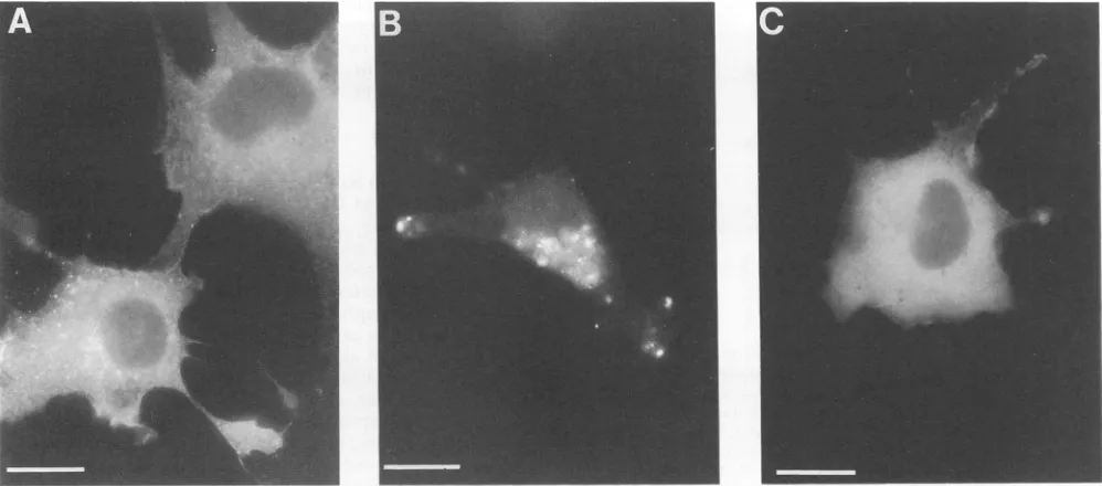
ClaI, HindIII,BglII,and PR- mutantGag proteinscould not bedistinguishedfrom wtpatterns. Incontrast,cells express-ing theAccI mutant showed punctate staining asymmetri-callylocatedaround cellnuclei (Fig. 4B).Thepatternof the AccI mutantwas different from thatwith the Myr- protein,
which localized to the cytoplasm of transfected cells (Fig. 4C). These resultssuggest thatAccI mutants demonstrated reduced particle assembly because the Gag proteins were
trapped intracellularly.
Characterization of virusparticles. The results of Table 1 indicate that many of our HIV gag mutants were poorly
infectious, while data in Table 2 show that, for most
mu-TABLE 2. HIV Gag protein release fromcellsa
Total
Gaf
Construct Expt protein in
Medium/cell
%wtratio' Medium Cells
HIVgpt 1 4.96 3.35 1.46 100
2 31.88 24.0 1.33 100
3 9.89 15.2 0.65 100
4 3.62 6.0 0.60 100
Myr- 1 <0.32 19.6 0.016 <1
ClaI 2 30.6 9.8 3.12 234
AccI 1 0.72 6.0 0.12 8
2 3.74 9.2 0.41 30
3 1.57 16.0 0.10 15
HindIII 2 10.8 10.0 1.08 81
3 6.66 19.6 0.34 52
PvuII 4 2.91 7.2 0.40 67
dl.NsiPst 2 24.47 7.2 3.40 255
SpeI 3 9.20 4.6 2.0 130
ApaI 3 4.43 7.2 0.62 95
BglII 2 8.32 32.4 1.49 112
3 34.4 18.6 1.85 286
PR- 2 18.23 9.6 1.90 143
aSupernatant and cell samples ofwtand mutant constructs wereanalyzed by immunoblot techniques as described in Materials and Methods. Gag proteins from mediumorcellsampleswerequantitated by scanningPr55, p41, andp24 band densitiesfromimmunoblots. Theresults of fourexperiments werecompiled.
bThe sum ofarbitrary density units(Pr55 plus p41 plus 24) indicated the totalGag proteinin the medium orinside the cells.Inthecaseofthe Myr-mutant, noGag proteins could be detected inthe medium(the0.32value indicated the minimal detectablesignal by densitometry).
cRatios of totalGag proteininthemediumversustotalGag inside the cells wereindicators of virusparticle assembly and release. Forcomparisonwith release ofwtvirus,the ratio of each mutant wasdividedby the ratioof wt in eachparallelexperimentandwasmultiplied by100.
tants, virion releasewas unimpaired. Anobvious
explana-tion for this difference is that the virus particles assembled byour mutantGagproteinsweredefective forone reason or another. Because reversetranscription is essential for HIV
replication,gagmutantsalsowerecharacterizedby measur-ingRTactivitylevels.Assayswereperformedasdescribed inMaterials andMethods, and in each case, thecountsper minute ofincorporatednucleotidewas atleastthreefoldover backgroundlevels(Table 3).Whilenucleotideincorporation counts yielded some information on RT levels, to obtain
specificactivities for each mutant, the ratios of normalized counts per minute versus densitometer-determined virus-associatedGag proteinlevelswerecomparedwithwtlevels in parallel experiments. The results in Table 3 show that most mutantsshowedatleast50%of thewtRTactivity.The
only exceptions to this ruleweretheSpeI, BglII, and PR-mutants. FortheBglIIandSpeI mutants, which retained 30 to60% ofwtactivity (Table 3),reduced RT levels could be due to reduced stability, processing, or incorporation of
Gag-Pol proteins into virions. With ourprotease-defective
mutant,specificRTactivitieswere30 and50%ofwtlevels,
consistent withpreviousreports(35). Although unprocessed Gag-Polfusionproteinsmay resultinreduction of
enzymatic
on November 9, 2019 by guest
http://jvi.asm.org/
[image:5.612.310.549.91.376.2]FIG. 4. Indirectimmunofluorescence detection of HIV Gag proteins inCOS7 cells.COS7 cellsgrown oncoverslipsweretransfected with
wtHIVgpt (A), Gag AccI (B), orMyr- (C)mutant constructs. Forty-eight hours after transfection, cellswerefixed and permeabilized for immunofluorescence assays asdescribed in Materials and Methods. The primary antibodywas amouseanti-p24 monoclonal antibodyata
1:1,000dilution, and the secondary antibodywas a 1:100dilution ofrhodamine-conjugated rabbit anti-mouse antibody. Mock-transfected
COS7 cells and cellsnotexposedtotheprimary anti-Gagantibody yieldednosignals (datanotshown). Bars, 20 ,um.
activity(23, 24, 26), itisconceivable that the lowenzymatic activityof theprotease mutantmaybe duetothestability of immature viruscores (seebelow), and inefficient detergent release ofGag-Pol fusion proteins during RTassays.
Previous studies have shown thatafunctionalproteaseis required forretrovirus infectivity (13, 20, 35), suggesting a
requirementfor HIV Gag protein processing. Weanalyzed processingof HIVgagmutantsby immunoblotting of parti-cle-associated proteins. Figure 5 shows thatmost mutants
were processed as well as wt HIVgpt, in which p249ag is present, but precursor forms Pr55gag and p41gagwere also
observed. Asexpected, the clearest exceptionwasthe
PR-mutant,inwhich theprocessingwasblockedcompletely. In thisexperiment, medium AccI andHindIIIGaglevelswere
belowourlevels ofdetection, consistent with theirreduced
release from cells, but results in Fig. 2 show that at least
someprocessing of these proteinsoccurred. The dl.NsiPst
constructwitha56-amino-aciddeletion in thecapsiddomain
stillcould beprocessedand detectedasbands of49, 35,and
18kDa,correspondingtothewtPr55fag,p419a9, andp249ag proteins,respectively.Toassesstheprocessing efficiencyof
eachmutant,wequantitated thep24-associated Gagproteins
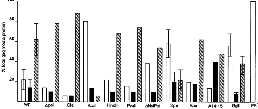
in mediumsupernatantsamplesby densitometryandplotted the ratio ofeachGag productversustotal Gag proteins.As
illustrated in Fig. 6, and asobserved in Fig. 2 and5, most mutantsdisplayedaprocessingpatternsimilartothatofwt. However, there were several exceptions. In particular,
mutantsAccI, SpeI,andBglIIwereprocessedincompletely,
suggestingeither thatthesemutantGagproteinswere
resis-tant to cleavage, that mutant Gag-Pol proteins possessed defectiveproteasemoieties,orthatmutantGag-Pol proteins entered virions at reducedefficiencies (seeDiscussion).
Of all our mutants, onlyPR- was completely devoid of
mature virus particle-associated mature proteins. This al-lowedustoinvestigatestructural differences between
imma-ture andmature HIVparticles. Previous studies have
dem-TABLE 3. RTactivities of HIV gag mutantsa
Relativeactivity
Construct cpmincorporated (%
Wtib
HIVgpt 3,090 100
5,890 100
1,781 100
4,868 100
ClaI 5,700 101
2,621 56
AccI 1,450 66
HindIII 5,420 83
1,380 54
NsiPst 5,180 113
3,453 90
SpeI 1,810 45
1,526 43
A14-15 1,838 63
BglII 3,010 30
5,710 64
PR- 1,310 30
1,352 50
aPreparationofsupernatantsand RT assayswereperformedasdescribed inMaterials and Methods. Note that activities(cpmincorporated)were at
least threefold higherthan that of background(396 + 6 cpm). For each sample,virus-associatedGag proteinlevelsweredeterminedasdescribedin Table 2 footnotes. Results offourseparatetransfectionexperimentsaregiven.
bRelativeactivitiesweredeterminedaspercentages ofwtactivitiesbythe equation100x[(mutantcpm -background)/mutantGagprotein/(wtcpm
-background)/wtGag protein].
on November 9, 2019 by guest
http://jvi.asm.org/
[image:6.612.65.564.81.301.2] [image:6.612.324.561.401.661.2]A B C D E F G H J K L M
Pr55
-p41
-
p24-C
6 V m m o w
[image:7.612.61.278.75.230.2]~~~~~~E FIG. 5. Assembly and processing of HIVgagmutants. COS7 cellsweretransfected withwtHIVgpt andmutantplasmids. Forty-eight hours after transfections, supernatants were collected and
preparedfor proteinanalysisasdescribed in Materials and Methods. Sampleswerefractionatedby SDS-10% PAGE followed by immu-noblotanalysis with anti-p24 antibodyasdescribed in the legendto
Fig. 2. Lanes: A, wt (HIVgpt); B, Psi-; C, ClaI; D, AccI; E, HindIII;F,NsiPst; G, SpeI; H, ApaI;I,A14-15;J, BglII; K, Pro-;
L, mock; M, standard. HIV Pr55, p41, and p24 Gag proteins are
designatedatthe left. Note that particlerelease ofAccl andHindlll
mutantswaslow inthisexperiment and thatprocessing levelswere
determinedfromother transfections.
onstrated that immature virions of avian sarcoma-leukosis virusandMLVprotease mutantswereresistanttononionic detergent (14a, 42).Toinvestigate whether theHIVprotease mutanthadasimilarproperty,culturesupernatantsof trans-fectedcellsweretreated with0.5% Triton X-100 followedby
centrifugation through 20%sucrosecushionsasdescribedin
Materials and Methods. Gag proteinsrecovered in the pel-letswereanalyzed byimmunoblotting. As shown inFig. 7, mostprotease mutant virions were recovered in the pellet
either with or without treatment of Triton X-100 (Fig. 7, lanes C and D). Incontrast,p24&ag in particles produced by wtHIVgptwassolubilized byTritontreatment(Fig. 7,lanes
A andB). However, itwas noted thatwt HIVgpt medium
Pr55&ag proteins appearedresistanttoTriton X-100 solubili-zation, suggesting either that proteolytic processing is very
rapidonce it is initiated or that immature and matureGag proteinswerenotpresent within thesameparticles.
DISCUSSION
Tronoetal. (46) have reported that coexpression of HIV gagmutantswithwtHIV could resultinadrasticreduction of virus infectivity, suggesting that mutant Gag proteins wereincorporated intowtvirusparticles and interfered with subsequent viral replication. We have analyzed the infectiv-ity of HIVgag mutants by cotransfection withan ampho-tropic envelope expression plasmid, SV-A-MLV-Env, into COS7 cells. Titers ofgenerated virus particleswere deter-minedby infection and selection of drug-resistant colonies. Although HIV and MLV Env proteins do not share
signifi-canthomology, Page et al. (33)reported that titers of virions generated by coexpression of HIVgpt and MLV Envwere generallyhigher than those of virions generated by coexpres-sion of HIVgpt and HIV Env in parallel transfections, indicating thatpseudotypingof MLV EnvwithHIVgpt is an efficient process. However, a recent study demonstrated thatthe HIV matrix domain may be involved in the incor-poration of HIV envelopes into virions (51), and we must note that because of our utilization of a murine envelope protein, we would be unable to detect specificperturbation ofputative HIV Gag and Env interactions in our system.
We weresurprised to discover the number of mutants that were able to assemble virus particles. To a certain extent, thismay be attributable to use of ahigh-expression COS cell system.However, Western immunoblotanalysis indicated a failure of mutants AccI andMyr- toproduce high levels of virus particles (Fig. 2 and Table 2). Our findingswith the
Myr- mutant corroborate previous reports that myristyla-tion of HIVGagisrequiredfor virusassembly and infectiv-ity (5, 34). Also, previous work has shown that deletion
mutations around the MA AccI region could block virus assembly, because very little RTactivitywas released into the culture medium(51); however,these mutants were not
.0
0.
.A 60
0~
40
10
20-wr Apsi CIa Acci Hindlil PvuII ANsiPst Spe Apa A14-15 BgIll PR
FIG. 6. ProcessingofHIVgagmutants.Gag proteinsinmedium supernatants werequantitatedby scanningdensitometry asdescribedin thelegendtoTable2.Thepercentages ofPr55gag(openbars),p419a9(solidbars),and p249ag (hatchedbars) were obtained by dividingdensity unitsofindividual bands bythetotalGag proteindensityunits and multiplying by 100. The wt values were derived from six independenttrials,
mutantsSpeIandBglIIwerefrom threeexperimentseach, and all others were derived from one experiment each.
on November 9, 2019 by guest
http://jvi.asm.org/
[image:7.612.84.514.502.684.2]A B C D
Pr55gag-
p24gag-WT
PR-FIG. 7. Virus particle sensitivity to nonionic detergent treat-ment. Supernatants from wt or protease mutant transfected COS7 cellswere filtered through a 0.45-pum-pore-size filter. Cell
superna-tantsthenweremock treated (lanes A and C) or treatedwith0.5% Triton-X100(lanes B and D). Aftertreatment, sampleswere centri-fuged through2 ml of20%sucrose at 274,000 x g and 4°Cfor45min. Pellets were solubilized in sample buffer and separated on an SDS-10% polyacrylamide gel and wereelectroblotted onto a nitro-cellulose filter.Immunodetection of Gag proteins wasperformedas describedinthe legend to Fig. 2. Core protein precursorPr55gagand
maturep249aB proteinbands areindicated on the left.
further characterized. Indirect immunofluorescence re-vealed that mutant AccI Gag proteins appeared trapped around the perinuclear area (Fig. 3),indicating that mutation at this region may block a normal Gag transport route or cause the protein to mislocalize. Previous studies have suggested that the matrix domain of Moloney MLV Gag protein may interact with intracellular membranes prior to transporttothe cell surface (15). Thus, it isconceivable that the HIV matrix domain also may interact with intracellular membranes under some circumstances.
Our linker-generated mutants ClaI (near the MA amino terminus, after the 15th codon ofgag) and PvuII(after codon 120, 12 codons from the CA coding region) did not affect virus assembly or processing and had significant exogenous
templateRTactivities, consistent with previous reports(50, 51). Our infectivity analysis showed that mutants ClaI and PvuII were somewhat infectious (PvuII) or up to half as infectious as wt (ClaI). However, we cannot exclude the possibilitythat the matrix domain may be involvedin in vivo replication processes.
Several lines of evidence have suggested that retroviral
capsid domains are key regions responsible for interactions among theGag polyproteins (9, 37). Surprisingly, our HIV mutantcontaining a56-amino-acid CA deletion did not affect virus assembly, budding, or processing (Fig. 2 and 6). The dl.NsiPst mutant RT activity was comparable to that ofwt (Table3), and this mutant even exhibited acharacteristicwt retrovirus density of 1.16 to 1.18 g/ml (Fig. 3). Apparently, the amino-terminal HIV Gag CA of this region may be
dispensablefor HIV core assembly. The other capsid muta-tion (SpeI) containing only a two-amino-acid insertion,
ap-parently eliminated viral infectivity (Table 1). Incomplete
processing(Fig. 2 and 6) and low enzymatic activity (Table
3) mayhave rendered this mutant noninfectious. However, early postbinding steps in the infection process also maybe affected by theSpeI mutation.
Both nucleocapsid mutants A14-15 and ApaI could
assem-ble, release, and process Pr55gag (Fig. 2 and 6) but showed
reduced or no infectivity (Table 1). These observations are consistent with previous work, suggesting that NC (p7) does not play a role in core assembly (1). The inability of mutant A14-15 toreplicate appears tobe duetodefective packaging of viral RNA into virions, because previous quantitation of viral RNA by slot blot hybridization (1) showed a nearly complete blockin RNA encapsidation(1). However, prelim-inary resultswithourApallinker insertion betweenCys-His finger motifs suggest that RNAincorporation is notinhibited (datanot shown), although the mutation reducedinfectivity. As for the PR- mutant (Bcll), our findings support previ-ous studies (13, 35) which showed that protease-defective mutants are blocked for virus infectivity but not for assem-bly. The fact that maturation of immature virions by viral protease is required for viral infectivity is consistent with our findingswithmutantsAccI,SpeI,andBglII,in whichvirions are both incompletely processed and noninfectious orpoorly infectious (Table 1 and Fig. 6). It is possible that these mutations may lead to conformation changes in Gag precur-sors and subsequently may interfere with the exposure of cleavage sites to protease. Alternatively, conformation changes ofGag-Pol fusion proteins induced by these mutants may interfere with dimer formation of Gag-Pol molecules, which is required for activation of viral protease (22, 31,49). The low RT activity of mutants AccI, SpeI, andBglII, also may be a consequence of incomplete processing, defective Gag-Polprotein dimerization, orinsufficient incorporation of Gag-Pol into virions (22, 31, 42).
Because the majority of our HIV gag mutants assembled noninfectious or poorly infectious particles, these studies strongly support the notion that retroviral Gag proteins function not only in driving assembly for the construction of virus particles but also in a variety of postassembly, post-processing events. It will be of interest to dissect theeffects of these and other mutations on the processesof endogenous template reverse transcription, nuclear localization, and integration events.
ACKNOWLEDGMENTS
Weareindebted tomembersof Epitope, Inc. (Beaverton, Oreg.) ingeneraland Rick Bestwick in particular for advice and assistance. Thanks aregiven to D. Littman and N. Landau for the gifts of the HIVgptandSV-A-MLV-env plasmids. Many thanks are also given toA.Aldovini, R. Young, and L. Ratner for the gifts of the HIV-1 mutant constructs. We gratefully acknowledge the assistance of laboratorymembersMark Hansen, Jason McDermott, Jenny Stege-man-Olsen, and YaquingZhang.
This work could not have been accomplished without support fromthe National Institutes of Health (NCI grant 5RO1 CA47088-05) andstarting support from the Oregon Medical Research Foun-dation (grant 2-2-614-521).
REFERENCES
1. Aldovini, A., and R. A. Young. 1990. Mutations of RNA and protein sequences involved in human immunodeficiency virus type 1packaging result in production of noninfectious virus. J. Virol.64:1920-1926.
2. Bess, J. W.,Jr., P.J.Powell,H. J.Issaq, L. J. Schumack, M. K. Grimes,L.E.Henderson,and L.0.Arthur. 1992. Tightly bound zinc in human immunodeficiency virus type 1, human T-cell leukemiavirustypeI, andother retroviruses. J. Virol. 66:840-847.
3. Bolognesi, D. P., R. Luftig,andJ.H.Shaper. 1973. Localization of RNAtumorviruspolypeptides. I. Isolation of further virus substances. Virology56:549-564.
4. Bowerman, B., P.0. Brown, J. M. Bishop, and H. E. Varmus. 1989. A nucleoprotein complex mediates the integration of retroviral DNA. GenesDev.3:469-478.
on November 9, 2019 by guest
http://jvi.asm.org/
[image:8.612.139.236.79.229.2]5. Bryant, M., and L. Ratner. 1990. Myristoylation-dependent replication and assembly of human immunodeficiency virus 1. Proc.Natl. Acad. Sci. USA87:523-527.
6. Cann, A. J., and J. Karn. 1989. Molecular biology of HIV-1: newinsightsinto the virus life cycle. AIDS 3(Suppl. 1):S19-S34. 7. Cohen, E. A., E. F. Terwilliger, J. G. Sodroski, and W. A. Haseltine. 1988. Identification of a protein encoded by the vpu geneofHIV-1. Nature(London) 334:532-534.
8. Crawford, S., and S. P. Goff. 1984. Mutations in Gag proteins P12 and P15 of Moloney murine leukemia virus block early stagesof infection. J. Virol. 49:909-917.
9. Ehrlich, L. S., B. E. Agresta, and C. A. Carter. 1992. Assembly of recombinant human immunodeficiency virus type 1 capsid protein in vitro. J. Virol. 66:4874-4883.
10. Gheysen, D., E. Jacobs, F. de Foresta, C. Thiriart, M. Francotte, D. Thines, and M. De Wilde. 1989. Assembly and release of HIV-1 precursor pr55gagvirus-likeparticles from recombinant baculovirus-infected insectcells. Cell 59:103-112.
11. Gorelick, R. J., S. M. Nigida, Jr., J. W. Bess, Jr., L. 0. Arthur, L. E. Henderson, and A. Rein. 1990. Noninfectious human immunodeficiency virus type 1 mutants deficient in genomic RNA. J. Virol.64:3207-3211.
12. Gottlinger, H. G., T. Dorfman, J. G. Sodroski, and W. A. Haseltine.1991.Effectof mutationsaffecting the p6 gag protein onhumanimmunodeficiencyvirus particle release. Proc. Natl. Acad.Sci. USA88:3195-3199.
13. Gottlinger, H. G., J. G. Sodroski, and W. A. Haseltine. 1989. Role of capsid precursor processing and myristoylation in morphogenesis and infectivity ofhuman immunodeficiency vi-rus type 1. Proc. Natl.Acad. Sci. USA 86:5781-5785. 14. Graham, R., and A. van der Eb. 1973. Anewtechnique forthe
assay of infectivity of human adenovirus 5 DNA. Virology 52:456-467.
14a.Hansen, M.,etal.Submitted for publication.
15. Hansen, M., L. Jelinek, S. Whiting, and E. Barklis. 1990. Transport and assemblyof Gag proteins into Moloneymurine leukemiavirus. J.Virol. 64:5306-5316.
16. Hayashi,T., T.Shioda, Y.leakura,andH. Shibuta.1992. RNA packaging signal of human immunodeficiency virus type 1. Virology188:590-599.
17. Henderson, L. E., M. A.Bowers, R. C. Sowder II, S. A. Serabyn, D.G. Johnson, J. W. Bess, Jr., L.0.Arthur, D. K.Bryant,and C. Fenselau. 1992. Gag proteins ofthe highly replicative MN strain of human immunodeficiency virus type 1: posttransla-tional modifications, proteolytic processing, and complete aminoacidsequences.J. Virol. 66:1856-1865.
18. Jones, T. A., G. Blaug, M. Hansen, and E. Barklis. 1990. Assembly ofgag-p-galactosidaseproteinsinto retrovirus parti-cles.J. Virol.64:2265-2279.
19. Klimkait, T., K.Strebel, M. D. Hoggan, M. A. Martin, and J.M.
Orenstein. 1990. Thehumanimmunodeficiency virustype 1-spe-cific proteinvpu isrequired forefficientvirus maturation and release. J.Virol.64:621-629.
20. Kohl, N. E., E. A. Emini, W. E. Schleif, L. J. Davis, J. C. Heimbach, R. A. F. Dixon, E. M. Scolnick, and I. S.Sigal. 1988. Active humanimmunodeficiencyvirus protease is requiredfor viralinfectivity. Proc. Natl.Acad. Sci. USA85:4686-4690. 21. Laemmli, U.K.1970.Cleavage of structuralproteinsduring the
assembly of the head ofbacteriophage T4. Nature (London) 227:680-685.
22. Lapatto, R., T. Blundell, A. Hemmings, J. Overington, A. Wilderspin, S. Wood, J. R. Merson, P. J. Whittle, D. E. Danley,
K. F. Geoghegan, S. J. Hawrylik,S. E. Lee, K. G.Scheld,and
P.M. Hobart.1989. X-ray analysis of HIV-1 protease at 2.7 A resolution confirms structural homology among retroviral en-zymes. Nature(London)342:299-302.
23. Le Grice, S. F. J., R. Ette, J. Mills, and J. Mous. 1989. Comparisonof the humanimmunodeficiencyvirus type 1 and 2 proteasesby hybrid gene construction and trans complementa-tion. J.Biol. Chem.264:14902-14908.
24. Le Grice, S. F. J., J. Mills, and J. Mous. 1988. Active site mutagenesisof the AIDS virus protease and its alleviation by
transcomplementation. EMBO J. 7:2547-2553.
25. Leis,J., D.Baltimore, J. B. Bishop, J. Coffin, E. Fleissner, S. P. Goff,S.Oroszlan,H.Robinson, A. M. Skalka, H. M.Temin, and V. Vogt. 1988. Standardized and simplified nomenclature for proteinscommon toallretroviruses.J. Virol. 62:1808-1809. 26. Leuthardt, A., and S. F. Le Grice. 1988. Biosynthesis and
analysis of a genetically engineered HIV-1 reverse tran-scriptase/endonuclease polyprotein in Escherichia coli. Gene 68:35-42.
27. Lever, A., H. Gottlinger, W. Haseltine, and J. Sodrosky. 1989. Identification ofasequencerequired forefficient packagingof human immunodeficiency virus type 1 RNA into virions. J. Virol.63:4085-4087.
28. Maniatis, T., E. F. Fritsch, and J. Sambrook. 1982. Molecular cloning: alaboratorymanual. ColdSpringHarborLaboratory, ColdSpring Harbor, N.Y.
29. Mervis, R. J., N. Ahmad, E. P. Lillehoj, M. G. Raum, F. H. R. Salazar, H. W. Chan,and S. Venkatesan. 1988. The gag gene products of human immunodeficiency virus type 1: alignment within thegagopenreading frame, identification of posttrans-lationalmodifications, and evidenceforalternative gag
precur-sors.J.Virol. 62:3993-4002.
30. Mulligan, R. C., and P. Berg. 1981. Selection foranimal cells that express the Escherichia coli gene coding for xanthine-guaninephosphoribosyltransferase. Proc. Natl. Acad.Sci. USA 78:2072-2076.
31. Navia, M. A., P. M. D. Fitzgerald, B. M. Mckeever, C. T. Leu, J. C.Heimbach,W. K.Herber, I. S.Sigal,P. L.Darke, and J. P. Springer. 1989. Three-dimensional structure of aspartyl
pro-teasefromhumanimmunodeficiency virusHIV-1.Nature (Lon-don) 337:615-620.
32. Overton, H. A., Y. Fuji,I.R.Price, andI.M.Jones.1989. The proteaseand gag geneproductsof thehumanimmunodeficiency virus:authentic cleavageandpost-translational modification in
aninsectcellexpressionsystem. Virology170:107-116. 33. Page, K. A., N. R. Landau, and D. R. Littman. 1990.
Construc-tion and use of a human immunodeficiency virus vector for analysisof virus infectivity.J. Virol.64:5270-5276.
34. Pal, R., M. S. Reitz, Jr., E. Tschachler, R. C. Gallo, M. G. Sarngadharan, and F. D. M. Veronese. 1990. Myristylationof gagproteins ofHIV-1playsanimportantrole in virusassembly. AIDS Res. Hum. Retroviruses6:721-730.
35. Peng, C., B. K. Ho, T. W.Chang, and N. T. Chang. 1989. Role ofhuman immunodeficiencyvirus type 1-specific protease in
coreproteinmaturation and viralinfectivity. J. Virol. 63:2550-2556.
36. Ratner, L.,W. Haseltine, R. Patarca, K. J.Livak, B.Starcich, S. F.Josephs,E.R.Doran,J.A.Rafalski,E. A.Whitehorn, K. Baumeister, L.Ivanoff,S. R. Petteway, Jr., M. L. Pearson,J. A. Lautenberger, T. S. Papas, J. Ghrayeb, N. T. Chang, R. C. Gallo,andF.Wong-Staal.1985.Completenucleotide sequences of theAIDSvirus,HTLV-III. Nature (London) 313:277-284. 37. Schwartzberg, P.,J. Colicelli, M. L. Gordon, and S. P. Goff.
1984. Mutationsinthe gag gene ofMoloney murine leukemia virus:effectsonproductionof virions and reversetranscriptase. J.Virol. 49:918-924.
38. Sharova,N., and A. Bukrinskaya. 1991.p17andp17-containing Gag precursors of input human immunodeficiency virus are
transportedinto the nuclei of infected cells. AIDS Res. Hum. Retroviruses7:303-306.
39. Shioda, T.,and H. Shibuta. 1992.Productionof human immu-nodeficiencyvirus(HIV)-like particlesfrom cellsinfectedwith recombinant vaccinia viruses carrying the gag gene of HIV. Virology175:139-148.
40. South,T.L., P. R. Blake, R. C. SowderIII,L.0.Arthur, L.E.
Henderson, andM.F. Summers.1990. Thenucleocapsid protein isolated from HIV-1particles binds zinc and forms retroviral-type zincfingers. Biochemistry29:7786-7789.
41. Stephens, E., and R.W. Compans. 1988. Assemblyof animal virusesatcellular membranes. Annu. Rev. Microbiol. 42:489-516.
42. Stewart, L., G. Schatz, and V. M. Vogt. 1990. Properties of avian retrovirus particles defective in viral protease. J. Virol. 64:5076-5092.
on November 9, 2019 by guest
http://jvi.asm.org/
43. Strebel, K., T. Klimkait, andM. A.Martin.1988. A novelgene
of HIV-1,vpu,and its 16-kilodalton product. Science
241:1221-1223.
44. Terwilliger,E. F., E.A. Cohen,Y. Lu, J.G. Sodroski, and W. A.
Haseltine. 1989. Functional role of human immunodeficiency virustype 1vpu.Proc. Natl.Acad.Sci. USA 86:5163-5167.
45. Towler,D.A.,S. R. Eubanks, D. S. Towery,S. P. Adams, and
L. Glaser. 1987. Amino-terminal processing of proteins by N-myristoylation. Substrate specificity of N-myristoyl
trans-ferase. J. Biol. Chem.262:1030-1036.
46. Trono, D., M. B. Feinberg,andD. Baltimore. 1989. HIV-1gag
mutants can dominantly interfere with the replication of the
wild-type virus. Cell 59:113-120.
47. Wain-Hobson,S., P. Sonigo,0.Danos, S.Cole, and M. Alizon. 1985. Nucleotide sequence of the AIDS virus, LAV. Cell
40:9-17.
48. Wilcox, C., J.-S. Hu, and E. N. Olson. 1987. Acylation of proteins with myristic acid occurs cotranslationally. Science 238:1275-1278.
49. Wlodawer, A., M. Miller, M. Jaskolski, B. K. Sathyanarayana, E. Baldwin,I.T.Weber, L. M.Selk, L. Clawson, J.Schneider, andS. B. Kent.1989.Conserved folding in retroviralproteases:
crystal structure ofa syntheticHIV-1 protease. Science 245:
616-621.
50. Yu, X.,Q. Yu, T. Lee, and M. Essex. 1992. The Cterminus of
humanimmunodeficiency virustype1 matrixprotein is involved inearlystepsof the virus life cycle. J. Virol. 66:5667-5670. 51. Yu, X., X. Yuan, Z. Matsuda, T. Lee, and M. Essex.1992. The
matrix protein of human immunodeficiency virus type 1 is required for incorporation of viral envelope protein intomature
virions. J. Virol. 66:4966-4971.