0022-538X/94/$04.00+0
Copyright © 1994,American Societyfor Microbiology
Interactions of Normal and Mutant Vesicular Stomatitis Virus Matrix
Proteins with the Plasma Membrane and Nucleocapsids
LISA D. CHONGANDJOHN K. ROSE*
Departments of Pathology and Cell Biology, Yale University School of Medicine, New Haven, Connecticut 06510
Received 17 March 1993/Accepted 4 October 1993
Wedemonstratedrecently that a fraction of the matrix (M) proteinofvesicularstomatitis virus(VSV) binds
tightly
tocellular membranes in vivo when expressed in the absence of other VSV proteins. This membrane-associated M protein was functional in binding purified VSV nucleocapsids in vitro. Herewe showthat the membrane-associated Mprotein is largely associated with a membrane fraction having the density of plasma membranes, indicating membrane specificity in the binding. In addition, we analyzed truncated forms of M protein toidentify
regions responsible for membrane association and nucleocapsid binding. Truncated M protein lacking the amino-terminal basic domain still associated with cellular membranes, although not as tightly aswild-type
Mprotein, and could not bindnucleocapsids. In contrast, deletion of the carboxy-terminal 14aminoacids did not disrupt stablemembrane association or nucleocapsid interaction. These results suggest that the aminoterminus of M protein either interacts directly with membranes and nucleocapsids or stabilizes aconformation that is required for M protein to mediate both of these interactions.Vesicular stomatitis virus (VSV) is an enveloped negative-strandRNAviruscontaining five proteins. The genomicRNA is encapsidated by the nucleoprotein (N) and is tightly associ-ated with the viral RNA polymerase proteins L and NS(P) (11). This core complex is surrounded by a lipid bilayer derived from the plasma membrane and contains the viral transmem-brane glycoprotein (G). The matrix (M) protein of VSV lies beneath thelipid bilayer and presumably bridges between the viral envelope and the nucleocapsid core.
The presence of M protein at the inner surface of the plasma membrane of infected cells(4, 23,27) and the rapid association ofafraction ofMprotein with membranes after synthesis on cytosolic polyribosomes (1, 10, 19) have been well docu-mented, but it was not clear whether these were membranes already involved in viral assembly.We recently used subcellu-lar fractionation studies of transfected HeLa cells to show that M
protein
canassociatetightly
with membranes invivo,
inthe absence of other VSV proteins, but the nature of the mem-branestowhich Mprotein
boundwas not determined in that study. Furthermore, membrane-associated M protein was functional in binding nucleocapsid cores to membranes in vitro. On the basis of these findings, we proposed a VSV assembly pathway that could be initiated by membrane-asso-ciatedMprotein(9).
M
protein
lacks a distinct stretch ofhydrophobic
residues indicative ofamembrane-associatingregion, and modification by fatty acid has not been demonstrated (22, 29). However, labeling studies withahydrophobic reagent suggested that the amino-terminal domain penetrates the bilayer(22).
Interest-ingly, atrypsin-resistant coreofMprotein
lacking the amino-terminal 43 residues could also associate with artificiallipo-somes,
suggesting
that otherregions
of Mprotein
may be involvedin membraneinteraction(26). Here,wereportstudies using subcellular fractionation to show that Mprotein
ex-pressed in cells binds largelyto aplasma
membrane fraction rather than nonspecifically to all cellular membranes.Addi-*Correspondingauthor.Mailingaddress:DepartmentofPathology,
YaleUniversitySchool ofMedicine, NewHaven,CT06510. Phone: (203) 785-6184.Fax: (203)785-7467.
tional studies on truncated M
proteins
show that the amino-terminal basic domain of M is notrequired
forbinding
M protein to membranes but is required for stable membrane association. Inaddition,
we demonstrate that this domain is required formembrane-associated M proteinto bind nucleo-capsid cores in vitro. We propose that this amino-terminal region either interactsdirectly
with membranes andnucleo-capsid
cores orstabilizesaconformation which isrequired
for Mprotein
to support these interactions.MATERUILSANDMETHODS
Viruses and cell culture.
Preparation
of radiolabeled VSV (Indiana serotype, San Juan strain) was performed in baby hamster kidney (BHK-21) cells as described previously (9). The recombinant vaccinia virus vTF7-3(14)
wasprepared
as describedby Whittet al.(35).
Plasmid construction and antibodies. Plasmids pBSG, pBSM, and pBSN,encoding the G, M, andN
proteins
of VSV (Indiana serotype, San Juanstrain),
are described elsewhere (9). Plasmid pBSsCD4KDELcontaining
DNAencoding
sCD4KDEL has been described
previously
(7).
Rabbit anti-VSVserum(9)
andanti-sCD4KDELantibody
(7)
have been described elsewhere. PlasmidspBSMN15 and pBSMC14weregenerated
by the PCRmethod,using synthetic
oligonucleotide
primers and
plasmid
pARM. PlasmidpARMwasgenerated
by cloningaBamHI fragment containing
theMprotein
genefromplasmid
pMZ10(10)
into theunique
BamHI site ofpAR-2529
(14,
30).
PlasmidpBSMN15
was madeby
using
oneprimer
(5'
-GCGGGATCCATCATGAGTAAGAAATTAGGGA
TCGCA-3') containing
aBamHI restriction site(underlined)
and an initiation codon
(in
boldface)
followedby
sequencecorresponding
to the DNAencoding
residues 18 to 23 ofMprotein
and a secondprimer
(5'-CAACTCAGCTTlCCTIf
CGGGC-3')
corresponding
todownstreamvectorsequence in pAR-2529. The PCRproduct
wasdigested
with BamHI and ligatedintotheuniqueBamHI
siteofpBS-SK(+)
(Stratagene,
La Jolla,
Calif.).
PlasmidpBSMC14
was madeby using
one primer(5'-ATCGGATCCTCACTTIV1llCTCGACAATCAG
GCCAAACATTAAGGC-3')
corresponding
to DNAencod-ing
residues205 to 215 of Mprotein
andintroducing
astop 441on November 9, 2019 by guest
http://jvi.asm.org/
codon (bold letters) to truncate M protein at residue 215, followed by a
BamHI
restriction site (underlined), and a second primer (5'-TAATACGACTCACTATAGGG-3') over-lapping the sequence encoding part of the T7 RNA polymerase promoter in pAR-2529 located upstream from theBamHI
cloning site. The PCR product was digested with BamHI and ligated into the unique
BamHI
site of pBS-SK(+). DNA sequences were confirmed by dideoxynucleotide sequencing (31) using Sequenase (U.S. Biochemical Corp., Cleveland, Ohio).Expression, radiolabeling, and immunoprecipitation of M proteins. Procedures have been described elsewhere (9). Briefly, HeLa cells
(106
cells per 3.5-cm-diameter plate) were infected for 30min
at37°C
with vTF7-3 at a multiplicity of infection of 10 in 100RI
of Dulbecco's modified Eagle's medium (DMEM) with the cationic liposome reagent Trans-fectACE(Bethesda
Research
Laboratories,Gaithersburg,
Md.). At 4 h postinfection, cells were labeled for 1 h with 50
,uCi
of[35S]methionine
per ml of methionine-free DMEM. When indicated, labeling medium was replaced with 2 ml of DMEM supplemented with 2.5 mM unlabeled methionine. Proteins were immunoprecipitated from cell lysates with anti-VSV serum(28). Labeled proteins were resolved by polyacryl-amide gel electrophoresis (PAGE) on 10% polyacrylamide gels containing sodium dodecyl sulfate (SDS) (21) and visual-ized by fluorography (6).Analysis of membrane-associated M proteins. Procedures have been described elsewhere (9). Briefly, HeLa cells (5 x
106
cells) were infected with vTF7-3 at a multiplicity of infection of 10 for 30min
at37°C
and then transfected with 10,ug
of plasmid DNA per 2.5 x10⁶
cells in 2 ml of DMEM for 4 h. Cells were then labeled for 1 h with[35S]methionine
(50,uCi/ml)
and chased for 1 h in DMEM supplemented with 2.5 mM unlabeled methionine. Cells were harvested and disrupted in a Dounce homogenizer (9). The postnuclear lysate was adjusted to 80% sucrose and fractionated on an 80%-65%-10% (wt/wt) sucrose step gradient by centrifugation at 35,000 rpm for 18 h at4°C.
Fractions were collected from the top, diluted with detergent solution, and immunoprecipitated with anti-VSV serum (28). Labeled proteins were analyzed by SDS-PAGE (10% gel).Reconstitution of RNP cores with membranes. Total cellular membranes from transfected and radiolabeled HeLa cells (1.5 x
107
cells) containing associated matrix protein were pre-pared according to the sucrose membrane flotation gradient method described previously (9). One unit of ribonucleopro-tein (RNP) cores, prepared from radiolabeled VSV virions (9), was incubated with the indicated membranes for 1 h at 37°C with periodic shaking. The suspension was then subjected to buoyant density analysis by centrifugation on a continuous 10 to 70% (wt/wt) sucrose gradient at 150,000 x g for 45mim at0°C.
Fractions were collected from the bottom, diluted with detergent solution, and immunoprecipitated with anti-VSV serum as described above. Labeled proteins were analyzed by SDS-PAGE (10% gel).Subcellular fractionation and assays. Total cellular mem-branes were isolated from the indicated transfected and radio-labeled HeLa cells (1.5 x
107
cells) by the sucrose membrane flotation gradient (9) and then further fractionated according to a method modified from that of Frangioni et al. (13). Isolated membranes were resuspended in 200pLI
of 0.25 M sucrose buffer containing 10 mM Tris-HCl (pH 7.4), 5 mMKCl,
1 mM EDTA, and 100 kallikren units of aprotinin per ml. The membrane suspension was applied to the top of a sucrose step gradient consisting of 2.0 M sucrose (bottom), 1.2 M sucrose (center), and 0.25 M sucrose (top), and the gradientTop
Fraction Number Bot
2 4 6 8 10
CD4KDEL
VSV-G
VSV-M
0
'64 -*--VSV-M--X VSV-G
--s CD4KDEL
- mannosidaseII
2 4 6 8 10
Top Bot
Fraction Number
FIG. 1. Fractionation ofcellularmembranes. Total cellular mem-branes were isolated from 1.5 x 10⁷ transfected and radiolabeled HeLa cells expressing sCD4KDEL,VSV G protein, or VSV Mprotein and fractionated on a sucrose gradient by isopycniccentrifugation as described in Materials andMethods. Gradient fractionswerecollected fromthe top and divided in half. One half of the gradient fractionswas diluted with detergent solution, immunoprecipitated with the appro-priate antibody(28), andanalyzed by SDS-PAGE (10%gel). Quanti-tation of total labeled protein in each fraction was carried out by scanning densitometry of autoradiographs. The other half of the gradient fractions was analyzed forcx-mannosidaseII activity bythe method of Storrie and Madden (33). The graph was generated from data collected from two experiments of cells expressing VSV G or sCD4KDEL protein, three experiments of cells expressing VSV M protein, and seven gradients for oa-mannosidase II activity. Fractions are numberedfrom the top (fraction 1) to the bottom (fraction 10).
wascentrifugedat 100,000 x gfor 2.5 h at4°C.Fractionswere collected from the top. Eachfractionwasdivided in
half;
one half wasimmunoprecipitated with the indicated antibody, and labeled proteins were resolved by SDS-PAGE (10%gel).
Quantitation oftheamountoflabeledprotein in eachfraction was carried out byscanning densitometry ofautoradiographs. The other half of the gradient fractions was assayed for cx-mannosidaseII activity by the method ofStorrie and Mad-den (33).
RESULTS
M protein associates with a plasma membrane-enriched membrane fractioninvivo. Previously,weexpressedwild-type M protein in transfected HeLa cells by using a recombinant vaccinia virus/T7 RNA polymerase-based expression system (14) and reportedits associationwithcellular membranes
(9).
To determinewhether there was specificity to the membrane association invivo, total cellular membranescontaining radio-labeled Mprotein wereisolatedfromtransfectedHeLacellsby equilibrium membrane flotation in asucrose gradient (9) and thenfurther separatedbyisopycnic centrifugation with appro-priate markers (13). This fractionation protocol was chosen because it is known to give good separation of membranes
IMPRIA
on November 9, 2019 by guest
http://jvi.asm.org/
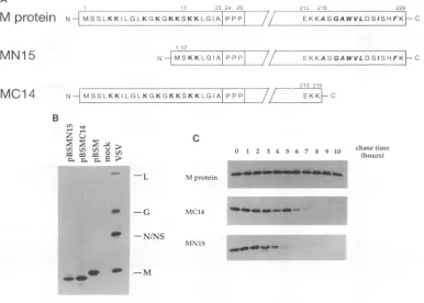
[image:2.612.326.547.75.305.2]NH
MSSLKKILGLKGKGKKSKKLGIAIPPPI
/ EKKASGAWVLDSISHFKFCMN1 5
N I MS K K L G Al P P P//
MC14
NI
MSSLKKILGLKGKGKKSKKLGIAI
PPPB
L "
z! u
04 m mni O >
EKKASGAWVLDSISHFKFC
213 215
/f
EKKF Cchase time (hours)
-L M protein
-G MC14
-N/NS
MN15
-M
1 2 3 4 .
FIG. 2. Construction,expression, and stability of mutated M proteins. (A) Diagram of the N-terminal and C-terminal deletions in M protein. MutantMN15lacks thefirst 15amino acids following the initial methionine residue. Lysineresiduesareindicated in boldface. Mutant MC14 lacks 14residuesattheextremecarboxyl terminus. Nonpolar and hydrophobic residuesareindicated by boldface italics. (B) HeLa cellswere infected
witharecombinant vaccinia virus (vTF7-3). After 30 min, infected cellsweretransfected with 5 ,ug of pBSMN15 (lane 1), pBSMC14 DNA (lane
2), and pBSM DNA (lane 3)or weremocktransfected (lane 4). At 4 h postinfection, cellswere labeled for 1 h with 50 ,uCi of[35S]methionine
perml.Immunoprecipitation of proteins from cell lysateswasperformed with anti-VSVserum,andlabeledproteinswereanalyzed bySDS-PAGE. VSVprotein markers from solubilized virionsareshownatthe right. Letters indicate theprotein designation. (C) HeLa cellswereinfected with
vTF7-3 for 30min and then transfected with the appropriate plasmid DNA. Cellswerepulse-labeled with 50 ,uCi of[35S]methionineperml for I hand thenchased inmedium containingexcesscoldmethioninefor 1-h intervalsupto 10 h.Immunoprecipitation of proteins from cell lysates
wasperformed with anti-VSVserum, and labeledproteinswereresolvedbySDS-PAGE.
derived from endoplasmic reticulum (ER), Golgi apparatus, and the plasma membrane.
Radiolabeled cells expressing M protein were lysed with a
Douncehomogenizer, and nucleiwereremoved. The
superna-tantwasadjusted to80%sucroseand placedatthebottom of
a sucrose gradient. Membraneswere fractionated awayfrom cytosol by equilibrium flotation during ultracentrifugation. Total cellular membranes were collected from the 10O%-65%
sucrose interface and thensubjectedtoasecondseparation by
isopycniccentrifugation (13).
The extent ofenrichment for ER, plasma membrane, and Golgi apparatus in the gradient fractions was determined by
assaying for specific markers. The protein sCD4KDEL,which accumulates in the ER of transfected HeLa cellsas aresult of
aspecific retention signal(7),wasusedasamarker fortheER.
When expressed in HeLa cells and analyzed by this method, sCD4KDEL was found predominantly in the high-density region near the 1.2 M-2.0 M sucrose interface (Fig. 1). In
contrast,thetransmembraneglycoprotein Gof VSVexpressed atthesurface oftransfected HeLa cellswas usedas amarker for plasma membrane. The majority of the total G protein localized to the 0.25 M-1.2 M interface (Fig. 1), although a
small amount was found at the higher-density interface. The natureofthishigher-density fraction containing G isnotclear, but itmightrepresentGinendosomes.Distribution of
ox-man-nosidase II activity, a marker for the Golgi membranes, was
between theERand plasmamembrane markers, aswouldbe
expected forthesemembranes ofintermediatedensity(Fig. 1). When we examined the subcellular localization of
mem-brane-bound M protein under thesameconditions, itshowed
a distributionvirtually identicalto that ofGprotein,with the majorityin thelightmembrane fraction andasmallamountin the high-density fraction. These results indicate that the frac-tion ofMproteinthatbindsmembranes isspecifically targeted to the plasma membrane rather than binding membranes nonspecifically.
We also used indirect immunofluorescence microscopy to attempt to examine the localization of M protein. Although
many cells appeared to have some M protein bound to the plasma membrane,thehigh backgroundofcytosolicMprotein madedefinitive localization impossible. However,others have successfullyshown Mprotein stainingattheplasmamembrane ofinfected cellsbyalternative cell disruptionmethods (23) as
wellasquick-freezinganddeep-etch replicamethods(25),but
we have not appliedthese methods totransfected cells. Expression oftwo truncated forms of M protein.To define the sequencerequirements forthe membrane association and nucleocapsid binding of M protein, we decided to generate mutants. Althoughthe three-dimensional structureofM
pro-tein isnotknown,computermodelingof thestructurepredicts A
M
protein
C
0 1 2 3 4 5 6 7 8 9 10
on November 9, 2019 by guest
http://jvi.asm.org/
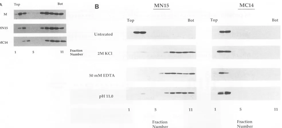
[image:3.612.117.503.84.360.2]A Top Bot
B
MN15 MC14M
Top Bot Top
MN15
Bot
=
.-Untreated
MC14
5 11 Fraction
Number 2MKCI
50mMEDTA
pH11.0 5 *- _ _ 1_
5 11 1 5 11
Fraction Fraction
Number Number
FIG. 3. Analysis of the membrane association ofwild-type Mprotein, mutantMN15, and mutant MC14bysucroseflotationgradients.(A) Lysates from 5 x 10⁶transfected and radiolabeled HeLa cells were prepared as described inMaterialsand Methods. Lysates were made to80% sucrose,placedatthe bottom of a Beckman SW41centrifuge tube,and overlaid with65%(wt/wt) (5 ml)and10%(wt/wt) (2.5 ml)sucroselayers. The stepgradients were thencentrifugedtoequilibriumat35,000 rpm for 18 h at4°C. Fractions werecollectedfrom the top, diluted withdetergent solution,andimmunoprecipitatedwithrabbit anti-VSV serum, and labeledproteinswereanalyzedbySDS-PAGE(10% gel).Shown aregradients from cells expressing wild-type Mprotein,MN15, or MC14.Fractions are numbered from the top(fraction 1)tothe bottom(fraction 11).(B)Total cellular membranes from 1.5 x 10⁷HeLacellscontainingassociatedmutantMN15 ormutantMC14 fromtransfected and[35S]methionine-labeled HeLacells wereprepared by the sucroseflotationmethod as described inMaterialsand Methods. Membranes were treated with 2 MKCI-10mM Tris-HCl(pH7.4)or50 mMEDTA-Tris-HCl (pH 7.4)for 1 h at25°C, extractedwith carbonatebuffer (pH 11.0)for 30 min at0°C(15),orleft untreated. Samplesweremadeto80%sucrose,and membraneswerereisolatedonasecondsucroseflotationgradient.Fractionswerecollected from the top, diluted with detergent solution, immunoprecipitated with rabbit anti-VSV serum, and analyzed by SDS-PAGE. Fractions are numbered from the top(fraction1)tothe bottom(fraction 11).
aglobularcorewith very littlesecondary structureatits amino or carboxyl terminus. We reasoned that ifboth terminiwere free, theymight constitute domains interacting with the nucle-ocapsid and the membrane. The amino-terminal 19 residues contain eight lysines and are followed by a triple-proline region, suggesting that they defineaseparatedomain(29).The carboxyl terminus is somewhat hydrophobic when analyzed by the program of Kyte and Doolittle
(20).
We therefore con-structed twoplasmids, pBSMN15
andpBSMC14,
which dif-fered fromthecomplete Mgeneclone pBSMby deletions of 15 amino acids at the amino terminus and 14 amino acids at thecarboxyl terminus ofMprotein. Aschematic representa-tion of the truncatedproteins ispresented
inFig. 2A.Both mutant proteins were expressed in HeLa cells, as shownbyimmunoprecipitation from lysates of transfected and radiolabeled cells with anti-VSV serum(Fig. 2B). Both mutant MN15 and mutant MC14had mobilities onSDS-PAGE con-sistent with the deletion sizes (Fig. 2B, lanes 1 and 2). To compare thestability of mutant proteins MN15 and MC14 with that of wild-type M protein, a pulse-chase experiment was conducted in whichtransfected HeLa cells were labeled with
[35S]methionine
for 30 min and then incubated in medium supplementedwith unlabeledmethionine for a period of 1 h to 10 h(Fig. 2C). Wild-type Mprotein was stable through 10 h of chase time. However,mutantMC14 exhibited a half-life of 5 to 6h,
andmutantMN15exhibitedahalf-life of3to4h. A chase time of 1 h was chosen in subsequent experiments so that similar levels of labeledwild-type and mutant proteins could be studied.Membrane association ofmutants MN15 and MC14. We
expressed mutants MN15 and MC14 in HeLacells and ana-lyzed membrane association by subcellular fractionation. Total cell lysateswere prepared from transfected and radiolabeled cells expressing M
protein,
mutant MN15, or mutant MC14, and membranes were fractionated from cytosolic material by equilibrium flotation during ultracentrifugation as previously described(9).
Approximately 10% of the total M protein, MN15 protein, or MC14 protein was associated with cellular membranes (Fig. 3A). This result indicated that despite dele-tionsateitherterminus,both mutantproteins were capable of membrane association.The amino terminus of M protein is required for stable membrane association. Membranes containing radiolabeled mutant MN15 orMC14 were isolated from transfected HeLa cells
by
the sucrose flotation gradient method as previously described(9).
Membrane samples were then extracted with 2 MKCl,
50 mM EDTA, or pH 11.0 carbonate buffer and subjectedtofractionationon asucroseflotation gradient(Fig.3B).
All of these conditions removed mutant MN15 protein fromcellularmembranes,andthedislodged MN15proteinwas detected atthe bottom of the gradient. However,allof these conditions failed to release mutant MC14 from membranes, similar to what was previously observed with membrane-associatedwild-type M protein (9).The aminoterminus is required for membrane-associated M protein to bind RNP cores. To determine whether the deletions affected theability of MN15 or MC14 to bind RNP coresin
vitro,
membranescontaining radiolabeled Mprotein, mutantMN15,ormutantMC14 were isolated fromtransfected HeLa cells by the sucrose flotation gradient method. Mem-qoow.10ii,
on November 9, 2019 by guest
http://jvi.asm.org/
[image:4.612.62.554.70.294.2]B
BotaidSE:
5L-07
D
BotN/NS
-10
FIG. 4. Analysisof theinteraction of RNPcoreswith membrane-associated Mprotein, mutantMC14, andmutantMN15 bysucrosedensity
gradients. Membraneswere isolatedfrom 1.5 x 10⁷transfected andradiolabeled HeLa cells expressing M protein (A), mutantMC14 (B),or
mutantMN15 (C)orfrom mock-transfectedHeLacells (D)asdescribed in Materials and Methods.Membraneswereincubatedwith 1Uof RNP
coresfor 1hat37°C. Eachmixturewasthensubjectedtogradient analysis by centrifugationonacontinuous 10to70% (wt/wt)sucrosegradient
at 150,000 x g for 45 min at 0°C. Fractionswere collected from the bottom, diluted with detergent solution, and immunoprecipitated with anti-VSVserum. Labeled proteinswereresolved by SDS-PAGE. Fractionsare numbered from the bottom (fraction 10)tothe top(fraction 1).
brane sampleswere thenincubated with RNPcoresprepared
from radiolabeled VSVvirions. As observed previously, these RNPcorescontainedL, N, and NS proteins andtrace amounts of Mprotein(8, 26).Bindingof RNPcorestomembraneswas
assayed by the colocalization of RNP core proteins with
membranes on sucrose density gradients. RNP cores
associ-atedwithmembranescontaining wild-typeMprotein(Fig. 4A)
ormutantMC14(Fig. 4B) butnotwithmembranes containing mutant MN15 (Fig. 4C). RNP cores did not bind to
mem-branes lacking VSV M protein (Fig. 4D), indicating that the trace amount ofnucleocapsid-bound Mprotein did not facili-tate association with membranes. Because MN15 protein remained associated with membranes after RNP binding was
attempted (Fig. 4C),weconcludedthatlack ofbindingwasnot due to displacement of the MN15 protein from membranes during the assay.
DISCUSSION
Our earlierstudyonmembrane binding of the VSV matrix protein in vivo(9) showed that about 10% of the VSV matrix protein expressed inVSV-infected cellsorexpressed incells in
the absence ofVSV proteins wasvery tightly associated with
membranes and had the characteristics of an integral
mem-brane protein. In addition, membranes containing M protein
wereabletobind VSVnucleocapsidsin vitro.Furthermore,we
suggestedthat themembrane-bound M proteinfraction
might
be required to initiate the budding process by
tethering
nucleocapsids to the membrane and perhaps also
interacting
with G.
Theexperimentsreportedhereweredesignedtoaddress the specificity of M protein's membrane association, because M protein involved in viral assembly would be expected to localizeat the plasma membrane. These membrane fraction-ation studies, carried outwith markers for ER, plasma
mem-brane, and Golgi membranes, showed thatmembrane-bound M protein had a distribution virtually identical to that of a
plasma membrane marker. It istherefore reasonable to spec-ulate that this Mprotein fraction mightdirect viral assemblyat the plasma membrane. The basis for the membrane
binding
specificity is not known because the natureof the membrane attachment itself is unclear. M protein does not contain any
significant hydrophobic amino acid domains that resemble membrane-spanningdomains,nordoes it haveanyknown
lipid
modification. Ourcurrentmodel is thatafraction of M
protein
may fold into a conformation that is capable of membrane
Bot Top Top
A
.
-N/NS
-M
-C
L
-N/NS -Bot
-M
1 Fraction Number
Top
-MC14
1 Fraction
Number
Top
10
-MN15
Fraction Number
Fraction
NCtniber
on November 9, 2019 by guest
http://jvi.asm.org/
[image:5.612.84.541.77.418.2]insertion and
perhaps
also interact with aplasma
membrane component.The other studies described herewere
designed
toidentify
membrane and
nucleocapsid binding
domains in Mprotein.
We have shown that neither the amino-terminal 15 residues nor the
carboxyl-terminal
14 residues of the VSV Mprotein
are essential for a normal extent of Mprotein binding
to membranes in vivo.However,
the presence of the amino-terminalresidues isrequired
toachieve the normalstability
of membrane association seen withwild-type
Mprotein.
It ispossible
thatmembrane association is stabilizedby
penetration
of the
lipid bilayer
with the aminoterminus(22).
Other studies demonstrated that removal of the initial 43 amino acids ofMprotein by trypsin
did not prevent thetrypsin-resistant
core fromassociating
with artificialliposomes
invitro, although
thenature of this interaction wasnot defined
(26).
It ispossible
that a
region
in thetrypsin-resistant
core of Mprotein
mediates an electrostatic interaction with the
lipid bilayer
whilethe amino terminus actsas amembrane anchor. Such a
mechanism has
recently
beenproposed
for the membrane association of the neuronalprotein synapsin
I withsynaptic
vesicles
(3).
Itis alsopossible
thatthe amino terminus formsamembrane-associating
domain with anotherregion
ofM pro-tein. Amutation in measles virus matrixprotein
outside of thepredicted hydrophobic carboxyl
terminus affected membraneassociation, suggesting
that otherregions
in measles matrixprotein
may beinvolved in membranebinding (16).
Membrane-boundM
protein lacking
theamino-terminal 15 residues lost theability
to bindnucleocapsids
in vitro. Thesimplest interpretation
of these data is that the amino-terminal domain isrequired
fornucleocapsid
binding. However,
the role of the amino terminus ofMprotein
in interaction withnucleocapsids
isnotyet clear. Directinteractionwassuggested
from the
ability
of asynthetic oligopeptide corresponding
to the first 20amino acids ofMprotein
topreventtranscription
inhibition
by
Mprotein
in vitro(32). Kaptur
et al.(18)
suggested
that the amino terminus of Mprotein
isexposed
when bound to
nucleocapsids
because protease could cleavenucleocapsid-bound
Mprotein
atpositions
19 and 20. How-ever, it is not clear from thatexperiment
whether residuespreceding position
19 interact with thenucleocapsid.
Theinability
ofMN15protein
tobindnucleocapsids
wouldalso be consistent with thesuggestion by
Kaptur
et al.(18)
thatproteolytic
cleavage
at the amino terminus may cause a conformationalchange
that isdisruptive
to a downstreamnucleocapsid binding region (26, 32).
Removal of criticalphosphorylation
sites inthe amino terminus orthe alteration ofotherphosphorylation
sites in Mprotein
due to a confor-mationalchange
may affectnucleocapsid and/or
membrane association(2, 17).
Although
thecarboxyl-terminal
14residues ofMprotein
areslightly
hydrophobic,
theirremoval hadnoeffectontheextent ofstability
of membranebinding
in vivo ornucleocapsid
binding by
membranescontaining
the truncated Mprotein.
This
region
therefore maynotbecriticaltoassembly, although
other studies have indicated that the C terminus may be
required
for thecytopathic
effectscausedby
Mprotein
(5, 24).
TheassociationofM
protein
withtheplasma
membranein vivoand theability
ofmembrane-bound Mprotein
tointeract withnucleocapsid
cores invitro(9),
takentogether,
supporta model of VSVassembly
in which membrane-boundMprotein
could nucleate sitesfor viral
assembly
at the cell surface(9).
The cell-free system in which
nucleocapsid
cores can bindto membrane-bound Mprotein
may therefore be useful for furtherinvestigation
of thebudding
process.ACKNOWLEDGMENTS
Wethank B.Crise, M. Whitt, C. Hammond, and all othermembers of the laboratory for advice during thecourseof thiswork.
This work was supported by grant A124345 from the National Institutes of Health.
REFERENCES
1. Atkinson,P.H.,S. A.Moyer,and D. F. Summers. 1976.Assembly ofvesicular stomatitis virus glycoprotein and matrix proteininto HeLacellplasma membranes. J. Mol. Biol. 102:613-631. 2. Beckes, J. D., L. C.Childers,andJ. Perrault. 1989.
Phosphoryla-tion of vesicular stomatitis virus M protein: evidence fora second virion-associatedprotein serine kinase activity. Virology 169:161-171.
3. Benefanati, F.,P. Greengard,J. Brunner, and M. Bahler. 1989. Electrostatic and hydrophobic interactions of synapsin I and synapsin I fragments with phospholipid bilayers. J. Cell Biol. 108:1851-1862.
4. Bergmann, J. E., and P. J. Fusco. 1988. The Mprotein of vesicular stomatitisvirus associates specificallywith the basolateral mem-branes ofpolarized epithelial cells independently of the G protein. J.CellBiol. 107:1707-1715.
5. Blondel, D., G. G. Harmison, and M. Schubert. 1990. Role of matrixprotein in cytopathogenesisof vesicular stomatitis virus. J. Virol. 64:1716-1725.
6. Bonner,W.M.,and R.A.Laskey.1974. Afilm detectionmethod for tritium-labelled proteins and nucleic acids in polyacrylamide gels.Eur.J.Biochem. 46:83-88.
7. Buonocore, L., and J. K. Rose. 1990. Prevention of HIV-1 glyco-protein transport by soluble CD4 retained in the endoplasmic reticulum. Nature(London) 345:625-628.
8. Carroll,A.R., and R. R. Wagner. 1979. Role of the membrane(M) protein in endogenous inhibition of in vitro transcription by vesicular stomatitis virus. J. Virol. 29:134-142.
9. Chong, L. D., and J. K. Rose. 1993. Membrane association of functionalvesicular stomatitis virusmatrixprotein in vivo.J.Virol. 67:407-414.
10. David,A. E. 1973.Assemblyof the vesicularstomatitisenvelope: incorporation of viral polypeptides into the host cell plasma membrane. J. Mol. Biol. 76:135-148.
11. Emerson,S.U.,and Y. H.Yu.1975.Both NS and Lproteinsare required for in vitroRNAsynthesis byvesicular stomatitisvirus. J. Virol.23:708-716.
12. Florkiewicz,R., and J. K. Rose.Unpublisheddata.
13. Frangioni,J. V.,P. H. Beahm, V. Shifrin, C. A. Jost, and B. G. Neel.1992. ThenontransmembranetyrosinephosphatasePTP-1B localizes to the endoplasmic reticulum via its 35 amino acid C-terminal sequence. Cell 68:545-560.
14. Fuerst, T. R., E. G. Niles, F. W. Studier, and B. Moss. 1986. Eukaryotic transient expression system based on recombinant vaccinia virus thatsynthesizes bacteriophageT7RNApolymerase. Proc.Natl. Acad. Sci. USA 83:8122-8126.
15. Fujiki, Y.,A. L. Hubbard, S. Fowler, and P. B. Lazarow. 1982. Isolation of intracellular membranes by means of sodium carbon-atetreatment: applicationtoendoplasmicreticulum. J. Cell Biol. 93:97-102.
16. Hirano, A.,A. H.Wang,A. F.Gombart, and T. C. Wong. 1992. The matrixproteinsof neurovirulent subacute sclerosing panencepha-litis virus and itsacutemeaslesprogenitorarefunctionally
differ-ent.Proc.Natl.Acad. Sci. USA 89:8745-8749.
17. Kaptur,P.E.,B.J. McCreedy,and D.S. Lyles. 1992.Sites of in vivophosphorylationof vesicularstomatitis virus matrix protein. J. Virol. 66:5384-5392.
18. Kaptur,P.E.,R. B.Rhodes,and D.S.Lyles. 1991. Sequencesof the vesicularstomatitis virus matrixproteininvolved inbindingto nucleocapsids.J.Virol. 65:1057-1065.
19. Knipe, D. M., D. Baltimore, and H. F. Lodish. 1977. Separate pathwaysof maturation of the majorstructuralproteins of vesic-ularstomatitis virus. J. Virol. 21:1128-1139.
20. Kyte,J.,and R.F. Doolittle.1982. Asimple method for displaying thehydrophobiccharacter ofaprotein.J.Mol.Biol. 157:105-132. 21. Laemmli,U. K. 1970.Cleavageofstructural proteins during the
on November 9, 2019 by guest
http://jvi.asm.org/
assembly of the head of bacteriophage T4. Nature (London) 227:680-685.
22. Lenard, J., and R. Vanderoef. 1990. Localization of the mem-brane-associated region of vesicularstomatitis virus M protein at the N terminus, using the hydrophobic photoreactive probe '25I-TID. J. Virol. 64:3486-3491.
23. McCreedy, B. J., and D. S. Lyles. 1989.Distribution of M protein and nucleocapsid protein of vesicular stomatitis virus in infected cell plasmamembranes. Virus Res. 14:189-206.
24. Moyer, S. A., S. C. Baker, and J. L. Lessard. 1986. Tubulin: a necessary factor for the synthesis of both Sendai virus and vesicular stomatitis virus RNAs. Proc. Natl. Acad. Sci. USA 83:5405-5409.
25. Odenwald, W. F., H. Arnheiter, M. Dubois-Dalcq, and R. A. Lazzarini. 1986. Stereoimages of vesicular stomatitis virus assem-bly. J. Virol. 57:922-932.
26. Ogden, J. R., R. Pal, and R. R. Wagner.1986. Mapping regions of the matrix protein of vesicular stomatitis virus which bind to ribonucleocapsids,liposomes, and monoclonal antibodies. J. Virol. 58:860-868.
27. Ohno, S., and N. Ohtake. 1987. Immunocytochemical study of the intracellular localization of M protein of vesicular stomatitis virus. Histochem. J. 19:297-306.
28. Rose, J. K., and J. E. Bergmann. 1983. Altered cytoplasmic domains affect intracellular transport of the vesicular stomatitis
virus glycoprotein.Cell 34:513-524.
29. Rose, J. K., and C. L.Gallione. 1981.Nucleotide sequences of the mRNAsencoding the vesicular stomatitis virus G and M proteins determined from the cDNA clones containing the complete coding regions. J. Virol. 39:519-529.
30. Rosenberg,A. H.,B.N.Lade,D.-C.Chiu,S.-W.Lin,J.J. Dunn, and F. W.Studier.1987. Vectorsforselectiveexpression of cloned DNAs by T7 RNApolymerase. Gene 56:125-135.
31. Sanger, F.,A. R.Coulson,B.J. Barrell,A.J.H.Smith,and B. A. Roe. 1980. Cloning insingle-stranded bacteriophage as anaid to rapid DNAsequencing. J. Mol. Biol. 143:161-178.
32. Shipley, J. B., R. Pal, and R. R. Wagner. 1988. Antigenicity, function, and conformation ofsynthetic oligopeptides correspond-ing toamino-terminal sequences ofwild-type and mutantmatrix proteins of vesicular stomatitis virus. J. Virol. 62:2569-2577. 33. Storrie, B., and E. A. Madden. 1990. Isolation of subcellular
organelles. MethodsEnzymol. 182:203-225.
34. VanderSluijs, P.,M.Hull,L. A.Huber,P.Male,B.Goud,andI. Mellman. 1992. Reversible phosphorylation-dephosphorylation determines the localizationof rab4duringthe cellcycle.EMBO J. 11:4379-4389.
35. Whitt, M. A., L. Chong, and J. K. Rose. 1989. Glycoprotein cytoplasmic domain sequences required for rescueofavesicular stomatitis virusglycoprotein mutant.J.Virol. 63:3569-3578.