0022-538X/95/$04.0010
Copyrightq1995, American Society for Microbiology
Mutations That Specifically Impair the DNA Binding Activity
of the Herpes Simplex Virus Protein UL42
CONNIE S. CHOWANDDONALD M. COEN*
Committee on Virology and Department of Biological Chemistry and Molecular Pharmacology, Harvard Medical School, Boston, Massachusetts 02115
Received 12 June 1995/Accepted 8 August 1995
The herpes simplex virus DNA polymerase is a heterodimer consisting of a catalytic subunit and the protein UL42, which functions as a processivity factor. It has been hypothesized that UL42 tethers the catalytic subunit to the DNA template by virtue of DNA binding activity (J. Gottlieb, A. I. Marcy, D. M. Coen, and M. D. Challberg, J. Virol. 64:5976–5987, 1990). Relevant to this hypothesis, we identified two linker insertion mutants of UL42 that were unable to bind to a double-stranded-DNA–cellulose column but retained their ability to bind the catalytic subunit. These mutants were severely impaired in the stimulation of long-chain-DNA synthesis by the catalytic subunit in vitro. In transfected cells, the expressed mutant proteins localized to the nucleus but were nonetheless deficient in complementing the growth of a UL42 null virus. Thus, unlike many other processivity factors, UL42 appears to require an intrinsic DNA binding activity for its function both in vitro and in infected cells. Possible mechanisms for the activity of UL42 and its potential as a drug target are discussed.
The herpes simplex virus (HSV) UL42 gene encodes one of seven viral products essential for viral DNA synthesis and replication (34, 39, 41). UL42 was first identified as an abun-dant double-stranded-DNA (dsDNA)-binding protein in HSV-infected cells (2, 42). It has also been shown to coelute with the catalytic subunit Pol through various chromatographic proce-dures (7, 14, 52). Indeed, UL42 and Pol form a heterodimer which possesses processive DNA polymerase activity (19, 22). Gottlieb et al. (19) hypothesized that UL42 functions as a tether between Pol and the DNA primer template to increase the number of nucleotides incorporated by Pol per binding event. Consistent with this hypothesis, we and others have previously demonstrated that the interaction between Pol and UL42 is required for long-chain-DNA synthesis (8, 9, 45).
If this hypothesis is correct, UL42 can be distinguished from other well-studied processivity factors such as thebsubunit of
Escherichia coli Pol III, or proliferating cell nuclear antigen
(PCNA) of polymerased in eukaryotes by its intrinsic DNA binding activity. X-ray crystallography and biochemical studies suggest thatbsubunits form a dimer ring and PCNA forms a trimer ring around the DNA strand (30, 31, 46). Therefore, while these processivity factors appear to clamp the poly-merase to the DNA template by virtue of topology, the current hypothesis predicts that UL42 interacts with the template by directly binding to DNA.
Previous studies of UL42 mutants have failed to identify mutations that specifically impair DNA binding without also affecting Pol binding (9, 48), preventing a test of the role of DNA binding in UL42 function. We therefore constructed additional mutants of UL42 and tested them for their ability to bind DNA, to bind Pol, and to stimulate Pol activity. Two mutants were unable to bind DNA while retaining Pol binding activity. We present the analyses of these mutants, which iden-tify UL42 as a novel processivity factor and pose interesting questions regarding the mechanism of its function.
MATERIALS AND METHODS
Linker insertion mutagenesis.The pINGUL42 plasmid (10) contains a copy of the KOS strain UL42 gene, including 150 bp upstream and 725 bp downstream of the UL42 coding sequence. Complementation by pINGUL42 of a UL42 null virus (described below) was in the same range as the complementation by a plasmid carrying the entire intergenic region (;475 bp) of UL41 and UL42 (5). The mutants I-203 and I-206 were constructed by partially digesting pINGUL42 with AluI (New England Biolabs [NEB]) or fully digesting pINGUL42 with HpaI (NEB), respectively, and then ligating to the 12-bp oligonucleotide TGCATCG ATGCA (10). The oligonucleotide creates an in-frame insertion after the num-bered codon and encodes the amino acids CIDA in both mutants. Identities of the mutants were confirmed by DNA sequencing with Sequenase 2.0 according to manufacturer’s instructions (United States Biochemical). The mutant I-160, which is specifically defective for Pol binding, was constructed similarly and is described in reference 10. d129-163, which has codons 129 through 163 deleted, and its corresponding wild-type parental plasmid, pLBN 19A, were generously provided by S. Monahan and D. Parris (37).
In vitro transcription and translation.The wild-type pINGUL42 plasmid and mutant plasmids derived from the same vector were linearized with HindIII (NEB), while d129-163 was linearized with XbaI (NEB). Linearized plasmids were transcribed in vitro by SP6 RNA polymerase (Stratagene), and the resulting mRNA was translated in a rabbit reticulocyte system (Promega) in the presence of35S-labeled methionine (New England Nuclear) as previously described (10).
dsDNA-cellulose chromatography.DNA-cellulose chromatography was per-formed as described previously (9). Briefly, 10-ml aliquots of UL42 mRNA-programmed reticulocyte lysate were diluted into 100ml of TM buffer (20 mM Tris-Cl [pH 7.6], 5 mM MgCl2) containing 50 mM NaCl and 2ml of RNace-it
(Stratagene). After 20 min of incubation at room temperature, the mixture was applied to a dsDNA-cellulose (Sigma) column with a 0.5-ml bed volume. The column was then washed stepwise with 2 bed volumes each of TM buffer con-taining various concentrations of NaCl. The eluates were ethanol precipitated in the presence of 40mg of bovine serum albumin (BSA) and analyzed by sodium dodecyl sulfate-polyacrylamide gel electrophoresis (SDS-PAGE). The gel was fixed, treated with Amplify (Amersham), dried, and exposed to X-ray film. Figures were generated by manipulation in Adobe Photoshop and Canvas.
Coimmunoprecipitation of Pol and UL42.Immunoprecipitations were per-formed as described previously (9). Briefly, 5ml of in vitro-expressed, [35
S]me-thionine-labeled UL42 protein was incubated at room temperature for 1 h with 100 ng of purified Pol derived from recombinant baculovirus-infected cells (gen-erous gift of K. Weisshart) (35, 55) or 100 ng ofb-galactosidase. The mixture was then diluted by the addition of 100ml of IP buffer (100 mM KCl, 50 mM Tris-Cl [pH 7.6], 5 mM MgCl2, 0.02% NaN3) containing 0.1% Nonidet P-40 and 1 mM
phenylmethylsulfonyl fluoride, 50ml of a 10% (wt/vol) suspension of protein A-Sepharose (Sigma) as described elsewhere (9), and 1ml of PP5 antibody, which was raised against a Pol–b-galactosidase (b-Gal) fusion protein (56). Immune complexes were allowed to form overnight at 48C on a rotator. Protein A-Sepharose beads were collected by centrifugation and washed twice with 750
ml of IP buffer containing 1% Triton-X 100, 0.1% SDS, and 1% sodium
deoxy-* Corresponding author. Phone: (617) 1691. Fax: (617) 432-3833. Electronic mail address: [email protected].
6965
on November 9, 2019 by guest
http://jvi.asm.org/
cholate and once with 750ml of IP buffer including 0.1% Nonidet P-40. Aliquots of the boiled mixture were fractionated by SDS-PAGE and visualized by auto-radiography.
Pol assay.UL42 mutants expressed in reticulocyte lysates were assayed for
their ability to stimulate Pol on a poly(dA)zoligo(dT) template obtained from Pharmacia or prepared as follows. A total of 50ml of poly(dA) (Sigma) at a concentration of 1 mg/ml was mixed with 5ml of oligo(dT) (0.1 mg/ml) in the presence of 100 mM NaCl in a total volume of 100ml, heated at 708C, and allowed to cool to room temperature gradually. To assay the ability of UL42 to stimulate long-chain-DNA synthesis, aliquots of UL42-programmed rabbit re-ticulocyte lysate were added to a 50-ml reaction mixture containing 100 mM (NH4)2SO4, 20 mM Tris-Cl (pH 7.5), 3 mM MgCl2, 0.1 mM EDTA, 0.5 mM
dithiothreitol, 4% glycerol, 40mg of bovine serum albumin (BSA) per ml, 40mM dTTP, 13mM [a-32
P]dTTP, 5 ng of purified HSV Pol, and 25 ng of poly(dA)z
oligo(dT) template. After a 30-min incubation at 378C, a 10-ml aliquot was taken to monitor the integrity and relative amounts of the polypeptides by SDS-PAGE while the remaining reaction mixture was diluted with 50ml of stop solution containing 1% SDS, 20 mM EDTA, and 10mg of sheared salmon sperm DNA per ml. The DNA was ethanol precipitated in the presence of 1 M ammonium acetate, and the products were fractionated on a 1.5% alkaline agarose gel. Products were visualized by autoradiography.
Complementation assay.Mutant UL42 plasmids were tested for their ability to complement the growth of a UL42 null virus in a transient-transfection assay as described previously (4, 9). A total of 100 ng of purified test plasmid was transfected into a 35-mm-diameter well containing Vero cells at approximately 80% confluence by the DEAE-dextran method. Eighteen to twenty-two hours later, cells were infected with the UL42 null virus (CgalD42 (generous gift of P. Johnson and D. Parris) (26) at a multiplicity of infection of 3. After adsorption at 378C for 1.5 h, cells were washed with an acid-glycine saline solution (4) for 2 min to remove nonadsorbed virus. At 24 h postinfection, progeny virus was har-vested and titers were determined on the permissive cell line V9 (kindly provided by P. Johnson and D. Parris) (26), and virus was also plated on Vero cells to check for recombinants. Percent complementation of test plasmids was calcu-lated as [(titer on V9 - titer on Vero)mutant/(titer on V9-titer on Vero)wild type]3
100. Values obtained are the averages for at least two independent experiments with different plasmid preparations.
Indirect immunofluorescence.For each transfection, 1mg of UL42 test plas-mid DNA was transfected into Vero cells seeded on 18-mm-diameter coverslips by the calcium phosphate precipitation method (28). Cells were infected the next day with CgalD42 at a multiplicity of infection of 20. Some coverslips were infected with KOS at the same multiplicity of infection for comparison. Cells were fixed at 6 h postinfection in 2.5% formaldehyde (Fluka) in 13 phosphate-buffered saline (PBS) containing 0.5 mM MgCl2z6H2O and 88mM CaCl2for 10
min and permeabilized for 15 min with 0.5% Triton X-100 in PBS. Cells were double stained with a 1:100 dilution of the anti-UL42 monoclonal antibody Z1F11 (kind gift of H. Marsden and H. Ludwig) (38, 44) and with a 1:75 dilution of the anti-ICP8 polyclonal antibody 3-83 (generous gift of David Knipe) (29). Secondary antibodies used were fluorescein isothiocyanate-conjugated goat anti-rabbit and Texas red-conjugated goat anti-mouse antibodies (Cappel Laborato-ries). UV fluorescence photomicroscopy was performed on a Zeiss microscope with standard equipment. Images were recorded on Kodak TMX 400 film.
RESULTS
UL42 mutants I-203 and I-206 fail to bind dsDNA. We previously reported on the effects of a panel of mutants in
UL42 that were constructed to map its various functions. Of
the 17 linker insertion mutants of UL42 on which we reported, only I-160, which contains a 4-amino-acid insertion at codon 160, was defective for Pol binding, while none was defective for DNA binding (9). To gain more insight into UL42 function, additional mutagenesis was performed. In this article, we re-port the characterization of two new mutants in detail.
The insertion mutants I-203 and I-206 contain a 12-bp linker at codons 203 and 206 of UL42, respectively, resulting in the insertion of the amino acids CIDA. Plasmids carrying these genes were transcribed in vitro, translated in a rabbit reticulo-cyte system, and labeled with [35S]methionine. The DNA
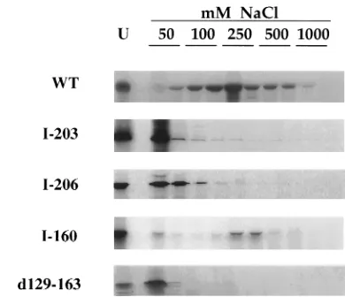
bind-ing activity of these mutants was assayed by DNA-cellulose chromatography. Labeled proteins were applied to a dsDNA-cellulose column and eluted stepwise with 2 bed volumes each of salt solution at increasing concentrations. As shown in Fig. 1, most of the wild-type protein was retained on the dsDNA column and the majority of the protein was eluted at 250 mM NaCl. Only a small fraction was eluted in the first 50 mM fraction. In contrast, the insertion mutants I-203 and I-206
failed to bind to the column, with the bulk of eluted protein being found in the 50 mM NaCl fractions. The insertion mu-tant I-160 was retained on the column, and as with the wild-type protein, the majority of the mutant polypeptide was eluted from the column at 250 mM salt, as previously reported (9).
The UL42 mutant d129-163 was reported to coimmunopre-cipitate with Pol but was unable to stimulate the synthesis of DNA by Pol (37, 43). To determine if its inability to stimulate Pol could be due to a loss of DNA binding activity, we sub-jected the mutant protein to dsDNA-cellulose chromatogra-phy. As with mutants I-203 and I-206, the majority of the eluted protein was found in the first fraction. Therefore, we conclude that d129-163 failed to bind dsDNA.
I-203 and I-206 can coimmunoprecipitate with Pol.Several of our previously reported UL42 mutants were deletion mu-tants. These mutants could not bind DNA and were also im-paired for Pol binding, suggesting that those deletions either affected regions involved in both activities or otherwise caused a global misfolding of the protein (9). We wished to determine if the mutants I-203 and I-206 were also affected in terms of their ability to bind Pol. Therefore, labeled UL42 proteins expressed in vitro were mixed with purified Pol or withb-Gal, which served as a control for nonspecific association. Protein complexes were immunoprecipitated with an antibody directed against ab-Gal–Pol fusion protein. As shown in Fig. 2, a large percentage of input wild-type UL42 associated with Pol but only a small amount boundb-Gal, indicating a specific inter-action. Similarly, the mutants I-203 and I-206 specifically co-immunoprecipitated with Pol but not b-Gal. On the other hand, as reported before (9), the mutant I-160 failed to bind to Pol specifically. Since the mutants I-203 and I-206 were not impaired in their Pol binding activity, we conclude that their defect in DNA binding is specific and not due to global mis-folding of the protein.
[image:2.612.331.526.69.237.2]The mutant d129-163 did not associate with Pol any more than it did with b-Gal (Fig. 2), indicating a lack of specific binding to Pol. This observation contrasts with the conclusion of Monahan et al. that d129-163 bound Pol, which was based on the finding that when the two proteins were cotranslated, a low percentage of d129-163 could be coimmunoprecipitated
FIG. 1. Elution profiles of UL42 from a dsDNA-cellulose column. A 10-ml aliquot of rabbit reticulocyte lysate programmed with UL42 mRNA was diluted in 100ml of TM buffer with 50 mM NaCl and 2ml of RNace-it. The treated lysate was applied to a dsDNA-cellulose column equilibrated in the same buffer. Elu-tion was carried out by washing with TM buffer containing the indicated con-centrations of NaCl. BSA (400 ng) was added to the eluate, which was then ethanol precipitated. Fractions were analyzed by SDS-PAGE and autoradiogra-phy. WT, wild-type protein; U, unfractionated input protein.
on November 9, 2019 by guest
http://jvi.asm.org/
with Pol by using the anti-UL42 antibody 834 (37). However, this procedure did not lend itself to the use of a control for nonspecific interactions. Recent unpublished experiments by that group have also reexamined the activity of d129-163. In-sect cells were coinfected with a recombinant baculovirus which expresses the d129-163 UL42 mutant protein and with either a recombinant baculovirus expressing Pol or one which expressesb-Gal. When antibody 834 was incubated with ex-tracts from these infected cells, it coimmunoprecipitated Pol with the mutant UL42 protein no better than it didb-Gal (49). Thus, d129-163, like our internal deletion mutants (9), may have removed amino acids involved in both DNA binding and Pol binding or may simply be misfolded.
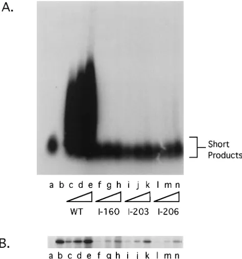
I-203 and I-206 are impaired for stimulation of Pol.The mutants I-203 and I-206 allowed us to examine whether the specific loss of DNA binding activity affected processive DNA synthesis. We therefore assayed our mutants for the ability to stimulate long-chain-DNA synthesis by Pol in vitro using a poly(dA)zoligo(dT) primer template. While this assay does not directly measure processive DNA synthesis, a good corre-lation exists between the ability of a functionally active Pol-UL42 complex to generate long products and its processivity (20). We tested UL42 mutants for Pol stimulation by incubat-ing the appropriate programmed reticulocyte lysate with puri-fied Pol in the presence of [a-32P]dTTP and the primer
tem-plate. The resulting products were fractionated on an alkaline agarose gel (Fig. 3A). The amount of UL42 added to the reactions was assessed by SDS-PAGE and autoradiography (Fig. 3B). As previously reported (11, 20), Pol alone generates short products (Fig. 3A, lane a). When cell-free translated wild-type UL42 was added to the reaction, longer chains of DNA were synthesized (Fig. 3A, lanes c through e). This stim-ulation was dependent on the amount of UL42 added (Fig. 3B, lanes c through e). Moreover, mutant I-160, which is unable to associate with Pol (9), did not stimulate long-chain-DNA syn-thesis in this assay (Fig. 3A and B, lanes f through h).
The ability of mutants I-203 and I-206 to enhance the activity of Pol on the primer template was also greatly impaired. Ad-dition of amounts of I-203 or I-206 protein similar to the amount of wild-type UL42 protein (Fig. 3B, compare lanes c through e with lanes i through k and l through n) to the reaction mixture had little effect on Pol activity on the
poly(dA)zoligo(dT) template (Fig. 3A, lanes i through k and l through n): there was only a minor increase in the amount of longer products. When the stimulation of Pol-mediated long-chain-DNA synthesis by I-203 and I-206 was quantified and normalized for the amount of UL42 protein in the reaction mixture, I-203 and I-206 were each found to have less than 3% of wild-type UL42 activity. Like the mutant I-160, I-203 and I-206 had no effect on the amount of short products generated by Pol alone (Fig. 3A, lanes a and f through n). In addition, the presence of I-203 or I-206 did not reduce the stimulatory effect of wild-type UL42 on Pol (5). Thus, while these mutants were able to complex with Pol, they were impaired for stimulating long-chain-DNA synthesis by Pol, without inhibiting basal polymerase activity. These data are consistent with the hypoth-esis that the DNA binding activity of UL42 is required for Pol processivity in vitro.
Mutants I-203 and I-206 fail to complement the growth of a
UL42 null virus.The results described above indicated that I-203 and I-206 were severely impaired but not entirely inactive for processive DNA synthesis in vitro. To see whether such low activity was sufficient for viral replication, and to examine if DNA binding was required for viral DNA synthesis, the mu-tants I-203 and I-206 were tested for their ability to support the growth of the UL42 null virus CgalD42. This virus can replicate only in the presence of an exogenous source of functional UL42, such as a transfected plasmid or a resident copy of the
UL42 gene in the stable cell line V9 (26). The
transient-FIG. 2. Coimmunoprecipitation of UL42 with Pol. UL42 expressed in vitro and labeled with [35
[image:3.612.317.554.365.618.2]S]methionine was mixed with eitherb-Gal (2) or purified Pol (1). After a 1-h incubation, the antibody PP5 and protein A-Sepharose were added to the protein mixture in the presence of IP buffer containing 0.1% Nonidet P-40 and 1 mM phenylmethylsulfonyl fluoride. This mixture was incu-bated overnight on a rotator at 48C. The beads were collected by centrifugation and washed twice in a high-stringency buffer and once in a low-stringency buffer. Following boiling in SDS sample buffer, samples were fractionated by SDS-PAGE. An aliquot of programmed reticulocyte lysate equivalent to the amount of input protein was run at the same time (T). WT, wild-type protein.
FIG. 3. Stimulation of Pol-mediated long-chain-DNA synthesis. UL42 mu-tants expressed in reticulocyte lysates were assayed for their ability to stimulate Pol on a poly(dA)zoligo(dT) template. Increasing amounts of wild-type (WT)-or mutant-mRNA-programmed lysate were added to a 50-ml reaction mixture as described in Materials and Methods. After a 30-min incubation, a 10-ml aliquot was taken to monitor the integrity of the polypeptides by SDS-PAGE (B). DNA in the remaining reaction mixture was ethanol precipitated, and the products were fractionated on a 1.5% alkaline agarose gel (A). Products were visualized by autoradiography. Lane a, no UL42 added; lane b, reaction mixture with no Pol and 1ml of wild-type UL42 reticulocyte lysate; lanes c through e, 0.25-, 0.5-, and 1-ml aliquots of wild-type UL42; lanes f through h, 0.25-, 0.5-, 1-ml aliquots of I-160; lanes i through k, 0.25-, 0.5-, and 1-ml aliquots of I-203; lanes l through n, 0.25-, 0.5-, and 1-ml aliquots of I-206.
on November 9, 2019 by guest
http://jvi.asm.org/
complementation assay was performed as follows. Vero cells were transfected with the mutant plasmids and superinfected with CgalD42 the next day. Cells were harvested 18 h postin-fection, and titers of progeny viruses on V9 cells were deter-mined. Typically, the titer of CgalD42 is 2 to 3 orders of magnitude higher in Vero cells transfected with the wild-type plasmid than in mock-transfected cells.
The complementation index for the plasmid carrying the wild-type UL42 gene was set at 100% as described in Materials and Methods. As previously reported, the mutant I-160 plas-mid was inefficient in complementing the growth of CgalD42 (9), in that its complementation index was less than 0.5%. The mutants I-203 and I-206 also had complementation indices of less than 0.5%. The inability of I-203 and I-206 to complement the replication of the null mutant suggests that the low level of processivity observed in vitro was insufficient to support viral replication in vivo.
I-203 and I-206 can localize to the nucleus.One possible explanation for the inability of mutants I-203 and I-206 to support the growth of the UL42 null virus is improper nuclear compartmentalization. To study whether these mutations which resulted in the loss of DNA binding activity also affected nuclear localization, we performed immunofluorescence stud-ies. Vero cells were transfected with the plasmids used in the complementation assays and then infected with the virus CgalD42 to induce UL42 expression. Cells were double stained with anti-ICP8 (3-83) and anti-UL42 (Z1F11) antibodies (29, 38, 44). As controls, cells were mock transfected and infected with CgalD42 or infected with the wild-type strain KOS. Re-sults are shown in Fig. 4.
In agreement with previous studies, the wild-type UL42 (Fig. 4a) and ICP8 (Fig. 4b) proteins in KOS-infected cells colocal-ized to the nucleus in stained patches known as replication compartments (17). In mock-transfected, CgalD42-infected cells, the anti-UL42 antibody Z1F11 gave only very faint non-specific nuclear staining (Fig. 4i). As CgalD42 fails to replicate DNA, ICP8 exhibited punctuate staining characteristic of pre-replicative sites (Fig. 4j) (17). In cells transfected with a plas-mid carrying the wild-type or mutant I-203 or I-206 UL42 gene and then infected with CgalD42, nuclear staining of UL42 was observed (Fig. 4c, e, and g). These cells also expressed ICP8, a marker for viral infection (Fig. 4d, f, and h). (Often, staining of UL42 in transfected cells was much more prominent than the signal for ICP8 under our assay conditions.) Nuclear staining of UL42 was also observed in cells transfected with a plasmid carrying the I-160 mutation (data not shown). These studies suggest that nuclear localization of UL42 does not require its DNA binding activity and that the lack of complementation of I-203 and I-206 was not due to the failure of the mutant proteins to translocate to the nucleus.
DISCUSSION
Two UL42 mutants with four amino acid insertions either at codon 203 or at codon 206, while impaired for DNA binding, were active for Pol binding. However, they were greatly re-stricted in their ability to stimulate Pol-mediated long-chain-DNA synthesis and were unable to support viral replication in a transient-complementation assay despite proper nuclear compartmentalization. Although we cannot exclude the formal possibility that these mutations affected some unknown activity of UL42, the simplest interpretation of our results is that the DNA binding activity of UL42 plays a vital role in processive DNA synthesis in vitro and in viral replication. Assuming that this interpretation is correct, we discuss the nature of DNA
binding by UL42, how it could function in processivity, and whether it could serve as a potential drug target.
[image:4.612.326.533.69.491.2]DNA binding activity of UL42.Previous studies have used amino- and carboxy-terminal deletion mutations to identify the first 315 amino acids of UL42 as sufficient for its DNA binding activity and all other known activities associated with UL42 (9, 15, 48). However, the specific involvement of internal regions of UL42 in DNA binding has eluded discovery. The isolation of mutants I-203 and I-206 suggests a starting point for the identification of an internal region in UL42 which may be involved specifically in DNA binding and required for Pol stimulation. Indeed, alignment of UL42 homologs from pseu-dorabies virus, varicella-zoster virus, and equine herpes virus identified the phenylalanine at residue 203 as absolutely served and the residue immediately upstream as highly con-served among these alphaherpesviruses (3). We suggest that
FIG. 4. Nuclear localization as demonstrated by indirect immunofluores-cence. Vero cells were infected with wild-type strain KOS (a and b), mock transfected (i and j), or transfected with plasmids encoding the indicated UL42 gene (c through j) and superinfected the next day with CgalD42. At 6 h postin-fection, cells were fixed and stained with UL42- and ICP8-specific antibodies as described in Materials and Methods. WT, wild-type plasmid.
on November 9, 2019 by guest
http://jvi.asm.org/
either residues 203 and 206 lie at the UL42-DNA interface or alterations at these residues act indirectly to disrupt DNA binding.
There is evidence to support the involvement of noncontig-uous regions of UL42 in DNA binding. We reported previously that alterations to the extreme amino terminus of UL42 re-sulted in a biphasic elution profile from the dsDNA-cellulose column (though these alterations did not appear to have any effect on processive DNA synthesis or viral replication). Fur-thermore, the amino-terminal-deletion mutant DN281 was shown to be eluted from the dsDNA-cellulose column at a higher salt concentration than the wild-type protein (9). This suggests that the region encompassing amino acids 281 through 315 may constitute a region capable of binding to DNA but with properties different from those of full-length UL42. Consistent with this, Owsianka et al. (40) reported five overlapping 15-residue peptides between amino acids 269 and 303 that could bind DNA. We suggest that multiple regions of UL42, including the region around residues 203 to 206, conjoin to confer DNA binding activity on UL42.
Monahan et al. (37) have described a mutant, i206, with a 12-bp insertion at codon 206 which encodes RGSA. In contrast to the behavior of our mutant with an insertion at the same location (I-206, insertion of CIDA), their mutant was reported to stimulate Pol almost as well as wild-type UL42 when activity was measured by [3H]dTTP incorporation into activated calf
thymus DNA. Furthermore, Reddig et al. found that i206 com-plemented the growth of CgalD42 at 10% of wild-type levels (43), while I-206 was unable to complement the growth of the null virus (,0.5% complementation). Analysis based on the Robson-Garnier secondary-structure prediction algorithm (16) suggests that residues 206 to 209 lie in a flexible region of the protein. Residues introduced by our insertions at codons 203 and 206 appear to limit the flexibility of this region, while the effect of the mutation in i206 is predicted to be less pro-nounced. This may explain why the i206 RGSA insertion is better tolerated, although other explanations are also tenable. Structural information would be of great assistance in under-standing how these mutations affect the local architecture and would shed more light on the different phenotypes of the mutants.
When assaying for the ability of extensively overlapping 15-mers spanning UL42 to inhibit HSV DNA polymerase activity, Owsianka et al. (40) found that at a concentration of 50mM, peptides derived from the region encompassing amino acids 103 and 223 had no effect on the functional interaction of Pol and UL42. (Peptide 46, corresponding to amino acids 209 to 223, was insoluble and not tested). In light of the inability of mutants I-203 and I-206 to stimulate Pol, we suggest either of the following explanations. (i) The peptides did not assume a stable conformation analogous to the protein, perhaps because of their short length. To illustrate, a peptide corresponding to the last 15 amino acids of Pol had a fivefold-lower helical content than a similar peptide only 3 amino acids longer. The 18-mer was also approximately 50-fold more potent in inhib-iting the functional interaction of Pol and UL42 (11). (ii) The region around amino acids 203 and 206 may not be surface exposed, i.e., the mutants I-203 and I-206 affect DNA binding indirectly, and hence the peptides did not block the ability of UL42 to bind DNA. Further study is required to determine if the region encompassing residues 203 to 206 directly interacts with DNA.
Mechanism of Pol stimulation.In order for HSV to repli-cate efficiently in the cell, processive DNA synthesis must oc-cur. Whereas some organisms such as Thermus aquaticus have evolved single-subunit processive DNA polymerases (25), HSV
relies on an accessory factor to boost the processivity of its catalytic subunit. Pol is a nonprocessive DNA polymerase at physiological monovalent cationic concentrations; when asso-ciated with UL42, the enzyme becomes highly processive (19, 21, 22). This heterodimerization requires neither ATP nor the presence of DNA. This is similar to the tight association of T7 gp5 and its processivity factor thioredoxin (23, 24, 36, 47). However, thioredoxin does not appear to have DNA binding activity (36) and is therefore likely to employ a mechanism of stimulation different from that of UL42.
How does UL42 increase processivity? Current and previous results support the model that UL42 acts as a tether between Pol and DNA by virtue of its ability to bind both the catalytic subunit and the DNA template. Consistent with this model are two recent observations: (i) the nuclease protection of a prim-er-template junction by the Pol-UL42 complex was more ex-tensive than that of Pol alone, and (ii) the affinity of the Pol-UL42 complex for such DNA was greater than that of either protein alone (18, 54). The tethering function of pro-cessivity factors is exemplified by thebsubunit of E. coli Pol III and PCNA of the replicative DNA polymerasedin eukaryotes. Like UL42, they are believed to clamp the catalytic subunit to the DNA template by binding DNA. However, these proteins differ from UL42 in important ways (30, 31). First,bsubunit and PCNA both require the assistance of cofactors for loading onto DNA, presumably because of the negative electrostatic potential on the surfaces of these molecules, whereas the in-trinsic affinity of UL42 for DNA and Pol dispenses with this requirement. Second, the association of the b subunit or PCNA with DNA is topological: duplex DNA is predicted to pass through the toroidal processivity factors with little, if any, direct contact with side chains of residues lining the inside surface. These properties allow the processivity factors to ‘‘slide’’ along the isopotential phosphate backbone of DNA without significantly slowing the movement of the polymerase. Given the intrinsic affinity of UL42 for DNA and its impor-tance for processivity, there arises the question of whether the stabilizing interaction between UL42 and DNA, while provid-ing an anchor for Pol on the DNA template, might not also act as a brake, decreasing the translational speed of the replication fork.
This potential paradox in the action of UL42 may not be peculiar to the HSV system. Indeed, the same problem has been proposed for the Epstein-Barr virus DNA Pol accessory subunit BMRF1, which preferentially binds dsDNA and in-creases the processivity of the catalytic subunit BALF5 (50, 51). Moreover, studies with other herpesvirus systems, includ-ing human and murine cytomegalovirus and human herpesvi-rus 6, suggest a converging theme in their mechanism of DNA replication, in that a DNA-binding protein acts as a Pol acces-sory factor (1, 13, 27, 33). In human cytomegalovirus, the functional analog of UL42 is UL44 (13, 53). There is a corre-lation between the loss of dsDNA binding activity in human cytomegalovirus UL44 and Epstein-Barr virus BMRF1 mutant proteins and the loss of stimulation of the cognate catalytic subunits (27, 53). However, it is not clear that the effects on DNA binding were specific, since binding to the cognate Pols was not assessed. Thus, the requirement for the DNA bind-ing activity of these Pol stimulatory factors for their function has yet to be rigorously demonstrated. Nevertheless, it is tempting to suggest that herpesvirus processivity factors em-ploy a mode of action which is evolutionarily divergent from that of other organisms studied thus far. It is likely that the elucidation of the mechanism of action of UL42 will provide a prototype for the function of processivity factors in other her-pesviruses.
on November 9, 2019 by guest
http://jvi.asm.org/
The protein-DNA interface as a drug target.Since the DNA binding activity of UL42 appears to be required for HSV DNA replication, one approach to the design of an anti-HSV drug could be the interruption of the UL42-DNA interaction. The use of peptides and peptidomimetics to disrupt protein-protein interactions has been shown to be effective for inhibiting HSV ribonucleotide reductase in vitro and in vivo (6, 12, 32) and the Pol-UL42 complex in vitro (11). Perhaps the DNA-UL42 in-terface can be similarly affected and exploited. Since UL42 binds DNA nonspecifically, an antiviral agent mimicking the DNA binding region of UL42 is likely to have nonspecific effects on cellular processes as well. Therefore, it is more reasonable to develop an antiviral agent that is complementary to the DNA binding region of UL42. Since UL42 does not appear to have mechanistic analogy or sequence homology to the eukaryotic processivity factor PCNA, such an antiviral agent could be HSV specific and not affect cellular polymerase functions. This strategy may also apply to other herpesvirus processivity factors for reasons mentioned above. Rational de-sign of such compounds may require further elucidation of the structure of the DNA binding regions of UL42 as well as the mechanism of action of the Pol-UL42 complex.
ACKNOWLEDGMENTS
We thank D. Parris and C. Crumpacker for communicating unpub-lished results and helpful comments, D. Parris and P. Johnson for CgalD42 virus and the cell line V9, H. Ludwig and H. Marsden for antibodies, K. Weisshart for purified Pol, and W. Bebrin and S. Up-richard for critically reading the manuscript.
This work was supported by grants RO1 AI19838 and UO1 AI26077 from the National Institutes of Health.
REFERENCES
1. Agulnick, A. D., J. R. Thompson, S. Iyengar, G. Pearson, D. Ablashi, and R. P. Ricciardi.1993. Identification of a DNA-binding protein of human herpesvirus 6, a putative DNA polymerase stimulatory factor. J. Gen. Virol. 74:1003–1009.
2. Bayliss, G. J., H. S. Marsden, and J. Hay. 1975. Herpes simplex virus proteins: DNA-binding proteins in infected cells and in the virus structure. Virology 68:124–134.
3. Berthomme, H., S. J. Monahan, D. S. Parris, B. Jacquemont, and A. Epstein. 1995. Cloning, sequencing, and functional characterization of the two sub-units of the pseudorabies virus DNA polymerase holoenzyme: evidence for specificity of interaction. J. Virol. 69:2811–2818.
4. Cai, W., S. Person, C. DebRoy, and B. Gu. 1988. Functional regions and structural features of the gB glycoprotein of herpes simplex virus type 1. An analysis of linker insertion mutants. J. Mol. Biol. 201:575–588.
5. Chow, C. S., and D. M. Coen. Unpublished results.
6. Cohen, H. A., P. Gaudreau, P. Brazeau, and Y. Langelier. 1986. Specific inhibition of herpesvirus ribonucleotide reductase by a nonapeptide derived from the carboxy terminus of subunit 2. Nature (London) 321:441–443. 7. Crute, J. J., and I. R. Lehman. 1989. Herpes simplex-1 DNA polymerase.
Identification of an intrinsic 59-39exonuclease with ribonuclease H activity. J. Biol. Chem. 264:19266–19270.
8. Digard, P., W. R. Bebrin, K. Weisshart, and D. M. Coen. 1993. The extreme C terminus of herpes simplex virus DNA polymerase is crucial for functional interaction with processivity factor UL42 and for viral replication. J. Virol. 67:398–406.
9. Digard, P., C. S. Chow, L. Pirrit, and D. M. Coen. 1993. Functional analysis of the herpes simplex virus UL42 protein. J. Virol. 67:1159–1168. 10. Digard, P., and D. M. Coen. 1990. A novel functional domain of ana-like
DNA polymerase: the binding site on the herpes simplex virus polymerase for the viral UL42 protein. J. Biol. Chem. 265:17393–17396.
11. Digard, P., K. P. Williams, P. Hensley, I. S. Brooks, C. E. Dahl, and D. M. Coen.1995. Specific inhibition of herpes simplex virus DNA polymerase by helical peptides corresponding to the subunit interface. Proc. Natl. Acad. Sci. USA 92:1456–1460.
12. Dutia, B. M., M. C. Frame, J. H. Subak-Sharpe, W. N. Clark, and H. S. Marsden.1986. Specific inhibition of herpesvirus ribonucleotide reductase by synthetic peptides. Nature (London) 321:439–441.
13. Ertl, P. F., and K. L. Powell. 1992. Physical and functional interaction of human cytomegalovirus DNA polymerase and its accessory protein (ICP36) expressed in insect cells. J. Virol. 66:4126–4133.
14. Gallo, M. L., D. H. Jackwood, M. Murphy, H. S. Marsden, and D. S. Parris.
1988. Purification of the herpes simplex virus type 1 65-kilodalton DNA binding protein: properties of the protein and evidence of its association with the virus encoded DNA polymerase. J. Virol. 62:2874–2883.
15. Gao, M., S. F. DiTusa, and M. G. Cordingley. 1993. The C-terminal third of UL42, a HSV-1 DNA replication protein, is dispensable for viral growth. Virology 194:647–653.
16. Garnier, J., D. J. Osguthorpe, and B. Robson. 1978. Analysis of the accuracy and implications of simple methods for predicting the secondary structure of globular proteins. J. Mol. Biol. 120:97–120.
17. Goodrich, L. D., P. A. Schaffer, D. I. Dorsky, C. S. Crumpacker, and D. S. Parris.1990. Localization of the herpes simplex virus type 1 65-kilodalton DNA-binding protein and DNA polymerase in the presence and absence of viral DNA synthesis. J. Virol. 64:5738–5749.
18. Gottlieb, J., and M. D. Challberg. 1994. Interaction of herpes simplex virus type 1 DNA polymerase and the UL42 accessory protein with a model primer template. J. Virol. 68:4937–4945.
19. Gottlieb, J., A. I. Marcy, D. M. Coen, and M. D. Challberg. 1990. The herpes simplex virus type 1 UL42 gene product: a subunit of DNA polymerase that functions to increase processivity. J. Virol. 64:5976–5987.
20. Hamatake, R. K., M. Bifano, D. J. Tenney, W. W. Hurlburt, and M. G. Cordingley.1993. The herpes simplex virus type 1 DNA polymerase acces-sory protein, UL42, contains a functional protease-resistant domain. J. Gen. Virol. 74:2181–2189.
21. Hart, G. J., and R. E. Boehme. 1992. The effect of the UL42 protein on the DNA polymerase activity of the catalytic subunit of the DNA polymerase encoded by herpes simplex virus type 1. FEBS Lett. 305:97–100. 22. Hernandez, T. R., and I. R. Lehman. 1990. Functional interaction between
the herpes simplex-1 DNA polymerase and UL42 protein. J. Biol. Chem. 265:11227–11232.
23. Himawan, J. S., and C. C. Richardson. 1992. Genetic analysis of the inter-action between bacteriophage T7 DNA polymerase and Escherichia coli thioredoxin. Proc. Natl. Acad. Sci. USA 89:9774–9778.
24. Huber, H. E., S. Tabor, and C. C. Richardson. 1987. Escherichia coli thi-oredoxin stabilizes complexes of bacteriophage T7 DNA polymerase and primed templates. J. Biol. Chem. 262:16224–16232.
25. Innis, M. A., K. B. Myambo, D. H. Gelfand, and M. A. Brow. 1988. DNA sequencing with Thermus aquaticus DNA polymerase and direct sequencing of polymerase chain reaction-amplified DNA. Proc. Natl. Acad. Sci. USA 85:9436–9440.
26. Johnson, P. A., M. G. Best, T. Friedmann, and D. S. Parris. 1991. Isolation of a herpes simplex virus type 1 mutant deleted for the essential UL42 gene and characterization of its null phenotype. J. Virol. 65:700–710.
27. Kiehl, A., and D. I. Dorsky. 1995. Bipartite DNA-binding region of the Epstein-Barr virus BMRF1 product essential for DNA polymerase accessory function. J. Virol. 69:1669–1677.
28. Kingston, R. E., C. A. Chen, and H. Okayama. 1990. Transfection of DNA into eukaryotic cells, p. 9.1.1-9.1.9. In F. M. Ausubel, R. Brent, R. E. King-ston, D. D. Moore, J. G. Seidman, J. A. Smith, and K. Struhl (ed.), Current protocols in molecular biology. Wiley Interscience, New York.
29. Knipe, D. M., D. Senechek, S. Rice, and J. Smith. 1987. Stages in the nuclear association of the herpes simplex virus transcriptional activator protein ICP4. J. Virol. 61:276–284.
30. Kong, X. P., R. Onrust, M. O’Donnell, and J. Kuriyan. 1992. Three-dimen-sional structure of theb-subunit of E. coli DNA polymerase III holoenzyme: a sliding clamp. Cell 69:425–437.
31. Krishna, T. S. R., X. P. Kong, S. Gary, P. M. Burgers, and J. Kuriyan. 1994. Crystal structure of the eukaryotic DNA polymerase processivity factor PCNA. Cell 79:1233–1243.
32. Liuzzi, M., R. Deziel, N. Moss, P. Beaulieu, A. Bonneau, C. Bousquet, J. G. Chafouleas, M. Garneau, J. Jaramillo, R. L. Krogsrud, L. Lagace, R. S. McCollum, S. Nawoot, and Y. Guidon.1994. A potent peptidomimetic in-hibitor of HSV ribonucleotide reductase with antiviral activity in vivo. Na-ture (London) 372:695–698.
33. Loh, L. C., W. J. Britt, C. Raggo, and S. Laferte. 1994. Sequence analysis and expression of the murine cytomegalovirus phosphoprotein pp50, a homolog of the human cytomegalovirus UL44 gene product. Virology 200:413–427. 34. Marchetti, M. E., C. A. Smith, and P. A. Schaffer. 1988. A
temperature-sensitive mutation in a herpes simplex virus type 1 gene required for viral DNA synthesis maps to coordinates 0.609 through 0.614 in UL. J. Virol.
62:715–721.
35. Marcy, A. I., P. D. Olivo, M. D. Challberg, and D. M. Coen. 1990. Enzymatic activities of overexpressed herpes simplex virus DNA polymerase purified from recombinant baculovirus-infected insect cells. Nucleic Acids Res. 18: 1207–1215.
36. Modrich, P., and C. C. Richardson. 1975. Bacteriophage T7 deoxyribonu-cleic acid replication in vitro. Bacteriophage T7 DNA polymerase: an en-zyme composed of phage- and host-specific subunits. J. Biol. Chem. 250: 5515–5522.
37. Monahan, S. J., T. F. Barlam, C. S. Crumpacker, and D. S. Parris. 1993. Two regions of the herpes simplex virus type 1 UL42 protein are required for its functional interaction with the viral DNA polymerase. J. Virol. 67:5922– 5931.
on November 9, 2019 by guest
http://jvi.asm.org/
38. Murphy, M., P. Schenk, H. M. Lankinen, A. M. Cross, P. Taylor, A. Owi-sianka, R. G. Hope, H. Ludwig, and H. S. Marsden. 1989. Mapping of epitopes on the 65K DNA-binding protein of herpes simplex virus type 1. J. Gen. Virol. 70:2357–2364.
39. Olivo, P. D., N. J. Nelson, and M. D. Challberg. 1989. Herpes simplex virus type 1 gene products required for DNA replication: identification and over-expression. J. Virol. 63:196–204.
40. Owsianka, A. M., G. Hart, M. Murphy, J. Gottlieb, R. Boehme, M. Chall-berg, and H. S. Marsden.1993. Inhibition of herpes simplex virus type 1 DNA polymerase activity by peptides from the UL42 accessory protein is largely nonspecific. J. Virol. 67:258–264.
41. Parris, D. S., A. Cross, L. Haarr, A. Orr, M. C. Frame, M. Murphy, D. J. McGeoch, and H. S. Marsden.1988. Identification of the gene encoding the 65-kilodalton DNA-binding protein of herpes simplex virus type 1. J. Virol. 62:818–825.
42. Powell, K. L., and D. J. M. Purifoy. 1976. DNA-binding proteins of cells infected with herpes simplex virus type 1 and type 2. Intervirology 7:225–239. 43. Reddig, P. J., L. A. Grinstead, S. J. Monahan, P. A. Johnson, and D. S. Parris.1994. The essential in vivo function of the herpes simplex virus UL42 protein correlates with its ability to stimulate the viral DNA polymerase in
vitro. Virology 200:447–456.
44. Schenk, P., S. Pietschmann, H. Gelderblom, G. Pauli, and H. Ludwig. 1988. Monoclonal antibodies against herpes simplex virus type 1-infected nuclei defining and localizing the ICP8 protein, 65K DNA-binding protein and polypeptides of the ICP35 family. J. Gen. Virol. 69:99–111.
45. Stow, N. D. 1993. Sequences at the C-terminus of the herpes simplex virus type 1 UL30 protein are dispensable for DNA polymerase activity but not for viral origin-dependent DNA replication. Nucleic Acids Res. 21:87–92. 46. Stukenberg, P. T., P. S. Studwell-Vaughn, and M. O’Donnell. 1991.
Mech-anism of the sliding beta-clamp of DNA polymerase III holoenzyme. J. Biol. Chem. 266:11328–11334.
47. Tabor, S., H. E. Huber, and C. C. Richardson. 1987. Escherichia coli thi-oredoxin confers processivity on the DNA polymerase activity of the gene 5 protein of bacteriophage T7. J. Biol. Chem. 262:16212–16223.
48. Tenney, D. J., W. W. Hurlburt, M. Bifano, J. T. Stevens, P. A. Micheletti, R. K. Hamatake, and M. G. Cordingley.1993. Deletions of the carboxy terminus of herpes simplex virus type 1 UL42 define a conserved amino-terminal functional domain. J. Virol. 67:1959–1966.
49. Thorton, K., S. Monahan, and D. S. Parris. Personal communication. 50. Tsurumi, T. 1993. Purification and characterization of the DNA-binding
activity of the Epstein-Barr virus polymerase accessory protein BMRF1 gene products, as expressed in insect cells by using the baculovirus system. J. Virol. 67:1681–1687.
51. Tsurumi, T., T. Daikoku, R. Kurachi, and Y. Nishiyama. 1993. Functional interaction between Epstein-Barr virus DNA polymerase catalytic subunit and its accessory subunit in vitro. J. Virol. 67:7648–7653.
52. Vaughn, P. J., D. J. M. Purifoy, and K. L. Powell. 1985. DNA-binding protein associated with herpes simplex virus DNA polymerase. J. Virol. 53:501–508. 53. Weiland, K. L., N. L. Oien, F. Homa, and M. W. Wathen. 1994. Functional analysis of human cytomegalovirus polymerase accessory protein. Virus Res. 34:191–206.
54. Weisshart, K., and D. M. Coen. Unpublished observations.
55. Weisshart, K., A. A. Kuo, B. C. Hwang, K. Kumura, and D. M. Coen. 1994. Structural and functional organization of herpes simplex virus DNA poly-merase investigated by limited proteolysis. J. Biol. Chem. 269:22788–22796. 56. Yager, D. R., A. I. Marcy, and D. M. Coen. 1990. Translational regulation of
the herpes simplex virus DNA polymerase. J. Virol. 64:2217–2225.