0022-538X/95/$04.0010
Copyright 1995, American Society for Microbiology
PREPs: Herpes Simplex Virus Type 1-Specific Particles
Produced by Infected Cells When Viral DNA
Replication Is Blocked
D. J. DARGAN,* A. H. PATEL,ANDJ. H. SUBAK-SHARPE
Medical Research Council Virology Unit, University of Glasgow, Glasgow G11 5JR, United Kingdom
Received 2 February 1995/Accepted 16 May 1995
Herpes simplex virus (HSV)-infected cells produce not only infectious nucleocapsid-containing virions but also virion-related noninfectious light particles (L-particles) composed of the envelope and tegument
compo-nents of the virus particle (J. F. Szila´gyi and C. Cunningham, J. Gen. Virol. 62:661–668, 1991). We show that
BHK and MeWO cells infected either with wild-type (WT) HSV type 1 (HSV-1) in the presence of viral DNA
replication inhibitors (cytosine-b-D-arabinofuranoside, phosphonoacetic acid, and acycloguanosine) or with a
viral DNA replication-defective mutant of HSV-1 (ambUL8) synthesize a new type of virus-related particle that
is morphologically similar to an L-particle but differs in its relative protein composition. These novel particles we term pre-viral DNA replication enveloped particles (PREPs). The numbers of PREPs released into the culture medium were of the same order as those of L-particles from control cultures. The particle/PFU ratios
of different PREP stocks ranged from 63105
to 3.83108
, compared with ratios of 33103
to 13104
for WT L-particle stocks. Sodium dodecyl sulfate-polyacrylamide gel electrophoresis and Western immunoblot anal-yses revealed that true late proteins, such as 273K (VP1-2), 82/81K (VP13/14), and gC (VP8), were greatly reduced or absent in PREPs and that gD (VP17) and 40K proteins were also underrepresented. In contrast, the amounts of proteins 175K (VP4; IE3), 92/91K (VP11/12), 38K (VP22), and gE (with BHK cells) were increased. The actual protein composition of PREPs showed some cell line-dependent differences, particularly
in the amount of gE. PREPs were biologically competent and delivered functional Vmw65 (VP16;aTIF) to
target cells, but the efficiency of complementation of the HSV-1 (strain 17) mutantin1814 was 10 to 30% of that
of WT L-particles.
From about 3 h postinfection, herpes simplex virus type 1 (HSV-1)-infected BHK cells produce both infectious virions and noninfectious light particles (L-particles) in approximately equal numbers (26) throughout the virus replication cycle (20). HSV-1 L-particles are composed of the virus envelope and tegument components but lack the capsid and DNA of the virion particle. Consequently, L-particles can be readily sepa-rated from virions by centrifugation through Ficoll gradients (26). L-particles analogous to HSV-1 L-particles have been isolated from BHK and other cell lines infected with the fol-lowing alphaherpesviruses: pseudorabies virus from BHK cells, equine herpesvirus 1 from BHK cells (15), bovine herpesvirus 1 from MDBK cells, varicella-zoster virus from CV-1 cells, and HSV-2 from BHK cells (5a). It appears highly probable that at least in tissue culture, L-particles are produced during repli-cation of all alphaherpesviruses. HSV-1 L-particles have been suggested to have potential for vaccine materials.
Comparative analyses of the protein compositions of L-par-ticles and virions made by wild-type (WT) HSV-1, pseudora-bies virus, and equine herpesvirus 1 showed that L-particles possessed most, if not all, of the virus tegument and envelope proteins but lacked the nucleocapsid proteins (15, 26). HSV-1 L-particles have been reported to contain five phosphoproteins
not detected in virion particles (26). Recently, Szila´gyi and
Berriman (25) identified by cryoelectron microscopy smooth membrane-bound inclusion vesicles in the tegument of a large proportion of HSV-1 L-particles, but inclusion vesicles were
not detected in virions; therefore, they suggested that some or all of the phosphoproteins unique to L-particles are associated with these inclusion vesicles.
The role played by L-particles in alphaherpesvirus infection is not known. However, McLauchlan et al. (14) demonstrated that HSV-1 L-particles were as effective as virions in supplying at least two functional tegument proteins (vhs and Vmw65) to target cells, indicating that L-particles might be involved in boosting the infectious process by elevating the levels of such proteins in coinfected cells.
Using ts1201, an HSV-1 temperature-sensitive UL26 gene mutant (19), Rixon et al. (20) demonstrated that L-particles could be generated independently of virion maturation. Under nonpermissive conditions, the packaging of ts1201 viral DNA into nucleocapsids was blocked and L-particles but no infec-tious progeny virions were produced. Nevertheless, the ts1201 L-particles generated under nonpermissive conditions were identical in morphology and protein composition to typical WT L-particles.
How L-particles are formed is still unclear, as is the impor-tance of their various protein components for morphogenesis. As several L-particle proteins are known to be made in appre-ciable amounts only after DNA synthesis, we investigated the ability of HSV-1-infected cells to produce virus-related parti-cles under conditions in which viral DNA synthesis is blocked either genetically or biochemically. In the absence of viral DNA synthesis and hence of the production of infectious virus, we find that HSV-1-infected cells release into the growth me-dium virus-related particles that are superficially similar to HSV-1 L-particles but characteristically differ in relative pro-tein composition, complementation efficiency, and
morphol-* Corresponding author. Mailing address: Medical Research Coun-cil, Virology Unit, University of Glasgow, Church St., Glasgow G11 5JR, United Kingdom. Phone: (141) 339 8855. Fax: (141) 337 2236.
4924
on November 9, 2019 by guest
http://jvi.asm.org/
ogy. We term these novel particles pre-viral DNA replication enveloped particles (PREPs).
MATERIALS AND METHODS
Cells.BHK-21 and human fetal lung cells (HFL; Flow 2002) were propagated in Glasgow modified Eagle’s medium supplemented with 10% newborn calf serum (EC10) or 10% fetal calf serum (EFC10), respectively; human malignant melanoma cells (MeWO cells) (3, 5, 7) were grown in Dulbecco’s modified Eagle’s medium supplemented with 10% fetal calf serum (DFC10); Vero A26 cells (17a) were grown in DFC10 containing 300mg of Geneticin (G418) per ml. For infectivity titrations, following infection BHK cells were overlaid with EC10 containing 1.25% methylcellulose.
Viruses.The viruses used were WT HSV-1 strain 17 and ambUL8, a mutant of HSV-1 (strain 17) with an amber translational stop codon inserted in frame at codon 267 of the UL8 gene, one of the seven HSV-1 genes required for viral DNA replication (27). HSV-1 ambUL8 is propagated in a Vero cell line (A26) transformed with WT HSV-1 sequences encoding the UL8 gene product (along with those of UL6, UL7, UL9, and UL10) (17a). ambUL8 replicates in A26 cells with WT HSV-1 kinetics and achieves WT-equivalent titers. in1814 is a mutant of HSV-1 (strain 17) which has an in-frame 12-bp insertion of DNA into the UL48 gene, which encodes VP16 (also known as Vmw65). This insertion abol-ishes the ability of Vmw65 to transinduce immediate-early transcription without affecting the structural role of the protein (1, 2).
Preparation of virions and L-particles.Purified virions and L-particles were prepared as described by Szila´gyi and Cunningham (26). BHK and MeWO monolayers grown on 80-oz (1 oz528.350 g) roller bottles (23108cells per
roller) were infected at a multiplicity of infection of 1/300 (PFU/cell). After 3 to 4 days of incubation at either 31 or 378C, the cell-released virus and/or L-particle yields were harvested and separated on 5 to 15% Ficoll gradients. Banded virions and/or L-particles were collected, diluted in Eagle’s medium lacking phenol red (Epr2), and pelleted by centrifugation at 19,0003g for 16 h at 48C. Pellets were
gently resuspended in Epr2and stored at2708C.
WT L-particle preparations are typically contaminated with virions at a level of 1 to 5% of total particle numbers. To eliminate virion infectivity, L-particle preparations were treated with UV light as described by McLauchlan et al. (14), who found that this treatment had no apparent effect on the ability of L-particles to deliver functional tegument proteins to cells.
Preparation of PREPs.BHK-21 and MeWO cells (;23108
cells per roller) were treated with either 10mM acycloguanosine (ACV), 300mg of phosphono-acetic acid (PAA) per ml, or 100mg of cytosine-b-D-arabinofuranoside (ara-C) per ml 1 h before and throughout infection with WT HSV-1. Cells were infected at a multiplicity of infection of 5 PFU per cell in 10 ml of EC5 (containing the appropriate inhibitor) and allowed to absorb the virus for 2 to 3 h at 378C. Then the inoculum was decanted and the cell sheet was treated with the following to inactivate residual inoculum infectivity: first, 20 ml of 0.14 M NaCl; then, 20 ml of 0.1 M glycine in 0.14 M NaCl, pH 3.0, for 1 min; finally, EC5 to neutralize the acid (21). Cell layers were subsequently overlaid with 30 ml of EC5 containing the appropriate inhibitor per roller bottle and incubated either at 318C for 48 h or at 378C for 24 h, and then supernatants were harvested.
PREPs were prepared from ambUL8 in the same way, except that no DNA inhibitor was present. Purification of PREPs from growth medium supernatants was the same as that described above for L-particles.
Polyacrylamide gel electrophoresis (PAGE), silver staining of proteins, and Western immunoblotting.Purified virions, L-particles, and PREPs were solubi-lized, and 23109particle equivalents (determined by direct counting of particles
with an electron microscope) were loaded onto either 5 to 12.5% gradient or 9% single-concentration sodium dodecyl sulfate (SDS)-polyacrylamide gels (12). Proteins were visualized by silver staining as described by McLean et al. (16).
For Western immunoblotting, proteins separated by SDS-PAGE were trans-ferred to Hybond-ECL nitrocellulose sheets (Amersham), were treated over-night with blocking buffer (phosphate-buffered saline [PBS] containing 0.05% Tween 20 [PBS-T] and 2% Marvel), and after being washed with PBS-T were incubated with test mouse monoclonal antibodies (MAbs) or rabbit polyclonal antibodies prepared in PBS-T containing 1% bovine serum albumin for 2 h at 188C. After further washes with PBS-T, blots were treated with anti-mouse or anti-rabbit immunoglobulin G-horseradish peroxidase (as appropriate), and tagged proteins were detected by enhanced chemoluminescence (Amersham). The antibodies used were MAB 4846 (anti-gD), MAB 3114/109 (anti-gE), and rabbit polyclonal antibodies 94497 (anti-82/81K) (provided by A. Cross) and R47 (anti-gC) (kindly provided by G. Cohen and R. Eisenberg).
Solubilization of envelope proteins.Approximately 431010
virions, L-parti-cles, and PREPs were treated with 1% Nonidet P-40 in EPr2for 30 min on ice. Soluble (envelope) and insoluble (tegument) fractions were then separated by centrifugation at 13,000 rpm for 5 min in an MSE Microfuge. After solubilization in sample buffer (12), volumes equivalent to 43109
particles were loaded into individual gel lanes.
Complementation ofin1814 by PREPs and L-particles.HFL cell monolayers on 24-well tissue culture dishes were infected with in1814 at 0.1, 1.0, and 10 PFU per well. After a 1-h absorption period, virus that was not taken up was removed by washing cells with PBS–5% fetal calf serum, and then monolayers were
treated for 1 h at 378C with 0.1, 1.0, 10, and 100 particles of either HSV-1 L-particles or PREPs per cell. After three washes with PBS–5% fetal calf serum to remove unbound particles, monolayers were overlaid with EFC5, incubated at 378C for 48 h, fixed, and stained, and the numbers of plaques were counted.
Negative-stain electron microscopy.Five-microliter samples of L-particles and PREPs were spotted onto a Formvar-coated electron microscopy grid and al-lowed to dry. Then the grid was treated with 5ml of phosphotungstic acid for 5 s, excess phosphotungstic acid was removed by blotting, and the grid was sub-jected to electron microscopic examination with a JEOL 101 electron micro-scope.
RESULTS
The ability of HSV-1-infected cells to produce and release virus-related particles has been investigated under conditions in which viral DNA replication has been blocked either bio-chemically with appropriate antimetabolites or genetically by using ambUL8, a conditional lethal mutant of HSV-1 defective in viral DNA synthesis.
The growth medium from BHK and MeWO cells infected with WT HSV-1 (multiplicity of infection, 5 PFU per cell) and
incubated either at 318C for 48 h or at 378C for 72 h yielded
characteristic L-particle and virion bands upon Ficoll density gradient centrifugation. However, BHK and MeWO cells
in-fected at 5 PFU per cell with WT virus and grown at 318C for
48 h or at 378C for 24 h in the continuous presence of DNA
synthesis inhibitors (either 10 mM ACV, 300mg of PAA per
ml, or 100 mg of ara-C per ml) or with the DNA-negative
ambUL8 mutant yielded supernatant medium which gave only
a single diffuse band on Ficoll gradients. This band’s position approximated that of WT control L-particles banded on par-allel gradients. No virion band was seen on gradients contain-ing extracellular material from cultures in which viral DNA synthesis was blocked (Fig. 1).
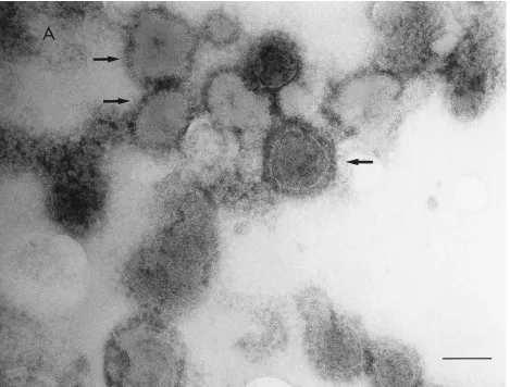
Ficoll-banded materials were collected through side punc-ture of the centrifuge tube and examined by electron micros-copy after negative staining. Banded materials from WT con-trol cultures yielded typical virions and L-particles (26), while bands from cultures in which DNA synthesis was blocked con-tained high numbers of particles that superficially resembled WT control L-particles, except for fewer and/or shorter enve-lope spikes (Fig. 2). These particles, made in HSV-1-infected cells in the absence of DNA replication, have been designated PREPs.
[image:2.612.316.555.72.229.2]Although present in insufficient numbers to constitute a visible band on PREP-containing gradients, some virion par-ticles were detected by electron microscopy in samples from the region corresponding to the WT control virion band. Table
FIG. 1. WT HSV-1 virions and L-particles (A) and ambUL8 PREPs (B) banded on 5 to 15% Ficoll density gradients.
VOL. 69, 1995 NOVEL HSV-1-SPECIFIC PARTICLES 4925
on November 9, 2019 by guest
http://jvi.asm.org/
1 gives the particle and infectivity titers of virions, PREPs, and L-particles from BHK and MeWO cells infected in parallel either with WT virus in the presence or absence of drugs or with ambUL8. Particle numbers were determined from elec-tron microscopy counts. The infectivities associated with banded particles were determined by titration on BHK or A26 monolayers, as appropriate. It is clear that HSV-1 PREPs were produced in numbers similar to those of WT control L-parti-cles by both BHK and MeWO cultures, regardless of whether viral DNA synthesis was blocked genetically or biochemically. Small amounts of infectivity were found in all PREP prepara-tions, but the particle/PFU ratios obtained were routinely 100-to 10,000-fold greater than those of WT control L-particle preparations. The high particle/PFU ratios obtained for the PREP band and (region-located) virions from PREP gradients indicated that most of the virions present were noninfectious, probably representing adsorbed, acid-inactivated, inoculum vi-rus that persisted in these cultures despite the washing proce-dures and was later released from the cell surface.
The small amounts of infectious virus found in cultures treated with DNA synthesis inhibitors or infected with amb-UL8 may be explained by the following: preexistent drug-resistant variants present in the WT virus stock, WT revertants or recombinants present in the ambUL8 stock, incomplete drug-induced block of viral DNA synthesis, or adsorbed
per-sistent infectious virus inoculum which survived the acid inac-tivation treatment and was later released from the cell surface. To check out the possibility of drug-resistant virus, virions banded from ACV-, PAA-, and ara-C-treated infected cell cultures were titrated in the presence of the drug used in the production of PREPs. In every case, a number of tiny abortive plaques were observed on lower-dilution plates; however, after picking five representative plaques from cultures treated with each drug and replating them in the presence or absence of the appropriate drug, no progeny plaques were obtained (data not shown). This makes drug resistance an unlikely explanation.
The small amounts of infectivity associated with ambUL8 PREPs (MeWO cells) were similarly investigated by compar-ing the titers of virion and PREP band materials on A26 cells
(4.43106and 1.33105PFU/ml, respectively). The titers of
ambUL8 virions and PREPs determined separately on BHK
cells were 6.33104and 93102PFU/ml, respectively.
There-fore, a very low level of WT virus appears to be present in
ambUL8 PREP yields; this is probably due to recombination
between ambUL8 and HSV-1 sequences in A26 cells. The particle/PFU ratios (determined from the BHK cell titer and electron microscopy particle number given in Table 1) were 8.6
3104for the virion band and 1.13109for the PREP band.
[image:3.612.74.543.71.427.2]We consider the small amounts of infectivity contaminating
FIG. 2. Electron micrographs showing negatively stained WT L-particles (A) and ambUL8 PREPs (B). Particles bearing envelope glycoprotein spikes are indicated by arrows. Bar, 100 nm.
on November 9, 2019 by guest
http://jvi.asm.org/
banded PREPs to be unlikely to have any significant effect on the results obtained in the subsequent experiments.
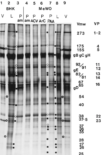
The protein composition of PREPs made in BHK and MeWO cells was examined by SDS-PAGE with both 5 to 12.5% gradient gels and 9% single-concentration gels. In over-all appearance, the PREP gel polypeptide pattern was similar to that obtained with WT L-particles (Fig. 3; compare lanes 2 and 3 as well as 8 and 4). However, the following differences
between the gel protein profile of L-particles and that of PREPs were consistently observed, regardless of whether the preparations were obtained from infected BHK or MeWO cells. Bands of the 273K (VP1-2), 82/81K (VP13/14), 57K (VP17; gD), and 40K proteins were clearly reduced, while bands of the 175K (VP4; IE3), 92/91K (VP11/12), and 38K (VP22) proteins were increased (Fig. 3).
[image:4.612.73.543.71.426.2]The polypeptide profiles obtained for L-particles and
TABLE 1. HSV-1 virion, L-particle, and PREP infectivity measures and the particle/PFU ratios of Ficoll-banded yields from BHK and MeWO cells
Type of
cells Virus Drug
L-particle or PREP band Virion region
Pa
/ml PFU/ml P/PFU P/ml PFU/ml P/PFU
BHK WT None 9.031011 8.23107 1.13104 6.531011 4.93109 133
WT ACV 3.631011 1.03104 3.63107 1.13109 2.03104 5.53104
WT ara-C 3.531011 1.63104 2.23107 6.63109 1.53105 4.43104 ambUL8 None 1.831011 1.03104 1.83107 ,108 3.53104 .33103
MeWO WT None 2.331011 7.43107 3.13103 1.331011 3.43109 38
WT ACV 3.031011 5.03105 6.03105 4.33109 4.03104 1.13105
WT ara-C 10.031011 2.73103 3.83108 8.73108 2.03103 4.33105
WT PAA 3.931011 1.83104 2.23107 8.831010 5.43105 1.63105 ambUL8 None 9.631011 1.33105 7.43106 5.43109 4.43106 1.23103 a
[image:4.612.58.555.591.718.2]P, particles visible by electron microscopy.
FIG. 2—Continued.
VOL. 69, 1995 NOVEL HSV-1-SPECIFIC PARTICLES 4927
on November 9, 2019 by guest
http://jvi.asm.org/
PREPs on SDS-polyacrylamide gels were quantitated by den-sitometric scanning of these gels. The relative amounts of the 273K (VP1-2), 175K (IE3), 92/91K (VP11/12), 82/81 (VP13/ 14), 57K (VP17; gD), 40K, and 38K (VP22) proteins in L-particles and PREPs from three independent experiments, in which L-particle controls and PREPs were made by the same batch of MeWO cells, are shown in Table 2. The amount of each protein was standardized to the amount of 65K (VP16;
aTIF) protein in each lane; the average percentage of total
particle protein represented by 65K (VP16;aTIF) protein in
L-particles and ACV-, ara-C-, PAA-, and ambUL8 PREPs from three experiments was 9.2% (8.8, 8.0, and 10.8%), 9.0% (9.5, 10.1, and 7.3%), 8.6% (9.7, 9.1, and 7.1%), 8.5% (10.8, 9.3, and 5.3%), and 8.6% (8.3, 10.5, and 6.9%), respectively. The average amounts of total L-particle protein represented by the 273 (VP1-2), 175K (IE3), 92/91K (VP11/12), 82/81 (VP13/
14), 57K (VP17; gD), 40K, and 38K (VP22) proteins were 2.8, 0.8, 4.2, 8.1, 14.3, 4.0, and 3.1%, respectively.
Table 2 shows that the amount of the true late 273K (VP1-2) protein was reduced in PREPs, ranging from below the level of detection to 35% of the L-particle level. The amounts of the 175K (IE3) protein in different preparations of L-particles varied over a ninefold range; however, in all experiments, the amount of this protein was increased in PREPs (from 116 to 1,050% of the L-particle level), and within any individual ex-periment, the increase for individual PREP preparations var-ied only over a 2.2-fold range. The amount of the 92/91K (VP11/12) protein also consistently increased in PREPs (from 138 to 400% of that found in L-particles); the range in increase for individual PREP preparations varied less than twofold for any experiment. The amount of the 57K (VP17; gD) protein was consistently reduced in PREPs (from 27 to 76% of the L-particle level), and the range in decrease for individual PREP preparations varied less than twofold for any experi-ment. The amount of the 40K protein was consistently reduced in PREPs (from below the level of detection to 76% of the corresponding L-particle level), and the decrease for individual PREP preparations varied over a 1.5-fold range in experiments 1 and 2. The amount of the 38K (VP22) protein was consis-tently increased in PREPs (from 128 to 262% of the corre-sponding L-particle level), and in any experiment, the variation between PREP preparations was only 1.3-fold. Similar but only fragmentary results (data not shown) were obtained for L-particles and PREPs made in BHK cells. The true late 82/81K (VP13/14) protein appeared to be absent from PREPs (Fig. 3); this was confirmed by Western immunoblotting (see below).
Additional differences in minor bands were observed be-tween PREPs and L-particles prepared in MeWO cells (Fig. 3). The intensities of the bands of the 37.5K, 36K, 28K, and 27K proteins were reduced, while those of the 37K and 18K proteins were increased (Fig. 3; data for 18K protein not shown). BHK-produced PREPs contained much more gE and gD than those produced in MeWO cells. Cell line-dependent differences in protein composition between L-particles and PREPs require more extensive study and evaluation.
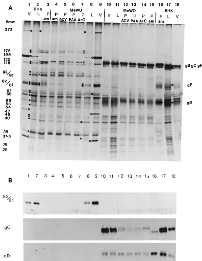
To further investigate the composition of PREPs, the enve-lopes of PREPs and L-particles were removed by solubilization with the nonionic detergent Nonidet P-40. The resultant de-enveloped PREP- and L-particle-derived particulate structures (15) were pelleted, and the separated tegument (pellet) and envelope (supernatant) fractions were then investigated by SDS-PAGE (Fig. 4A) and Western immunoblotting (Fig. 4B). The amounts of the following proteins were increased in the tegument fractions of PREPs: 175K (multiple band) (VP4; IE3), 120K, 118K, 92/91K (VP11/12), gE (BHK cells only), 67K (VP15) (MeWO cells only), and 38K (VP22). The amounts of the 273K (VP1-2), 82/81K (VP13/14), 40K (MeWO cells only), and 37.5K (MeWO cells only) proteins were decreased. In the envelope fractions, the amounts of the bands representing gB, gC, and gH show little or no difference in L-particles and PREPs. However, the amount of gD in PREPs was reduced. gE appeared to be either missing or present only in trace amounts in all types of particles made in MeWO cells, while the amount of gE detected in ambUL8 PREPs produced in BHK cells was clearly increased, compared with that of the L-particle control. The minor and lower-molecular-weight pro-tein bands detected in MeWO-produced PREPs (Fig. 3) were mostly associated with envelope fractions, and some of these are probably host proteins (Fig. 4A).
[image:5.612.74.287.70.392.2]We have begun to identify by Western immunoblotting some proteins whose abundances appeared to be altered in PREPs (Fig. 4B [82/81K, gC, and gD]). We had polyclonal antibody
FIG. 3. Polypeptide profiles of BHK- and MeWO cell-produced HSV-1 viri-ons, L-particles, and PREPs on a silver-stained SDS–9% PAGE gel. HSV-1 virions (V), L-particles (L), and ambUL8 PREPs (P am) prepared in BHK cells (lanes 1 through 3, respectively) or in MeWO cells (lanes 9, 8, and 4, respec-tively). PREPs were also prepared from MeWO cells infected with WT HSV-1 in the presence of ACV, ara-C, and PAA (lanes 5 through 7, respectively).F, L-particle protein;{and■, increased and decreased intensity of PREP protein band, respectively, compared with that of L-particle protein;E, capsid protein. Each lane was loaded with 23109
particles (determined by electron microscope counting). The protein bands consistently found to be altered in their amounts in PREPs were quantitated by densitometric scanning of the gel and standardized against the level of the 65K (VP16) protein. The bands with increases in ambUL8 PREPs made in BHK cells, compared with those of the corresponding L-particle bands, were (percentages shown parenthetically) 175K (347), 92/91K (162), gE (322), and 38K (VP22) (111). Those with decreases were (percentages shown parenthetically) 273K (33), 82/81K (6), gD (67), and 40K (28). The data for particles produced in MeWO cells in this experiment are shown in Table 2 (experiment 2).
on November 9, 2019 by guest
http://jvi.asm.org/
raised against the 82/81K protein, a true late protein, available (16); with it, we identified the 82/81K protein in the tegument of virions and L-particles made in either cell line, but as ex-pected, we did not detect the 82/81K protein in PREPs (Fig. 4B). Another true late protein, gC (18), present in substantial amounts in the envelope fractions of both L-particles and viri-ons was detected in much smaller amounts in PREPs by poly-clonal antibody (R47), suggesting either that a small amount of viral DNA synthesis took place or that some gC was produced from the input genomes (Fig. 4B). Anti-gD MAb recognized gD in the envelope fractions of all particles; however, the amount of gD was slightly reduced in PREPs (Fig. 4B). West-ern immunoblotting with anti-gE MAb confirmed that gE was apparently absent from virions, L-particles, and PREPs made in MeWO cells (data not shown). This interesting finding is being investigated further.
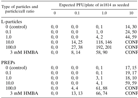
The fact that the polypeptide compositions of PREPs and L-particles differed so significantly made it important to com-pare their biological competence. To do this, we assayed the ability of PREPs to complement the HSV-1 Vmw65 (VP16;
aTIF)-defective mutant in1814 (1, 2). We have already
dem-onstrated that the Vmw65 tegument protein appears to be present in similar quantities (9.2 and 8.7%, respectively) in L-particles and PREPs (Fig. 3 and 4A, lanes 1 through 9). McLauchlan et al. (14) have shown that L-particles are as effective as virions at complementing the in1814 mutant.
HFL cells were infected with in1814 at 0.1, 1.0, and 10 PFU per plate, treated with WT L-particles or ambUL8 PREPs at
0.1, 1.0, 10, and 100 per cell, and incubated for 2 days at 378C,
after which the resulting virus plaques were fixed, stained, and counted (Table 3). Treatment with 3 mM hexamethylene bisac-etamide (HMBA), which increases the plaquing efficiency of
in1814 on HFL cells by about 100-fold (13), was included as a
further control. Neither L-particles nor PREPs by themselves
had any detectable infectivity, but each was able to comple-ment the in1814 mutant. However, the efficiency of PREP complementation was about 10 to 30% of that of control L-particles. Interestingly, 100 PREPs per cell gave rise to about the same numbers of PFU as did treatment with 3 mM HMBA in cultures infected with 1 PFU of in1814 per plate (61 and 88 and 66 and 74, respectively).
DISCUSSION
PREPs are generated by HSV-1-infected cells in which viral DNA replication is either biochemically or genetically blocked and in which normal progeny virion assembly does not take place. Morphologically, PREPs resemble HSV-1 L-particles and exhibit rather similar banding behavior on Ficoll gradients. By definition, PREPs can result only from the transcription products of input virus genomes (5 PFU per cell); thus, it was surprising to find that PREPs were consistently produced in approximately equal numbers to those of L-particles and viri-ons from control cultures. It suggests that transcription from progeny genomes has little relevance for (i) the formation and morphogenesis of the common particle skeleton on which both L-particles and PREPs are assembled and for (ii) the processes that result in extrusions of the formed particles into the super-natant medium.
[image:6.612.61.555.83.317.2]PREPs were differentiated from other virions and L-parti-cles on the basis of their electron microscope morphology and polypeptide composition. Comparison of negatively stained preparations of L-particles and PREPs by electron microscopy disclosed that the envelope glycoprotein spikes of PREPs were less noticeable than those of L-particles, with reduced numbers and lower density of packing. These observations are in accord with the finding that PREPs contain greatly reduced amounts of gC and somewhat lower levels of gD. Stannard et al. (24)
TABLE 2. Densitometric quantitation of proteins from particles produced in MeWO cellsa
Protein band Expt no. Density
b
L-particles ACV-PREPs (%) AraC-PREPs (%) PAA-PREPs (%) ambUL8 PREPs (%)
273K (VP1-2) 1 0.26 0.03 (11.5) NDc 0.03 (11.5) 0.09 (35)
2 0.28 0.02 (7) 0.02 (7) ND 0.08 (28)
3 0.33 ND ND ND ND
175K (IE3) 1 0.08 0.35 (437) 0.33 (412) 0.18 (225) 0.40 (500)
2 0.18 0.41 (227) 0.36 (200) 0.21 (116) 0.28 (155)
3 0.02 0.21 (1,050) 0.21 (1,050) 0.16 (800) 0.12 (600)
92/91K (VP11/12) 1 0.17 0.49 (288) 0.47 (276) 0.37 (217) 0.68 (400)
2 0.52 0.86 (165) 0.85 (163) 0.73 (140) 0.72 (138)
3 0.43 1.23 (286) 1.16 (269) 1.70 (395) 1.06 (246)
57K (VP17; gD) 1 1.64 1.19 (72) 0.98 (60) 0.89 (54) 1.25 (76)
2 1.80 0.74 (41) 0.75 (42) 0.71 (39) 0.65 (36)
3 1.18 0.61 (52) 0.40 (34) 0.67 (57) 0.32 (27)
40K 1 0.52 0.20 (38) 0.21 (40) 0.21 (40) 0.16 (31)
2 0.55 0.18 (33) 0.27 (49) 0.20 (36) 0.20 (36)
3 0.25 0.07 (28) 0.10 (40) 0.19 (76) ND
38K (VP22) 1 0.46 0.77 (167) 0.86 (187) 0.69 (150) 0.70 (152)
2 0.32 0.60 (187) 0.66 (206) 0.41 (128) 0.53 (165)
3 0.23 0.84 (262) 0.79 (246) 0.63 (196) 0.72 (225)
82/81Kd 1 0.80 0.08 (10) 0.05 (6) 0.04 (5) 0.12 (15)
2 1.01 0.06 (6) 0.10 (10) 0.03 (3) 0.11 (11)
3 0.79 0.09 (11) 0.12 (15) 0.20 (25) 0.07 (9)
a
Silver-stained SDS-polyacrylamide gels were loaded with 23109
L-particles or PREPs per lane (numbers were determined by electron microscope counting).
b
Data are the relative amounts of each protein, standardized against the amount of the 65K (VP16;aTIF) protein present in each lane. Parenthetical values are the relative amounts of proteins in PREPs expressed as percentages of the same proteins in the corresponding L-particle controls. The virus used was WT strain 17 or the ambUL8 mutant of strain 17 as indicated.
c
ND, not detected.
d
Figure 4B shows that the 81/82K (VP13/14) protein was undetectable in PREPs by Western immunoblotting, though it was clearly present in L-particles.
VOL. 69, 1995 NOVEL HSV-1-SPECIFIC PARTICLES 4929
on November 9, 2019 by guest
http://jvi.asm.org/
FIG. 4. Polypeptide profiles (A) and immunoblot analysis (B) of the tegument and envelope fractions of HSV-1 virions, L-particles, and PREPs. Envelope components were solubilized from virions, L-particles, and PREPs by treatment with Nonidet P-40, and de-enveloped tegument structures were separated by pelleting. (A) Silver-stained proteins. Tegument (lanes 1 through 9) and envelope (lanes 10 through 18) fractions of HSV-1 virions (V), L-particles (L), and ambUL8 PREPs (P am) were prepared in BHK cells (lanes 1 through 3 and 16 through 18) and MeWO cells (lanes 4, 8 through 11, and 15). PREPs were also made in WT HSV-1-infected MeWO cells treated with ACV, PAA, and ara-C (lanes 5 through 7 and 12 through 14, respectively).F, L-particle protein;{and■, increased and decreased intensity of PREP protein band, respectively, compared with that of L-particle protein;E, capsid protein. To aid in the interpretation of these gels, some gel lanes have been rearranged. (B) Western immunoblots. Proteins were from duplicate SDS-polyacrylamide gels containing the same protein extracts and were loaded into lanes in the same order as in panel A. Blots were probed with rabbit polyclonal antibody raised against 82/81K (VP13/14), rabbit polyclonal antibody (R47) against gC, and MAb against gD (VP17). Antibody-tagged proteins were detected by enhanced chemoluminescence (Amersham).
on November 9, 2019 by guest
http://jvi.asm.org/
have shown by immunogold electron microscopy of negatively stained HSV-1 virions that gC is present only in the longest envelope spikes, while gD is found in shorter spikes which form irregular patches, giving an indistinct fringed appearance when viewed longitudinally. However, PREPs were not devoid of envelope spikes; the characteristic T-shaped gB spike (24) was also present.
Although the overall protein content of PREPs resembled that of L-particles, these two types of particles were clearly distinguished by several differences in relative polypeptide composition (Fig. 3 and 4) (Table 2). Characteristically, PREPs contained less of the 273K (VP1-2), gC (VP8), 82/81K (VP13/14), gD (VP17), and 40K polypeptides and more of the 175K (VP4; IE3), 92/91K (VP11/12), and 38K (VP22) proteins than L-particles did, regardless of whether they had been gen-erated in BHK or MeWO cells.
Differences in the polypeptide compositions of L-particles and PREPs can be expected as a consequence of inhibition of viral DNA synthesis.
First, true late HSV-1 proteins, which are thought to be transcribed almost exclusively from progeny genomes, are an-ticipated to be absent from PREPs or present only in trace amounts. gC and the 273K (VP1-2) and 82/81K proteins are classified as true late HSV proteins (16–18). Small amounts of gC (UL44) and the 273K (UL36) protein, but not of the 82/ 81K (UL47) protein, were detected in PREP preparations. Various explanations are possible. Some UL44 and UL36 tran-scription, but not UL47 trantran-scription, from input genomes takes place; the UL36 and UL44 transcripts or the 273K (VP1-2) and gC proteins are synthesized in greater amounts or are more stable; the antibody-recognized gC epitope is more stable than that of the 82/81K protein; or the anti-gC poly-clonal antibody used is more efficient than the 82/81K MAb in Western blotting.
Secondly, early/late proteins, such as gD, which require DNA replication to boost their levels of expression (10) might consequently be present in reduced amounts in PREPs.
The relative amounts of some proteins may be determined by the way that the particle is assembled. Some tegument proteins may actually be assembled as protein-protein com-plexes, e.g., Vmw65 (VP16) and vhs have been shown to form a complex with roles in particle assembly and modulation of
vhs activity (23). Either the absence or presence in reduced amounts of a DNA replication-dependent protein could result in the exclusion or underrepresentation of one (or more) other protein(s) involved in such a complex.
In addition, some aspects of the relative protein composition of PREPs may be intrinsically flexible and thus able to vary, either reflecting the abundance of particular nonessential pro-teins or allowing preferential incorporation of protein X to replace protein Y during PREP particle assembly. HSV-1 mu-tants with genes UL46 (VP11/12) and UL47 (VP13/14) (29) deleted were investigated by Zhang and McKnight (28); they found that virions deleted of UL46 contained 2.5-fold more VP13/14 than control virions did, while UL47-deleted virions contained twice as much VP11/12 and also had a 30% increase
in the amount of Vmw65 (VP16;aTIF). A mutant deleted of
both UL46 and UL47 contained 30 to 40% increases in the amounts of VP16 and gB-gC-gH in virions (28).
In general, tegument composition seems to be complex but not totally fixed. Under conditions of WT HSV infection, par-ticles with a maintained protein stoichiometry are produced, but as shown by Zhang and McKnight (28), variations in the relative amounts of at least some tegument proteins can be tolerated without impairing the efficiency of virion assembly. PREPs and L-particles provide further indications of the HSV particle’s potential for alterations in tegument composition, a property that could be manipulated and exploited for vaccine production.
Our finding that the overrepresentation of 92/91K (VP11/ 12) in PREPs is 2.45-fold, compared with that in L-particles, argues for nonrandom incorporation of at least some tegument proteins during assembly of PREPs, because the early/late temporal classification of the UL46 gene (8, 28) which encodes these proteins suggests that they should be present in reduced amounts in WT virus-infected cells treated with an inhibitor of viral DNA synthesis or in cells infected with the ambUL8 virus. Zhang and McKnight (28) have suggested that essential tegu-ment proteins, such as VP16, provide a fixed framework around which nonessential proteins assemble. Studies of vi-ruses deleted of particular tegument proteins may well uncover protein-protein interactions during particle assembly, and it is interesting that the data of Zhang and McKnight (28) suggest that there is interaction not only between tegument proteins but also between tegument proteins and envelope glycopro-teins. In this context, interactions between matrix proteins and envelope glycoproteins which play an essential role in virus particle assembly have been reported for vesicular stomatitis virus and Sendai virus (11, 22).
Despite their reduced levels of gC and gD, PREPs were biologically competent, delivering functional tegument pro-teins to cells (Table 2). However, the efficiency of complemen-tation of in1814 by PREPs was reduced to about 10 to 30% of
the L-particle efficiency. As the amounts of Vmw65 (aTIF)
protein contained in PREPs and L-particles appeared to be about equal (9.2 and 8.7%, respectively), the relative comple-mentation efficiencies are likely to reflect differences in the efficiency by which these two types of particles attach to or enter cells.
[image:8.612.56.299.91.263.2]Both gC and gD are known to play roles in the initial stages of infection. gC interacts with the heparin sulfate moieties of cell surface proteoglycans which serve as receptors for HSV-1 adsorption, and gC also appears to have a role in the rate of virus penetration into cells (9). gD is required for fusion be-tween the virus envelope and the plasma membrane during virus entry (4, 6). Thus, the lower levels of gC and gD in PREPs are probably responsible for their reduced efficiency of entry into cells; indeed, Herold et al. (9) have reported that gC
TABLE 3. Complementation of Vmw65-defective mutant in1814 by L-particles and PREPsa
Type of particles and particle/cell ratio
Expected PFU/plate of in1814 as seeded
0 0.1 1.0 10
L-particles
0 (control) 0, 0 0, 0 0, 1 14, 30
0.1 0, 0 0, 0 1, 0 24, 50
1.0 0, 0 0, 0 4, 2 44, 59
10.0 0, 0 14, 25 118, 146 CONFb
100.0 0, 0 27, 38 192, 201 CONF
3 mM HMBA 0, 0 8, 14 58, 90 CONF
PREPs
0 (control) 0, 0 0, 0 0, 1 17, 15
0.1 0, 0 0, 0 0, 1 19, 17
1.0 0, 0 0, 0 3, 1 18, 10
10.0 0, 0 0, 0 4, 1 59, 59
100.0 0, 0 4, 4 61, 88 CONF
3 mM HMBA 0, 0 13, 13 66, 74 CONF
a
Experiments were performed in duplicate.
b
CONF, monolayers were confluent with cytopathic effect.
VOL. 69, 1995 NOVEL HSV-1-SPECIFIC PARTICLES 4931
on November 9, 2019 by guest
http://jvi.asm.org/
enhances infectivity by about a factor of 10, so the low levels of gC in PREPs could alone account for our findings.
HSV-1 L-particles have been viewed as candidates for vac-cine production. By comparison, we consider PREPs to repre-sent potentially more advantageous new vaccine candidates because they are easier to prepare and purify, they have a 100-to 10,000-fold-greater particle/PFU ratio than that of WT trol L-particle preparations, and with them, HSV-1 DNA con-tamination is expected to be at a minimal level (i.e., only some surviving input genomes).
As prepared here, PREPs lack or have reduced levels of some important tegument and envelope proteins. It has not escaped our attention that manipulation of promoter and con-trol sequences, deletion of certain HSV genes, and possible insertion of non-HSV genes will allow us to generate PREPs with improved levels of those proteins important for biological and immunological activities and with the advantages of addi-tional, built-in safety features.
Finally, the fact that PREPs are produced demands reexam-ination of and will lead to new insights into the general prob-lem of HSV particle morphogenesis. Clearly, PREPs demon-strate that de novo expression of at least some, if not all, of the true late proteins is not a prerequisite for the synthesis, envel-opment, and egress of these particles and, by inference, L-particles.
ACKNOWLEDGMENTS
Thanks are due to J. Aitken, who carried out all of the electron microscopy, to G. Cohen and R. Eisenberg, who kindly provided an-tibody R47 (anti-gC), and to A. Cross and Howard Marsden, who supplied all of the other antibodies used. We also thank F. Rixon, J. McLauchlan, and H. Marsden for helpful discussions.
REFERENCES
1. Ace, C. I., M. A. Dalrymple, F. H. Ramsay, V. G. Preston, and C. M. Preston. 1988. Mutational analysis of a herpes simplex virus transinducing factor Vmw65. J. Gen. Virol. 69:2595–2605.
2. Ace, C. I., T. A. McKee, M. Ryan, J. M. Cameron, and C. M. Preston. 1989. Construction and characterisation of a herpes simplex virus type 1 mutant unable to transinduce immediate early gene expression. J. Gen. Virol. 63: 2260–2269.
3. Bean, M. A., B. R. Bloom, R. B. Herberman, L. J. Old, H. F. Oettgen, G. Klein, and W. D. Terry.1975. Cell-mediated cytotoxicity for bladder carci-noma: evaluation of a workshop. Cancer Res. 35:2902–2913.
4. Campadelli-Fiume, G., E. Avitabile, D. Stirpe, M. Arsenakis, and B. Roiz-man.1988. Herpes simplex virus glycoprotein D is sufficient to induce spon-taneous pH-independent fusion in a cell line that constitutively expresses the glycoprotein. Virology 166:598–602.
5. Carey, T. E., T. Takashashi, L. A. Resnick, H. F. Oettgen, and L. J. Old. 1976. Cell surface antigens of human malignant carcinoma: mixed hemad-sorption assays for humoral immunity to cultured autologous melanoma cells. Proc. Natl. Acad. Sci. USA 73:3278–3282.
5a.Dargan, D. J., and J. H. Subak-Sharpe. Unpublished data.
6. Fuller, A. O., and P. G. Spear. 1987. Anti-glycoprotein D antibodies that permit adsorption but block infection by herpes simplex virus type 1 prevent virion cell-fusion at the cell surface. Proc. Natl. Acad. Sci. USA 84:5454– 5458.
7. Grose, C., and P. A. Brunell. 1978. Varicella-zoster virus: isolation and propagation in human melanoma cells at 36 and 328C. Infect. Immun. 19: 199–203.
8. Hall, L. M., K. G. Draper, R. J. Frink, R. H. Costa, and E. K. Wagner. 1982. Herpes simplex virus mRNA species mapping in EcoRI fragment I. J. Virol. 43:594–607.
9. Herold, B. C., D. WuDunn, N. Soltys, and P. G. Spear. 1991. Glycoprotein C of herpes simplex virus type 1 plays a principal role in the adsorption of virus to cells and in infectivity. J. Virol. 65:1090–1098.
10. Johnson, P. A., C. McLean, H. S. Marsden, R. G. Dalziel, and R. D. Everett. 1986. The products of gene US11 of herpes simplex virus type 1 is expressed as a true late gene. J. Gen. Virol. 67:871–883.
11. Lyles, D. S., M. McKenzie, and J. W. Parce. 1992. Subunit interactions of vesicular stomatitis virus envelope glycoprotein stabilized by binding to viral matrix protein. J. Virol. 66:349–358.
12. Marsden, H. S., I. K. Crombie, and J. H. Subak-Sharpe. 1976. Control of protein synthesis of herpesvirus-infected cells: analysis of the polypeptides induced by wild type and sixteen temperature sensitive mutants of HSV strain 17. J. Gen. Virol. 31:347–372.
13. McFarlane, M., J. I. Daksis, and C. M. Preston. 1992. Hexamethylene bisacetamide stimulates herpes simplex virus immediate early gene expres-sion in the absence of trans-induction by Vmw65. J. Gen. Virol. 73:285–292. 14. McLauchlan, J., C. Addison, M. C. Craige, and F. J. Rixon. 1992. Non-infectious L-particles supply functions which can facilitate infection by HSV-1. Virology 190:682–688.
15. McLauchlan, J., and F. J. Rixon. 1992. Characterisation of enveloped teg-ument structures (L-particles) produced by alphaherpesviruses: integrity of the tegument structure does not depend on the presence of capsid or enve-lope. J. Gen. Virol. 73:269–276.
16. McLean, G., F. Rixon, N. Langeland, L. Haarr, and H. Marsden. 1990. Identification and characterisation of the virion protein products of the herpes simplex virus type 1 gene UL47. J. Gen. Virol. 71:2953–2960. 17. McNabb, D. S., and R. J. Courtney. 1992. Characterisation of the large
tegument protein (ICP1/2) of herpes simplex virus type 1. Virology 190:221– 232.
17a.Patel, A. H. Unpublished data.
18. Peake, M. L., P. Nystrom, and L. I. Pizer. 1982. Herpesvirus glycoprotein synthesis and insertion into plasma membranes. J. Virol. 42:678–690. 19. Preston, V. G., J. A. V. Coates, and F. J. Rixon. 1983. Identification and
characterization of a herpes simplex virus gene product required for encap-sidation of virus DNA. J. Virol. 45:1056–1064.
20. Rixon, F. J., C. Addison, and J. McLauchlan. 1992. Assembly of enveloped tegument structures (L-particles) can occur independently of virion matura-tion in herpes simplex virus type 1 infected cells. J. Gen. Virol. 73:277–284. 21. Rosenthal, K. S., M. D. Leuther, and B. G. Barisas. 1984. Herpes simplex virus binding and entry modulate cell surface protein mobility. J. Virol. 49:980–983.
22. Sanderson, C. M., H.-H. Wu, and D. P. Nayak. 1994. Sendai virus M protein binds independently to either the F or the HN glycoprotein in vivo. J. Virol. 68:69–76.
23. Smibert, C. A., B. Popova, P. Xiao, J. P. Capone, and J. R. Smiley. 1994. Herpes simplex virus VP16 forms a complex with the virion host shutoff protein vhs. J. Virol. 68:2339–2346.
24. Stannard, L. M., A. O. Fuller, and P. G. Spear. 1987. Herpes simplex virus glycoproteins associated with different morphological entities projecting from the virion envelope. J. Gen. Virol. 68:715–725.
25. Szila´gyi, J. F., and J. Berriman.1994. Herpes simplex virus L-particles contain spherical membrane-enclosed inclusion vesicles. J. Gen. Virol. 75: 1749–1753.
26. Szila´gyi, J. F., and C. Cunningham.1991. Identification and characterisation of a novel non-infectious herpes simplex virus-related particle. J. Gen. Virol. 62:661–668.
27. Wu, C. A., N. J. Nelson, D. J. McGeoch, and M. D. Challberg. 1988. Iden-tification of herpes simplex virus type 1 genes required for origin-dependent DNA synthesis. J. Virol. 62:435–443.
28. Zhang, Y., and J. L. C. McKnight. 1993. Herpes simplex virus type 1 UL46 and UL47 deletion mutants lack VP11 and VP12 or VP13 and VP14, re-spectively, and exhibit altered viral thymidine kinase expression. J. Virol. 67:1482–1492.
29. Zhang, Y., D. A. Sirko, and J. L. C. McKnight. 1991. Role of herpes simplex virus type 1 UL46 and UL47 inaTIF-mediated transcriptional induction: characterization of three viral deletion mutants. J. Virol. 65:829–841.