LINEAR DERMATOSES
– A PROSPECTIVE STUDY
Dissertation Submitted In
Fulfillment of University Regulation For
MD DEGREE IN
DERMATOLOGY, VENEREOLOGY AND
LEPROLOGY
(BRANCH XII A)
THE TAMILNADU
DR. M. G. R. MEDICAL UNIVERSITY
CHENNAI
CERTIFICATE
Certified that this dissertation entitled “LINEAR
DERMATOSIS – A PROSPECTIVE STUDY” is a bonafide work
done by DR. P. PRABAHAR, Post graduate student of Department of
Dermatology and Leprology and Institute of Venereology, Madras Medical College, Chennai- 3, during the academic year 2004 – 2007. This work has not previously formed the basis for the award of any degree or diploma.
Prof. Dr. B. PARVEEN, M.D., D.D., Professor and Head,
Department of Dermatology and Leprology, Madras Medical College,
Chennai- 3
Prof. Dr .KALAVATHI PONNIRAIVAN, B. Sc., M.D., The DEAN, Madras Medical College,
DECLARATION
I, DR. P. PRABAHAR, solemnly declare that the Dissertation titled LINEAR DERMATOSIS – A PROSPECTIVE STUDY is a bonafide work done by me during 2004 – 2007 under the guidance and supervision of Prof. Dr. P. PARVEEN M.D.,D.D., Professor and Head of the Department of Dermatology, Madras Medical College, Chennai.
The Dissertation is submitted to The Tamilnadu Dr. M. G. R Medical University towards partial fulfillment of requirement for the award of
M.D Degree in Dermatology Venereology and Leprology (Branch XII A)
Place:
Date:
SPECIAL ACKNOWLEDGMENT
My sincere thanks to
Prof. Dr .KALAVATHI PONNIRAIVAN, B. Sc., M.D.,
The DEAN, Madras Medical College for allowing me to do this
Dissertation and utilize the institutional facilities.
ACKNOWLEDGEMENT
I am gratefully indebted to Prof. Dr. B. Parveen M.D.,D.D., Professor and Head of Department of Dermatology for her invaluable guidance, motivation and help though out the study. I would like to express my sincere and heartfelt gratitude to Prof. Dr. V.S. Dorairaj, M.D.,D.V., Director In charge, Institute of Venereology. I wish to thank Dr. N. Gomathy M.D., D.D., former Professor, Department of Dermatology and Dr. N. Usman M.D., D.V., Ph.D., former Director, Institute of Venereology for their constant support and motivation.
I am very grateful to Dr. S. Jayakumar M.D., D.D., Additional Professor, Department of Dermatology for his invaluable guidance and help. I sincerely thank Dr. C. Janaki M.D., D.D., Reader of Dermatology (Mycology) for her priceless support.
I xpress my earnest gratefulness to Dr. D. Prabavathy M.D., D.D., Professor and Head of Department of Occupational Dermatology and Contact Dermatitis for her constant motivation and guidance. I thank
Dr. V. Somasundaram M.D., D.D., Additional Professor, Department of Occupational Dermatology and Contact Dermatitis for his benevolent help and support.
I incline to thank Dr. R. Priyavathani M.D., D.D., D.N.B., Dr. V. Anandan M.D.,(Derm), D.Ch., D.N.B.,(Paed) and Dr. K. Tharini M.D., Dr. M. Vijayanand M.D.,D.D., Assistant Professors, Department of Dermatology for their kind support and encouragement.
I thank Dr. A. Hameedullah M.D., D.D., Dr. S. Kumaravelu M.D., D.D., Dr. J. Manjula M.D., D.N.B., (Derm) and Dr. Aftab Jameela Wahab M.D.,D.D., for their support and help.
My sincere thanks to Dr. S. Mohan M.D, D.V. former Registrar, Dr. K. Venkateswaran M.D., D.V., Dr. P. Elangovan M.D., D.V., Dr. S. Thilagavathy M.D., D.V., Dr. V. Thirunavukkarasu M.D., D.V., Dr. D. Ramachandra Reddy M.D., D.V., Dr. P. Mohan M.D., D.V., Dr. S. Arunkumar M.D.,D.V., and Dr. S. Kalaivani M.D.,D.V., Assistant Professors, Institute of Venereology for their help and suggestions.
I am also thankful to Dr. K. Manoharan M.D., D.D., and Dr. V. Sampath M.D., D.D., for their continuing guidance and support.
I duly acknowledge the paramedical staff and my colleagues for their help and favours.
CONTENTS
Sl.No
Title
Page No.
I.
INTRODUCTION 1
II. REVIEW OF LITERATURE 3
III. AIMS OF THE STUDY 39
IV. MATERIALS AND METHODS 40
V. OBSERVATIONS AND RESULTS 42
VI. DISCUSSION 59
VII. CONCLUSION 66
BIBLIOGRAPHY
PROFORMA
INTRODUCTION
Skin is a very important and largest organ of the body. It is the only organ which is visible and is in direct contact with the environment.
In the examination of the skin, the morphology of individual lesions, their overall pattern and spatial relationship to each other, and their body site distribution are helpful and provide an easily recognizable clue to a rapid visual diagnosis. Indeed, clinical diagnosis is more precise than laboratory tests in many disorders.
Skin lesions present with innumerable patterns like Discoid, Petaloid, Arcuate, Annular, Polycyclic, Livedo, Reticulate, Target, Stellate, Digitate, Linear, Serpiginous, Whorled, etc.
The mechanisms or anatomical factors dictating the Linearity are of the following groups:
- Linear configurations determined by the course of blood vessels, lymphatics or nerve trunks
- Linear lesions of developmental origin - Linear lesions following Dermatomal pattern
- Linear lesions caused by External factors like Plants, Allergens, Chemicals, Thermal and Physical factors (includes Koebner’s phenomenon).
- Linear configurations due to other determinants
REVIEW OF LITERATURE
LINES OF BLASCHKO
ALFRED BLASCHKO (1858 - 1922) private practitioner of dermatology in Berlin, whose interest ranged from leprosy to occupational skin disease. He presented his findings on distribution patterns of linear skin disorder at the German Dermatological Society meeting in Breslau in the year 19011,2. He examined more than 140 patients with linear
lesions such as epidermal naevi, sebaceous naevi and nevus lipomatosus and carefully transposed the pattern in each patient on to dolls and statues1,2.
A composite diagram of these distribution patterns was then drawn that has subsequently been referred to as the lines of Blaschko.
In 1976, Jackson2 provided a detailed review of the 1901 publication
and introduced the concept of the lines of Blaschko into the English literature, although it had been well known in the European community for decades. These lines do not correspond to other patterns such as Langer’s lines of cleavage3,Voigt’s lines (borders between areas of innervations by
peripheral cutaneous nerves4),Embroyonic clefts5, Pigmentary demarcation
Although the distribution is linear, the curvature of the lesions does not support the hypothesis that these lines represent Koebner’s phenomenon. Most commonly, Blaschko’s lines are confused with dermatomes, the segments of skin that are defined by sensory innervation7. A major reason
for these confusion is that both distribution patterns are characterized by a striking demarcation of cutaneous lesions at the midline. As a reflection of these confusion, several diseases that follow Blaschko’s lines are referred to as dermatomal or zosteriform, for example, zosteriform Porokeratoses, zosteriform Lichen planus and zosteriform Lentiginous naevi.
The Blaschko’s lines were most apparent on the trunk with arcs on the upper chest, S-shapes on the abdomen, a V-shape as the lesions approach the posterior midline and spirals on the scalp are seen.
Occasionally, however, the lesions of herpes zoster do appear to have a more figurate arrangement, raising the possibility that the migration of cutaneous nerves may influence the pattern of Blaschko’s lines. On the lateral foot, the lines of Blaschko respect the junction between plantar skin and hair bearing skin and therefore overlap with Wallace’s line8.
autonomic innervations to the dermal viscera, that is, the visceral afferents, as opposed to the sensory afferents (Edmund S. Crelin, PhD, Personal Communication, June 1992). The possibility has also been raised that Blaschko’s lines simply represent stretching of the skin during embryogenesis; an analogy given is the pattern seen when newspaper print is superimposed on Silly Putty, then stretched (Lawrence Solomon, MD, Personal Communication, July or August 1993). However, involvement of fat and blood vessels is difficult to explain with this theory9.
Although the lines of Blaschko clearly do not correspond to the distribution of hair tracts, the possibility of overlap with hair whorls has been raised. This debate is, in part, based on the fact that those lines of Blaschko are less well-defined on the head and neck. Happle et al10, 11 have
added lines to the posterior scalp, whereas we have attempted to delineate further the lines on the lateral aspect of the face and neck. Brown and Gorlin12 reviewed the literature in 1960 and mentioned vertical striations in
the lips, linear midline lesions on the hard and soft palate, and linear unilateral and / or midline bands on the tongue in patients with epidermal naevi.
Albinism can have a striated pigmentary pattern in the peripheral retina13 in
addition to an alternating spokewheel - like pigmentation of the iris (alternating normal pigmentation and hypopigmentation14). Female carriers
of X - linked cataracts and X - linked Lowe’s (oculocerebrorenal) syndrome have sectorial cataracts and lens opacities with an irregularly radiated pattern15. Of note, similar sectorial cataracts and radial patterns in
the lens have been described in women with X-linked dominant chondrodysplasia punctata16. Witkop17, also described alternating vertical
bands of opaque white and translucent ( normal – appearing ) enamel on the central incisors of women heterozygous for X-linked hypomaturation Amelogenesis imperfecta.
CHIMERISM
It denotes the presence of two or more genetically distinct cell population in an individual derived from two different zygotes.
MOSAICISM
Mosaicism describes an individual with two or more cell lines of different genotypes derived from the same zygote. In health, all females exhibit functional mosaicism with regard to their X chromosomes. One of the two chromosomes in the cells of normal females undergoes inactivation at an early stage of embryonic development (12 – 16 days after fertilization), a process known as LYONIZATION. In 1961, Mary Lyon reported striped patterns for some X-linked color genes in mice. She hypothesized that the stripes reflect two populations of cells, one expressing maternal X chromosome and the other paternal. In 1965, Curth and Warburton18 applied
the Lyon hypothesis to the X-linked Incontinentia Pigmenti which is characterized by lesions following Blaschko’s lines. In 1977, Happle19
CAUSES OF MOSAICISM AND CORRESPONDING PATHOGENESIS20 CAUSES OF MOSAICISM PATHOGENESIS Half Chromatid Mutation
A mistake in DNA polymerization during the first meiotic division of gametogenesis, whereby the wrong base is synthesized at one point, resulting in a mismatched double strand. If this mismatched chromosome is passed on to the next generation, the first time it separates in mitosis it will provide two templates that are not exactly complementary, giving rise to two different lines of daughter cells.
Lyonization
The hypothesis, proposed by Mary Lyon, states that only one X chromosome is active in each female cell, with the other forming the Barr body. Whether the paternal or maternal X chromosome is inactivated is random, but once the choice has been made it is the same in all daughter cells.
Post zygotic (somatic) Mutation
A mutation occurring after fertilization
Chromosomal non-disjunction
The failure chromosome to separate correctly during either meiosis or mitosis, resulting in daughter cells with aberrations of chromosome number or structure
Chimerism
THE DIFFERENT PATTERNS OF MOSAICISM
21TYPE 1: LINES OF BLASCHKO Fountain like pattern - back.
S-figure - lateral and ventral aspect of trunk. Spiral - scalp.
These lines reflect the dorsoventral outgrowth of embryonic cells from the neural crest. Their proliferation interfere with the longitudinal growth and increasing flexion of the embryo, resulting in a characteristic arrangement
Head and neck -variable pattern, tent to intersect at an angle of 90˚ TYPE 1.a -narrow bands (e.g.: Incontinentia pigmenti).
TYPE 1.b -broad bands (e.g.: McCune- Albright syndrome).
TYPE 2: CHECKBOARD PATTERN
Flag like area with a sharp midline separation (distributed in a random way and not alternating regularly).
E.g.: speckled lentiginous naevus, Becker nevus
Pattern of patchy hairiness as noted in women heterozygous for X-linked hypertrichosis
TYPE 3: PHYLLOID PATTERN
Leaf- like patches and oblong macules (midline separation is not always present)
TYPE 4: LARGE PATCHES WITHOUT MIDLINE SEPERATION
E.g.: congenital giant melanocytic nevi, with or without neurological Involvement (clonal origin)
Acquired melanocytic nevi
TYPE 5: LATERALIZATION PATTERN
E.g.: CHILD syndrome (X- linked dominant, male lethal-rait) CHILD nevus- one half of the body, with a sharp midline demarcation.
X-inactivation coincides with the origin of a clone of organizer cells controlling a large developmental field.
Term ZOSTERIFORM NEVI
A zosteriform arrangement corresponds to the system of dermatomes but all nevi are dermatomal but follow the lines of Blaschko.
LINES THAT DO NOT INVOLVE MOSAICISM
Lines of Voigt – boundaries of peripheral cutaneous innervations
Matsumoto line (also Futcher’s line) pigment demarcation line on arms and legs.
PATTERNS OF CUTANEOUS MOSAICISM
Lines of Blaschko Lines of Blaschko
Narrow Bands Broad Bands Checkerboard
ANATOMICAL AND CAUSATIVE FACTORS IN LINEAR LESIONS 23
Blood vessels - Thrombophlebitis, Mondor’s disease - Eczema related to varicose veins - Temporal arteritis
Lymphatics - Lymphangitis
- Sporotrichosis, Fish tank granuloma
Dermatome - Herpes zoster, zostiform naevus, zostiform Darier’s disease,
- Zostiform metastases
Nerve trunks - Leprosy (thickened cutaneous nerves) Developmental - Pigmentary demarcation line, linea nigra (Blaschko lines) - Epidermal naevi, Incontinentia pigmenti, Lichen striatus, Hypomelanosis of Ito, Linear psoriasis,
Linear lichen planus,
Skin stretching - Striae due to growth spurt ( on lower back ) Infestation - Scabies, Larva migrans ( serpiginous ) External factors
Plants - Phytophotodermatitis
Allergens - Elastoplast, nail varnish (neck), necklace) Chemicals - Caustics, eg. Phenol
Thermal - Burns
Physical - Trauma to previous normal skin
Keloid scar, bruising, dermatitis artifacta, Amniotic constriction bands
Trauma to skin with a pre-existing dermatosis
Purpura (cryoglobulinaemia, amyloid, vasculitis ) Blisters (epidermolysis bullosa, porphyria )
Koebner phenomenon
Psoriasis, lichen planus, lichen nitidus,
vitiligo,Lichen sclerosus,Pityriasis rubra pilaris. Inoculation : Molluscum contagiosum
Other mechanism : Scar sarcoid Other determinants - Linear scleroderma (limb, forehead) - Senear-Caro ridge (on hands in psoriasis) - Dermatomyositis (dorsum pf fingers)
LICHEN STRIATUS
Lichen striatus is an inflammatory papular eruption with a distinctive linear distribution, often following Blaschko’s line, which should be differentiated from many other cutaneous disease with linear pattern25.26
Variants of this disorder has also been called blaschkitis, Blaschko linear acquired inflammatory skin eruption, zonal dermatosis, linear neurodermatitis, linear dermatosis, linear lichenoid dermatosis, lichenoid eruption, systematized lichenification and linear eczema .
DEFINITION
Lichen striatus is an uncommon self limiting linear dermatosis with unknown aetiology and spontaneous regression. It primarily occurs in children from 5-15 years of age. The average age at diagnosis is 3 years25, 26.
It may also be seen in adults. Cases in two extremes of age have been reported. Females are affected more than males. They are affected 2 or 3 times as frequently as males27.
AETIOLOGY
The development of lesion along Blaschko’s lines raises the possibility of a cell-mediated autoimmune reaction to an abnormal clone of cells. Blaschko’s lines are believed to represent the direction along which epidermal growth centers expand during early skin development30. It has
been suggested that the distribution of lesions in lichen striatus may reflect a post zygotic abnormality such as somatic mutation affecting localized stem cells.30 Manifestation of atopy with abnormal immune response34D,35 About
80% of patients have the family history of atopy32.
PATHOGENESIS
In lichen striatus it has been found that the inflammatory cells reaching the epidermis are CD8+ (suppressor-cytotoxic) T-lymphocytes33 with the
Langerhans cells population in the epidermis either decreased or increased. These findings suggest a cell-mediated immunologic mechanism where cytotoxic events against keratinocytes could be taking place during the evolution of the disease33.
Immunohistochemistry has shown that most of lymphocytes in the upper dermis and epidermis are CD7+12, and most of the lymphocytes in the
epidermis are CD8+ T-cells expressing HLA-DR+ antigen on their surface.34
study CD1a Langerhans’ cells were either decreased or increased or normal in the epidermis
.
34CLINICAL FEATURES
The morphology of the lesions is distinct. They appear as small, pink, lichenoid papules which are at first discrete, but coalesce rapidly into plaques following Blaschko’s lines25, 26. The lesions start suddenly and
extend over the course of a week or more to become dull red slightly scaly bands.
The width is usually 2mm to 2cm and is often irregular. The bands may broaden into plaques. The length may vary from few centimeters to several centimeters, or may extend the entire length of the limb. The bands may be continuous, interrupted, parallel or zosteriform.25, 26
The lesions occur commonly on one arm or leg or on the neck, but may develop on the trunk, abdomen, buttocks or thighs25, 26. Rarely the lesions
may be multiple and bilateral27, 28. The lesions are normally asymptomatic,
but pruritus of moderate to severe degree may be experienced29.
Variations- verrucous lesions with confluence (by Johnson) 35
Light to yellow coloured grouped papules (by Netherton) 36
Papules, vesicles and crusting (by Felix pinkus) 38
Hypo pigmentation may be prominent, especially in dark skinned persons. In pigmented skin, post inflammatory hypo pigmentation is a useful sign for distinguishing lichen striatus from linear lichen planus. When lesions extend to the ends of digits, nail involvement may range from fraying to total nail loss39. Seasonal occurrence and simultaneous
involvement of siblings has suggested an infectious cause, perhaps viral28.
Differential Diagnoses
The differential diagnoses of lichen striatus include linear lichen planus, linear lichen nitidus, linear epidermal nevus, inflammatory linear verrucous epidermal nevus, linear psoriasis and linear lichen simplex chronicus.
HISTOPATHOLOGY 40
Although lichen striatus has been recognized by its variable histologic picture, some constant microscopic findings may be present. Lichen striatus
is chronic lichenoid dermatitis. There is usually a superficial perivascular inflammatory infiltrate of lymphocytes admixed with a variable number of histiocytes. Plasma cells and eosinophils are rarely seen34. Focally, in the
alteration of the basal layer and necrotic keratinocytes. In these areas, the papillary dermis occasionally contains melanophages30,32. Additional
epidermal changes consist of spongiosis and intracellular edema often associated with exocytosis of lymphocytes and focal parakeratosis.
Less frequently, there are scattered necrotic keratinocytes in the spinous layer as well as sub corneal spongiotic vesicles filled with Langerhans cells30,33. A very distinctive feature is the presence of
inflammatory infiltrate in the reticular dermis around hair follicles and eccrine glands. An unusual perforating variant of lichen striatus has been described, which shows Tran epidermal elimination of clusters of necrotic keratinocytes. `
TREATMENT
Because the lesions are self-limited and resolve spontaneously within one year usually there is no treatment necessary. Reassuring the patient is essential. Post inflammatory hypo pigmentation may persist longer`
ADULT BLASCHKITIS41
(BLASCHKO LINEAR ACQUIRED INFLAMMATORY SKIN ERUPTION; BLAISE)
eczematous (spongiotic) than lichenoid. It would be difficult to distinguish from linear Grover’s disease42. Taieb et al43. considered that ‘adult
Blaschkitis’ represents and adult version of lichen striatus, and proposed the acronym BLAISE to cover both. BLAISE should perhaps be regarded as a description rather than a diagnosis, a useful category for many of the disorders in this section, pending more precise identification.
LINEAR PSORIASIS
Atleast three clinical entities have been described as “linear psoriasis”. All follow Blaschko’s lines. The first, and most common, is an ILVEN that may resemble psoriasis clinically. Since Woringer’s first report44 in 1936,
the psoriasiform nature of some epidermal nevi has been recognized45. These
lesions often exhibit a characteristic histologic picture of areas of hypergranulosis and orthokeratosis alternating with agranulosis and parakeratosis, although features of psoriasis may also be present.
The existence of third true “linear psoriasis” that does not fit into other types is controversial. Nonetheless, at least two children have been described with extensive lesions following Blaschko’s lines that resembled psoriasis both clinically and histologically47.
In neither case was a pre - existing nevus present, nor were there any signs of psoriasis elsewhere on the patient’s skin. It has been suggested that psoriasis in such patients arises by somatic recombination, giving rise to the pattern following Blaschko’s lines.
The lesion could be distinguished from invasion of a verrucous epidermal nevus by psoriasis as a result of the isomorphic phenomenon and from dermatitic epidermal naevi, by their minimal pruritus and their therapeutic response to ultraviolet radiation. Linear psoriasis is easily confused with ILVEN45.
LINEAR LICHEN PLANUS
Linear lichen planus was first described by Devergie in 1854 and accounts for 0.24-0.62% of all the patients with lichen planus and was found to be more common in Japan48.
Scattered linear lesions often occur in patients with lichen planus and are a result of a combination of scratching and the Koebner’s phenomenon. Less commonly, unilateral streaks or bands of LP are seen that are longer and wider than the trauma induced lesions47. In a review of 18 cases of this
linear variant of LP48, the average age of onset was 33 years and lesions
developed over a period of 1 week to several months. More than half the patients had pruritus.
In the majority of cases, the streaks were composed of polygonal violaceous papules and coalescing plaques. Surface shows Wichkam’s striae (white lines). It mainly occurs on the front of wrists, lumbar region, around the ankles and glans penis. If they extend to the end of digit there may be associated nail dystrophy. Palms, soles, nails and oral mucous membranes may also affected.
Histopathology of a typical papule of lichen planus shows compact orthokeratosis, wedge shaped hypergranulosis contributing clinically to Wichkam’s striae, irregular acanthosis and pointed lower ends of the rete ridges, giving them a ‘saw – toothed’ appearance. Liquefactive degeneration of the basal layer, with formation of civatte or colloid bodies. Dense band like inflammatory infiltrate composed of lymphocytes and histiocytes, closely hugging the lower end of the epidermis and also present perivascularly are present. Melanophages are seen in the upper dermis. Lichen planus is an immunologically mediated dermatosis as evidenced by the immunofluorescence studies51.
EPIDERMAL NAEVI
(
VERRUCOUS NEVUS; NEVUS UNIUS LATERIS)
Epidermal nevus is a developmental malformation of the epidermis in which an excess of keratinocytes, sometimes showing abnormal maturation, results in a visible lesion with a variety of clinical and histopathological patterns52. Epidermal nevus may present either as keratinocytic epidermal
nevus, or it may have differentiation towards sweat gland, sebaceous gland or hair follicle.
Verrucous epidermal nevus is a congenital, non inflammatory cutaneous hamartomas composed of keratinocytes. They are divided into epidermolytic and non-epidermolytic type21
lines. If plaques are minimally elevated and multiple, can be mistaken for Linear and Whorled Nevoid Hypermelanosis.
Epidermal nevi can involve the palms and soles as well as the oral mucosa. When it occurs over the palm, it may be confused with linear keratoderma. Additional findings include Wooly hair nevus, straight hair nevus and nail dystrophy.
HISTOPATHOLOGY
Hyperkeratosis, papillomatosis and acanthosis with elongation of rete ridges. Epidermolytic hyperkeratosis or focal Acantholytic dyskeratosis53.
The salient histological features of epidermolytic hyperkeratosis are A) perinuclear vacuolization of the cells in the stratum spinosum and in the stratum granulosum. B) Peripheral to the vacuolization irregular cellular boundaries C) an increased number of irregularly shaped, large keratohyaline granules and D) compact hyperkeratosis in the stratum corneum.
INFLAMMATORY LINEAR VERRUCOUS EPIDERMAL NEVUS
(ILVEN; DERMATITIS EPIERMAL NEVUS)
Altman and Mehregan54 coined the phrase “ILVEN” to describe a
These nevi comprise a unique variety of keratinocytic epidermal nevus which exhibit both psoriasiform and inflammatory features. These nevi follow Blaschko’s lines.
Etiology include: mosaicism of a dominant mutation, partial expression of Retroposons, clonal dysregulation of growth triggered by HIV infection, absence of involucrin expression in epidermis53
Diagnostic criteria include the following;
Early age of onset
Female predominance (4:1)
Frequent left lower extremity involvement
Pruritus
Classical biopsy finding and
Lesional persistence with refractoriness
PATHOGENESIS
Electron microscopy and immunohistochemical studies have shown that keratinocytes differentiation is altered in the parakeratotic areas.
epidermis are widened by deposits of an electron-dense homogeneous substance54.
The cytoplasm of parakeratotic corneocytes contains remnants of nucleus and membrane structures and a few lipid droplets. The marginal band formation inside the plasma membrane is incomplete, suggesting a deficient Keratinization. The majority of the epidermal infiltrating T-lymphocytes are CD4 negative.
The lesions occur primarily on the lower extremities, with a slight preference for the left leg. Sometimes multiple Blaschko-distributed linear verrucous plaques most commonly on legs, pelvis and buttock may be seen. Although they Involucrin expression has a very characteristic pattern in ILVEN. The orthokeratotic epidermis shows almost negative staining for involucrin. This pattern differs from that of psoriasis, in which involucrin is expressed prematurely in most of the suprabasal keratinocytes54.
orthokeratosis with a thickened granular layer. Underneath the parakeratotic areas, there is mild exocytosis of lymphocytes and slight spongiosis. The papillary dermis shows a mild to moderate perivascular inflammatory infiltrate of lymphocytes and histiocytes55.
Clinically ILVEN are stable and do not respond to UV light or topical medications. Treatment of ILVEN is often challenging. The presence of overlapping psoriasis component will respond to antipsoriatic treatment. Other cases have improved with topical steroids/ retinoid, oral retinoids, and destructive modalities such as excision, ablative laser, cryotherapy, and dermabrasion. Surgical excision is an option, but the risk of scarring needs to be considered. Unlike the non-inflammatory epidermal nevi, ILVEN is not associated with neurological defects.
Rarely, there are Ipsilateral skeletal abnormalities, usually reduction deformities. But these abnormalities may instead represent examples of CHILD or Conradi-Hunermann syndromes. An alternative acronym that has been used to describe this association is PENCIL (Psoriasiform Epidermal Nevus with Congenital Ipsilateral Limb defects)
In 1975, Plewig and Christophers described an entity they termed Nevoid Follicular Epidermolytic Hyperkeratosis55 in which unilateral
epidermolytic hyperkeratosis in the follicular and sebaceous duct epithelium; however, it may not be necessary to consider this a separate entity.
TYPES OF EPIDERMAL NEVUS (described by Solomon and Esterly) 56
1. Nevus Unius Lateralis – most common type
2. Icthyosis hysterix
3. Acantholytic Epidermal Nevus
4. Linear Nevus Sebaceous
5. Localized linear verrucous nevus
6. Velvety Epidermal Nevus of Axilla
7. Mixed type
EPIDERMAL NEVUS SYNDROME 56
(Solomon syndrome)
ETIOLOGY AND PATHOGENESIS
Epidermal nevi are organoid nevi that arise from pluripotent germinative cells in the basal layer of the embryonic epidermis. Histological variation is common with different cell components seen in different areas of the same lesion and hence Solomon and Esterly suggest the term epidermal nevus to encompass all the variants.
Epidermal nevus appears to be a form of genetic mosaicism. The concept of AD lethal gene surviving in only mosaic state was proposed by Happle to explain the genetic basis of several syndromes under this condition. Approximately 5 – 10% of Epidermal nevus may show features of Epidermolytic hyperkeratosis which is a Forme fruste of Bullous icthyosiform erythroderma.
CLINICAL FEATURES
Most Epidermal nevi are isolated and can occur at any site but usually do not involve the head and neck but commonly present in these areas as Nevus Sebaceous of Jadassohn. Lesions are generally found along Blaschko lines and seldom cross the midline. Whorled patterns may be seen.
within one year of age. The lesions extend beyond their original distribution but reach stability in late adolescence without any further progression.
SYSTEMIC ABNORMALITIES57
1. Skeletal–Bone deformities, cysts, atrophies and hypertrophies.
2. Neurological – Mental retardation, seizures (due to hydrocephalus,
cerebrovascular malformation and intracranial calcification) and
Ocular defect.
3. Risk of Malignancy – generally limited to Nevus Sebaceous of Jadassohn. Visceral malignant associations include Wilms tumor, Nephroblastoma, adenocarcinoma of salivary glands / esophagus and stomach.
DIFFERENTIAL DIAGNOSIS
1. Schimmel penning syndrome – sebaceous nevi associated with cerebral abnormalities, coloboma and conjunctival lipodermoid.
2. Nevus comedonicus syndrome – cataracts, skeletal defects and ECG abnormalities.
4. Proteus syndrome – soft flat epidermal nevi, limb gigantism, skeletal hyperplasia and subcutaneous Hamartomas.
5. CHILD syndrome
6. Phakomatosis pigmentoketatotica - sebaceous nevus, contralateral speckled lentiginous nevus.
The importance of this syndrome is screening of patients for associated systemic manifestations. The epidermolytic pattern of histology is a mosaic pattern due to keratin 1 and 10 involvement. Genetic counseling is required for the Epidermolytic type.
NEVUS SEBACEOUS (OF JADASSOHN)
Sebaceous nevi are present in approximately 0.3% of newborns and appear as a waxy to verrucous plaques. Typically, there is a yellow to orange hue that reflects hyperplasia of sebaceous glands. Most commonly over head and neck but also on extremities and trunk58. Distribution is along lines of
Blaschko, but difficult to appreciate on scalp, face or neck.
Ocular: epidermal nevus involving the eyelid (or) conjuntiva,
choristomas, cotical blindness, microphthalmia, macrophthalmia, anophthalmia, corneal opacities and cataracts. Colobomas and lipodermoid tumors of conjunctiva or sclera59.
CNS: seizures, mental retardation and intracranial Hamartomas.
Skeletal: hypophosphatemic osteomalacia / rickets.
HISTOPATHOLOGY61
Increased number of sebaceous glands, apocrine sweat glands, in the dermis, absent or hypoplastic hair follicles and papillomatosis of the epidermis. Cords of undifferentiated cells resembling the embryonic stage of hair follicle are present in the dermis.
LINEAR MORPHOEA AND PARRY-ROMBERG SYNDROME
It is a form of localised Morphoea which is a disorder of unknown cause characterised by localized sclerosis of the skin.
They can follow the lines of Blaschko. Trauma and immobilization may be provocative factors. It has also occurred following BCG vaccination62, Varicella, injections of vitamin K and at the site of
with Borrelia burgdorferi63. Familial incidence is noticed. Frontal type of
Morphoea appears to have a genetic basis.
It may be associated with phenylketonuria and has occurred in those on treatment with penicillamine and bromocriptin. Autoimmune aetiology is incriminated as evidenced by increased incidence of organ-specific auto antibodies in patients and relatives and associated with idiopathic thrombocytopenic purpura. Females are 3 times more affected than males. Peak incidence is between 20 and 30 years of age group, although 15% being below 10 years of age64.
Linear morphoea is different from plaque morphoea, with respect to age of onset, distribution, clinical outcome and also serology. Encoup de sabre (sabre hit) 65 is linear morphoea of the forehead. One distinct aspect of
linear morphoea is the frequent association with high titers of ANA, including or biochemical analysis of an individual lesion is indistinguishable from that of classical plaque morphoea.
Linear morphoea tends to involve the underlying fascia, muscle and tendons. This leads not only to muscle weakness but shortening of the muscles and fascia impairs joint motility. Linear morphoea is especially dangerous when extending over joints, as this almost invariably results in disabling joint immobilization. In some patients, involvement is somewhat circular rather than linear and results in progressive atrophy of the limb similar to the Parry-Romberg variant of facial morphoea.
HISTOPATHOLOGY
i) Early inflammatory stage – found particularly at the violaceous border of enlarging lesions, the reticular dermis shows interstitial lymphoplasmacytic infiltrates among slightly thickened collagen bundles.
ii) Intermediate stage – infiltrates surround eccrine coils. More infiltrate is found in dermis and subcutaneous fat. Large areas of subcutaneous fat are replaced by newly formed collagen, composed of fine, wavy fibers. Endothelial swelling and edema of walls of vessels are seen.
are fibrotic with a narrowed lumen. Elastic fibers are thickened and arranged parallel to epidermis and collagen fibers.
iv) The fascia shows fibrosis and sclerosis66
The en coup de saber types of morphoea represent linear morphoea of the head. It is normally unilateral and extends from the forehead into the frontal scalp. It may start either as a linear streak or a row of small plaques that coalesce. A paramedian location is more common than a median location. Like plaque morphoea, it may initially be surrounded by a discrete lilac ring that extends longitudinally and may reach the eyebrows, nose and even the cheeks. The waning inflammation leaves a linear, hairless crevice that in some patients is more sclerotic, while in others is more atrophic.
En coup de sabre morphoea can also involve the underlying muscles and osseous structures. Rarely, the inflammation and sclerosis progress to involve the meninges and even the brain, creating potential focus for seizures. Alternatively, very slowly progressing inflammation, indistinguishable from the inflammatory process of linear morphoea, leads to gradual involution of the skin, fatty tissues and underlying bones.
Linear Scleroderma (either homogenous or speckled type) 67. Anti-U1RNP
antibodies may be present68. Anti-single stranded antibody and Anti - histone
antibody may be present which may indicate the aggressiveness of the disease69. Rheumatoid factor may be positive70.
Serum concentration of procollagen type - I carboxy terminal propeptide level is a useful indicator of disease severity with localized Scleroderma71. Elevated soluble CD23 levels in these sera are a new
serological indicator of the severity72. Hereditary deficiency of complement
factor C2 is reported. 20MHZ B –mode ultrasound scanner, non invasive method can be used to know the thickening and sclerosis of the skin in monitoring the course and treatment of localized Sclerodrema73.
Various drugs are used in the treatment. D-penicillamine and systemic corticosteroids are beneficial in the active phases of the disease. Phenytoin may help sometimes. Vitamin - D analogues both orally and topically may help, by fibroblast inhibition. Calcipotriol (50 microgm/kg) twice daily topical application at night has shown marked decrease in erythema associated telangiectasia, dyspigmentation and induration by the end of 3 months74. Chloroquine or Hydroxychloroquine, Methotrexate have been
beneficial in some cases76. Cyclophosphamide or Azathioprine is reserved
steroids for morphoea with elevated titres of ANA and anti-ds DNA may be helpful. UVA1 phototherapy and PUVA bath photochemotheraphy have also been tried. Tranilast in case of contractures, an anti-allergic drug, support the concept that mast cells have a role in increasing collagen synthesis in the disease76.
Active and passive stretching exercises of limbs in Linear Morphoea, surgical release of contracture, plastic surgery for ‘En coup de sabre’ and tissue expansion for Morphoea of face and scalp are done. Dental treatment is needed if jaw is involved77.
SEGMENTAL VITILIGO
Vitiligo is another multifactorial disorder that occasionally occur in a segmental distribution78. Compare with symmetric vitiligo, the linear type is
earlier in onset, less likely to spread to other areas of the body, and less frequently associated with other autoimmune diseases79. The lesion tend to
Segmental vitiligo may be confused with nevus depigmentosus, but the latter is characterized by hypopigmentation (rather than depigmentation), a stable size and shape (relative to the growth of the child), and is present at birth or noted soon thereafter. The presence since birth of a linear band with complete absence of pigment raises the possibility of mosaicism for piebaldism.
LINEAR POROKERATOSIS
Linear porokeratosis is a linear lesion composed of multiple annular plaques of typical porokeratosis80. It is often congenital and usually lifelong.
A rare case of squamous cell carcinoma arising from the linear lesions has been reported 81.
LINEAR BENIGN TUMOURS
AIMS OF STUDY
1. To study the Incidence of Linear Dermatoses at the Skin Out Patient Department, Government General Hospital, Madras Medical
College, Chennai, during the period Sep-2004 to Sep-2006
2. To study the age and sex distribution.
3. To study the symptomatology and predisposing factors.
4. To study the various sites of distribution.
5. To study the histopathological pattern.
MATERIALS AND METHODS
This study includes 70 cases of linear dermatoses. They were assessed clinically and histologically and routine investigations like blood, urine and motion examinations. X-ray tests were done wherever necessary.
Detailed and complete history in all the 70 cases studied was taken. Their address, occupation and socioeconomic status were noted. Special reference regarding the marital status of parents was kept in mind to rule out a genetic basis. Sibling history was taken in all cases to rule out an infectious origin if other siblings were affected by same dermatosis.
Great care was taken to find out associated skin disorders like Alopecia Areata or nail changes. Special importance was given to rule out Koebnerization. Other special findings like Auspitz sign for psoriasis and Wickham’s striae for Lichen Planus were noted.
All cases of Lichen Planus, Psoriasis and Lichen Striatus were tested for HBsAg, VDRL and ELISA for HIV. Patients with Linear Morphoea were referred to Rheumatology for further evaluation and for ANA titer.
Complete physical examination was done for each patient with special reference to lymphadenopathy, mucosal changes, Hair changes and Nail changes. Palms and Soles were also examined.
OBSERVATIONS
INCIDENCE OF LINEAR DERMATOSES
No. of new cases attending our skin OPD, GGH, Chennai- per day
70
Total number of new cases attending our OPD during the period of Sep-2004 to Sep-2006
4368
Total cases of Linear Dermatoses during this period 70
Incidence of Linear Dermatoses 0.32 %
Among the study group of 70 cases, 54 were asymptomatic and reported for cosmetic reasons.
Age and sex distribution in this study of 70 patients with Linear lesions are shown in Table -I (n = 70)
Total Male Cases = 37
Total Female Cases = 33 Male: Female Ratio = 10: 9
Age(years) Male Female Total
1- 10 7 4 11
11-20 20 17 37
21-30 6 7 13
31-40 0 4 4
41-50 1 0 1
>50 3 1 4
Most of the linear Dermatoses were in the age group between 10 to 30 years.
The clinical entities among the study group are shown in Table -II (n =70)
Clinical Entities No. cases %
Lichen Striatus 20 28.6
Linear Lichen planus 17 24.3
Linear Epidermal Nevus 17 24.3
Linear morphoea 9 12.9
Linear vitiligo 4 5.7
Linear psoriasis 2 2.8
Linear lichenoid dermatitis 1 1.4
Total 70 100
Among the 70 cases, Lichen Striatus was the most common presentation followed by Linear Lichen Planus and Linear Epidermal Nevus in this study. Family history of similar lesions was not present in any of these patients. Out of the 70 cases sixty four cases showed a unilateral distribution and only the remaining 6 showed bilateral distribution of lesions in a linear pattern. Fifty cases had lesions mainly over the extremities corresponding to the lines of Blaschko.
LICHEN STRIATUS
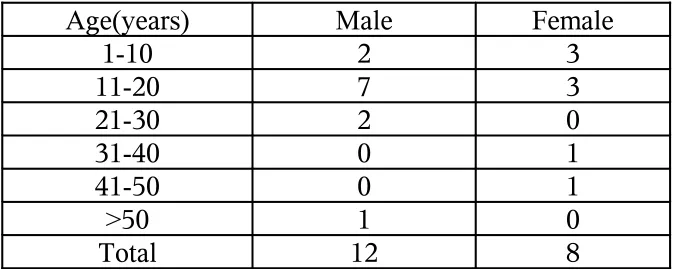
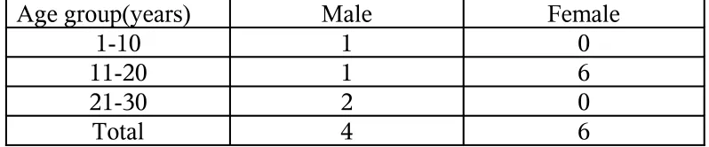
Age and sex distribution among patients with lichen striatus Table -III (n = 20)
Age(years) Male Female
1-10 2 3
11-20 7 3
21-30 2 0
31-40 0 1
41-50 0 1
>50 1 0
Total 12 8
Ten out of twenty patients in this group were in the age group between 10 and 20 years. Eighteen patients were asymptomatic and mainly came for cosmetic reasons, only two patients presented with itching. Eight patients had hypopigmented macules and papules, and another eight patients had skin coloured tiny papules and the remaining had erythematous macules. Fourteen out of twenty cases had an interrupted linear pattern of lesions and in the remaining six cases, lesions were continuous.
Most of these lesions were non scaly and only five cases had mild scaling. The duration of the lesions ranged from 1 week to 15 years, with an average of 4 months. Among these 20 cases, four patients had 2nd and 3rd
[image:54.612.156.495.125.260.2]The lesions were present mainly over the extremities and length varying from 4cms to 30cms, with a mean length of 20cms.
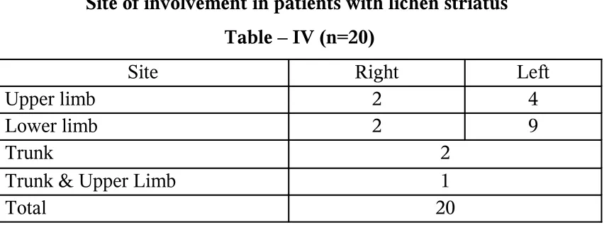
Site of involvement in patients with lichen striatus Table – IV (n=20)
Site Right Left
Upper limb 2 4
Lower limb 2 9
Trunk 2
Trunk & Upper Limb 1 Total 20
Most of the cases had unilateral distribution predominantly over the left side.
Some of the associated skin conditions seen in these patients included Tinea versicolor, varicose veins, xerosis of hands and feet and photosensitivity.
[image:55.612.113.539.143.302.2]Skin biopsy was done for thirteen cases. Nine cases showed a chronic dermatitis picture consisting of mild to moderate acanthosis, mild spongiosis, and perivascular lymphohistiocytic infiltrates in the dermis. Two cases showed psoriasiform dermatitis like picture consisting of mild hyperkeratosis, regular acanthosis, and sparse inflammatory infiltrates in the upper dermis and blood vessels with sparse inflammatory cells seen in the mid dermis. Two cases showed lichenoid dermatitis like picture consisting of flaky hyperkeratosis, mild to moderate acanthosis, indistinct dermo-epidermal junction, and subdermo-epidermal collections of chronic inflammatory infiltrates.
LINEAR LICHEN PLANUS
In this study group seventeen patients presented with Linear lichen planus. The age group ranged between 2 years and 62 years with an average of 20 years.
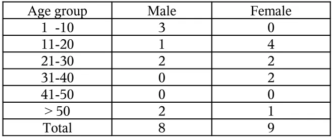
Age and sex distribution in patients with linear lichen planus Table –V (n =17)
Age group Male Female
1 -10 3 0
11-20 1 4
21-30 2 2
31-40 0 2
41-50 0 0
> 50 2 1
Total 8 9
Fourteen out of the seventeen cases, presented with itching with duration of symptom ranging from 3 days to 3 years.
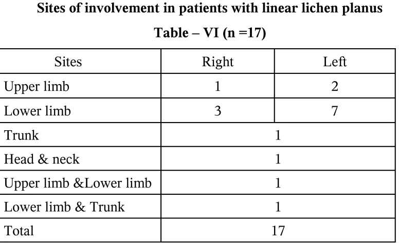
[image:57.612.150.480.167.304.2]Sites of involvement in patients with linear lichen planus Table – VI (n =17)
Sites Right Left
Upper limb 1 2
Lower limb 3 7
Trunk 1
Head & neck 1
Upper limb &Lower limb 1
Lower limb & Trunk 1
Total 17
The lesions were common over the lower extremities especially the left. One patient presented with multiple linear lesions over the trunk and extremities.
Associated conditions
1. Becker’s nevus 2. Tinea versicolor 3. Insect bite allergy 4. Eczema
5. Post inflammatory hyper pigmentation 6. HBsAg sero positivity
[image:58.612.138.526.73.310.2]Ten out of the seventeen patients’ were biopsied from the recent lesions. Nine specimens showed the classical features of Lichen planus like orthokeratosis, focal hypergranulosis, saw toothed rete ridges, irregular acanthosis, basal cell degeneration, band like lympho histiocytic infiltrates hugging the epidermis and pigment incontinence. Two specimens showed the classical colloid bodies.
The remaining one had the features of lichenoid dermatitis like basal cell degeneration, superficial mononuclear cell infiltrate in upper dermis, colloid bodies and pigment incontinence.
LINEAR EPIDERMAL NEVUS
Age and sex distribution among the patients with
Linear epidermal nevus -Table -VII (n = 17)
Age group Male Female
1 - 10 1 2
11- 20 3 6
21- 30 3 0
31- 40 0 2
Total 17
5 out of 17 had lesions since birth. Other cases developed lesions later in life (mostly 1 yr to 18 years).
All patients except one had gradual progression of lesions.14 out of 17 cases presented with hyperpigmented, verrucous papules and plaques which coalesced to form a linear pattern and 3 of them had smooth, hyperpigmented papules and the remaining 3 had erythematous lesions.
Among these 17 patients, 4 were born of 2nd and 3rd degree
Sites and involvement in patients having Linear epidermal nevus
Table – VIII (n=17)
Sites Right Left
Upper limb 2 1
Lower limb 1 0
Trunk 2
Head & Neck 5
Lower limb & Trunk 2
Upper limb & Lower limb & Trunk 1
Upper limb & Trunk 1
Total 17
The nevi were more common over the face and neck followed by extremities. Four out of seventeen cases had nevi involving trunk implicating systematized form of verrucous epidermal nevus.
Neurological and ophthalmological evaluations were normal for all patients. One had associated Nevus sebaceous of Jadassohn over the scalp and another had scabies.
increased pigment basal layer, and patchy inflammatory infiltrates in upper dermis. Two of them showed the features of ILVEN like hyperkeratosis with foci of parakeratosis, moderate acanthosis, elongation and thickening of the rete ridges with a ‘psoriasiform’ appearance, papillomatosis, and slight spongiosis with exocytosis of lymphocytes.
LINEAR MORPHOEA
In this study 10 cases presented with Linear Morphoea in the age group between 4 and 22 years with an average of 20.
Out of the 10 cases 4 were males and 6 were females forming a Male: Female ratio of 4: 6.
[image:62.612.99.498.508.591.2]Age and sex distribution in patients with Linear Morphoea
Table – IX (n =10)
Age group(years) Male Female
1-10 1 0
11-20 1 6
21-30 2 0
The lesions started in early childhood or in adolescence. The duration of lesions varied from 1 year to 4 years. Nine out of the ten cases presented without any specific complaints and the remaining one had pain over the Morphoea plaque. Only one patient had prior history of intramuscular injection over the lesional site.
Only two cases were born of consanguineous marriage (second degree consanguinity). There was no family history of similar illness.
Three patients had lesions involving the lower limbs (left side) while all the other patients had lesions over the head and neck
The lesions were skin coloured to brownish, atrophic, indurated plaques of the size of 3cms to 25cms arranged in a linear pattern following Blaschko’s lines. Three cases of Linear Morphoea were found to be fixed to the underlying structures. All of them had hair loss over the plaques. None of them showed either mucous membrane or nail involvement.
eosinophilic, oedematous collagen bundles in upper dermis and cut section of eccrine duct and arrector pilorum muscle.
Two specimens showed the features consistent with Late Morphoea like atrophic epidermis, collagen bundles appeared homogenous, thickened, and hypo cellular, atrophic eccrine glands narrowed blood vessels and elastic fibers are thickened and arranged parallel to epidermis and collagen fibers. LINEAR PSORIASIS
SEGMENTAL VITILIGO
In this study group four male patients presented with segmental vitiligo between the age group of 8 years and 19 years. Two patients had lesions over the upper limb, one had over the lower limb and the other over the face.
The lesions were isolated macules to patches of size 0.5cm to 10cms. All the lesions were unilateral in distribution along the lines of Blaschko. The duration of lesions varied from 4 months to 1 year. The lesions were asymptomatic in all patients. None of them had vitiligo elsewhere or any other associated auto immune disorders.
Two patients had leukotrichia over the vitiligo patches. Examination of nails and mucosa were found to be normal.
DISCUSSION
In this study of 70 cases with a linear distribution of the lesions which did not exhibit Koebner’s phenomenon, none of the cases seemed to follow the linearity determined by the Nerves, vascular or lymphatic structure and it has been suggested that these lesions develop in the lines of Blaschko.
Hence the various nevoid and acquired conditions which are supposed to follow the lines of Blaschko, which are thought to be due to a form of human mosaicism were included in this study. Most of the patients were asymptomatic and mainly came for cosmetic reasons, except the Linear lichen planus patients and few of Lichen striatus patients who had attended our skin Out Patient Department for intense itching.
LICHEN STRIATUS
Lichen striatus formed the majority of cases amounting to 20 in this study. The condition is said to occur commonly in the age group of 5 -15 years, where as in the present study, majority of patients were in the age group between 10 – 20 years. It was more commonly observed in males in this study group with a Male: Female ratio of 3: 2 as also been documented by Hauber et al82.The lesions are normally asymptomatic, with occasional
predisposing factors in any of the patients, as Lichen striatus is of unknown etiology.
Most of the patients in the study group had lesions over the extremities, but few patients had lesions also over the trunk as recorded in the literature, (25, 26) which complied with the variable sites of expression. All
the patients had unilateral distribution of the lesions. None of the patients showed nail changes among this group, although changes in the form of longitudinal ridging, subungual hyperkeratosis, splitting and onycholysis have been documented.
Atopy was found to be associated with Lichen striatus in 80% of patients, although none in this study group had personal or family history of atopy but associated lesions like Tinea versicolor, xerosis and photosensitivity were seen, which were not documented so far and may be coincidental.
There were no systemic abnormalities noted in any of the Lichen striatus patients in this study group.
LINEAR LICHEN PLANUS
Linear lichen planus formed the next common condition in this study group consisting of 17 cases. Among them most of the patients were in the age group between 11- 40 years and the average age at the time of diagnosis was 25 years. In this study Male: Female ratio of 8: 9 was noted, showing a slight female preponderance as recorded in the literature83.
There was no history of contact with any chemicals or trauma but 2 patients had history of intake of NSAID and another 2 were treated for chronic suppurative otitis media prior to the skin lesions.
There was no history of similar lesions in their family members or any other associated autoimmune disorders. It has been suggested that the linear distribution seen could be due to the tendency of Lichen planus to develop, with the formation of a clone of predisposed or vulnerable cells, which is predetermined during embryogenesis,
no nail changes or mucous membrane involvement suggesting that they were cases of isolated Linear lichen planus.
In most of the cases, the lesions were found on the extremities. Two patients showed multiple linear lesions following the lines of Blaschko, but no immuno compromised state was noted as shown in literature. The length of the lesions ranged from 5cms – 50cms and 2 patients had lesions extending the entire length of the limb.
On histopathological examination, most of the lesions showed the classical features of Lichen planus as described in the literature74 and one
picture showed normal epidermis, basal cell degeneration, superficial mononuclear cell infiltrate in upper dermis, colloid bodies and pigment incontinence which was fit into the features of lichenoid dermatitis.
HBsAg sero positivity was found in one of the patient, which has not been reported in literature. Some of the associated features were Becker’s nevus, Tinea versicolor, Insect bite allergy and Eczema which may be coincidental.
LINEAR EPIDERMAL NEVUS
contrary to equal sex incidence given literature54. The patients with this
disorder mainly came for cosmetic reason. No family history of similar lesion was recorded.
The lesions manifested as hyperpigmented verrucous papules arranged in a linear continuous or interrupted bands and were present since birth in some of the cases, but in most of the patients the lesions became apparent later in life.
In this group 4 patients had lesions involving trunk and extremities, implicating systemized form of verrucous epidermal nevus.
LINEAR MORPHOEA
10 cases of Linear morphoea were recorded, among whom 8 were less than 20 years of age. Generally the peak incidence of this condition is between 20 and 30 years of the age group 64.
Out of the 10 cases, 4 were males and 6 were female patients forming a Male: Female ratio of 4: 6, correlating with the female preponderance of this condition as recorded in the literature64.
There was no history of any provocative factors like trauma or drug intake, etc. prior to the onset of lesion except one who had prior history of intramuscular injection over the lesional site.
There was no history of similar lesion in the family members. Most of the patients presented with an asymptomatic atrophic plaques except one who had pain over the plaque.
Most of the patients had lesions over the head and neck region and only 3 patients had lesions over the lower limb, which is the commonest site of involvement shown in literature84.
LINEAR PSORIASIS
In this study, one had presented with hypopigmented scaly plaque of 6 months duration over the extremity. There was no evidence of any trauma preceding the lesion. The patient had fine, regular pitting over the finger nails. On histopathological examination the lesion showed a characteristic feature of psoriasis, with which, the diagnosis was revised from lichen striatus to linear psoriasis.
Another patient presented with asymptomatic, erythematous plaques over the extremity but on histopathological examination, it was found to have hypergranulosis and orthokeratosis, alternating with absent granular layer and parakeratosis and the other characteristic features of psoriasis.
SEGMENTAL VITILIGO
In this study group, 4 male patients presented with segmental vitiligo between the age group of 8 years and 19 years, which was earlier than the other types of vitiligo.
theory of vitiligo and it could be also a clonal susceptibility of melanocytes to neurons.
CONCLUSION
1. The Incidence of Linear Dermatoses in our skin Out Patient Department,
Govt. General Hospital, Madras Medical College, Chennai during the period of Sep- 2004 to Sep-2006 --- 0.32 %
2. Among the Linear Dermatoses, Lichen striatus was found to be more common
3. The other Dermatoses following Blaschko’s lines, in the descending order of frequency seen in this study were Linear Lichen Planus, Linear Verrucous
Epidermal Nevus, Linear Morphoea, Linear Vitiligo, Linear Psoriasis and Linear Lichenoid Dermatitis.
4. In this study, on the whole, slight Male preponderance was noted.
5. Majority of patients showed unilateral distribution in a linear pattern, more often on the extremities.
7. The lesions were more of a cosmetic concern in most of the cases in this study.
BIBLIOGRAPHY
1. Blaschko A. Die Nervenverteilung in der Haut ihrer Beziehung zu den Erkrankungen der Haut. Wien: Wilhelm Braumuller, 1901.
2. Jackson R. Blaschko’s lines: a review and reconsideration of observations on the cause of certain unusual linear conditions of the skin. Br. J Dermatol 1976; 95:349-60.
3. Langer K. On the anatomy and physiology of the skin. I. the cleavability of the
cutis. Br. J Plast Surg 1978; 31:3-8. (Translation of Zur Anatomie und
Physiologie der Haut. I. Uber die Spaltbarkeit der Cutis. Sitzungbericht der
Mathematisch-naturwissenchaftlichen Classe der Kaiserlichen Academie der
Wissenchaften, 1861; 44:19.)
4. Siemens HW. Extent, shape and distribution. In:Siemens HW, ed General diagnosis and therapy of skin diseases. Chicago: University of Chicago Press, 1958:137-9. (Trans by K Wiener.)
5. Tessier P. Anatomical classification of facial, cranio-facial and laterofacial clefts. J maxillofac Surg 1976; 4:69-92.
Selmanowitz VJ, Krivo JM. Pigmentary demarcation lines: comparison of Negroes with Japanese. Br J Dermatol 1975; 93:371-7. 6. Keegan JJ, Garrett FD. The segmental distribution of the cutaneous
nerves in the limbs of man. Anat rec 1948; 102:409-37.
7. Rowland Payne CME, Branfoot AC. Wallae’s line. Br J Dermatol1986; 114:513-4.
8. Jackson FW, Tension lines, cleavage lines and hair tracts in man. J Anat Lond 1941; 75:248-50.
10. Happle R. Absence de bipolarite dans les lignes de Blaschko.[Letter] Ann Dermatol Venereol 1990;117:397.
11. Brown HM, Gorlin RJ. Oral mucosal involvement in nevus unius lateris (ichthyosis hystrix): a review of the literature and report of a case. Arch Dermatol 1960; 81:509-15
12.Lang GE, Rott H-D, Pfieffer RA. X linked ocular albinism: characteristic pattern of affcection in female carriers. Ophthal Paediatr Genet 1990; 11:265-71.
13.Maguire AM, Maumenee IH. Iris pigment mosaicism in carriers of X-linked ocular albinism cum pigmento. Am J Ophthalmol 1989; 107:298-9.
14.Koniszewski G, Rott H-D. Der Lyon-effekt an der lines: Konduktorinnenbefunde bei X-chromosomal gebundener katarakt und beim Lowe-syndrome. Klin Monatbsl Augenheilkd 1985; 187:525-8. 15. Happle R, Kuchle HJ. Sectorial cataract: a possible example of
lyonisation. Lancet 1983; 2:919-20.
16. Happle R. Mosaicism in human skin: understanding the pattern and mechanism. Arch Dermatology 1993; 129:1460-70.
17.Curth Ho. Warburton D. The genetics of IP. Arch Dermatol 1965; 92:229-35.
18.Happle R. Genetische Bedeutung der Blaschkosclen Lineiu Z Hautke 1977; 52:935-44.
19.Moss C, Savin J, Glossary. In:Dermatology and the New Genetics, Blackwell Science, Oxford 1995; 18691.
20. Happle R: New aspect of cutaneous mosaicism. J Dermatol 29: 681-692, 2002
22.Text Book of Dermatology, L. Bolognia / Joseph L Jorizzo, First edition .Vol.1; 872
22. C.M. Lawrence and N.H. Cox, Physical signs in Dermatology, 2nd
edition,London; Mosby, 2002(2).
24. Text Book of Dermatology, L. Bolognia / Joseph L Jorizzo, First edition.
Vol.1; 869, 870
25. Taieb A, El Youbi A, Grosshans E, et al. Lichen striatus: a Blaschko linear acquired inflammatory skin eruption. J Am Acad Dermatol 1991; 25:637-42.
26. `Sittart JA, Pegas JR, Sant’Ana LA, et al. Lichen striatus: epidemiologic study. Med Cutan Ibero Lat Am 1989; 17:19-21.
27. Rook’s Textbook of Dermatology-Tony Burns/ Stephen Breathnach, 7th Edition – Vol 1; P17.43.
28. Kennady, D, Rogers, M (1996) Lichen striatus, Clin lab Invest, 13, 95-96.
29. Sittar, J.A., Pegas, J.R.,Sant Ana, L.A.et al (1989) Lichen straitus; epidemiologic study. Med Cuten Iber Lat Am, 17, 19-21.
30. Taieb,A., El Youbi, A., Grosshans, E ,et al (1991).Lichen straitus:a Blaschko linear acquired inflammatory skin eruption. J Am Acad Dermatol, 25, 637-642.
31. Rook’s Textbook of Dermatology-Tony Burns/ Stephen Breathnach, 7th Edition – Vol 1; P17.44.