Presentation, Etiology and Risk Factors in
Patients with Focal and Segmental
Glomerulosclerosis
A dissertation submitted to the Tamil Nadu Dr. M.G.R.Medical
University in partial fulfillment of the University regulations for the
award of
D . M . D e g r e e ( N e p h r o l o g y )
DEPAR T M ENT O F NEP HROLOGY
Certificate
Certificate
This is to certify that “Presentation, Etiology and Risk
Factors in Patients with Focal and Segmental
Glomerulosclerosis” which is submitted as thesis
requirement of the DM Branch III examination of the
Dr. MGR Medical University is the bonafide work of
the candidate:
Dr. Manish Gupta
Dr. George T. John,
Professor and Head,
Acknowledgement
With a sense of deep regard, I offer my heartfelt gratitude to my guide, Dr. George T. John, under whose guidance and strict supervision this dissertation is made possible. He helped me in
formulating and executing the protocol and took a lot of effort in going through the analysis of the data and the text of the dissertation.
I am indebted to my teacher and former Head of our Department Dr. Chakko K. Jacob for his valuable suggestion, encouragement and guidance throughout the study.
I express my sincere thank to Dr V. Tamilarasi for extending her support and kind co-operation
in this study.
I thank Nithya Neelakantan, my Biostatistician who took extraordinary pains in analyzing the
data and without her help the analysis would have been impossible.
I acknowledge the co-operation of all my colleagues, friends and my family who obliged me whenever needed.
Contents
Contents
TITLE
PAGE NO.
List of Tables
i
List of Figures
ii
Abstract
iii
Introduction
1
Review of Literature
3
Aims
35
Patients and methods
36
Results
40
Discussion
52
Conclusions
55
Bibliography
56
Appendix 1 - Proforma
65
List of Tables
List of Tables
Table
No.
Title
Page
No.
1.
Etiologic Classification of FSGS
4
2.
Demographic profile at diagnosis
40
3.
Clinical, laboratory findings and histopathological
characteristics at biopsy: nephrotics vs
non-nephrotics
42
4.
Clinical, laboratory findings and histolopathological
characteristics of nephrotic patients at biopsy:
progressor vs non-progressor
44
5.
Multivariate analysis of prognostic factors: low
albumin
45
6.
Multivariate analysis of prognostic factors: normal
albumin
46
7.
Multivariate analysis of risk factors for progression
to ESRD: (Logistic regression model)
47
List of Figures
List of Figures
Figure
No.
Title
Page
No.
1.
Study design
39
2.
Cumulative probability of renal survival vs risk
factors: progressor vs non-progressor
48
3.
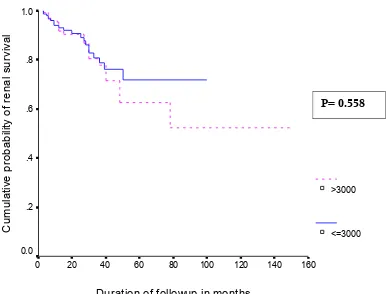
Cumulative probability of renal survival:
nephrotic vs non- nephrotic
49
4.
Cumulative probability of renal survival vs risk
factors: degree of interstitial fibrosis
50
5.
Cumulative probability of renal survival:
additive risk
51
Objectives:
To identify prognostic factors that predict the risk of progression of renal failure in adult patients at the time of diagnosis of primary focal and segmental glomerulosclerosis (FSGS) and to
calculate cumulative renal survival rate based on identified factors.
ii
Focal and segmental glomerulosclerosis (FSGS) is defined as a clinical-pathologic syndrome manifesting proteinuria, usually of nephrotic range, associated with
lesions of focal and segmental glomerular sclerosis and foot process effacement. Although hyaline insudation is common, the condition lacks glomerular immune complex deposits. Early in the disease process, the pattern of glomerular sclerosis is focal,
involving a subset of glomeruli, and segmental, involving a portion of the glomerular tuft. As the disease progresses, a more diffuse and global pattern of sclerosis evolves.
Alterations of the podocyte cytoarchitecture constitute the major ultrastructural findings. FSGS came to be viewed as a distinct pathologic entity by 1970. In recent years it has become clear that FSGS is seen more usefully as a clinicopathologic syndrome,
comprising diverse distinct diseases with different etiologies. These include genetic mutation (likely showing both Mendelian and non-Mendelian modes of inheritance), viral
infection, relative overabundance of a pathogenic plasma factor, and hyperfiltration injury. Doubtless, there are other disease entities within the FSGS syndrome that remain undefined.
Among the biopsy proven renal disease in adults, focal and segmental glomerulosclerosis is the commonest histological category in all age groups (16.8%)1.
Although the pathogenesis of primary focal and segmental glomerulosclerosis (FSGS) is unknown, it is one of the most common causes of primary glomerular disease that terminate in end stage renal disease (ESRD). In most series, the 10-year survival is
between 40-60%.2-5
Various studies have shown that clinical, biochemical and pathological indicators
presentation, gender, amount of proteinuria, hypertension, renal dysfunction, percentage of sclerosed glomeruli and interstitial fibrosis, location of segmental sclerosis and
therapeutic response to steroids6,7. Whether these prognostic factors and survival rate
mentioned in the western literature are relevant to Indian population is unknown.
In the present study, we analysed the clinical, laboratory, and histopathological
Fahr (1925) showed that patients with lipoid nephrosis who progressed to renal failure showed focal glomerular damage. Rich (1957) examined autopsy tissue from 20
children with nephrotic syndrome and otherwise typical lipoid nephrosis and described progressive sclerosis of glomeruli, affecting first the juxtamedullary glomeruli.
McGovern (1964) and Hayslett et al8 (1969) showed that in some patients whose initial
biopsy examination showed minimal changes, a later biopsy examination showed focal sclerosing glomerulonephritis. Churg et al9 (1970), writing for the International Study of
Kidney Disease in Children, described biopsy examination findings in 127 children with nephrotic syndrome and found that focal sclerosing glomerular lesions were the second
most common finding, after minimal changes.
The diagnosis of FSGS is complicated by the existence of a primary (or idiopathic) form and many secondary forms. Secondary FSGS caused by
structural-functional adaptations mediated by intrarenal vasodilatation and by increased glomerular capillary pressures and plasma flow rates. Such maladaptive glomerular hemodynamic
alterations can arise through: (1) a reduction in the number of functioning nephrons (such as after unilateral renal agenesis, surgical ablation, oligomeganephronia, or any advanced primary renal disease), or (2) mechanisms that place hemodynamic stress on an initially
normal nephron population (as in morbid obesity, cyanotic congenital heart disease, and sickle cell anemia). Finally, primary and secondary FSGS also must be differentiated
Table 1
.Etiologic Classification of FSGS
Primary (idiopathic) FSGS C1q nephropathy
HIV-associated nephropathy Heroin nephropathy
Familial FSGS
Mutations in α-actinin 4 (autosomal dominant) Mutations in podocin (autosomal recessive) Mitochondrial cytopathies
Drug toxicity
Pamidronate Lithium Interferon-α
Secondary FSGS
Reduced renal mass Oligomeganephronia Unilateral renal agenesis Renal dysplasia
Reflux nephropathy
Sequela to cortical necrosis Surgical renal ablation
Any advanced renal disease with reduction in functioning nephrons Chronic allograft nephropathy
Initially normal renal mass Diabetes mellitus
Hypertension Obesity
Cyanotic congenital heart disease Sickle cell anemia
Nonspecific pattern of FSGS caused by renal scarring
Hereditary nephritis Diabetic nephropathy
Hypertensive arterionephrosclerosis Membranous glomerulopathy Thrombotic microangiopathies
Pathologic Classification:
FSGS comprises a number of morphologic subtypes that may have different
prognostic and therapeutic implications. These morphologic variants were defined at a recent consensus conference of renal pathologists in New York City.
FSGS not otherwise specified (NOS) constitutes the generic lesion of FSGS. The
synonyms classic FSGS or FSGS of the usual type often are applied. This category requires that other morphologic categories (perihilar, cellular, tip, and collapsing) be
excluded. FSGS (NOS) is the most common morphologic pattern of FSGS. Evidence from repeat biopsy examinations suggests that other variants may evolve into this pattern in the course of disease progression and increasing chronicity.
Morphologically FSGS (NOS) is characterized by Lesions of sclerosis can affect the perihilar (ie, vascular pole) region or the periphery of the tuft. In some glomeruli,
segmental lesions may affect more than one lobule, involving both the perihilar and peripheral regions. According to one study using serial sections, peripheral lesions tend to be more common in childhood FSGS than the adult disease.10 Any number of glomeruli
can be affected by segmental sclerosis, with or without associated global sclerosis. There may be segmental glomerular basement membrane collapse without podocyte
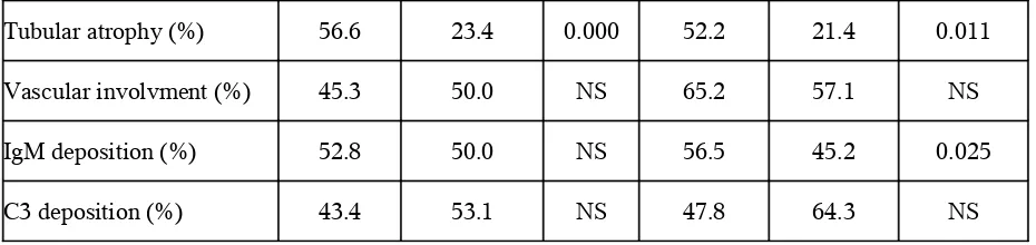
By immunofluorescence, there is typically focal and segmental granular deposition of IgM, C3, and more variably C1 in the distribution of the segmental
glomerular sclerosis and hyalinosis . More generalised weak mesangial deposition of IgM also may be present.
By electron microscopy, the lesion of segmental sclerosis display wrinkling and
retraction of glomerular basement membrane and accumulation of inframembranous hyaline, with resulting narrowing or occlusion of the glomerular capillary lumina. The
electron dense hyaline material is usually more waxy in appearance than true immune complex deposits and tends to pool beneath the GBM, conforming to the contours of the delimiting membrane. Endocapillary foam cells appear as large intracapillary cells
containing abundant electron lucent vacuoles.
Directly overlying the lesions of segmental sclerosis there usually is complete
effacement of foot processes, accompanied by podocyte alterations that include hypertrophy, increased organellar content, and focal microvillous transformation. This microvillous appearance is caused by the formation of slender cellular projections
resembling villi along the surface of the podocytes facing the urinary space. The hypertrophied podocytes display rounded cell bodies that adhere smoothly to the
glomerular basement membrane (GBM), with frequent loss of primary processes.
The major ultrastructural finding involving nonsclerotic glomerular capillaries is foot process effacement. The degree of foot process fusion observed overlying these open
glomerular capillary surface area. In general, the degree of fusion correlates roughly with the severity of the proteinuria, such that patients with subnephrotic proteinuria tend to
have less foot process fusion than those who are fully nephrotic. In the areas of foot process effacement, there usually is loss of recognizable slit diaphragms and mat-like condensations of cytoskeletal filaments oriented parallel to the direction of the GBM
itself. Thus, although the lesions of FSGS are focal at the light microscopic level, the podocyte alterations are relatively diffuse at the electron microscopic level
FSGS perihilar variant category requires that the cellular variant, tip variant, and collapsing variant be excluded. It is defined by the presence of perihilar sclerosis and hyalinosis involving greater than 50% of segmentally sclerotic glomeruli.
Glomerulomegaly and adhesions are common. Podocyte hypertrophy and hyperplasia may be present but typically are less frequent than in the other variants. Other glomeruli
may show lesions of segmental and/or global glomerulosclerosis, as described for FSGS (NOS) earlier.
Perihilar variant of FSGS may occur in primary FSGS. However, when
accompanied by glomerulomegaly, it is particularly common in patients with secondary forms of FSGS mediated by an adaptive response to increased glomerular capillary
The cellular variant of FSGS was first described by Schwartz and Lewis in 1985.
The cellular variant is defined by the presence of at least one glomerulus with segmental
endocapillary hypercellularity involving at least 25% of the tuft and causing occlusion of the capillary lumen. Any segment (perihilar or peripheral) may be affected. When numerous glomeruli are affected, the process may mimic focal proliferative
glomerulonephritis.11
By immunofluorescence there is focal and segmental glomerular positivity for
IgM and C3. At the ultrastructural level, the cellular variant usually displays severe foot process effacement, correlating with the generally high levels of proteinuria . Cellular lesions consist of segmental occlusion of glomerular capillaries by endocapillary
hypercellularity including foam cells and monocytes. The glomerular basement membrane is intact, without evidence of rupture.
Compared with FSGS (NOS), the cellular variant is characterized by more severe proteinuria and a shorter time course from clinical onset of renal disease to biopsy examination, suggesting an early phase in the evolution of the segmental sclerosis.
Schwartz and Lewis found a shorter interval between onset of proteinuria (3.4 versus 71.9 mo) in patients with cellular versus classic FSGS. Moreover, 90% of patients in the
cellular group had urine protein levels greater than 3 g/d, compared with 49% of those without cellular lesions. Similarly, the incidence of full nephrotic syndrome at presentation was significantly higher (70% versus 23%). Patients with cellular FSGS
The cellular variant may be responsive to immunosuppressive therapy.12 This
favourable treatment response probably relates to the early and relatively active stage of
glomerular injury in the cellular variant.
The tip variant of FSGS is defined by the presence of at least one glomerulus with a segmental lesion involving the tip domain (ie, the peripheral 25% of the glomerular tuft
next to the origin of the proximal tubule). There must be either adhesion between the tuft and Bowman's capsule at the tubular lumen or neck, or confluence of podocytes with
parietal epithelial or tubular epithelial cells at the tubular pole or neck.
Tip lesions may arise as a nonspecific response of the peritubular segment of the glomerular tuft to fluxes of protein-rich filtrate in the setting of nephrotic syndrome.
The designation of collapsing variant is applied to cases of FSGS in which at least one glomerulus displays segmental or global obliteration of the glomerular capillary
lumina by wrinkling and collapse of GBMs associated with podocyte hypertrophy and hyperplasia. Collapse involving a single glomerulus is considered significant, such that the presence of any glomerular collapse pre-empts the other morphologic categories of
FSGS.
There are 2 pathologic findings that together define the collapsing variant of
FSGS.13,14 First, there is implosive wrinkling and retraction of the GBM. Second, there is
epithelial cells frequently contain protein resorption droplets and may appear detached from the underlying GBM. Lesions of collapsing FSGS often coexist in the same biopsy
specimen with other patterns of FSGS, including cellular lesions and discrete segmental scars typical of the classic form of FSGS.
The changes seen in collapsing FSGS are not confined to glomeruli. There are
typically widespread tubular degenerative changes including luminal ectasia, cytoplasmic simplification and vacuolization, loss of brush border, nuclear pleomorphism with
prominent nucleoli, and multiple mitotic and apoptotic figures. Proximal tubules also display protein resorption droplets. In almost half of cases, tubular microcysts are seen and typically display a proteinaceous filtrate that stains positively with the periodic acid
Schiff stain. Interstitial edema and a mild to moderate chronic inflammatory infiltrate characteristically accompany the tubular degenerative changes. Rare foci of mild tubulitis
may be apparent. With progression of disease, tubular atrophy and interstitial fibrosis intervene.
In the setting of collapsing FSGS, immunofluorescence typically reveals
positivity for IgM and C3 in the distribution of the lesions of FSGS. The intensity of staining is typically trace to 1+ but may be up to 2+ (scale: 0, trace, 1-3+). Staining for
IgG, IgA, and κ and λ light chains is negative.
vesicles, microvillous transformation, lipid and protein resorption droplets, and extensive foot process fusion. Although the podocyte changes normally are diffuse, glomeruli
containing lesions of collapsing FSGS will display more prominent podocyte changes as well as wrinkling and retraction of the GBM. Within the capillary lumina, focal hyaline insudation and rare endocapillary foam cells may be apparent. Electron dense deposits
typically are absent although rare mesangial deposits should not be a deterrent from making the diagnosis of collapsing FSGS.
Trends in the Epidemiology of FSGS:
FSGS is currently a leading cause of nephrotic syndrome in adults. Previous studies from the 1970s and early 1980s list FSGS as being responsible for 15% to 20% of
cases of idiopathic nephrotic syndrome (NS) in adults. Korbet et al15 found that among
primary renal diseases, FSGS accounted for 29% of all adult patients presenting with nephrotic range proteinuria between 1975 and 1985, but accounted for 38% of such
patients between 1985 and 1994. Hass et al also observed similar increases in the proportion of FSGS in adults undergoing a biopsy examination for proteinuria or
nephrotic syndrome.16 The incidence of FSGS as a cause of end-stage renal disease
(ESRD) also is increasing. The increase in the fractional contribution of FSGS to the etiology of primary nephrotic syndrome could also be caused by changes in the
demographics of patients undergoing renal biopsy examinations or a decline in the incidence of other diseases such as membranous glomerulopathy or minimal change
Racial background has a strong influence on the propensity to develop FSGS. Black subjects have
an increased risk for idiopathic FSGS, as has long been recognized.17 In a logistic regression analysis of
adult patients with nephrosis, race remained the only significant predictor for FSGS, with black subjects 4
times more likely to have FSGS than white subjects.Black individuals are also at increased risk for Human
immunodeficiency virus (HIV)-associated FSGS.18 Occult HIV infection is unlikely to explain the
increasing frequency of FSGS. Although screening for HIV is often not performed in patients with
glomerulonephritis, most studies that show an increasing incidence of FSGS in black individuals exclude
patients with known HIV, intravenous drugs usage, or the presence of tubuloreticular structures. In at least
one study with serologic testing for HIV, a high frequency of idiopathic FSGS also was shown in black
subjects. Other infectious agents such as parvovirus B1919and SV4020 have been linked to the development
of FSGS. Whether these viruses or other as yet unknown infectious agents may contribute to the increasing
occurrence of FSGS in susceptible populations is yet to be determined.
Another consideration is the increase in the collapsing variant of FSGS. Barisoni et al21 found an increasing incidence of FSGS with collapsing features among
HIV-negative patients. This entity was not seen before 1979 and has more than doubled from 11% of all idiopathic FSGS between 1979 and 1985 to 24% from 1990 to 1993. The same group found that patients with collapsing FSGS were predominantly of black race.
The median renal survival from time to biopsy examination to end stage renal disease was shorter in the collapsing group (13 mo) compared with the other FSGS group (63
mo). Other investigators confirmed the higher incidence of the collapsing variant in black subjects and the poor renal outcome associated with this condition.22 However, these
investigators reported that the collapsing variant only accounted for 4% of all FSGS and
progression to renal failure, the collapsing variant likely contributes disproportionately to the FSGS patients reaching end stage kidney disease. This may in part account for a
higher incidence of black subjects with FSGS end stage renal disease.
Genetic Basis of FSGS:
The role of genetic factors in the development of FSGS in humans has become
increasingly apparent in recent years. Genetic studies also have helped strengthen the notion that glomerular visceral epithelial cell (or podocyte) disorders lead to a spectrum of clinical presentations, from congenital nephrotic syndrome (CNF), to minimal change
disease (MCD), and FSGS. Four siblings with nephrotic syndrome were described in a 1957 report. Pathology showed minimal change disease in some children, FSGS in
others. The absence of disease in the parents suggested recessive inheritance. Additional scattered reports of both single-generation and multigeneration disease have continued to appear in the case literature.24
Congenital nephrotic syndrome of the Finnish type, or CNF, is a geographically widespread disease characterized by the development of severe nephrosis in utero and
autosomal-recessive inheritance. Affected neonates have on the order of 20 to 30 g/d proteinuria and typically die from complications of the nephrotic syndrome at a young age. Subsequent to mapping the CNF gene to chromosome 19q13 by a means of a
immunoglobulin C2 motifs, and a single transmembrane segment. Nephrin localizes to the slit diaphragm in the podocyte. Nephrin appears to play a role in regulating signaling
pathways.25
Most of the congenital nephrotic syndrome in Finland is caused by 2 specific NPHS1 mutations, Fin major (the deletion of nucleotides 121-122 leading to a frameshift) and Fin
minor (encoding a premature termination signal at amino acid 1109). The incidence of CNF is 1 in 500 live births in this group.
The identification of NPHS1 has improved the antenatal diagnosis of CNF.
Fuchshuber et al described a form of nephrosis characterized by recessive transmission, early onset, resistance to steroid therapy, and rapid progression to end-stage
kidney failure. The majority of the affected children showed an FSGS pattern on renal biopsy. The gene for this recessive form of FSGS was mapped to chromosome 1q25-31
and subsequently cloned. NPHS2, the responsible gene, encodes podocin, a 383 amino acid integral membrane protein.
Autosomal-dominant forms of FSGS typically present later and are more slowly
progressive than recessive forms. Mutations in ACTN4, encoding α-actinin-4, cause a slowly progressive form of disease with dominant inheritance, nonnephrotic proteinuria,
ACTN4 mutations identified in FSGS families are all missense and increase the affinity of the encoded protein to actin filaments.
FSGS also is seen as part of well-defined inherited syndromes. The spectrum of disease seen with WT1 mutations is the best studied of these disorders. Frasier syndrome and Denys-Drash syndrome are related and overlapping syndromes caused by mutations
in WT1. Both syndromes are characterized by glomerular disease and the development of male pseudohermaphroditism.
Frasier syndrome is caused by donor splice mutations in intron 9 of WT1. Frasier syndrome can present as FSGS in 46, XX females in association with gonadal malignancy.26,27
Denys-Drash syndrome is defined by diffuse mesangial sclerosis on renal biopsy examination, genitourinary tumors, and pseudohermaphroditism. A different spectrum of
mutations is seen in Denys-Drash syndrome, most commonly within exon 9 of WT1.28
Nail-Patella syndrome generally is regarded as a disease of the basement membrane rather than the podocyte, though it is probably both. An altered glomerular
basement membrane typically predominates on histologic analysis, the glomerulopathy is variable and can present as nephrotic syndrome.Defects in the lmx1b transcription factor
Permeability Factors in FSGS:
The theory that idiopathic nephrotic syndrome, minimal change nephrotic syndrome (MCNS) or FSGS represents “a systemic abnormality in lymphocyte function
resulting in the secretion of a circulating chemical mediator toxic to an immunologically innocent glomerular basement membrane” was proposed formally by Shalhoub29 in 1974.
This proposal was based on the absence of evidence for a humoral antibody response, on clinical remissions after treatment with steroids or cyclophosphamide and after measles infection, and on the occurrence of nephrotic syndrome in patients with Hodgkin's
lymphoma. The presence of a circulating factor in FSGS was proposed by Hoyer et al30
after they observed that nephrotic proteinuria recurred promptly after transplantation of a
normal kidney into a recipient who had FSGS as the cause for renal failure. This idea was supported by the observation of Zimmerman31 that injection of serum from a patient with
recurrent FSGS into the aorta of a rat resulted in an immediate onset of proteinuria and
albuminuria. No proteinuria occurred after infusion of sera from a patient with prior FSGS but no posttransplant recurrence or from 10 patients with other forms of nephrotic
syndrome. Treatment of patients with recurrent FSGS in renal allografts with plasmapheresis, immunoadsorption, or low-density lipoprotein apheresis may result in decreased proteinuria and prolongation of allograft survival.
Work to clarify the relationship of a circulating proteinuric factor to FSGS remained dormant until the early 1990s when seminal observations from several centers
recurrent FSGS. In 1994, Savin et al reported that incubation in medium containing serum or plasma from patients with recurrent FSGS increased albumin permeability (Palb)
of glomeruli isolated from normal rats and that the capacity to increase Palb was reduced
by plasmapheresis.32,33 During that same year, other investigators reported that injection
of eluate obtained from plasma of patients with recurrent FSGS and eluted from protein
A-immunoadsorbent material induced albuminuria in rats.34 In vitro testing using
functional assays of permeability based on glomerular volumetric responses to oncotic
gradients has been essential to understand the role of permeability factors in FSGS.
The mechanism by which the FSGS factor increases glomerularalbumin permeability in vitro or
in vivo remains to be elucidated. Increasedglomerular albumin permeability after 10 min of incubation in vitro raises the possibility that the FSGS factor interacts with membrane components of the exposed
glomerular epithelial cells. Glomerular epithelial cellshave been shown to play a crucial role in maintaining
the filtration barrier. The magnitude of the effect of FSGS sera on glomerularpermeability in vitro was
comparable to that of anti-Fx 1a antibody, superoxide, hydroxyl ion, or tumor necrosis factor α. The
rapidity of increase in glomerular permeability suggeststhat the immediate effect of the FSGS factor is
unlikely tobe mediated by metalloproteinase-3 or through charge neutralizationby protamine35, as these
agents required prolonged incubationsof more than 4 h and 1 h, respectively.
It appears that the mechanism by which the FSGS factor increases glomerular permeability
depends on active cellular metabolism rather than charge neutralization. This conclusion is based on the
rapidity of the response, the small quantity of the FSGS factor required, its anionic rather than cationic
charge, and the fact that a number of inhibitors prevent the increase in permeability. The mechanism that is
most clearly documented relies on the action of cyclooxygenase. The permeability barrier is preserved by
the inclusion of indomethacin or the thromboxane synthase inhibitor furegrelate in the incubation medium.
Hyperfiltration and Glomerulosclerosis:
In 1932, Chanutin and Ferris described proteinuria and progressive glomerulosclerosis after major reductions in renal mass in the rat. Subsequent
investigators, notably Shimamura and Morrison, further characterized the experimental disease of the remnant kidney using ultrastructural approaches and described fusion of
epithelial cells as well as glomerular enlargement. Olson et al36 further detailed the
cellular and permselective responses, documenting detachment of podocytes from the basement membrane and loss of filtration size selectivity. These studies all used high
degrees of subtotal renal ablation to induce injury in the remnant glomeruli. Others have shown a graded response to removal of kidney parenchyma with even lesser degrees of
reduction accelerating damage but at a slower rate and with less severe damage. For example, even simple unilateral nephrectomy leads to an increased pace of sclerosis in remaining kidneys. With loss of renal tissue and before they sustain pathologic changes,
the remaining nephrons undergo a process conventionally termed compensatory hyperfunction, whereby their single nephron filtration rates increase and the nephrons
grow. The notion arose that this increase in single nephron filtration or hyperfiltration, though compensatory in the short term, caused subsequent pathology.
In humans, simple unilateral nephrectomy in otherwise healthy individuals seems
to lead to little in the way of adverse renal consequences. Perhaps the most complete study is one of men who lost a kidney owing to trauma during World War II. When these
proteinuria could be discerned. Furthermore, in a subset for whom autopsy tissue was available, no increased prevalence of glomerular injury was notable. As the investigators
cautioned, these servicemen were largely of European ancestry and healthy at the time of their initial loss of a single kidney. However, reviews of individuals who have donated a kidney for transplantation have in general revealed no substantial long-term
consequences. The donors were, of course, screened for serious underlying kidney disease or conditions predisposing to a renal injury. On the other hand, some suggestions
that losses of renal mass perhaps at susceptible periods of development or in susceptible individuals may be associated with subsequent injuries have been recorded. For example, unilateral renal agenesis is a relatively rare congenital condition but it has been associated
with serious proteinuria and sclerosis of the single kidney as an individual ages. Conceivably in this circumstance, the solitary kidney has subtle developmental defects
that render it susceptible to injury. Likewise, progressive damage to the remaining kidney after removal of a contralateral diseased kidney may just reflect unrecognized bilateral disease.However, with more extreme renal surgery, injury may be seen in humans. One
study of subtotal nephrectomy sustained owing to aggressive renal cancer surgery has suggested that sclerotic injury develops in the spared but hypertrophied glomeruli.
Perhaps as in the animal studies, some variations occur among different groups of people and susceptibility to loss of renal mass may be more pronounced in some individuals.
Fairly wide variation in the number of nephrons has been noted in human adult
and Mackenzie have argued that individuals with natively fewer nephrons are predisposed to renal disease, hypertension, and glomerular sclerosis. The concept fits in
part with Osmond's and Barker's hypothesis that lower birth weights predispose to cardiovascular disease in later life.
The potential for glomerular capillary pressures to induce progressive sclerotic
injury seems clear. Numerous studies have implicated all 3 of the major glomerular cell types in this process. Because of their similarities to vascular smooth muscle cells, which
have vigorous responses to arterial hypertension, and because of the prominent mesangial abnormalities with hyperfiltration, investigators have focused on the mesangial cell's response to altered glomerular hemodynamics.
Mesangial cells, when grown in culture on a pliable matrix, proliferate in response to stretching. This model simulating increased glomerular tension displays, in addition to
cellular hyperplasia, increased production of matrix substances such as collagens, laminin, and fibronectin. Cells along the periphery of cyclically stretched membranes, where deformation is most pronounced, develop the greatest degree of proliferation and
matrix production. This finding further supports the view that increased physical stretching of the mesangial cells contributes to responses reminiscent of the fundamental
processes of sclerosis in vivo
sclerosis, the failure of the mesangial cells to provide a tethering function to the overlying basement membrane in conjunction with podocyte insufficiency bares basement
membrane, which provides not only an egress for protein, but also a point at which the parietal epithelial cells may adhere to this exposed basement membrane.
After these seemingly adaptive increases in function, pathologic changes appear,
eventuating in glomerular sclerosis. Among the determinants of increased single-nephron filtration after renal mass reduction, the increase in glomerular capillary pressure seems
to be pre-eminent in aggravating the progressive sclerotic changes. Actions of the renin-angiotensin-aldosterone system probably underlie many of these hemodynamic changes. Other vasoactive systems also are likely to exert actions in the process but no one
element of these systems has yet proven to be a necessary component of heightened pressures. Various cellular responses follow from these higher glomerular pressures,
especially in association with the hypertrophy of the glomerular unit. Changes in mesangial cell function, relative deficiency of podocytes, and perhaps endothelial generation of vasoactive and fibroproliferative cytokines, all have been linked in the
chain connecting hemodynamic changes with glomerular sclerosis.
Human Immunodeficiency Virus (HIV) and FSGS:
Human immunodeficiency virus-associated nephropathy (HIVAN) was first
described by Rao et al37 in 1984 in 10 patients with acquired immune deficiency
moderate to massive proteinuria and all developed end-stage renal disease within 16 weeks. Biopsy examinations revealed focal segmental glomerular nephritis. Six of the 11
patients had no known risk factors for renal disease, and the association was made between infection with HIV-1 and a characteristic nephropathy.
The classic pathologic lesion of HIVAN is focal segmental glomerulosclerosis
with collapse of the glomerular tuft, associated with dilated tubules with microcysts. Tubuloreticular inclusions, once commonly observed by electron microscopy in as many
as 25% of patients, has become a rare finding, possibly owing to more effective therapy. Clinically, HIVAN patients present with heavy proteinuria and hypoalbuminemia. HIVAN patients also typically are normotensive. The absence of hypertension may be the
result of a tubular defect in fluid and electrolyte handling. Ultrasound shows enlarged, echogenic kidneys. The degree of azotemia varies, but HIVAN always has been
characterized by a rapid deterioration of renal function.
In early studies of the natural history of HIVAN, time from diagnosis of HIVAN to initiation of hemodialysis was reportedly from weeks to months, with most patients
dead within a year. More recent data show that despite improved survival of HIV-seropositive patients in general, patients with ESRD secondary to HIVAN have higher
mortality rates than those with ESRD of other causes. In a review of USRDS data from 1992 to 1997, 2-year survival was 36% for patients with HIVAN compared with 64% for all other patients with ESRD.HIVAN initially was believed to be a late manifestation of
counts and a history of opportunistic infections. There are now reported cases, however, of HIVAN developing in patients at the time of seroconversion, indicating that it also
may present early in the course of HIV.38,39
Renal biopsy examinations of patients have shown both proliferative and apoptotic changes. The primary process is uncertain, but because kidneys are enlarged,
proliferation is most likely the predominant process.
Transforming growth factor-β (TGF-β) is a fibrogenic cytokine that regulates
human immune function and has been shown to regulate HIV replication. A study of human kidneys found increased deposition of matrix proteins and increased levels of TGF-β in those kidneys with HIVAN compared with normal kidneys and with kidneys
with thin basement membrane and minimal change nephropathies. This was true even when compared with kidneys of HIV infected individuals without HIVAN.40
In a study comparing HIV transgenic mice with normal mice, transgenic kidneys had increased basic fibroblast growth factor (bFGF) and a greater number of bFGF binding sites. Transgenic tubular epithelial cells were found to express bFGF and
transgenic epithelial cells or nontransgenic cells treated with bFGF exhibited an increased rate of proliferation compared with controls.41
control sera. This effect was concentration dependent, and was inhibited in the presence of azidothymidine
Renal epithelial cells of transgenic mice42 and biopsy specimens from a human
subject with HIVAN43 showed an increased expression of markers of proliferation such as
the Ki-67 antigen, and a decreased expression of markers of differentiation such as
synaptopodin.
Collapsing Glomerulopathy or Malignant FSGS:
Collapsing glomerulopathy, also known as collapsing FSGS or malignant FSGS,
initially was described as a distinct clinicopathologic entity in 1986, when Weiss et al44
reported a group of 6 patients with nephrotic syndrome, rapidly progressive renal failure,
and glomerular collapse. Renal biopsy examination displayed the characteristic features of collapsing glomerulopathy: segmental and global collapse of the glomerular capillaries, wrinkling and retraction of the glomerular basement membrane, and marked
hypertrophy and hyperplasia of podocytes. Tubulointerstitial changes also were prominent and included tubular dilatation and degeneration, epithelial necrosis, and
interstitial fibrosis and edema. In this initial report, all 6 patients were black, and all presented with nephrotic syndrome and varying degrees of renal insufficiency. Five of the 6 patients also had a nonspecific febrile illness before presentation, but no clear
Most cases of collapsing glomerulopathy are either HIV-associated or idiopathic. In addition, a secondary cause of collapsing glomerulopathy is treatment with high-dose
pamidronate, a bisphosphonate used to treat osteolytic bone lesions and hypercalcemia of malignancy. In 2001, 7 patients who were white, HIV negative, and developed collapsing glomerulopathy after treatment of multiple myeloma or breast cancer with pamidronate
were reported. Patients were treated with pamidronate for 15 to 48 months before renal biopsy examination, and 5 of the 7 had received more than the recommended dosage of
90 mg intravenously per month. All patients developed renal insufficiency and nephrotic syndrome, with a mean creatinine level of 3.6 mg/dL and a mean 24-hour urinary protein excretion of 12.4 g/d. Three of 5 patients in whom pamidronate was withdrawn had
stabilization of their renal function.45 The same group later reported a case of collapsing
glomerulopathy in which the patient's proteinuria significantly decreased after withdrawal
of pamidronate but then worsened with reintroduction of this agent.75 Since the original
report of 7 patients, 10 additional cases of pamidronate-associated collapsing glomerulopathy have been seen.46 Other centers also have reported cases of FSGS and
collapsing glomerulopathy after treatment with pamidronate.47,48
Recent studies and case reports have suggested an association between collapsing
glomerulopathy and parvovirus B19 infection.49 This association is intriguing given the
establishment of HIV as a viral cause of collapsing glomerulopathy and the frequent reports of a nonspecific febrile illness before the development of collapsing
glomerulopathy. One study of 40 patients by Tanawattanacharoen et al19 reported a
reaction in patients with idiopathic FSGS and collapsing glomerulopathy compared with patients with membranous nephropathy and minimal change disease. A second study by
Moudgil et al50 analyzed biopsy specimens of 23 patients with collapsing glomerulopathy
for parvovirus B19 DNA by using polymerase chain reaction and compared the results with biopsy specimens of classic FSGS, HIVAN, and controls with other renal diseases.
Parvovirus B19 DNA was detected in 78.3% of biopsy specimens with collapsing glomerulopathy. This data suggest that infection of renal epithelial cells by parvovirus
B19 may be an etiology of collapsing glomerulopathy in susceptible patients.
Glomerular visceral epithelial cell injury underlies the pathogenesis of collapsing FSGS. Podocyte injury is characterized by increased cell turnover and reversion to an
immature state. In a study of 8 patients with collapsing glomerulopathy, Bariety et al51
showed that podocytes detach from the GBM, lose their normal podocyte markers
(vimentin, podocalyxin, and CR1), and acquire macrophage-associated epitopes (KP1, PG-M1, and M 18). Barisoni et al52 studied the expression of podocyte maturity markers
(WT-1, CALLA, C3b receptor, GLEPP-1, podocalyxin, synaptopodin) and the
proliferation marker Ki-67 in 10 cases of idiopathic collapsing glomerulopathy, 8 cases of HIVAN, 5 cases of membranous nephropathy, and 5 cases of minimal change disease.
In patients with idiopathic collapsing FSGS and HIVAN (but not in patients with other glomerular diseases), decreased expression of podocyte markers of maturity and increased expression of Ki-67 was observed. This data suggests that both idiopathic
In collapsing glomerulopathy, expression of cyclin A (a positive cell cycle regulatory protein) is increased while expression of synaptopodin, cyclin D1, and the
negative cell cycle regulatory proteins p27 and p57 are decreased.53 Shankland et al54
evaluated a series of 9 patients with collapsing glomerulopathy, 16 patients with HIVAN, and 37 patients with other causes of nephrotic syndrome. Expression of p27 and p57 was
uniformly decreased in collapsing glomerulopathy, cellular FSGS, and HIVAN.
Clinical Presentation and Course:
FSGS presents with nephrotic syndrome, defined variably by the classic tetrad of
proteinuria, hypoalbuminemia, edema, and hypercholesterolemia, or by the presence of edema and nephrotic-range proteinuria.. However, a significant number of patients
present with isolated proteinuria or with proteinuria and hematuria. The cause of proteinuria is uncertain. As is true for minimal change nephrotic syndrome (MCNS), proteinuria in FSGS occurs without apparent disruption of the glomerular filtration
barrier sufficient to account for the massive protein loss. Both MCNS and FSGS are described as showing decreased staining for glomerular polyanion, one possible
explanation for urinary loss of albumin, a negatively charged protein. Guasch et al describe a biphasic curve for glomerular permselectivity in patients with FSGS that includes decreased fractional clearance of smaller molecules, similar to what is observed
in MCNS, but also increased fractional clearance of larger macromolecules, suggesting the existence of a shunt mechanism for the clearance of larger proteins. Urinary proteins
clinical significance. These include IgG and opsonizing factors, whose loss may lead to susceptibility to infection by encapsulated bacteria; vitamin D-binding proteins and
25-OH-vitamin D3, causing bone demineralization; and iron-binding proteins, leading to
anemia.
An important consideration in the presentation of FSGS is the presence or absence
of the nephrotic syndrome. In children, a survey of several studies indicates that as many as 80% of patients are nephrotic at presentation. In adults, the percentage may be
somewhat lower. In a more recent retrospective study, adults were less likely to present with nephrotic syndrome than were children (55% versus 76%), but over time the incidence of nephrosis increased to greater than 80% in both groups.23 It has been
suggested that patients presenting with nephrosis are more likely to progress to chronic kidney failure. Nephrotic patients are more likely to be hypertensive, to have increased
serum creatinine levels, or to have hematuria.Hypertension and azotemia also are more likely in adults than in children. Although these findings may help define a population of patients, they are not useful for determining whether a patient has FSGS or is likely to
progress to renal failure. The only valid diagnostic determinant is the biopsy examination itself.
In the absence of nephrosis, FSGS may be found in as many as 30% to 50% of adult patients undergoing a biopsy procedure.5
The presenting feature in all patients with primary FSGS is proteinuria, frequently
to 25% of adults. In addition, hypertension, and renal insufficiency are common presenting features. The presentation for patients with primary FSGS may differ among
the histologic variants. In contrast to patients with classic FSGS, patients with the cellular or collapsing lesion are more often black, have more advanced renal insufficiency, and more severe proteinuria at presentation.55,56 Massive proteinuria (>10 g/d) at presentation
is much more common among patients with the cellular lesion compared with patients with classic FSGS (70% versus 10% of patients).
The degree of proteinuria at presentation is one of the most important prognostic features in patients with primary FSGS.2,4,5 Nephrotic patients with primary FSGS reach
ESRD over 5 to 10 years,4,5 and those patients with massive proteinuria (>10 g/24 h) have
an even more malignant course with essentially all patients progressing to ESRD within 5 years. This is in contrast to the more favorable prognosis in patients with nonnephrotic
proteinuria in whom a renal survival of over 80% is observed after 10 years.4,6
Additionally, the level of serum creatinine at presentation is prognostic with patients having a serum creatinine level greater than 1.3 mg/dL manifesting a significantly poorer
renal survival than those with a level of 1.3 mg/dL or less.4,6 Of the various pathologic
features that have been studied, the histologic feature that has most consistently been
predictive of a poor prognosis is the presence of advanced (>20%) interstitial fibrosis.6,58,59 Recent studies have now shown the presence of the cellular lesion is
associated with a significantly more rapid course to ESRD than that of classic FSGS.
resulting from hyperfiltration or functional adaptations such as reflux nephropathy or morbid obesity may differ somewhat from that of primary FSGS. Unlike patients with
primary FSGS, those with secondary FSGS often present with a more indolent course and rarely have hypoalbuminemia and nephrotic syndrome despite having nephrotic range or even massive proteinuria.60,61 In a series of 71 patients with obesity-related FSGS,
Kambham et al60 found that though 47% of obese patients presented with nephrotic range
proteinuria only 7% had nephrotic syndrome compared with patients with primary FSGS
in whom 66% of patients had nephrotic range proteinuria and 54% had nephrotic syndrome. On renal biopsy examination, patients with FSGS caused by obesity, as well as those caused by reduced nephron mass, reflux nephropathy, or sickle cell disease, were
found to have glomerulomegaly (over 30% greater diameter) and less extensive foot process fusion than those patients with primary FSGS.60-62 Finally, despite similar degrees
of renal insufficiency at presentation, patients with secondary FSGS have a less rapidly progressive course with a 5-year renal survival of approximately 80% compared with 50% for patients with primary FSGS.61
In primary FSGS, remission of proteinuria best predicts a favorable outcome in nephrotic patients.59,63 Less than 15% of patients entering a complete remission progress
to ESRD, whereas up to 50% of persistently nephrotic patients progress to ESRD over 5 years.
Even a partial remission is associated with a less rapid decline in renal function as
spontaneous remissions are rare, occurring in less than 5% of nephrotic patients with primary FSGS.However, patients receiving a course of treatment with steroids are 4 to 10
times more likely to enter a remission than untreated patients.59 Because no clinical or
histologic feature at presentation allows one to predict which patients will enter a remission, the response to a course of treatment becomes the best clinical indicator of
outcome.63
The Southwest Pediatric Nephrology Study Group reported that a significant
number of children with FSGS retained normal or near-normal function several years after diagnosis. The absence of nephrosis may be a highly favorable index of disease activity. In one study, 47% of nephrotic adults progressed to end-stage kidney disease
within 10 years, whereas only 8% of patients who were not nephrotic similarly progressed.64 Regardless of the conclusions of these studies, because patients with
asymptomatic proteinuria without nephrosis subsequently may become nephrotic or progress to end-stage without becoming nephrotic, it is not possible to predict outcome confidently from the symptoms at presentation.
Treatment of FSGS:
The use of angiotensin converting enzyme inhibitors (ACEIs) and or angiotensin II receptors blockers (AIIRBs) along with good blood pressure control should be part of
the therapeutic approach for all proteinuric patients with FSGS and should be considered the mainstay of therapy for patients with FSGS secondary to conditions associated with
otherwise progressive glomerulopathy.
A course of steroid therapy in primary FSGS is warranted in nephrotic patients
with reasonably well preserved renal function (serum creatinine levels ≤3 mg/dL) in whom it is not otherwise contraindicated. As an initial approach to treatment in adults, prednisone is given at a dose of 1 mg/kg/d (up to 80 mg) for 3 to 4 months. In the elderly
(≥60 y), an initial alternate-day regimen of prednisone (1-2 mg/kg up to 120 mg) for 4 to 5 months may be prudent. In patients showing a response to treatment (ie, a remission or
a ≥50% reduction in proteinuria), the dose can be slowly tapered over an additional 3 months. For patients unresponsive to the initial course of therapy, a more rapid taper, over 4 weeks, should be used to minimize further steroid exposure. Steroids should be
avoided in patients with familial FSGS or FSGS secondary to hyperfiltration and/or reduced nephron mass.
One differentiating factor, with corticosteroid treatment of adults,65 is response to
therapy. Pei et al63 found that only 42% of nephrotic adults with primary FSGS received
treatment as compared with 95% of children. However, over the past 20 years a more
optimistic experience has emerged with complete remission rates in excess of 30% being reported in over 80% of studies, with the majority showing complete remission rates of
40% or greater. The most obvious difference among studies was the duration of therapy because the initial dose of prednisone used was similar. The total duration of therapy in those studies with a poor response rate was 2 months or less (low-dose therapy)
treated with steroids for less than 4 months, whereas 61% of patients treated for 4 months or more entered a complete remission. Rydel et al64 found that, in addition to a longer
overall course of treatment (5 versus 3 mo), those patients achieving a remission had received an initial period of high-dose prednisone (≥60 mg/d) for a significantly longer duration than nonresponders (median time of 3 versus 1 mo, respectively). Thus, the
initial duration of high-dose treatment may be as important as the overall duration of therapy. Less than one third of adults who achieve a complete remission do so by 8
weeks of therapy. The median time to complete remission is 3 to 4 months, with the majority of patients reaching a complete remission by 5 to 9 months from the beginning of treatment.4,23,63 Based on this experience, it has now been proposed that steroid
resistance in adults be defined as the persistence of the nephrotic syndrome after a 4-month trial of therapy with prednisone at a dose of 1 mg/kg/d.
Cytotoxic agents along with steroids have been used as initial therapy in approximately 20% of adults, but this appears to confer no added benefit in attaining a complete remission when compared with steroids alone. However, their use may induce a
more stable remission than steroids alone.
There are essentially no data regarding the use and/or benefit of steroids in
Familial forms of FSGS are known to be steroid resistant and thus, steroids are of little value in these patients. In patients with secondary FSGS caused by hyperfiltration
and/or reduced nephron mass (especially in patients with obesity), steroids have no place in the management of these patients and should be avoided because the risk would be greater than any potential benefit. Additionally, they may exacerbate the underlying
disease (particularly in obesity-related FSGS) and accelerate the progression of renal disease.
In children, Arbus et al noted 3 clinical patterns after corticosteroid treatment of FSGS. Those who responded to steroids fared well, but those who never responded, or who developed resistance to treatment within 18 months, had a poorer prognosis. Other
factors associated with greater likelihood of progression include hypertension, interstitial inflammation and fibrosis, or African-American or Hispanic ethnicity. Massive
proteinuria has been associated with progression of steroid-resistant nephrotic syndrome, although the pathogenetic significance of the proteinuria remains unresolved.
Ponticelli et al.66 have performed the prospective trial in which patients were
randomized to cyclosporine (CSA) or supportive therapy only. Their definition of resistant was, however, only 6 weeks of prednisone therapy. Their age group was mixed,
with both children and adults included in the study. They received CSA for 6 months at full dose and in those that had either a partial or complete remission the drug was continued but the dose was tapered to zero over 6 months. Fifty-seven percent of their
these were still in remission at 2 years of follow-up evaluation although the details separating their FSGS from their minimal change patients was incomplete in the
published article. The highest rate of remission was in a recent study by Lee et al. However, the level of evidence was only 4, given that it was a descriptive study and there were only 5 patients with the biopsy specimen-proven diagnosis of FSGS. Even in this
group with 80% initial remission, relapse was high at 50% after 1 year off drugs.
Al-Lehbi et al67 administered mycophenolate (MMF) to 10 patients with FSGS
who were resistant to steroids and were either cyclosporine dependent or resistant. Patients received MMF 1.5-2 g/d plus prednisone for a total of 6 months. There was only a mild decrease of proteinuria from 12.6 to 10.8 g/d. All patients were still nephrotic at
the end of treatment. Matalon et al68 administered MMF to 11 adults with FSGS and
nephrotic syndrome, at a mean dose of 1,275 mg/d, for a mean period of 28 weeks. The
treatment induced a reduction in proteinuria from 6.8 to 5.7 g/d, which was statistically significant but of little clinical relevance. In no patients did proteinuria decrease to a level below 2.5 g/d.
Choi et al69 reported the outcome for 18 patients with FSGS dependent or resistant
to corticosteroids or cyclosporine, or with progressive renal insufficiency. Twelve
patients had renal insufficiency and 9 had nephrotic syndrome. These patients received MMF at initial doses of 1 to 1.5 g/d for at least 3 months, plus variable doses of steroids in 12 of them. The 24-hour urine protein to creatinine ratio significantly decreased from
patients 1 had a complete remission and 4 had a partial remission. Median serum creatinine level increased from 1.9 to 2.2 mg/dL. In the 12 patients receiving concomitant
steroid therapy, prednisone could be stopped completely without relapse in 8 cases. One patient relapsed and steroid treatment was resumed, and 3 others continued on low-dose steroid treatment. MMF generally was well tolerated
Ginsburg and Dau70 obtained a dramatic decrease in proteinuria, from 8.8 to 2.0 g/
24 hr, and in serum creatinine level, from 2.9 to 1.0 mg/dL, in an adult patient with
resistant FSGS and severe nephrotic syndrome, treated for 18 months with weekly plasmapheresis combined with moderate doses of prednisone and azathioprine. Mitwalli et al71 reported the outcome of 11 adult patients with FSGS resistant to prolonged
immunosuppression who were treated with plasmapheresis in a mean of 17 sessions over a period of 15 to 25 weeks, in combination with oral prednisolone 60 to 80 mg daily, and
intravenous cyclophosphamide, 5 to 10 mg/kg monthly, for 6 months. Eight patients responded to plasmapheresis with a stabilization of renal function, associated with a long-lasting complete remission in 6 patients (54.5%) and a partial remission in 2 other
patients (18.2%). The remaining 3 patients who did not respond to plasmapheresis developed progressive renal insufficiency and 2 of them reached end-stage renal failure.
No severe side effects were noted in any of the studied patients. Feld et al72 treated 8
steroid-resistant adults with 6 plasmapheresis sessions over 2 weeks. Proteinuria decreased in 2 patients, although only transiently in 1 of the 2 patients. Both the
treatment. No relationship between the circulating permeability factor and the development of remission was observed. Haas et al73 assessed the effects of
immunoadsorption on proteinuria and the predictive value of the permeability factor in serum measured with glomerular volume variation in 5 adults with FSGS. Immunoadsorption reduced proteinuria by more than 50% in 2 of 5 patients. In one
responder the glomerular volume variation test, which was positive before treatment, became negative after the first immunoadsorption cycle. The other 4 patients had
negative glomerular volume variation test results both before and after immunoadsorption.
Kidney transplantation is associated with 2 problems that are relatively specific
for FSGS. One of these is recurrence related to the presence of a circulating factor that enhances glomerular permeability.74 In children with FSGS in whom pre-emptive
transplantation has been attempted to avoid hemodialysis, a very high incidence of perioperative complications has been noted. These have been attributed to graft loss from thrombosis and may represent an effect of the nephrotic state rather than of FSGS per se.
For this reason, most pediatric centers now choose to dialyse all children with FSGS for a period of time before proceeding to transplant, using either coagulation studies or an
index of active nephrosis such as lipid abnormalities or serum albumin to determine whether the likelihood of thrombotic complications has diminished.
Aims:
primary focal and segmental glomerulosclerosis
2. To determine survival rates based on the factors identified
The biopsy reports and case records of all patients with FSGS (n=574) diagnosed at the Christian Medical College Vellore between January 1994 to December 2003 were
reviewed. The criteria that define primary FSGS are (1) A lesion involving only some of the glomeruli in the biopsy with others remaining uninvolved (2) the involved glomeruli
having a segment that has undergone collapse of capillaries with obliteration of capillary lumen with or without adhesion and (3) no pathologic evidence for primary disease that might produce secondary sclerosis (i e immune complex mediated glomerulopathy). 64
Inclusion Criteria:
a. Patient with Primary focal and segmental glomerulosclerosis b. Age ≥16 years
c. With ≥ 3 months of follow up after diagnosis
d. Glomerular filtration rate (GFR) by Modification of Diet in Renal Disease
(MDRD) formula of ≥ 10 ml / min at presentation.
Exclusion Criteria:
a. Patients with evidence of systemic disease, other disease associated with
glomerulopathy, or a history of vesicouretric reflux, nephrectomy, solitary kidney, or intravenous drug abuse
b. Age < 16 years
Among the secondary condition which lead to FSGS, 39 patients had history of
prolonged hypertension before developing proteinuric illness, 5 patients had renal agenesis, 5 patients had underlying vesicoureteric reflux, 2 patients were morbidly obese and 4 patients had past history of nephrectomy were excluded.
Based on these criteria, a total of 343 patients were identified and formed the basis of the present study.
Study design:
Patients were first divided into two groups based on the amount of proteinuria at the time of presentation, n=182, with proteinuria ≥3 grams per day and n=161,
proteinuria <3 grams per day. Nephrotic patients were subdivided on the basis of their serum albumin. Group 1 (n=117, with serum albumin <2.5 gram per dl) and Group 2 (n= 65, with serum albumin ≥2.5 grams/dl). Groups 1 and 2 were further analysed on the
basis of their decline in GFR as progressor (≥10 ml/min/year) and non-progressor (<10 ml/min/year).
Clinical Parameters:
Clinical and laboratory reports were collected for each patient at the time of presentation. Nephrotic range proteinuria was defined as ≥3 gm/24 hours. Non nephrotic
patients had proteinuria <3 gm/24 hours. Hypoalbuminemia was defined as serum albumin of <2.5 gm/dl. Hematuria was defined as >5 red blood cells per high-power
been taken as end stage renal disease.
Histopathological Parameters:
In addition to histopathological diagnosis of FSGS the other features recorded
were proportion of glomeruli with segmental scars and or global scars. In addition specific histopathological changes such as mesangial hypercellularity, interstitial fibrosis,
tubular atrophy and vascular involvement were evaluated according to semi-quantitative methods as absent, mild to moderate and severe. The immunofluorescence study included the following antisera conjugated with fluorescein isothiocyanate: IgG, IgM, IgA, C3.
For logistic regression analysis, absent and mild was given a value of 0, and moderate to severe, a value of 1.
Statistical Analyses:
Between group comparisons for continuous variables independent t-test for
normal data and Mann Whitney test for non-normal data were used. Pearson Chi-Square test was used for categorical variables. Logistic regression analysis (step-wise method) was carried out to find the factors associated with progression in both the groups (1A vs
1B and 2A vs 2B). Multivariate analyses were done on the factors whose Univariate logistic regression test had a p value of <0.25. Renal survival was calculated using the
N =343 patients
Non-nephrotic proteinuria (24hrUP<3.0gm/24hr) (n = 161)
Nephrotic proteinuria 24hrUP≥3.0gm/24hr) (n = 182)
Group 1B Non-progressor ΔGFR<10ml/min/yr (n=64)
Group 2A Progressor
ΔGFR≥10ml/min/yr (n=23)
Figure 1- Study design
Group 2B Non-progressor ΔGFR<10ml/min/yr (n=42)
Group 1A Progressor
ΔGFR≥10ml/min/yr (n=53)
Group 2 S alb≥2.5gm/dl (n = 65) Group 1