Pre-Tertiary Hospital Care of
Pre-Tertiary Hospital Care of
Patients with Chronic Kidney
Patients with Chronic Kidney
Disease Stage 5
Disease Stage 5
A dissertation submitted to the Tamil Nadu Dr. M.G.R.Medical
University in partial fulfillment of the University regulations for the
award of
D . M . D e g r e e ( N e p h r o l o g y )
Certificate
Certificate
This is to certify that “Evaluation of Pre-Tertiary
Hospital Care of Patients with Chronic Kidney Disease
Stage 5” which is submitted as thesis requirement of the
DM Branch III examination of the Dr. MGR Medical
University is the bonafide work of the candidate:
Dr. Santosh Varughese
Dr. George T. John,
Professor and Head,
Acknowledgement
Doing this study has been an interesting experience. I am indebted to a lot of people who made
this study possible by rendering their timely and expert guidance.
I would like to thank the Lord Jesus Christ who provided the right circumstances and enabled
me by His wisdom and grace to complete this work.
I extend my sincere gratitude to my guide, Dr. George T. John, who helped with the idea of the
study, advice, valuable suggestions, direction and much needed encouragement. It was indeed a great
privilege to have him as my guide.
I thank my teacher and former Head of our Department Dr. Chakko Korula Jacob for all the
guidance and mentorship received.
I also thank Dr. Tamilarasi for all her encouragement.
I thank Nithya Neelakantan, my able Biostatistician guide who painstakingly guided the
statistical analysis.
Thanks are also due to my colleagues of the Department of Nephrology for their help in sending
the patients to me for interviewing.
Contents
Contents
TITLE
PAGE NO.
List of Tables
i
List of Figures
ii
Abstract
iii
Introduction
1
Review of Literature
3
Aims and Objectives
40
Methodology
41
Results
43
Discussion
58
Conclusions
61
Bibliography
63
Appendix 1 - Proforma
71
Chronic Kidney Disease as a Public Health Problem
Chronic kidney disease (CKD) is a worldwide public health problem with a rising
incidence and prevalence of kidney failure, with poor outcomes and high cost1. There is an even higher prevalence of earlier stages of CKD, which often goes unnoticed.
Even in affluent western countries, despite the magnitude of the resources
committed to the treatment of Chronic Kidney Disease stage 5 (CKD 5) and the
substantial improvements in the quality of dialysis therapy; these patients continue to
experience significant mortality and morbidity, and a reduced quality of life. Survival
probabilities for dialysis patients at 1, 2, 5 and 10 years are approximately 80, 67, 40, and
18 percent, respectively2.
Moreover, 50 percent of dialysis patients have three or more comorbid conditions,
the mean number of hospital days per year is approximately 14 per patient, and
self-reported quality of life is far lower in dialysis patients than in the general population.3, 4 In the past two decades, there has been increasing evidence that the adverse
outcomes of CKD, such as kidney failure, cardiovascular disease, and premature death,
can be prevented or delayed. Earlier stages of CKD can be detected only through periodic
health check-ups and laboratory testing. Treatment of earlier stages of CKD is effective
in slowing the progression toward stage 5. Initiation of treatment for cardiovascular risk
factors at earlier stages of CKD should be effective in reducing cardiovascular disease
events both before and after the onset of kidney failure1. Unfortunately, CKD is “under- diagnosed” and “under-treated” resulting in lost opportunities for prevention.
There is therefore a need for uniform application of the current recommendations
comorbid medical conditions, such as cardiovascular disease, diabetes mellitus and lipid
disorders; and to decrease the complications secondary to progression of CKD, including
hypertension, anemia, secondary hyperparathyroidism, and malnutrition1.
Once the disease has progressed to CKD 5, patients have decreased quality of life,
high morbidity, and an annual mortality of about 22%.
The high morbidity and mortality seen in dialysis patients may decrease
significantly if patients were healthier at the time of initiating renal replacement therapy
(RRT).
Data from India is lacking on how patients with CKD 5 are treated in the
community. Data obtained about the pre-tertiary hospital care of patients with CKD 5
would form the background to see the pitfalls in treatment and would allow us to address
Consensus Definition
In 2000, the National Kidney Foundation (NKF) Kidney Disease Outcome
Quality Initiative (K/DOQI) Work Group set about to develop clinical practice guidelines
to define chronic kidney disease (CKD) and to classify stages in its progression. The
work group consisted of experts in nephrology, pediatric nephrology, epidemiology,
laboratory medicine, nutrition, social work, gerontology and family medicine. An
Evidence Review Team, consisting of nephrologists and methodologists, was responsible
for assembling the evidence.
Defining CKD and classifying the stages of severity would provide a common
language for communication among providers, patients and their families, investigators,
and policy-makers and a framework for developing a public health approach to affect
care and improve outcomes of CKD. This would also permit more reliable estimates of
the prevalence of earlier stages of disease and of the population at increased risk for
development of CKD. From this would come recommendations for laboratory testing to
detect earlier stages and progression. It was also possible to find associations of stages
with clinical manifestations of disease.
The staging of CKD is useful because it endorses a model in which primary
physicians and specialists share responsibility for the care of patients with CKD. This
classification also offers a common language for patients and the practitioners involved in
the treatment of the disease.
Based on these stages of CKD, one could now evaluate treatments to slow
progression or prevent other adverse outcomes.
the potential to cause either progressive loss of kidney function or complications resulting
from decreased kidney function.
CKD was thus defined as the presence of kidney damage or decreased level of
kidney function for three months or more, irrespective of diagnosis. (Table 1)
Table 1 - Definition of Chronic Kidney Disease
1Calculation of Glomerular Filtration Rate (GFR)
An essential requirement for the classification and monitoring of CKD is the
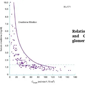
measurement or estimation of glomerular filtration rate (GFR). Serum creatinine is not an
ideal marker of GFR, because it is both filtered at the glomerulus and secreted by the
proximal tubule. Creatinine clearance (CrCl) is known to overestimate GFR by as much
as 40% in normal individuals and by even more in patients with CKD2.
Estimates of GFR based on 24-hour CrCl require timed urine collections, which
are difficult to obtain and often involve errors in collection. Classic methods for
measurements of GFR, including the gold-standard inulin clearance, are cumbersome,
require an intravenous infusion and timed urine collections, and are not clinically
feasible. In adults, the normal GFR based on inulin clearance and adjusted to a standard
body surface area of 1.73 m2 is 127 ml per minute per 1.73 m2 for men and 118 ml per minute per 1.73 m2 for women, with a standard deviation of approximately 20 ml per minute per 1.73 m2. After age 30, the average decrease in GFR is 1 ml per minute per 1.73 m2 per year1.
Equations based on serum creatinine but factored for gender, age, and ethnicity
Cockcroft-Gault equation3. This equation was developed to predict CrCl, but has been used to estimate GFR.
CrCl = [(140-age) x Weight (Kg)]/[S. Cr x 72] [x 0.85 in women]
The Cockcroft–Gault formula is reasonably accurate in mild renal impairment with a
GFR of around 50 ml/min but can overestimate GFR by up to 100 per cent when GFR is
less than 10 ml/min.
The Modification of Diet in Renal Disease (MDRD) Study equation was derived
on the basis of data from more than 500 patients [using the clearance of Iothalamate] with
a wide variety of kidney diseases and GFRs up to 90 ml per minute per 1.73 m2. The abbreviated MDRD equation4,5 is recommended for routine use and requires only serum creatinine, age, gender and race.
GFR = 186.3 x (S. Cr)-1.154 x (age)-0.203 x (0.742 if female) x (1.21 if black)
This formula is more accurate especially at low clearance and in preferred to the
Cockcroft-Gault method. A caveat is that this method has only been validated in Black
and White Americans and not in other racial groups.
Why “Kidney”?
The word “kidney” is of Middle English origin and is immediately understood by
patients, their families, providers, health care professionals, and the lay public of native
English speakers. On the other hand, “renal” and “nephrology,” derived from Latin and
Greek roots, respectively, commonly require interpretation and explanation.
Classification of CKD
Table 2 shows the classification of stages of CKD, including the population at
CKD and to improve outcomes in each stage.
Table 2 - Classification of Chronic Kidney Disease
The shaded area identifies patients who have CKD. The unshaded area designates
individuals who are at increased risk for developing CKD.
Kidney damage is defined as pathologic abnormalities or markers of damage,
including abnormalities in blood or urine tests or imaging studies.
* Includes actions from preceding stages.
Adverse outcomes of CKD are based on the level of kidney function and risk of
loss of function in the future. It has almost been a uniform observation that CKD tends to
worsen over time. Therefore, the risk of adverse outcomes increases over time with
disease severity. Many disciplines in medicine, including related specialties of
hypertension, cardiovascular disease, diabetes, and transplantation, have adopted
classification systems based on severity to guide clinical interventions, research, and
professional and public education. Such a model is essential for any public health
approach to disease.
The extent of the problem and Epidemiology of CKD
Chronic kidney disease is a worldwide public health problem with rising
incidence and prevalence of kidney failure, with poor outcomes and high cost. Data from
the United States suggests that the incidence and prevalence of CKD 5 have doubled in
the past 10 years and are expected to continue to rise steadily in the future.
The third National Health and Nutrition Examination Survey (NHANES III:
1988–1994) estimated that 3% of the population of the United States of America (USA)
of the population has a GFR of less than 60 ml/min.
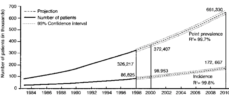
Figure 1 (given below) shows the incidence and prevalence of CKD 5 in the
United States based on the USRDS 2000 Annual Data Report7. Incident patients refers to new cases during the year. Point prevalent patients refers to patients alive on December
31st of the year. Projections for future years are based on extrapolation of regression
[image:11.612.93.545.266.458.2]equations.
Figure 1 - Incidence and prevalence of CKD 5 in United States
Data from the USRDS 2000 Annual Data Report
In the United Kingdom (UK), the prevalence of renal impairment (serum
creatinine of >1.36mg/dl) was 6.1% in known hypertensives, 12.6% in known diabetics
and 16.9% in patients with both these conditions in the age group 50–75 years8. It is estimated that the dialysis population will double in the next 15 years consisting
patients were detected by routine screening in a primary health care setting.
In developed countries like USA and UK, most patients are sent in a stable state
to the renal outpatient clinic. At the first visit a thorough medical history and physical
examination are obtained and the patient's previous case records are carefully scrutinized.
Additional investigations are carried out to fill in the gaps. Thus, a diagnosis can be
established and decisions about management may be made. Some patients present as an
'acute uraemic emergency' with a short history and no diagnosis; they are first
resuscitated and then investigated to detect or exclude an acute renal condition which will
require specific treatment. Thereafter, establishing a diagnosis can proceed at leisure in
those countries.
Epidemiology of Chronic Kidney Disease in India
Similar date regarding epidemiology of CKD is unavailable in our country. There
are only two population based studies from India to date. The first study was from Apollo
Hospital, Chennai by Dr. M. K. Mani.9 Prevalence of CKD in the surveyed community was 0.16% and other renal diseases (short of CRF) in 0.7% of patients. However, all the
patients were not evaluated with blood tests for urea and creatinine, and only those who
had some abnormality in the urine test or blood pressure and/or a positive response to a
questionnaire were subjected to a blood test for urea and creatinine.
The second study was from All India Institute of Medical Sciences by Dr. Sanjay
Agarwal10. This was a community based study done in urban Delhi where 4972 subjects were screened with blood urea and serum creatinine estimation with a specific aim to find
out the prevalence of CKD. The prevalence of CKD, defined as serum creatinine > 1.8mg
be 0.79% or 7852 per million population. Extrapolating the estimate of the number of
CKD 5 to be 10% of this number, the prevalence is about 785 per million population in
India.
Clinical Features and Investigations in Chronic Kidney Disease
When the patient presents with CKD and is referred early in the course of the
disease, the need for a diagnosis is self-evident. The difficulty of obtaining a precise
diagnosis increases as the patient approaches CKD 5 and the rewards diminish.
Consequently, a substantial proportion of patients starting dialysis have no more precise a
diagnosis than 'CKD – native kidney disease - unknown'.
Although it is virtually impossible to establish a firm diagnosis in some patients
presenting in CKD 5 with small fibrotic kidneys, it is prudent to pursue a vigorous
diagnostic approach in all others, since a precise diagnosis is invaluable in identifying the
groups of diseases causing CKD which affect patient management.
Etiology of Chronic Kidney Disease
Renal insufficiency can ensue from a primary renal disease or a systemic disease
which affects the kidneys.
Renal causes
Glomerulonephritides
These manifest as haematuria, signs of a nephrotic syndrome (tiredness, weight
gain, oedema, susceptibility to infection, hyperlipidaemia), and/or hypertension. With
more frequent, regular health screening, one of the most common presentations of
chronic glomerulonephritis (e.g. IgA nephropathy) is the detection of dipstix proteinuria
patient.
Reflux nephropathy
Usually diagnosed in childhood during investigation of urinary infection or
screening in families, it may, however, escape discovery until adult life when it is
detected during investigation of urinary infection, especially in pregnancy, hypertension,
or renal failure. Though often silent, there may be a history of recurrent episodes of flank
pain, dysuria, fever, and rigors. CKD in adult life peaks in the twenties and thirties and is
uncommon after the age of 50.
Secondary chronic pyelonephritis
This is the etiology in patients with CKD and a history of stone disease,
obstruction, or neuropathic bladder.
Medication related
Both over the counter as well as prescribed and recreational drugs are implicated
in this category. Chinese herb nephropathy (caused by Aristolochia fangchi) is an
example where fibrosis and tubulointerstitial changes persist for months or years after
discontinuation of the toxin though slow recovery may occur on stopping. Prolonged
consumption of non-steroidal anti-inflammatory drugs (NSAIDs) and selective COX-2
agents has also been implicated in the etiology of CKD (analgesic nephropathy). These
drugs also cause a reduction in GFR in patients with glomerulonephritis, due to inhibition
of vasodilatory prostaglandins, acute interstitial nephritis, nephrotic syndrome or
papillary necrosis. Angiotensin-converting enzyme (ACE) inhibitors and/or Angiotensin
II Receptor Blockers may cause renal insufficiency in patients with bilateral renal artery
Hereditary nephritis
Autosomal Dominant Polycystic Kidney Disease (APKD) is the most frequent
cause of CKD in hereditary nephritis. Renal failure usually occurs in the third to fifth
decade; family history is positive in ~ 75% cases. Similarly, a positive family history is
usual in the rarer types of renal cystic disease such as tuberous sclerosis and medullary
cystic disease. Alport's syndrome is another important hereditary nephritis characterized
by progressive nephritis with haematuria and sensori-neural hearing loss.
Infections
Renal tuberculosis is a rare cause of CKD. Classically, the presentation is of
dysuria, fever and sterile pyuria and is confirmed by urine culture and renal imaging
(calcified renal substance). Interstitial renal tuberculosis causing renal failure with small
kidneys on imaging and without the tell-tale renal calcification is diagnosed by renal
biopsy or inferred from evidence of tuberculosis in other systems. Another infection
associated with CKD 5 that shows a geographic variation is schistosomiasis which is an
important cause in Egypt.
Substance abuse
Heroin-associated nephropathy (was an important cause of CKD 5 in New York)
Systemic diseases
Diabetes mellitus
Diabetes mellitus is the most frequent cause of renal involvement in a systemic
disease. Renal involvement occurs as frequently in type II as in type I diabetes. The
accounted for by type II diabetic nephropathy11. Hence, the need for good control of blood sugars to prevent microalbuminuria and therafter ACE inhibitors and/or ARBs to
retard progression.
Hypertension
Long-standing 'benign' hypertension is also an important cause of CKD,
particularly in the elderly age group, but is difficult to differentiate from occult renal
diseases with secondary hypertension except by renal biopsy.
Systemic Lupus Erythematosus (SLE)
A diagnosis of SLE may be initially suspected clinically and then a renal biopsy is
done to assess the severity of renal illness or else it is diagnosed when a renal biopsy is
performed in any patient with proteinuria or abnormal urinary sediment. Therefore most
patients should have one at the time of their initial assessment for renal insufficiency.
Amyloidosis
Secondary amyloidosis is suspected when CKD complicates long-standing
inflammatory diseases (like severe rheumatoid arthritis) or chronic infections (such as
destructive lung tuberculosis). It presents as a nephrotic syndrome. Primary amyloidosis
occurs due to the proliferation of a single clone of plasma cells in middle and old age
presenting with renal insufficiency and proteinuria. In patients with plasma cell
dyscrasias, renal failure may also result from myeloma cast nephropathy, light chain
deposition disease or hyperviscosity syndrome.
Systemic vasculitis and Anti-glomerular basement membrane disease usually
present as rapidly progressive renal failure. A history of haemoptysis and dyspnoea,
together with rapidly deteriorating renal function and an active urine sediment, suggest
antiglomerular basement membrane disease (Goodpasture's syndrome) but can also occur
in systemic vasculitis. Other significant symptoms are of persistent sinusitis with
dyspnoea, cough, and haemoptysis (Wegener's disease) or only systemic symptoms
(microscopic polyangitis).
Thrombotic microangiopathy
This may occur due to haemolytic uraemic syndrome, thrombotic
thrombocytopenic purpura or anti phospholipids antibody syndrome.
Occupational renal diseases
Occupational renal diseases causing CKD is an extremely rare entity with the
exception of lead toxicity. Theses patients have hyperuricaemia, hypertension and small
kidneys. The diagnosis is best made by demonstrating an increase in urinary lead
excretion following an infusion of EDTA. More recently, long-term occupational or
environmental exposure to relatively low levels of cadmium has been shown to cause
CKD12.
Postrenal causes
Prostatic hypertrophy
This presents with obstruction of the lower urinary tract is common in elderly
males and is usually symptomatic. However, 40 per cent of men over the age of 65 have
some of the symptoms of hesitancy, slow and forked stream, urgency with or without
Pressure-flow measurements have shown that two-thirds of these patients have some degree of
outflow obstruction and are at risk of urinary retention14. A useful clinical clue to serious outflow obstruction is palpation of a distended bladder after micturition (confirmed more
reliably by ultrasonography before and after micturition).
Other causes of obstruction are retroperitoneal fibrosis, sloughed papillae and
renal calculi inducing hydronephrosis and a deterioration of renal function.
Clinical examination
At the patient's first visit, although a full standard physical examination, as taught
during undergraduate medical studies is recommended, it is seldom practiced in busy
outpatient clinics. The common clinical findings in practice are of anemia and fluid
overload. Other occasionally found features are those of overt uremia such as asterixes or
pericardial rub. Abdominal examination alerts the clinician to the presence of palpable
kidneys (in ADPKD or hydronephrosis) or palpable bladder in bladder oulet obstruction.
Rarer findings are of clues such as the body habitus of Lawrence Moon Biedl
syndrome (a rare cause of CKD), a saddle nose (in Wegener's disease), deafness (in
Alport's disease) and skin manifestations of Systemic Lupus Erythematosus. Per rectal
examination may reveal an enlarged prostate or a pelvic mass causing urinary
obstruction. The neurological examination may detect the neuropathies associated with
diabetes, vasculitis and and primary amyloidosis. Fundus examination (through a dilated
pupil) shows presence of retinal haemorrhages and exudates. There may also be corneal
calcification, pingueculae or rarely the 'uraemic red eye' of acute hypercalcaemia usually
Laboratory investigations
After history and clinical examination, the relevant investigations (urine, blood,
radiology and histology) are to be done to confirm the diagnosis.
Urine
Urinalysis and microscopy
Dipstick examination of a fresh mid-stream urine sample is useful to assess the
urinary pH and screen for leucocyturia, proteinuria, haematuria, and glucosuria. The pH
is usually low in CKD, unless the patient is on a very low protein diet. If the urine
contains non-albumin proteinuria, then the dipstick test will be negative with the 24-hour
quantification showing significant proteinuria.
Microscopy of the urine sediment may reveal erythrocytes (dysmorphic if
glomerular in origin), leucocytes and casts. Erythrocyte casts are seen in
glomerulonephritis (IgA, postinfectious, and SLE) and in cases of vasculitis. Leukocyte
casts identify pyuria as coming from the kidney and are found in stone disease,
tuberculosis, analgesic nephropathy and other causes of chronic interstitial nephritis.
Quantitation of proteinuria (24-hours)
Quantitation of proteinuria (24-hours) is used to measure proteinuria. Proteinuria
is almost universal in CKD with the proteinuria in the nephrotic range in conditions like
diabetic nephropathy. The accuracy of the collection is checked by quantitating total
urinary creatinine, which is fairly constant (range 10–14 mmol/day in females and 12–18
mmol/day in males). The degree of proteinuria can be also be “measured” by the spot
measures,15 although the accuracy decreases at extremes of creatinine excretion (e.g. muscular men who have high and cachextic patients who and low urinary creatinine
concentrations, respectively).
Blood
Hematology
A full hemogram is important to establish the type of anaemia in a patient with
CKD. A Coomb's test is done if clinically indicated to exclude autoantibody-induced
haemolysis (as can occur in SLE). A peripheral blood smear may show microangiopathic
haemolytic anaemia (fragmented and helmet-shaped erythrocytes and burr cells). If this
occurs in combination with thrombocytopenia, it is suggestive of the haemolytic uraemic
syndrome or thrombotic thrombocytopenic purpura.
Biochemistry
Serum creatinine concentration provides only a rough approximation of GFR as
the amount excreted increases with a decline in GFR16.
The electrolyte profile shows the presence of hyperkalemia and severe metabolic
acidosis. Blood sugar estimation and liver function tests provide information about
underlying diseases such as diabetes mellitus and liver failure.
Serology
Serological tests can give additional support in the assessment of a diagnosis and
underlying disease activity. Serum total haemolytic complement and C3 can be decreased
endocarditis), cryoglobulinaemia and lupus nephritis. Elevated titres of serum
anti-nuclear antibody and anti-double-stranded DNA support the diagnosis of lupus. In a
patient with rapidly progressive renal failure with an active urine sediment,
antiglomerular basement membrane antibodies confirm a diagnosis of Goodpasture's
disease while Anti-Neutrophil Cytoplasmic Antibodies support a diagnosis of systemic
vasculitis (proteinase 3 - specific for Wegener's granulomatosis or
Anti-myeloperoxidase - associated with microscopic polyangitis).
Virology
Patients with CKD are commonly tested for antibodies against Human
Imunodeficiency Virus (HIV) and Hepatitis C Virus (HCV) and Hepatitis B surface
antigen (HBsAg). HBsAg is associated with membranous glomerulopathy, IgA
nephropathy and mixed cryoglobulinaemia. HCV infection is associated with type 1
membranoproliferative glomerulonephritis and cryoglobulinaemia. About 95 per cent of
patients with mixed essential cryoglobulinaemia have evidence of HCV infection (by
testing for anti-HCV antibodies and HCV RNA).
Knowing the virology status is also important in vaccinating against Hepatitis B
virus to protect against infection from dialysis and blood transfusions.
Radiological investigations
Renal ultrasonography is invaluable in assessment of a patient of CKD. The renal
size, renal cortical thickness and echogenicity can be determined. The presence of cysts
urinary tract can be done followed by intravenous urography (if renal function is normal)
or non-contrast spiral computerized tomography (CT). CT with intravenous contrast and
arteriography (or magnetic resonance imaging with angiography) is required for
diagnosis of classical polyarteritis nodosa and renovascular disease. However,
angiogaphy remains the 'gold standard' for diagnosing renovascular disease.
Role of biopsy
In places where routine health checks are commonplace or if the patient has
presents early in the course of the illness, a renal biopsy is mandatory (in most) to
establish a diagnosis, to assess the extent of damage and to plan therapy. In others, it may
have been deemed unnecessary because the diagnosis was established on clinical
grounds, for example, in diabetes. If the kidneys are small, the hazards of the procedure
are increased and have to be weighed against the small chance of finding a reversible
cause.
Establishing chronicity in CKD
In a patient with no prior medical illness presenting with renal impairment, it is
necessary to establish chronicity based on clinical and laboratory evidence.
The factors that suggest chronicity are:
• Duration of symptoms for several months
• Nocturia
• Absence of severe symptoms despite very high urea and creatinine
• Bone disease
• Sexual dysfunction
• Skin disorders, nail changes and pruritus
• Neurological complications
• Small kidneys on renal imaging
Treatment of CKD
Attenuation of progression
Once a diagnosis of CKD is made, there are certain medical measures that can be
undertaken to attenuate the progression.
Blood pressure control
The importance of the control of blood pressure in patients with renal disease
cannot be overemphasised. Hypertension is common in patients with CKD and the rate of
decline in renal function increases with increasing blood pressure17,18 and also reduction in blood pressure attenuates the deterioration of renal function (first convincing
demonstration was in diabetic nephropathy).19,20
Two important factors contribute to the rise in blood pressure of patients with
CKD. First, most renal diseases are associated with sodium retention, which results in an
increase in extracellular fluid volume and an increase in peripheral vascular resistance.
Second, activation of the renin–angiotensin–aldosterone system results in increased
circulating angiotensin II, which in addition to being a potent vasoconstrictor, also
enhances sodium retention by the kidney. Hence, the two initial steps in the treatment of
use of diuretics, and treatment with agents that block the effects of angiotensin II, that is,
ACE inhibitors or ARBs. Other antihypertensives that are commonly prescribed are
calcium channel blockers, beta adrenergic antagonists, α-adrenoceptor blockers and
diuretics.
The target blood pressure in patients with CKD is 120/70 mmHg especially with
proteinuria of over 1 g/day21,22. Regular follow-up is essential as it has been demonstrated that patient compliance, efficacy of antihypertensive treatment, and retardation of renal
failure are clearly related to the number of outpatient visits23. Dietary recommendations
Sodium restriction
In patients with CKD, the ability to excrete sodium usually is limited. Thus, a
sodium-restricted diet of 6 g/day is a useful initial step in the treatment of hypertension.
Determining a 24 h urinary sodium excretion can check compliance with the sodium
restricted diet.
Sodium depletion may occur in patients with CKD due to tubulointerstitial disease
such as pyelonephritis, interstitial nephritis or hydronephrosis. In these, sodium has to be
monitored closely and often restriction is not advisable.
Protein restriction
The initial observations in the rat model with reduced renal mass had shown that
protein restriction attenuated the development and progression of renal failure.
Thereafter, some retrospective studies and several (but not all) randomized prospective
studies confirmed that a low protein diet might have the same effect in humans. In the
the largest trial to date on the effect of protein restriction on CKD in man) the effects of
dietary protein restriction and blood-pressure control on the progression of CKD, the
investigators reported a small beneficial effect of the low-protein diets on the course of
renal function after an average follow-up period of 2.2 years. When the initial 4 months
of low-protein diet were excluded from the analysis, the decline in GFR of
protein-restricted patients was attenuated24. This secondary analysis of the MDRD trial patients also revealed a high protein intake was associated with a more rapid decline in GFR. It
was calculated that each 0.2 g/kg body weight reduction in protein intake resulted in a 29
per cent reduction in the rate of decline in GFR.
The effect of protein restriction on the progression of CKD has been analysed in
two meta-analyses.25, 26
Altogether, it can be concluded from these studies that protein restriction causes a
modest reduction in the progression of CKD in man. It was concluded that dietary protein
restriction significantly reduced the risk for renal insufficiency or death with a relative
risk of 0.67 (95% confidence interval 0.50–0.89) in patients with non-diabetic renal
failure and 0.56 (95% confidence interval 0.40–0.77) in patients with diabetic
nephropathy. Currently, mild protein restriction to 0.6 to 0.8 grams per kg is
recommended. Calorie intake is kept at 30 kcal/kg body weight/day or more.
Dietary protein restriction also helps to alleviate the symptoms of uraemia in
those not being considered for dialysis in addition to attempting to slow the progression
of renal insufficiency without negatively jeopardizing nitrogen balance. Dietary
compliance is monitored most readily by measuring the serum urea : creatinine ratio
example, a 40 g protein diet should produce about 150 mmol of urea.
Fluid balance
In CKD, the regulatory capacity of the kidney is progressively reduced. Both
excretion and conservation of electrolytes and water are impaired; when sudden loads of
potassium, acid, or fluid has to be handled the limitations of renal functional reserve
become apparent and signs of decompensation may occur. Consequently, water and
electrolyte intakes must be adapted to renal excretory capacity.
In the subset of patients who are fluid overloaded, both sodium and water have to
be restricted. On the other hand, in patients with conditions that predominantly affect the
renal medulla (for example, interstitial nephritis and pyelonephritis), defective urinary
concentrating ability is particularly common and dehydration occurs easily in patients
with inadequate fluid intake, due to persistent diuresis despite fluid deprivation. Several
mechanisms are responsible for the inability to excrete concentrated urine, including the
increased solute load in remnant nephrons resulting in an osmotic diuresis, alteration of
medullary interstitial solute concentrations as a result of the damaged countercurrent
exchange system and impaired medullary blood flow. In addition, impaired sensitivity to
antidiuretic hormone causes decreased outward water transport in the distal nephron
segments. As a result of decreased concentrating capacity, urine osmolality is roughly
that of plasma, approximately 300 mOsm/kg H2O, in patients with CKD. If the
obligatory osmolar production in an adult is around 600 mOsm/kg H2O, daily urine output will be roughly 2 l per day. Fluid intake should therefore be approximately 2–3
l/day in order to ensure adequate urine flow rates and to prevent dehydration. In some
Potassium
Patients with CKD are usually able to maintain serum potassium within normal
limits until oliguria occurs or GFR is less than 5 ml/min. Preservation of normokalaemia
results from an adaptive increase in potassium excretion by remnant nephrons and
increased bowel loss. However, hyperkalaemia may be an early feature of renal failure in
patients with hyperchloraemic metabolic acidosis and hyporeninaemic
hypoaldosteronism, which occur particularly in patients with chronic tubulointerstitial
nephritis and diabetic nephropathy. Hyperkalaemia also complicates an acute potassium
load (e.g. blood transfusion, or medication, which interferes with potassium secretion, for
example, potassium sparing diuretics, ACE inhibitors, β-blockers, and NSAIDs) Foods
containing high levels of potassium like nuts, chocolate, fruits, wine and fruit juice and
salt substitutes (containing potassium) are particularly dangerous. Therefore a judicious
restriction of potassium rich diet and monitoring of serum potassium while on ACE
inhibitors and ARBs is warranted.
Other medications
Phosphate binders and calcitriol
Skeletal abnormalities occur early in renal failure, well before symptoms
develop.27 A variety of biochemical and radiological investigations are available to assist in the diagnosis and monitoring of renal osteodystrophy of which serum parathyroid
hormone (PTH) remains the single most useful biochemical test in predicting bone
histology in an individual patient.28
In early CKD, it would appear that adynamic bone disease is the principal type of
failure. As renal insufficiency progresses, higher levels of PTH are necessary for normal
bone remodeling. The cause of this 'skeletal resistance' to PTH in uraemia is probably
multifactorial. Inhibition of osteoclastic bone resorption appears to be the central
mechanism. Therefore a plasma PTH of two to three times the normal value is usually
required to maintain normal bone turnover.
When the patient is seen in the early phases of CKD, the objective is to maintain
normal bone turnover by maintaining serum calcium, phosphate, PTH and calcitriol and
blood pH in the normal range.
The mainstay in preventing secondary hyperparathyroidism is strict phosphorus
control. Some dietary phosphate restriction is usually required once GFR is less than 50
ml/min. Care must be taken in maintaining a sufficient protein intake, however, and
adequate nutrition must be maintained. Dietary restriction alone is usually inadequate in
controlling serum phosphate once GFR is less than 25 ml/min. Phosphate binders are then
added to reduce phosphorus absorption from the intestine. Calcium carbonate
(500-2000mg thrice daily) is effective and probably the most widely used phosphate
binder. It must be taken with food to give optimal phosphorus binding and to reduce the
risk of hypercalcaemia. If hyperphosphataemia persists despite administration of calcium
carbonate, excess dietary intake (e.g. dairy products) should be excluded and alternative
phosphorus binding agents substituted. Another commonly used calcium containing
binders is calcium acetate. It is a more effective binder than calcium carbonate and is less
likely to be associated with hypercalcaemia. The downside of using calcium containing
phosphate binders is the risk of a positive calcium balance which in dialysis patients is
associated with a higher relative risk of death. In the face of hypercalcemia, other
phosphate binders like sevelemar hydrochloride and lanthanum carbonate may need to
replace calcium based products. Others still being evaluated include polynuclear iron
preparations.
In some patients, aluminium-containing phosphate binders have to be resorted to.
If needed, they should be used only for a limited period of time since aluminium is
absorbed to a variable extent and can lead to aluminium overload, manifesting as anaemia
and aluminium-mediated bone disease.
In patients with lower stages of CKD, administration of 1,25-(OH)2 D3 (calcitriol) 0.25 μg/day causes a rise in serum calcium, a fall in serum phosphorus and alkaline
phosphatase, and retards the development of histological bone abnormalities.29
Careful monitoring of serum calcium is required, since hypercalcaemia may
accelerate the decline in renal function. Though new vitamin D metabolites are available
such as 22-oxacalcitriol, paracalcitriol (19 nor-1,25 dihydroxy-vitamin D2), and
doxercalciferol (1-hydroxy-vitamin D2), their benefits over conventional calcitriol and
alfacalcidol remain to be established30. Bone biopsy is generally reserved for patients with unusual biochemical and radiological evidence of bone disease.
Treatment of anemia
Anaemia is a predictable consequence of CKD and is directly related to its
severity. It frequently occurs early with one study reporting a prevalence of 45% in
patients with a serum creatinine ≤ 2 mg/dl31 Monitoring anaemia is important to determine if it becomes disproportionate to the stage of CKD. A haemoglobin of less than
onset of iron, folate or vitamin B12 deficiency. Functional iron deficiency is common
and should be confirmed by measurement of percentage of hypochromic red cells, serum
iron, transferrin and ferritin. As oral iron is often poorly tolerated, intravenous iron is
now frequently administered to predialysis patients. Occult gastrointestinal bleeding is
common in patients with advanced stages of CKD and is most commonly due to
superficial upper gastrointestinal lesions32.
Recombinant human erythropoietin (rhEPO) is effective in treating anaemia in
adults and children with CKD both prior to and while on dialysis. Benefits of correcting
anaemia include increased quality of life, reduced morbidity and improved survival. This
may be related to reduction in left ventricular mass and normalization of cardiac output
with partial correction of anaemia. There is also evidence to suggest that rhEPO therapy
may retard the progression of CKD and delay the onset of dialysis by as much as 6
months33, 34.
Treatment of hyperlipidemia
Hyperlipidaemia is often present in patients with CKD35. Nonetheless, there are only a limited number of studies, usually with a small number of patients, in which the
effects of treatment of hyperlipidaemia has been investigated. A meta-analysis by Freid et
al showed clearly that treatment of hyperlipidaemia ameliorates the progression of
CKD36, 37.
Other recommendations
Obesity is clearly associated with hypertension, and reduction of body weight
should be recommended to obese patients. Increasing physical exercise and reducing
exercised in severe calorie restriction because of the risk of catabolism.
Alcohol abuse can also contribute to hypertension and may also interfere with
adherence to antihypertensive or other therapy. It is advisable to limit alcohol intake to
less than 21 units in men and 14 units in women.
In patients with CKD, it has been shown that cigarette smoking enhances the rate
of progression of disease.38, 39 Thus, patients with CKD should be strongly advised to quit smoking.
Follow-up assessment and treatment
The aims of follow-up are the following:
1. Monitor the progression to CKD 5 and slow or arrest it where possible;
2. Detect and treat the complications of CKD and the primary disease;
3. Detect those symptoms of uraemia that call for dialysis and transplantation;
4. Plan and implement an orderly preparation for RRT.
5. Timely vascular access or CAPD catheter placement to allow a smooth transition
to dialysis
The ability to predict confidently the rate of progression of CKD is invaluable in
making these plans. This is largely, though not exclusively, dependent on early referral to
a nephrologist40. The factors most likely to speed progression that can be modified include control of blood pressure, diet, avoidance of unnecessary drugs, and, in some
instances, control of the underlying disease. Partial correction of anaemia with rhEPO
may also slow progression. Care can be shared with the general practitioner in monitoring
Monitoring decline in GFR
Serum creatinine concentration
Serum creatinine is the most widely used serial measurement of GFR but has
important limitations. When GFR declines, serum creatinine initially changes only
slightly.41 (See Fig 2 below). Minor changes in serum creatinine may, therefore, reflect major changes in GFR. When the GFR declines to less than 40 ml/min/1.73m2, the loss of nephrons over-rides the effects of enhanced tubular secretion and decreased generation of
creatinine, and large increases in serum creatinine correspond to small changes in GFR.
Thus, serum creatinine concentration is a poor indicator of renal function in
[image:32.612.100.385.307.594.2]patients with CKD and cannot be used to assess GFR accurately. In addition, serum
Figure 2
creatinine concentration is affected by other factors such as changes in muscle mass and
nutritional status, physical activity and gut metabolism of creatinine42.
Any reduction in serum creatinine in late CKD may well reflect a loss in muscle
mass rather than improvement in renal function. Another more suitable endogenous
marker for GFR is the protein cystatin C which is produced by all nucleated cells and
which is almost exclusively eliminated from the circulation by glomerular filtration43. It is sowly being tried in in clinical practice. Its use has yet to become routine.
Most clinicians use the Cockcroft–Gault formula but the MDRD formula being
more accurate especially at low clearance is now preferred.
Reciprocal of serum creatinine
In every CKD patient on follow up, the reciprocal of the serum creatinine against
time should be plotted. Linear regression of this produces a straight line in most patients
Figure 3 - The reciprocal plots of two patients with CKD on follow up
illustrating a linear progression in decline in renal function
Several limitations of the reciprocal creatinine plot must be kept in mind. The
accuracy of this prediction increases with the number of observations of serum creatinine.
Also, a proportion of patients do not have a linear decline in renal function. Nine per cent
of adults44 and 13 per cent of children45 deviated significantly from a straight line. Even among those that do follow a straight line, 20 per cent deviate from it at least once during
their follow-up46.
However, despite these limitations, the plot is useful in drawing attention to
episodes of acute-on-chronic kidney disease, monitoring response to treatment, and
predicting when stage 5 will be reached.
Preparing for RRT
Once the renal function is seen to be deteriorating, it is time to plan for RRT. A
checklist is useful at this stage in the management of the patient. The check-list given
From previous follow-up visits
Full blood count Calcium, magnesium, phosphate
Liver function tests Alkaline phosphatase Blood sugars
Other appropriate tests, e.g. complement, VDRL, HBsAg, CRP,serum electrophoresis, HbA1C,
24-h urinary protein
Midstream urine Radiography Renal ultrasound
ECG
Test Reason for test In preparation for dialysis and transplant
Ferritin, iron, transferrin Iron status to look for concomitant iron deficiency Folate and vitamin B 12 To rule out another cause of anaemia
PTH, vitamin D To assess degree of renal osteodystrophy and as baseline Aluminium level As a baseline for dialysis and to assess toxicity
Glucose tolerance test If fasting blood sugar is abnormal, to assess diabetic status HIV status If positive, will affect decisions regarding management.
Special precautions with blood letting
Hepatitis C status If positive, special precautions with blood letting Blood group, tissue typing
and cytotoxic antibodies Relevant for future transplant
Fasting lipids Patients are at risk of hyperlipidaemia
CMV serology Relevant to future transplant and donor CMV serology Skeletal survey To assess renal osteodystrophy
Micturating cystourethrogram
A routine in some centres to assess the bladder and exclude reflux before transplant
Dental assessment To look for occult dental sepsis pretransplant and for dental treatment predialysis Ophthalmology
assessment In diabetics
Urological assessment In certain conditions, e.g. reflux or stone disease
Family planning In females of child-bearing age regarding contraception and in males for consideration of sperm banking
Assessment by gastroenterologist
It is routine in some centres to examine pretransplant patients by endoscopy
Assessment by
cardiologist For ischaemic heart disease
worker
Assessment by dialysis
administrator To assess need for home adaptation for home haemodialysis Family interview For education and to assess possible family donors
Assessment by dialysis
[image:36.612.83.529.73.160.2]nursing staff To assess suitability for CAPD/haemodialysis and counselling
Table 3 Pre-renal replacement therapy assessment checklist
Social and psychological assessment
Assessment of the patient's circumstances including distance from the nephrology
clinic, cost and difficulty of travel determines the success of therapy. The occupational
history is of great importance in judging the patient's suitability for the various types of
RRT and the possible need for a change in occupation. It is important to discuss and
educate the patient with respect to all aspects of therapy including compliance to diet and
medication, prognosis and the various aspects of RRT. Once the patient has moderate
renal impairment, it is imperative to psychologically prepare him/her for RRT, especially
if renal transplantation is planned.
The patient entering a dialysis and transplant programme has to face many
changes in life style due to such factors as dependence and change of body image. The
patient must receive consistent and realistic advice about future treatment options. The
renal unit should function as a multi-disciplinary team of which the social worker is a
vital member, providing the social assessment on which many decisions hinge and
counseling the patient during an extremely stressful period. The social worker assesses
the patient's grasp of what is happening and establishes good communication. This will
depend on their intelligence, level of education, and linguistic skills. Also the patient’s
assessed. Their housing is important in deciding whether peritoneal dialysis or home
haemodialysis is a treatment option. Rehousing on medical grounds is a major issue even
in affluent countries and usually not a feasible idea in our country. The patient's
employment record and financial situation are needed to be explored so that the RRT is a
financially viable option. Interviewing the family is also part of the social assessment,
giving a better understanding of the patient and his or her social inter-relationships and
provides an opportunity to discuss the possibility of a live related transplant.
Hepatitis screening
All patients should be screened for hepatitis B and C. Patients who are negative
for HBsAg are to be offered vaccination if they do not have naturally acquired immunity.
As CKD reduces the effectiveness of vaccination, it is best to arrange this early. It is
preferred to use a double dose schedule of the Hepatitis B vaccine. The preferred
injection site is the deltoid in adults and the anterolateral thigh in children because the
buttock site is associated with a smaller chance of response. Approximately 75 per cent
of subjects who are HCV antibody positive also have viral particles in their blood and are
therefore infectious. In them, hepatitis C mRNA should be sought. Some strains
(genotype 1) are less responsive to treatment with interferon alpha.
HIV testing
It is advisable to test for HIV infection prior to dialysis with appropriate
counselling when indicated. HIVAN is the third leading cause of CKD5 in Blacks in the
age group 20–64 years after diabetes and hypertension in the United States47.
The most likely mode of transmission to hospital staff is through inoculation of
dialysis units. The majority of HIV infections are picked up by routine testing and if the
patient is known to be HIV positive, continuous ambulatory peritoneal dialysis (CAPD)
or home haemodialysis would be the treatment of choice in most cases. Blood taking
should be kept to a minimum and special care should be taken in handling, transporting,
and analysing samples. Healthcare workers need to be aware of the need for assessment
for postexposure HIV prophylaxis in the event of a needle stick injury.
Vascular access
The planning of vascular access is of great importance for a smooth start to
haemodialysis. If a fistula is required for haemodialysis, it should be created at least 3
months before the estimated date of starting dialysis in males and up to 6 months in a
female with small veins. This allows time for maturation of the fistula, prepares the
patient psychologically for the inevitable and avoids the need for temporary access such
as a jugular or femoral vein catheter with all the attendant risks of such procedures viz
trauma, septicaemia, venous stenosis, and thrombosis. Because of the high risk of
stenosis in the subclavian vein, which seriously jeopardizes future fistula attempts and
venous hypertension in the fistula, the use of subclavian catheters should be avoided48. Suitability for continuous ambulatory peritoneal dialysis
During the predialysis assessment period, suitability for CAPD must be assessed.
There are few absolute contraindications to CAPD though some, such as a major
diaphragmatic defects will only become apparent after CAPD has been started. A
colostomy, ileostomy, or nephrostomy is a firm contraindication and previous major
abdominal operations form relative contraindications. For young children, for those with
incoordination and for the mentally handicapped a family member such as the spouse or
parent will be trained, and thus the suitability of the helper will need to be assessed.
However, blind patients and some with severe arthritis have been trained to perform
CAPD successfully themselves.
Obesity and hyperlipidaemia get worse more frequently in CAPD patients than in
haemodialysis patients, so they are a relative contraindication to CAPD. Abdominal wall
hernias are aggravated by CAPD and should be repaired before CAPD is started. CAPD
will also result in an exaggerated lumbar lordosis, and if pre-existing lumbar disc disease
is present, this will almost always deteriorate on CAPD.
Suitability for pre-emptive transplantation
Pre-emptive renal transplantation, if contemplated, should obviously be planned
early in the assessment of the patient. When possible, it obviates all the attendant risks
and cost of dialysis. It is appropriate in patients with a predictable cause, such as diabetes,
so that stage 5 can be accurately dated. It is also easier to plan when a family donor is
available. Interestingly, pre-emptive transplantation compared to transplantation from
long-term dialysis is associated with better graft survival. In a study by Mange et al49, there was a 52 per cent reduction in the risk of allograft loss in the first year in patients
who underwent renal transplantion without prior dialysis.
Palliative care
Some patients approaching CKD 5 do not wish to have RRT and in others it may
be inappropriate if multiple other comorbid conditions coexist. In such cases, providing
good palliative care at the end of life is essential. Many symptoms can be alleviated with
nephrologists lack appropriate training in palliative care and a multidisciplinary approach
should be adopted drawing on facilities such as the local hospice, district nurses, and
general practitioners according to local circumstances.
Monitoring uraemic symptoms and when to start RRT
Monitoring the patient to assess when dialysis will be required is one of the main
aims of follow-up and one of the most difficult. The timing of the start of dialysis is
controversial, not least because of financial and practical implications and consequently
the practice varies widely. Recently there has been a general trend in the developed world
to initiate dialysis at an earlier stage of renal failure which would be in line with the
DOQI recommendations that dialysis should be commenced once the GFR declines to
less than 10.5 ml/min/1.73 m2 or the creatinine clearance is in the range of 9–14 ml/min. This guideline is based on opinion only. The decision of when to start dialysis should
involve a combination of clinical acumen in assessing symptomatology, discussed below,
and identifying early signs of malnutrition in association with measurements of declining
GFR discussed above. Evidence currently would also suggest that delaying dialysis till
the GFR is less than 6 ml/min irrespective of symptoms is unsafe.
The complaints of patients on starting dialysis (Malangone et al,)50 were:
• anorexia–nausea–vomiting 76%
• fatigue-weakness 72%
• pruritus 40%
• dyspnoea-orthopnoea 26%
• bleeding tendency 14%
• apathy—mental changes 12%
• asterixis-muscle twitching-cramps 11%
• dysguesia 8%
The authors found that none of these symptoms had a significant relationship to
biochemical measurements reflecting GFR (creatinine clearance, serum creatinine, and
urea) except bleeding tendency.
The onset of pericarditis or symptomatic peripheral neuropathy, with no
alternative explanation, is an indication that dialysis is already late and should be started
at once. Nausea and vomiting are signs that dialysis is needed very soon. Restless limbs
(a symptom which patients have difficulty in describing and often refer to as pain,
itching, or discomfort until questioned closely) are usually a sign that dialysis is needed
soon; a more generalized restlessness syndrome is an uncommon but dramatic indication
that it is overdue. Severe pruritus and excoriated rash is an indication for early dialysis if
not explained by drug allergy or coincidental skin disease. Other warnings that uraemia is
becoming troublesome are myoclonic jerks (particularly on falling asleep), sleep
disturbance, severe lethargy, mental clouding, loss of concentration and sexual
dysfunction in younger adults, unexplained by drug therapy. In a significant minority of
our patients, the trigger to starting dialysis is difficulty in controlling fluid balance and
blood pressure rather than the symptoms or biochemical evidence of severe uraemia. A
check for ankle oedema, crepitations at the lung bases, a raised jugular venous pressure
and the presence of cardiomegaly, a third heart sound and functional cardiac murmurs is
The National Kidney Foundation-Dialysis Outcomes Quality Initiative
(NKF-DOQI) guidelines recommends that dialysis is initiated when the nPNA (normalized
protein catabolic rate (nPNA) falls to below 0.8 g/kg per day. This is a measure of the
level of protein intake.
The problem of late referral to nephrology services
Timely referral of patients with CKD to specialist nephrological care is needed to
ensure the introduction of such measures early enough in the disease process to provide
benefit. However, worldwide 30–40% of patients are referred to a nephrologist at a very
late stage of renal disease.51, 52, 53, 54
In a study called “Late referral for dialysis: improving the management of chronic renal
disease”, Roderick et al found that one in six of all those starting RRT who were
avoidably late-referred, and might have benefited from earlier intervention under care of
a nephrologists54.
Late referral is likely to result in the patients having a lower serum calcium and
albumin and they were less likely to have a permanent vascular access in place at the start
of dialysis. They were also more likely to have a hematocrit less than 28% and were less
likely to have received Erythropoeitin55.Late referral has also been shown to influence early mortality56.
In the United Kingdom, the Renal Association has recommended that all patients
with serum creatinine > 150µmol/l (1.7mg/dl) be referred to a nephrologist57. While this might be an extreme, an early referral once the diagnosis of CKD is established is
advantageous. The patients can then be followed up on a regular basis by his personal
complementary.
Aim:
To evaluate patients with Chronic Kidney Disease Stage 5 [CKD 5] regarding:
1. Their pre-tertiary hospital care and
2. To assess their knowledge of the disease and its treatment
Objectives:
1. To assess the pre-tertiary hospital care of consecutive patients with CKD 5 who
presented themselves to the nephrology services of Christian Medical College,
Vellore.
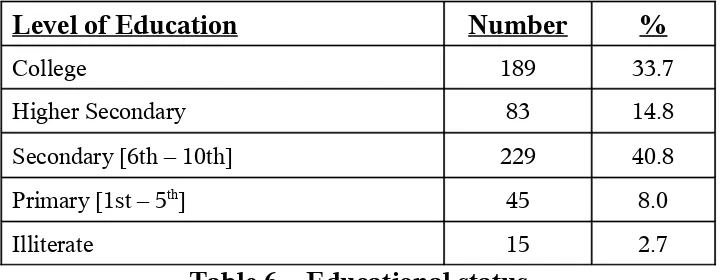
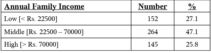
2. To describe the demographic profile, education, occupation, socio-economic
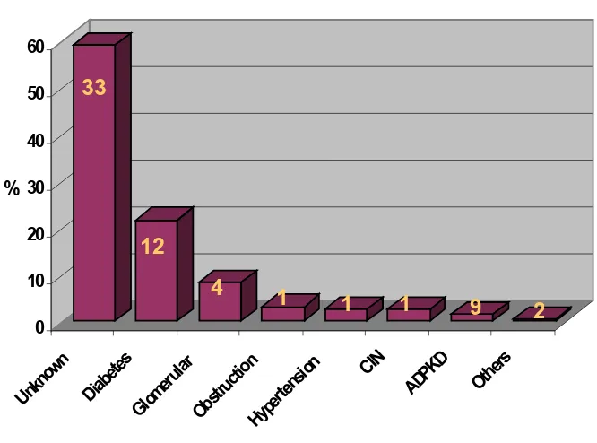
status, previous treating personnel, referral pattern, native kidney disease (if
known), previously done investigations, vaccination against Hepatitis B virus,
treatment received, prior patient education and decision towards renal
replacement therapy.
3. To determine if the care given prior to coming to a tertiary hospital was
appropriate and whether early referral to a nephrologist improved the adequacy of
Study Design:
The study was a cohort study on pre-tertiary hospital care of consecutive patients
with Chronic Kidney Disease stage 5 (CKD 5) who presented to the nephrology services
of Christian Medical College, Vellore.
Setting:
The study was conducted among the out-patients of the Department of
Nephrology, Units I and II of the Christian Medical College (CMC), Vellore, South India
which is a 1800 bedded tertiary care teaching hospital. Nephrology services have been
offered here for over 3 decades.
Subjects
Inclusion Criteria:
a. Subjects were newly diagnosed cases of CKD 5 based on history, calculated
glomerular filtration rate less than 15ml/min (by the Modification of Diet in Renal
Disease formula)
b. They had compatible ultrasonographic evidence of CKD 5.
Exclusion Criteria:
a. The study excluded subjects who had been diagnosed to have CKD at CMC prior to
the study period.
was in doubt and required verification by further investigations.
Evaluation:
Consecutive pts with CKD 5 presenting to Nephrology services (over an eight month period) were prospective enrolled upon making a diagnosis of CKD 5. Both the
patient and his relatives were interviewed by the investigator with regard to the
pre-tertiary hospital management. The data thus collected was then entered into an
electronically compatible proforma.
Statistical Analysis:
In this study on pre-tertiary hospital care of patients with CKD 5, there were a
total of 561 patients who were included in this study. During this period, 10843 patients
were seen as outpatients by the department of nephrology while 518 patients underwent
hemodialysis and 82 underwent renal transplantation.
Demographic Data
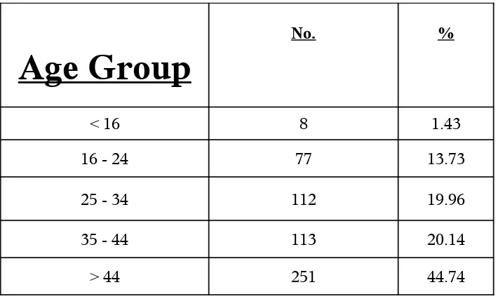
Age
Age Group
No. %
< 16 8 1.43
16 - 24 77 13.73
25 - 34 112 19.96
35 - 44 113 20.14
[image:46.612.130.485.351.569.2]> 44 251 44.74
Table – 4 - Age group profile
The mean age at presentation was 41.1 (± 14.5) years and the range was from 5
years to 79 years. It can be seen from Table 4 that the majority of patients were aged 35
years or above with 44.74% in more than 44 years age bracket. Figure 4 graphically
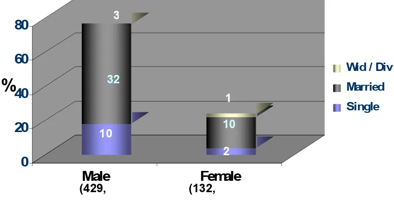
Gender and Marital Status
The population under study was predominantly male forming 76.4% of cases [429
[image:47.612.114.467.91.317.2]of 561 cases]. (Figure 5). Most of the men and the women in the study were married.
Figure 5 – Gender and marital status of patients
8 7 7 7 113 25 1
%
0 10 20 30 40 50 [image:47.612.102.502.432.636.2]< 16 16 - 24 25 - 34 35 - 44 > 44
Figure 4 - Age Groups
7 7 11 2 11 3 25 1 0 20 40 60 80 Male Female
Wid / Div Married Single
(429,