Copyright © 1997, American Society for Microbiology
Role of the First and Third Extracellular Domains of CXCR-4
in Human Immunodeficiency Virus Coreceptor Activity
ANNE BRELOT, NIKOLAUS HEVEKER, OLIVIER PLESKOFF, NATHALIE SOL,ANDMARC ALIZON*
INSERM U.332, Institute Cochin de Ge´ne´tique Mole´culaire, 75014 Paris, France
Received 17 January 1997/Accepted 4 March 1997
The CXCR-4 chemokine receptor and CD4 behave as coreceptors for cell line-adapted human immunode-ficiency virus types 1 and 2 (HIV-1 and HIV-2) and for dual-tropic HIV strains, which also use the CCR-5 coreceptor. The cell line-adapted HIV-1 strains LAI and NDK and the dual-tropic HIV-2 strain ROD were able
to infect CD41cells expressing human CXCR-4, while only LAI was able to infect cells expressing the rat
homolog of CXCR-4. This strain selectivity was addressed by using human-rat CXCR-4 chimeras. All chimeras tested mediated LAI infection, but only those containing the third extracellular domain (e3) of human CXCR-4 mediated NDK and ROD infection. The e3 domain might be required for the functional interaction of NDK and ROD, but not LAI, with CXCR-4. Alternatively, LAI might also interact with e3 but in a different way. Monoclonal antibody 12G5, raised against human CXCR-4, did not stain cells expressing rat CXCR-4. Chimeric human-rat CXCR-4 allowed us to map the 12G5 epitope in the e3 domain. The ability of 12G5 to neutralize infection by certain HIV-1 and HIV-2 strains is also consistent with the role of e3 in the coreceptor activity of CXCR-4. The deletion of most of the amino-terminal extracellular domain (e1) abolished the coreceptor activity of human CXCR-4 for ROD and NDK but not for LAI. These results indicate that HIV strains have different requirements for their interaction with CXCR-4. They also suggest differences in the interaction of dual-tropic HIV with CCR-5 and CXCR-4.
Human immunodeficiency virus types 1 and 2 (HIV-1 and HIV-2) infect cells by a membrane fusion process mediated by their envelope glycoproteins (gp120/gp41, or Env) that re-quires the interaction of gp120 with the cellular receptor CD4 (reviewed in references 27 and 41). The complete resistance of several nonhuman and human CD41cell lines to HIV-1
in-fection and to the formation of syncytia with Env1cells
sug-gests that other cellular factors are required for Env-mediated fusion (3, 6, 10, 13, 19, 25). The existence of such factors could also explain cell tropism differences between clinical isolates (primary strains) and cell line-adapted strains (23). Primary strains generally have a non-syncytium-inducing (NSI) pheno-type and replicate in peripheral blood mononuclear cells and macrophages but not in CD41 cell lines. Certain primary
strains, usually isolated at late stages of HIV infection, have a syncytium-inducing (SI) phenotype and can replicate in CD41
cell lines and peripheral blood mononuclear cells, but most HIV strains with these properties have been obtained by ex vivo adaptation. Finally, certain HIV strains, termed dual tropic, display features of both primary and cell line-adapted strains. These phenotypic differences among HIV strains are principally supported by genetic differences in their gp120 sur-face envelope proteins, in particular in the third hypervariable (V3) domain (reviewed in references 18, 27, and 41).
The cellular factors responsible for the permissiveness of CD41cells to HIV-1 entry were found to be members of the
chemokine receptor family. Chemokines are small (molecular weight, 8,000 to 10,000) soluble proteins that mediate leuko-cyte chemotaxis by interacting with a family of G-protein-coupled receptors with seven membrane-spanning (TM) do-mains. Chemokines are classified into two groups,a(or CXC) andb(or CC), depending upon the position of two conserved
cysteines in their amino-terminal domains (reviewed in refer-ence 28). A factor allowing entry of cell line-adapted HIV-1 strains was first identified by genetic complementation of mu-rine CD41cells and was named fusin (16). It was later shown
to be a receptor for stromal cell-derived factor 1 (SDF-1), a CXC chemokine, and renamed CXCR-4 (5, 30). This chemo-kine receptor can also be used for cell entry by HIV-2 strains, in both CD4-dependent (33) and CD4-independent (15) modes. It does not allow infection by NSI (or macrophage-tropic) HIV-1 strains, which were found to use CCR-5, a re-ceptor for the CC chemokines RANTES, MIP-1a, and MIP-1b
(2, 9, 11, 12, 14). Dual-tropic HIV-1 (9, 12), primary HIV-1 with an SI phenotype (37, 44), and HIV-2 strains such as ROD (38) can use both CXCR-4 and CCR-5 for cell entry. In addi-tion to these chemokine receptors, certain HIV-1 strains seem to be able to use CCR-3 and CCR-2b (9, 12).
The mechanism by which chemokine receptors participate in the process of HIV-1 entry is not entirely known, but different experiments suggest that gp120 interacts with CXCR-4 or CCR-5 (22, 40, 43). This interaction is markedly stronger in the presence of CD4, which suggests that chemokine receptors and CD4 behave as coreceptors for HIV. The identification of the domains of the chemokine receptors that interact with gp120 is important for understanding the process of HIV-1 entry. We have previously shown that human CXCR-4 was a coreceptor for the genetically divergent HIV-1 strains LAI (clade B) and NDK (clade D) and for HIV-2ROD. In contrast, the rat
ho-molog of CXCR-4 was able to efficiently mediate infection by LAI but not by NDK and ROD (33). This strain selectivity was used to identify domains of CXCR-4 supporting HIV corecep-tor activity.
MATERIALS AND METHODS
Cell lines and viral strains.The U373MG-CD4 cell line (19) and HeLa cell lines stably expressing wild-type or chimeric LAI Env (33, 36) have been de-scribed previously. HIV-1NDK(39) and HIV-1NL4.3(1) were propagated in the T-cell line CEM. HIV-1LAIwas produced by transfection of HeLa cells with a
* Corresponding author. Mailing address: INSERM U.332, Institut Cochin de Ge´ne´tique Mole´culaire, 22 rue Me´chain, 75014 Paris, France. Phone: 33-1-40 51 64 86. Fax: 33-1-40 51 77 49. E-mail: alizon @cochin.inserm.fr.
4744
on November 9, 2019 by guest
http://jvi.asm.org/
recombinant provirus (32). HIV-2RODwas produced by a chronically infected cell line (7).
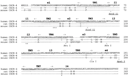
Construction of chimeric CXCR-4.The Rc/CMV vectors allowing expression of human and rat CXCR-4 from the cytomegalovirus immediate-early promoter have been described previously (33). The coding sequence of the rat CXCR-4 cDNA (kindly provided by R. S. Duman) was determined by direct automated sequencing of the expression vector. Several differences from the previously published sequence (42) (GenBank accession no. U54791) were noted, all re-sulting in identity of rat with human and/or mouse CXCR-4 (see Fig. 2). Con-structs A through G were obtained by substitution of rat and human CXCR-4 at one or two of the conserved restriction sites BstEII, HincII, and ApaLI (see Fig. 2 and 3). Constructs H, I, and J, were obtained by ligation of PCR fragments amplified from rat and human CXCR-4 into Rc/CMV between the HindIII and
NotI sites. The rat CXCR-4 fragments were amplified with T7 (59-AATACGA CTCACTATAGG) as the 59primer and either Nru-1 (59-CGAAGATGATGT CAGGG), Mfe-1 (59-TGGAACACCACCATCC), or Cla-1 (59-TCGATGCTG ATCCCC) as the 39primer. The human CXCR-4 fragments were amplified with either Nru-2 (59-CGAACGTCAGTGAGGCAG), Mfe-2 (59-ATTGCAGCACA TCATGGTTGG), or Cla-2 (59-TTCCTTCATCCTCCTGG) as the 59primer and SP6 (59-ATTTAGGTACACTATAG) as the 39primer. Constructs H to J were obtained by blunt-end ligation of PCR fragments T7/Nru-1 and Nru-2/SP6, T7/Mfe-1 and Mfe-2/SP6, and T7/Cla-1 and Cla-2/SP6, respectively, creating the restriction sites NruI at the TM4/e3 junction (construct H), MfeI at the e3/TM5 junction (construct I), and ClaI at the TM6/e4 junction (construct J). Construct K was derived by substitution of the ApaLI-NotI fragment from rat CXCR-4 into construct J. Construct L was obtained by blunt-end ligation of PCR fragments T7/Cla-1, amplified from construct A, and Cla-2/SP6, amplified from construct C. Construct M was obtained by blunt-end ligation of PCR fragments T7/Mfe-1, amplified from construct H, and Mfe-2/SP6, amplified from rat CXCR-4. Amino acid changes in the resulting protein, introduced by primers Mfe-1 and Mfe-2 (V194M and P198L), were shown to be detrimental to the coreceptor function and were corrected by site-directed mutagenesis with the oligonucleotide 59-CA TGATGTGCTGAAACTGGAACACAACCACCCACAAGTCATT on a sin-gle-stranded DNA template. Construct N was obtained by blunt-end ligation of PCR fragments amplified from human CXCR-4 with primers T7 and Nru-4 (59-CGAAGATGAAGTCGGGAA) and from construct I with primers Nru-3 (59-GGAATGTCAGCCAGGGGG) and SP6. In this chimera, the first amino acid of e3 corresponds to the sequence of human CXCR-4 (N176) and not to rat CXCR-4 (D173).
Mutations in human CXCR-4.TheDi4 mutant was obtained by blunt-end ligation between the DraI site (position 925 relative to the initial ATG codon) and a SmaI site of the pUC18 polylinker, creating a frameshift in the carboxy-terminal domain after F309, with the addition of 10 irrelevant amino acids. The N11Q and D4–41 mutants were obtained by site-directed mutagenesis on a single-stranded human CXCR-4 template with the oligonucleotides 59-GGTGT ATTGATCAGAAGTGT (BclI site underlined) and 59-GTAGATGGTGGGC CCCTCCATGGT (Bsp120I site underlined), respectively. TheD4–36 mutant was obtained by ligation of the partly complementary oligonucleotides 59-CAT GGAGGGGAATAAGATCTTCC and 59-CCGAGGAAGATCTTATTCCCCT into theD4–41 mutant digested with NcoI (position22 relative to the initial ATG codon) and Bsp120I. TheD2–9 mutant was obtained by digestion of the N11Q mutant with NcoI and BclI, filling in with T4 RNA polymerase, and blunt-end ligation.
Functional assays for CXCR-4.The U373MG-CD4 cells were transfected with wild-type or mutant CXCR-4 expression vectors in 24-well plates by calcium phosphate precipitation (overnight contact). Infections or cocultures were per-formed 24 h after transfection. The virus inoculum (10 to 30 ng of p24 antigen per well) was left in contact with cells for 36 to 40 h. Cocultures with Env1cells (1:1 ratio) were grown for 24 h. Cells were then fixed and stained forb -galac-tosidase activity with the X-Gal (5-bromo-4-chloro-3-indolyl-b-D -galactopyrano-side) substrate, as described previously (13). Blue-stained cells or foci were scored under a magnification of320.
Flow cytometry. COS cells were transfected with expression vectors for CXCR-4 and green fluorescent protein (GFP) (EGFP-N1 vector; Clontech, Palo Alto, Calif.) by calcium phosphate precipitation. Cells were detached with phos-phate-buffered saline containing 1 mM EDTA and pelleted 36 to 48 h after transfection. About 106cells were incubated for 1 h at 4°C with monoclonal antibody 12G5 (kindly provided by J. Hoxie) at a concentration of 7.5mg/ml in phosphate-buffered saline containing 2% bovine serum. Cells were washed three times and incubated for 1 h with phycoerythrin-conjugated rabbit anti-mouse immunoglobulins (Dako, Glostrub, Denmark). Stained cells were washed, fixed in 2% paraformaldehyde, and analyzed on an Epics Elite flow cytometer (Coul-tronics).
RESULTS
Strain specificity of rat CXCR-4.The ability of wild-type or
mutant chemokine receptors to behave as HIV coreceptors was tested by their expression in the CD41human glioma cell
line U373MG-CD4, which is naturally resistant to HIV-1 entry
and to fusion with Env1cells (19). This cell line was stably
trans-fected with a Tat-inducible lacZ reporter gene (LTRlacZ), allow-ing us to detect complementation for HIV infection and for fu-sion with Tat1 Env1cells by a simple in situ b-galactosidase
assay, as described previously (19, 33).
Upon expression of human CXCR-4 or its rat homolog, U373MG-CD4 cells were susceptible to infection with HIV-1LAIand fusion with cells stably expressing the LAI Env
pro-teins (Fig. 1). In contrast, only human CXCR-4 allowed infec-tion of U373MG-CD4 cells with HIV-1NDKor HIV-2RODand
fusion with cells stably expressing chimeric LAI Env with the V3 loop of ROD or NDK (Fig. 1). In all subsequent experi-ments, parallel results were obtained in infection assays with NDK or ROD and in syncytium formation assays with HeLa cells stably expressing chimeric LAI Env with the V3 loop of NDK or ROD.
Human-rat CXCR-4 chimeras. Human and rat CXCR-4
have more than 90% amino acid identity. The differences are found essentially in the extracellular domains, in particular e1 and e3, while the TM and intracellular (i) domains are highly conserved or identical (Fig. 2). A first set of chimeras (structs A to G) was constructed by using restriction sites con-served between the rat and human CXCR-4 cDNAs. The transfection of these chimeras into U373MG-CD4 cells al-lowed infection with HIV-1NL4.3(a recombinant HIV-1 strain
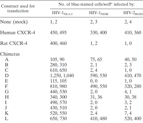
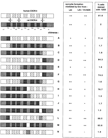
with LAI env) (Table 1) and fusion with HeLa cells stably expressing LAI Env (Fig. 3b). Infection mediated by chimeras A and E was less efficient while infection mediated by chimeras D and F, both with the TM7 and i4 domains from rat CXCR-4, was more efficient than infection mediated by human CXCR-4. These differences were not strain specific and did not seem to be supported by differences in cell surface expression (Fig. 3c). This first series of constructs allowed us to rule out a role for genetic differences in the e1 to TM3 and i4 domains in the selectivity of rat CXCR-4 for LAI and in the ability of human CXCR-4 to mediate infection by NDK or ROD. Accordingly, chimeric rat CXCR-4 with the i2 to e4 domains of human CXCR-4 (construct F) had the same phenotype as human CXCR-4. Since the i2, i3, and TM5 domains of human and rat CXCR-4 are identical, the phenotypic differences between hu-man and rat CXCR-4 can be supported only by genetic varia-tion in the TM4, e3, TM6, and e4 domains.
The role of these domains was addressed with another series of chimeric receptors (constructs H to J) with replacements at the TM4/e3, e3/TM5, and TM6/e4 junctions (Fig. 3). Only construct H was able to mediate the infection of U373MG-CD4 cells by NDK or ROD (Table 1) and their fusion with HeLa cells expressing chimeric LAI Env (Fig. 3b). However, its efficiency was markedly reduced relative to that of infec-tion mediated by human CXCR-4 or construct F. The com-parison of results obtained with chimeras H and I indicates that the e3 domain of human CXCR-4 was necessary for NDK and ROD infection, while experiments with constructs K and L showed that differences in the e4 domain had no apparent role in mediating infection (Table 1). The substi-tution of the e3 domain from rat CXCR-4 into human CXCR-4 (construct N) abolished the coreceptor activity of CXCR-4 for NDK but not for LAI (Table 2). The reciprocal chimera (rat CXCR-4 with human e3 [construct M]) was able to mediate infection of U373MG-CD4 cells with LAI (Table 2), NDK (Table 2), and ROD (data not shown). It also allowed fusion with cells expressing LAI Env with NDK V3 (Fig. 3b). The efficiency of NDK infection was lower (about 50%) when U373MG-CD4 cells expressed chimera M than when they expressed human CXCR-4. However, chimera M was able to mediate NDK infection much more
VOL. 71, 1997 FUNCTIONAL DOMAINS OF CXCR-4 4745
on November 9, 2019 by guest
http://jvi.asm.org/
efficiently than was chimera H. Also, its surface expression was apparently lower than that of chimera H or human CXCR-4 (Fig. 3c). The functional differences between chi-meras H and M could be due to conformational effects
[image:3.612.135.479.69.307.2]induced by chimerization. Overall, these experiments indi-cated that differences in the e3 domain were sufficient to account for the lack of coreceptor activity of rat CXCR-4 for NDK and ROD.
FIG. 1. Coreceptor activity of human and rat CXCR-4 for HIV-1 and HIV-2 strains. Target cells (U373MG-CD4 [LTRlacZ]) transfected with the indicated CXCR-4 expression vector were infected with HIV-1LAI, HIV-1NDK, or HIV-2ROD(a) or cocultured with HeLa cell lines stably expressing LAI Env or chimeric LAI Env with V3 from NDK or ROD (b). Infection with HIV or fusion with Env1cells results in transactivation of the LTRlacZ reporter gene and highb-galactosidase activity. Cells were stained with the X-Gal substrate 36 to 40 h after infection or 24 h after coculture. Bars represent the average numbers of blue-stained foci (log scale) in duplicate wells (24-well plate). Asterisks indicate that fewer than 10 foci were obtained.
FIG. 2. Alignment of the amino acid sequences of human, rat, and mouse CXCR-4. The human and rat CXCR-4 sequences were determined directly from the expression vectors used in this study. Asterisks indicate discrepancies with the previously established sequence of rat CXCR-4 (42). The mouse CXCR-4 sequence is from the work of Nagasawa et al. (29). The predicted TM domains are underlined. The positions of the restriction sites used for construction of rat-human chimeras are indicated. Natural restriction sites are underlined.
on November 9, 2019 by guest
http://jvi.asm.org/
[image:3.612.84.533.425.691.2]Deletion mutants.A series of deletions was engineered in human CXCR-4 cDNA by using natural restriction sites or site-directed mutagenesis. The deletion of the extracellular (e3 and e4) or intracellular (i2 and i3) loops abolished HIV core-ceptor activity, but epitope-tagged forms of these mutants were not detected at the cell surface (data not shown), suggesting that these deletions were not compatible with correct folding of CXCR-4. The deletion of the carboxy-terminal intracyto-plasmic domain (Di4) was fully compatible with coreceptor activity for LAI, NDK, and ROD (Table 3). Higher numbers of infected cells, or syncytia with Env1cells, were observed when
U373MG-CD4 cells were transfected with this mutant than with wild-type (WT) human CXCR-4.
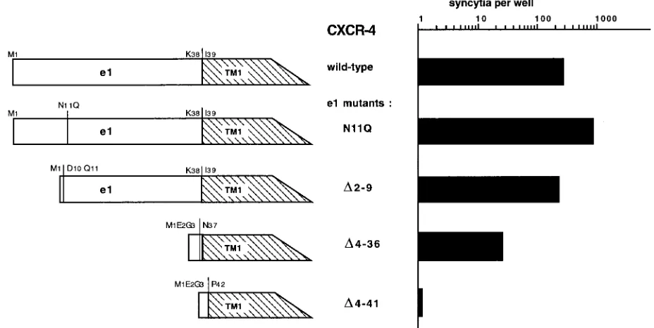
The other mutations were introduced in the amino-terminal domain (e1) of human CXCR-4. The N11Q mutation, which suppressed one of the two potential N glycosylation sites in CXCR-4, increased its coreceptor activity for LAI, NDK, and ROD (Fig. 4; Table 3). The other potential N glycosylation site, at the TM4/e3 boundary (N176), is not conserved in rat CXCR-4. It seems, therefore, that N glycosylation is not nec-essary for HIV coreceptor activity. The deletion of the first nine amino acids of e1 (yielding the D2–9 mutant) had no apparent effect on infection or cell fusion mediated by LAI Env and slightly reduced the efficiency of NDK and ROD infection (Table 3). Surprisingly, CXCR-4 with an almost com-plete deletion of e1 (D4–36 mutant) was able to mediate LAI infection and the fusion of U373MG-CD4 cells with HeLa cells expressing LAI Env (Fig. 4; Table 3). This mutant was less efficient than WT CXCR-4, but flow cytometry experiments also showed reduced cell surface expression (see below). The coreceptor activity of the D4–36 mutant was extremely weak for NDK and null for ROD (Table 3), which is more evidence that these strains and LAI have different requirements for their interaction with CXCR-4. TheD4–41 mutant mediated neither infection by LAI, NDK, and ROD nor fusion with Env1cells
(Fig. 4; Table 3). However, an epitope-tagged form was not detected at the cell surface (data not shown), suggesting that
the deletion, which extends beyond the predicted e1/TM1 boundary, was not compatible with correct folding of CXCR-4.
Mapping of the 12G5 epitope.Monoclonal antibody 12G5
was selected for its ability to block cell fusion mediated by HIV-2 Env and was later found to detect a conformational epitope of human CXCR-4 (15). It was also recently shown to neutralize infection by HIV-1 and HIV-2 strains (26). Flow cytometry experiments were performed on simian COS cells cotransfected with expression vectors for human or rat CXCR-4 and for GFP. More than 80% of cells transfected with human CXCR-4 (GFP positive) were stained with 12G5, while about 2% of cells transfected with rat CXCR-4 were stained (Fig. 3c). Therefore, the 12G5 epitope is not conserved in rat CXCR-4. The rat-human CXCR-4 chimeras were tested for their reactivity with 12G5; it stained only cells expressing chi-meras bearing the e3 domain of human CXCR-4, including construct M, in which e3 was the only domain from human CXCR-4. This indicated that the 12G5 epitope is located within the e3 domain of CXCR-4. However, it cannot be ruled out that the 12G5 epitope is formed by residues in e3 and in another domain conserved between human and rat CXCR-4. The percentage of stained cells and the mean fluorescence intensity were lower for cells transfected with chimera M than for cells expressing WT CXCR-4 (Fig. 3c and 5d). This could be due to reduced surface expression of the chimera or to less efficient presentation of the epitope in this context. Cells ex-pressing theD4–36 mutant were stained by monoclonal anti-body 12G5, although less efficiently than cells expressing WT CXCR-4 (Fig. 5f).
DISCUSSION
The CXCR-4 chemokine receptor behaves as a coreceptor for HIV-1 and HIV-2 strains adapted to replicate in immor-talized cell lines (15, 16, 33). It is also a coreceptor for dual-tropic HIV-1 strains (9, 12), for primary HIV-1 strains with an SI phenotype (37, 44), and for certain HIV-2 strains (38), all of which can also use CCR-5 as their coreceptor. The rat ho-molog of CXCR-4, differing from human CXCR-4 by a num-ber of residues in the extracellular domains, is a coreceptor for the cell adapted strain LAI but not for the cell line-adapted strain NDK or for ROD (which also uses CCR-5). A series of chimeric molecules was constructed from human and rat CXCR-4 and functionally tested by its expression in a CD41cell line (U373MG-CD4) naturally resistant to infection
by HIV-1 and HIV-2 and to Env-mediated cell-cell fusion. All chimeras tested allowed infection by LAI, indicating that they were folded and expressed at the cell surface, which was con-firmed by flow cytometry experiments. In the context of chi-meras, the third extracellular domain (e3) of human CXCR-4 was both necessary and sufficient to allow observation of co-receptor activity for NDK and ROD. The simpler interpreta-tion of these results is that e3 participates in the interacinterpreta-tion of human CXCR-4 with NDK and ROD but not with LAI. Al-ternatively, these strains might interact differently with e3, with only LAI being able to tolerate the amino acid differences between human and rat CXCR-4. It seems less likely that e3 substitutions can have indirect effects and modulate the inter-action of HIV with other domains of CXCR-4, although this possibility cannot be formally ruled out.
[image:4.612.58.298.91.297.2]Monoclonal antibody 12G5 (15) stained cells expressing hu-man, but not rat, CXCR-4. It reacted only with chimeras con-taining the e3 domain of human CXCR-4, indicating that e3 bears the 12G5 epitope or part of it. Antibody 12G5 can neutralize infection by HIV-1 and HIV-2 strains, but important variations depending upon the viral strains and cell types used
TABLE 1. Infection of U373MG-CD4 (LTRlacZ) cells expressing WT or chimeric CXCR-4 by selected HIV strains
Construct used for transfection
No. of blue-stained cells/wellainfected by: HIV-1NL4.3 HIV-1NDK HIV-2ROD
None (mock) 1, 2 2, 3 2, 4
Human CXCR-4 450, 495 330, 400 410, 360
Rat CXCR-4 400, 460 1, 2 1, 0
Chimeras
A 105, 90 75, 65 40, 50
B 280, 310 2, 1 2, 3
C 610, 650 2, 4 1, 0
D 1,250, 1,040 590, 530 410, 470
E 115, 105 0, 0 1, 0
F 810, 980 490, 550 320, 280
G 440, 530 2, 0 4, 1
H 340, 300 31, 36 30, 38
I 490, 570 2, 0 3, 2
J 430, 510 2, 0 2, 1
K 520, 550 2, 4 7, 4
L 650, 730 410, 480 520, 400
aValues for duplicate wells (24-well plates). Cells were stained with X-Gal for b-galactosidase activity 40 h after infection. All constructs were tested in the same experiment. Cell counts of.150 were obtained by extrapolation from randomly selected fields.
VOL. 71, 1997 FUNCTIONAL DOMAINS OF CXCR-4 4747
on November 9, 2019 by guest
http://jvi.asm.org/
FIG. 3. (a) Diagrams of CXCR-4 constructs; (b) functionality of chimeric constructs as HIV-1 coreceptors; (c) construct reactivity with antibody 12G5. Chimeric constructs were transfected into U373MG-CD4 cells, and syncytium formation assays with HeLa cells expressing WT or chimeric LAI Env were performed as described in the legend to Fig. 1. Results are shown semiquantitatively, relative to the number of syncytia observed in parallel transfections with WT human CXCR-4. Symbols:
11,.60%;1, 20 to 60%;2,,10%. To assess 12G5 reactivity, COS cells were stained 36 h after cotransfection with WT or chimeric CXCR-4 and GFP vectors and analyzed by flow cytometry as described in the legend to Fig. 5. The percentage of 12G5-stained cells among GFP-positive cells was determined.
on November 9, 2019 by guest
http://jvi.asm.org/
[image:5.612.76.536.85.672.2]seem to exist (26). Overall, it seems that cell line-adapted HIV-1 strains, such as LAI, are relatively resistant to neu-tralization by 12G5 compared to dual-tropic HIV-1 strains or to HIV-2 (26). If this result is confirmed, it could also suggest that the e3 domain is not required for the interac-tion of LAI with CXCR-4. However, the antiviral activity of 12G5 could have other mechanisms. The binding of 12G5 might exert steric hindrance effects on domains other than e3, or it might even induce CXCR-4 internalization. In that case, strains such as LAI might be less affected than others by the reduced expression of their coreceptor at the cell surface. At this time, it is difficult to sort out these possible mechanisms supporting the antiviral activity of antibody 12G5.
The mutation of a potential N glycosylation site in the ami-no-terminal domain (e1) increased the coreceptor activity of CXCR-4 for all strains tested. It can be envisioned that the absence of N-linked sugars facilitates coreceptor access. N glycosylation was also found to be dispensable for the corecep-tor activity of CCR-5 (34). The truncation of the carboxy-terminal intracytoplasmic domain (i4) increased the corecep-tor activity of CXCR-4. The i4 domain contains a number of
serine and threonine residues representing targets for phos-phorylation events modulating the activity of G-protein-cou-pled receptors (28). The coupling of CXCR-4 via its i4 domain is therefore dispensable for HIV coreceptor activity. The in-creased activity of the Di4 mutant was probably an indirect effect of the i4 deletion on, for example, the lateral mobility or the turnover of CXCR-4 at the cell surface. The intracellular loops i2 and i3 are also involved in coupling to the signal transduction machinery, but their deletion was not compatible with the surface expression of CXCR-4, due probably to severe conformational effects. A more detailed mutagenesis study of these domains will be required to address their possible role in HIV coreceptor activity.
[image:6.612.317.556.90.216.2]The deletion of the first nine amino acids of e1 had no apparent effect on the coreceptor activity of CXCR-4 for LAI and slightly reduced its activity for NDK and ROD. The almost complete deletion of e1 (yielding theD4–36 mutant) was com-patible with the cell surface expression and coreceptor activity
FIG. 4. Coreceptor activity of CXCR-4 with mutations or deletions in the amino-terminal e1 domain. Transfection of U373MG-CD4 cells and syncytium formation assays with HeLa cells stably expressing LAI Env were performed as described in the legend to Fig. 1.
TABLE 2. Infection of U373MG-CD4 cells expressing WT or chimeric CXCR-4 by two HIV-1 strains
Construct used for transfection
No. of blue-stained cells/wellainfected by:
HIV-1LAI HIV-1NDK
Human CXCR-4 2,400, 2,400 2,000, 2,000
Rat CXCR-4 2,400, NAb 29, 28
Chimera M 1,300, 1,300 640, 600
Chimera N 1,300, 1,200 26, 20
aValues for duplicate wells. This experiment was performed as described in Table 1, footnote a. The larger numbers of infected cells relative to those in Tables 1 and 3 are due to a higher transfection efficiency.
[image:6.612.57.298.91.163.2]bNA, not available.
TABLE 3. Infection of U373MG-CD4 cells expressing WT or mutant human CXCR-4 by selected HIV strains
Construct used for transfection
No. of blue-stained cells/wellainfected by: HIV-1NL4.3 HIV-1NDK HIV-2ROD
None (mock) 2, 2 1, 3 3, 5
WT CXCR-4 480, 530 460, 480 320, 270
CXCR-4 mutants
Di4 750, 660 820, 950 500, 620
N11Q 1,050, 1,250 850, 900 750, 810
D2–9 480, 560 370, 440 250, 220
D4–36 120, 150 5, 20 2, 3
D4–41 5, 2 7, 3 0, 0
aValues for duplicate wells. This experiment was performed as described in Table 1, footnote a.
VOL. 71, 1997 FUNCTIONAL DOMAINS OF CXCR-4 4749
on November 9, 2019 by guest
http://jvi.asm.org/
[image:6.612.74.538.472.705.2]of CXCR-4 for LAI, although both were reduced relative to those of WT CXCR-4. Notably, structure-function studies on the interleukin-8 receptor (CXCR-1) have suggested the exis-tence of a disulfide bridge between cysteines of e1 and e4 (20). These cysteines are conserved in other chemokine receptors, including CXCR-4. Our results with theD4–36 mutant indicate that the formation of a disulfide bridge between e1 and e4 is not absolutely necessary for the processing of CXCR-4 to the cell surface. TheD4–36 mutant had very low coreceptor activ-ity for NDK and did not mediate infection with ROD. The e1 domain might therefore be dispensable for the interaction of CXCR-4 with gp120 of LAI but required for the other strains. It can also be envisioned that LAI is affected differently by the reduced expression of the D4–36 mutant at the cell surface than is NDK or ROD. Similar studies with CCR-5 have shown that the e1 and e2 domains were necessary for coreceptor activity for primary and dual-tropic HIV-1 strains, while the e3 and e4 domains had no apparent role (4, 34). Therefore, dif-ferences in the interaction of dual-tropic HIV strains with CXCR-4 and CCR-5, as well as in the interaction of HIV strains with CXCR-4, seem to exist.
The HIV surface envelope protein gp120 seems to interact directly with the chemokine receptors CXCR-4 and CCR-5, this interaction being markedly reinforced by the presence of CD4 (22, 40, 43). The third hypervariable domain (V3) of gp120 was found to be necessary for the interaction of gp120 with CCR-5, although other domains of gp120 were apparently also involved in this interaction (40, 43). The role of V3 in the interaction of gp120 with CXCR-4 has not been directly ob-served but has been strongly suggested by different experi-ments. In cell line-adapted HIV-1, V3 substitutions resulted in a coreceptor switch from CXCR-4 to CCR-5 (9). We have observed that the V3 domain of LAI gp120 was required for utilization of the rat coreceptor CXCR-4 and conferred this property to chimeric NDK gp120 (33). A direct role for V3 in gp120–CXCR-4 interaction would be consistent with the greater sensitivity of cell line-adapted HIV-1 strains to neu-tralization by anti-V3 antibodies (reviewed in reference 35) or drugs targeting V3 (24, 31) compared with that of primary HIV-1 strains using the CCR-5 coreceptor. The V3 mutations associated with the switch of HIV-1 strains from the NSI to the SI phenotype usually increase the net positive charge of V3 (8, 17, 21). The e3 domain of CXCR-4 is negatively charged (net charge,23), while the homologous domain of CCR-5 is posi-tively charged (12). It is tempting to speculate that electro-static interactions take place between the V3 loop of SI strains and the e3 domain of CXCR-4. A detailed mutagenesis study of the charged residues in e3 could make it possible to address this hypothesis.
Even if a role for V3 in the interaction of gp120 with core-ceptors is probable, the very slight similarity between HIV-1 and HIV-2 in this domain suggests a role for a more conserved domain(s) of gp120. It seems unlikely to us that gp120 can accommodate totally distinct sites, conserved across strains and primate immunodeficiency viruses, which mediate the in-teraction with CCR-5, CXCR-4, and possibly other chemokine receptors. A simpler view might be that the same domain, or conformation, of gp120 has the ability to interact with different chemokine receptors, possibly via their amino-terminal do-mains. This interaction might be sufficient for an HIV strain to use CCR-5, but not CXCR-4, as its coreceptor, thus explaining the preferential use of CCR-5 by primary NSI strains. Muta-tions in other domains of gp120 (in particular, V3) might be required for a functional interaction with CXCR-4, possibly involving the e3 domain. The HIV strains bearing these mu-tations (primary SI or dual tropic) could therefore use either CCR-5 or CXCR-4. In cell line-adapted strains, mutations in gp120 resulting in a tighter interaction with CXCR-4 might be detrimental to interaction with CCR-5, thus preventing the use of this coreceptor. This model is consistent with the observa-tion that V3 mutaobserva-tions or substituobserva-tions conferring the NSI phenotype to a cell line-adapted HIV-1 strain were associated to a coreceptor switch from CXCR-4 to CCR-5 (9).
If a conserved domain of gp120 allows interaction with che-mokine receptors as different as CCR-5 and CXCR-4, it can be wondered why other chemokine receptors do not behave as HIV coreceptors (CCR-1, CCR-4, or CCR-2a) or can be used only by certain viral strains (CCR-3 and CCR-2b). The ability to interact with gp120 might be necessary but not sufficient for HIV coreceptor activity, which may require other properties, such as the ability to colocalize with CD4 either spontaneously or in the presence of gp120.
ACKNOWLEDGMENTS
[image:7.612.58.295.70.423.2]A. Brelot and N. Heveker made equal contributions to this work. We thank our colleagues J. Hoxie (University of Pennsylvania, Phil-adelphia) and R. Duman (Yale University, New Haven, Conn.) for
FIG. 5. Flow cytometry analysis of CXCR-4 expression. COS cells were transfected with a GFP expression vector and with WT or mutant CXCR-4, as indicated (1:6 ratio), and stained either with the anti-CXCR-4 monoclonal an-tibody 12G5 and a phycoerythrin-conjugated secondary anan-tibody or with the secondary antibody only (shaded curves in panels c to f). (a and b) Two-color analysis of 12G5 (x axis, red fluorescence) and GFP (y axis, green fluorescence) expression (arbitrary units); (c to f) distribution of 12G5 expression among GFP-positive cells (.102arbitrary units) (x axis, red fluorescence; y axis, cell numbers).
on November 9, 2019 by guest
http://jvi.asm.org/
generous gifts of reagents and I. Bouchaert and F. Letourneur (ICGM) for help with flow cytometry and sequencing.
This work was supported by the Agence Nationale de Recherches sur le SIDA and by Ensemble contre le SIDA.
REFERENCES
1. Adachi, A., H. E. Gendelman, S. Koenig, T. Folks, R. Willey, A. Rabson, and M. A. Martin.1986. Production of acquired immunodeficiency syndrome-associated retrovirus in human and nonhuman cells transfected with an infectious molecular clone. J. Virol. 59:284–291.
2. Alkhatib, G., C. Combadiere, C. C. Broder, Y. Feng, P. E. Kennedy, P. M. Murphy, and E. A. Berger.1996. CC CKR5: a RANTES, MIP-1a, MIP-1b receptor as a fusion cofactor for macrophage-tropic HIV-1. Science 272: 1955–1958.
3. Ashorn, P. A., E. A. Berger, and B. Moss. 1990. Human immunodeficiency virus envelope glycoprotein/CD4-mediated fusion of nonprimate cells with human cells. J. Virol. 64:2149–2156.
4. Atchison, R. E., J. Gosling, F. S. Monteclaro, C. Franci, L. Digilio, I. F. Charo, and M. A. Goldsmith.1996. Multiple elements of CCR5 and HIV-1 entry: dissociation from response to chemokines. Science 274:1924–1926. 5. Bleul, C. C., M. Farzan, H. Choe, C. Parolin, I. Clark-Lewis, J. Sodroski, and
T. A. Springer.1996. The lymphocyte chemoattractant SDF-1 is a ligand for LESTR/fusin and blocks HIV-1 entry. Nature 382:829–833.
6. Broder, C. C., D. S. Dimitrov, R. Blumenthal, and E. A. Berger. 1993. The block to HIV-1 envelope glycoprotein-mediated membrane fusion in animal cells expressing human CD4 can be overcome by a human cell component(s). Virology 193:483–491.
7. Brun-Ve´zinet, F., M. A. Rey, C. Katlama, P. M. Girard, D. Roulot, P. Ye´ni, L. Lenoble, F. Clavel, M. Alizon, S. Gadelle, J. J. Madjar, and M. Harzic. 1987. Lymphadenopathy-associated virus type 2 in AIDS and AIDS-related complex. Lancet i:128–132.
8. Callahan, L. 1994. HIV-1 virion-cell interactions: an electrostatic model of pathogenicity and syncytium formation. AIDS Res. Hum. Retroviruses 10: 231–233.
9. Choe, H., M. Farzan, Y. Sun, N. Sullivan, B. Rollins, P. D. Ponath, L. Wu, C. R. Mackay, G. LaRosa, W. Newman, N. Gerard, C. Gerard, and J. Sodroski.1996. The b-chemokine receptors CCR3 and CCR5 facilitate infection by primary HIV-1 isolates. Cell 85:1135–1148.
10. Clapham, P. R., D. Blanc, and R. A. Weiss. 1991. Specific cell surface requirements for the infection of CD4-positive cells by human immunode-ficiency virus types 1 and 2 and by simian immunodeimmunode-ficiency virus. Virology 181:703–715.
11. Deng, H., R. Liu, W. Elmeier, S. Choe, D. Unutmaz, M. Burkhart, P. Di Marzio, S. Marmon, R. E. Sutton, C. M. Hill, C. B. Davis, S. C. Peiper, T. J. Schall, D. R. Littman, and N. R. Landau.1996. Identification of a major co-receptor for primary isolates of HIV-1. Nature 381:661–666.
12. Doranz, B. J., J. Rucker, Y. Yi, R. J. Smyth, M. Samson, R. C. Peiper, M. Parmentier, R. G. Collman, and R. W. Doms.1996. A dual-tropic primary HIV-1 isolate that uses fusin and theb-chemokine receptors CKR-5, CKR-3, and CKR-2B as fusion cofactors. Cell 85:1149–1158.
13. Dragic, T., P. Charneau, F. Clavel, and M. Alizon. 1992. Complementation of murine cells for human immunodeficiency virus envelope/CD4-mediated fusion in human/murine heterokaryons. J. Virol. 66:4794–4802.
14. Dragic, T., V. Litwin, G. Allaway, S. R. Martin, Y. Huang, K. A. Nagashima, C. Cayanan, P. J. Maddon, R. A. Koup, J. P. Moore, and W. A. Paxton.1996. HIV-1 entry into CD41cells is mediated by the chemokine receptor CC-CKR5. Nature 381:667–673.
15. Endres, M. J., P. R. Clapham, M. Marsh, M. Ahuja, J. Davis Turner, A. McKnight, J. F. Thomas, B. Stoebenau-Haggarty, S. Choe, P. J. Vance, T. N. C. Wells, C. A. Power, S. S. Sutterwala, R. W. Doms, N. R. Landau, and J. A. Hoxie.1996. CD4-independent infection by HIV-2 is mediated by fusin/CXCR4. Cell 87:745–756.
16. Feng, Y., C. C. Broder, P. E. Kennedy, and E. A. Berger. 1996. HIV-1 entry cofactor: functional cDNA cloning of a seven-transmembrane G-protein coupled receptor. Science 272:872–877.
17. Fouchier, R. A. M., M. Groenink, N. A. Kootstra, M. Tersmette, H. G. Huisman, F. Miedema, and H. Schuitemaker.1992. Phenotype-associated sequence variation in the third variable domain of the human immunodefi-ciency virus type 1 gp120 molecule. J. Virol. 66:3183–3187.
18. Freed, E. O., and M. A. Martin. 1995. The role of human immunodeficiency virus type 1 envelope glycoproteins in virus infection. J. Biol. Chem. 270: 23883–23886.
19. Harrington, R. D., and A. P. Geballe. 1993. Cofactor requirement for human immunodeficiency virus type 1 entry into a CD4-expressing human cell line. J. Virol. 67:5939–5947.
20. He´bert, C. A., A. Chuntharapai, M. Smith, T. Colby, J. Kim, and R. Horuk. 1993. Partial functional mapping of the human interleukin-8 type A receptor. J. Biol. Chem. 268:18549–18553.
21. Kuiken, C. L., J.-J. de Jong, E. Baan, W. Keulen, M. Tersmette, and J. Goudsmit.1992. Evolution of the V3 envelope domain in proviral sequences
and isolates of human immunodeficiency virus type 1 during transition of the viral biological phenotype. J. Virol. 66:4622–4627.
22. Lapham, C. K., J. Ouyang, B. Chandrasekhar, N. Y. Nguyen, D. S. Dimitrov, and H. Golding.1996. Evidence for cell-surface association between fusin and the CD4-gp120 complex in human cell lines. Science 274:602–605. 23. Levy, J. A. 1993. Pathogenesis of human immunodeficiency virus infection.
Microbiol. Rev. 57:183–289.
24. Lynch, G., L. Low, S. Li, A. Sloane, S. Adams, C. Parish, B. Kemp, and A. L. Cunningham.1994. Sulfated polyanions prevent HIV infection of lympho-cytes but do not inhibit monocyte infection. J. Leukocyte Biol. 56:266–272. 25. Maddon, P. J., A. G. Dalgleish, J. S. McDougal, P. R. Clapham, R. A. Weiss, and R. Axel.1986. The T4 gene encodes the AIDS virus receptor and is expressed in the immune system and the brain. Cell 47:333–348. 26. McKnight, A., D. Wilkinson, G. Simmons, S. Talbot, L. Picard, M. Ahuja, M.
Marsh, J. A. Hoxie, and P. R. Clapham.1997. Inhibition of human immu-nodeficiency virus fusion by a monoclonal antibody to a coreceptor (CXCR4) is both cell type and virus strain dependent. J. Virol. 71:1692– 1696.
27. Moore, J. P., B. A. Jameson, R. A. Weiss, and Q. J. Sattentau. 1993. The HIV-cell fusion reaction, p. 233–289. In J. Bentz (ed.), Viral fusion mecha-nisms. CRC Press, Inc., Boca Raton, Fla.
28. Murphy, P. M. 1994. The molecular biology of leucocyte chemoattractant receptors. Annu. Rev. Immunol. 12:593–633.
29. Nagasawa, T., T. Nakajima, K. Tachibana, H. Iizasa, C. C. Bleul, O. Yoshie, K. Matsuhima, N. Yoshida, T. A. Springer, and T. Kishimoto.1996. Molec-ular cloning and characterization of a murine pre-B-cell growth-stimulating factor/stromal cell-derived factor 1 receptor, a murine homolog of the hu-man immunodeficiency virus 1 entry coreceptor fusin. Proc. Natl. Acad. Sci. USA 93:14726–14729.
30. Oberlin, E., A. Amara, F. Bachelerie, C. Bessia, J.-L. Virelizier, F. Arenzana-Seisdenos, O. Schwartz, J.-M. Heard, D. F. Legler, M. Loetscher, M. Bag-giolini, and B. Moser.1996. The CXC chemokine SDF-1 is the ligand for LESTR/fusin and prevents infection by T-cell-line-adapted HIV-1. Nature 382:823–835.
31. O’Brien, W. A., M. Sumner-Smith, S.-H. Mao, S. Sadeghi, J.-Q. Zhao, and I. S. Y. Chen.1996. Anti-human immunodeficiency virus type 1 activity of an oligocationic compound mediated via gp120 V3 interactions. J. Virol. 70: 2825–2831.
32. Peden, K., M. Emerman, and L. Montagnier. 1991. Changes in growth properties on passage in tissue culture of viruses derived from infectious molecular clones of HIV-1LAI, HIV-1MAL, and HIV-1ELI. Virology 185:661– 672.
33. Pleskoff, O., N. Sol, B. Labrosse, and M. Alizon. 1997. Human immunode-ficiency virus strains differ in their ability to infect CD41cells expressing the rat homolog of CXCR-4 (fusin). J. Virol. 71:3259–3262.
34. Rucker, J., M. Samson, B. J. Doranz, F. Libert, J. F. Berson, Y. Yi, R. J. Smyth, R. G. Collman, C. C. Broder, G. Vassart, R. W. Doms, and M. Parmentier.1996. Regions inb-chemokine receptors CCR5 and CCR2b that determine HIV-1 cofactor activity. Cell 87:437–446.
35. Sattentau, Q. J. 1996. Neutralization of HIV-1 by antibody. Curr. Opin. Immunol. 8:540–545.
36. Schwartz, O., M. Alizon, J. M. Heard, and O. Danos. 1994. Impairment of T cell receptor-dependent stimulation in CD41lymphocytes after contact with membrane-bound HIV-1 envelope glycoprotein. Virology 198:360–365. 37. Simmons, G., D. Wilkinson, J. D. Reeves, M. T. Dittmar, S. Beddows, J.
Weber, G. Carnegie, U. Desselberger, P. W. Gray, R. A. Weiss, and P. R. Clapham.1996. Primary, syncytium-inducing human immunodeficiency virus type 1 isolates are dual-tropic and most can use either Lestr or CCR5 as coreceptors for virus entry. J. Virol. 70:8355–8360.
38. Sol, N., and M. Alizon. Unpublished data.
39. Spire, B., J. Sire, V. Zachar, F. Rey, F. Barre´-Sinoussi, F. Galibert, A. Hampe, and J.-C. Chermann.1989. Nucleotide sequence of HIV1-NDK, a highly cytopathic strain of the human immunodeficiency virus, HIV1. Gene 81:275–284.
40. Trkola, A., T. Dragic, J. Arthos, J. M. Binley, W. C. Olson, G. Allaway, S. R. Martin, C. Cheng-Mayer, J. Robinson, P. J. Maddon, and J. P. Moore.1996. CD4-dependent, antibody-sensitive interactions between HIV-1 and its co-receptor CCR5. Nature 384:184–187.
41. Weiss, R. A. 1993. Cellular receptors and viral glycoproteins involved in retrovirus entry, p. 1–108. In J. A. Levy (ed.), The Retroviridae, vol. 2. Plenum Press, New York, N.Y.
42. Wong, M.-L., W. W. Xin, and R. S. Duman. 1996. Rat LCR1: cloning and cellular distribution of a putative chemokine receptor in the brain. Mol. Psychiatry 1:133–140.
43. Wu, L., N. P. Gerard, R. Wyatt, H. Choe, C. Parolin, N. Ruffing, A. Borsetti, A. A. Cardoso, E. Desjardin, W. Newman, C. Gerard, and J. Sodroski.1996. CD4-induced interaction of primary HIV-1 gp120 glycoproteins with the chemokine receptor CCR-5. Nature 384:179–183.
44. Zhang, L., Y. Huang, T. He, Y. Cao, and D. D. Ho. 1996. HIV-1 subtype and second-receptor use. Nature 383:768.
VOL. 71, 1997 FUNCTIONAL DOMAINS OF CXCR-4 4751