Copyrightq1997, American Society for Microbiology
Formation of Herpes Simplex Virus Type 1 Replication Compartments
by Transfection: Requirements and Localization
to Nuclear Domain 10
CHRISTOPHER J. LUKONISANDSANDRA K. WELLER*
Department of Microbiology, University of Connecticut Health Center, Farmington, Connecticut 06030
Received 4 October 1996/Accepted 19 November 1996
During infection, the seven essential herpes simplex virus type 1 (HSV-1) replication proteins are found in globular nuclear structures called replication compartments. Replication compartments form adjacent to ND10, nuclear matrix-bound domains which are present in most cell types but whose function is unknown (G. G. Maul, I. M. Ishov, and R. D. Everett, Virology 217:67–75, 1996). We now demonstrate that replication compartments can be formed by cotransfecting Vero cells with constructs expressing the seven essential viral replication proteins and a plasmid containing an HSV-1 origin of DNA replication. Like replication compart-ments in infected cells, replication compartcompart-ments formed by cotransfection contain all of the essential viral replication proteins, are sites of DNA synthesis, and are found adjacent to ND10. However, neither the viral origin-binding protein nor a plasmid containing an HSV-1 origin of DNA replication is individually required for the formation of transfection replication compartments, although the presence of each increases the efficiency of replication compartment formation. Further, we provide evidence that UL29 independently localizes adjacent to ND10 and so may play a role in directing replication compartments to these preexisting nuclear structures.
Herpes simplex virus type 1 (HSV-1) possesses a large, dou-ble-stranded DNA genome and replicates in the nuclei of in-fected cells. Genetic analysis has resulted in the identification of replication-defective mutants of genes representing seven complementation groups (reviewed in reference 55). Analysis of the proteins encoded by the seven genes has resulted in the assignment of functions to each; UL5, UL8, and UL52 code for a heterotrimeric helicase-primase complex, UL30 and UL42 code for a dimeric DNA polymerase holoenzyme, UL9 codes for an origin-binding protein, and UL29 codes for a single-stranded DNA-binding protein (reviewed in references 45, 55, and 56).
HSV-1 replication occurs in nuclear domains termed repli-cation compartments, initially identified on the basis of UL29 staining patterns in an immunofluorescence (IF) assay (48). Each of the seven essential viral replication proteins localizes to replication compartments (18, 32, 33, 37, 47). Cellular
fac-tors, including DNA polymerase a, proliferating cell nuclear
antigen, Rb, p53, and topoisomerase II, also localize to repli-cation compartments (13, 59). Three lines of evidence support the idea that replication compartments form at a limited, pre-determined number of nuclear sites in infected cells. (i) The number of replication compartments per nucleus is dependent on the multiplicity of infection (MOI); at an MOI of 0.01 PFU/cell, a maximum of two replication compartments per nucleus are seen (49). However, the number of replication compartments in infected cells does not increase appreciably beyond a dozen even at high MOIs, suggesting that the number of potential replication sites is finite and predetermined (24, 39). (ii) Infected binucleate cells, arising either spontaneously or after treatment with cytochalasin B, contain replication
compartments which are symmetrically arranged in daughter nuclei (8). (iii) Input viral DNA is deposited close to preexist-ing nuclear structures known as ND10, which are also present in symmetrical patterns in daughter nuclei (39). Viral replica-tion compartments remain associated with the periphery of ND10 during infection (24, 39).
ND10 are 0.3- to 0.5-mm structures that are present in most
cells with an average frequency of 10. A number of proteins, including Sp100 (1), NDP52 (31), and PML (12, 30, 54), are components of ND10. Of these, only PML has been function-ally characterized. PML was initifunction-ally identified as a fusion
partner with the retinoic acid receptorathat is expressed in
the lymphocytes of individuals with promyelocytic leukemia
(10, 16, 25, 26). The PML-retinoic acid receptorafusion
dis-organizes ND10 into hundreds of smaller structures. Retinoic acid treatment results in the restoration of normal ND10 dis-tribution and correlates therapeutically with leukemia regres-sion (12, 30, 54). This finding suggests that ND10 status may be related to basic cellular processes such as proliferation, a hy-pothesis supported by the finding that PML can act as a tumor suppressor (29, 43).
HSV-1 is not alone in its predilection for replicating at the periphery of ND10. Two other DNA viruses, adenovirus type 5 (Ad5) and simian virus 40 (SV40), replicate their genomes adjacent to ND10 (24). Replication adjacent to ND10 may thus be a common feature of DNA viruses. Additionally, SV40 and Ad5 encode proteins that localize adjacent to or colocalize with ND10. In the case of SV40, the multifunctional large tumor antigen (TAg) is found juxtaposed to ND10 in cells transiently transfected with a TAg expression vector (5). The Ad E1A protein colocalizes with ND10 in transiently trans-fected cells (5). Although the HSV-1-encoded transactivator ICP0 localizes to ND10 early in infection, it subsequently dis-rupts them (14, 38). The only herpesvirus-encoded protein known to stably localize to ND10 is Epstein-Barr virus nuclear antigen 5 (52a), a protein whose function is poorly understood. Although the seven essential HSV-1 replication proteins are
* Corresponding author. Mailing address: Department of Microbi-ology, University of Connecticut Health Center, 263 Farmington Ave., Farmington, CT 06030. Phone: (860) 679-2310. Fax: (860) 679-1239. E-mail: [email protected].
2390
on November 9, 2019 by guest
http://jvi.asm.org/
sufficient to catalyze replication of a plasmid containing an HSV-1 origin of DNA replication in a cotransfection assay (6, 61), the question of whether this replication occurs in replica-tion compartments has never been addressed. In this report, we show that replication compartments can be formed by co-transfection and that these structures form at the periphery of ND10. Additionally, we provide evidence that UL29 indepen-dently localizes adjacent to ND10 and so may play a role in directing replication compartments to these nuclear matrix-bound structures.
(This work constitutes part of the Ph.D. thesis of C.J.L. to be submitted to The University of Connecticut in partial fulfill-ment of the requirefulfill-ments for the degree.)
MATERIALS AND METHODS
Cells and reagents.African green monkey kidney fibroblasts (Vero cells; American Type Culture Collection) were propagated and maintained as de-scribed previously (57). Glycerol gelatin, 1,4-diazobicyclo-[2.2.2]octane (DABCO), phosphonoacetic acid (PAA), and 5-bromo-29-deoxyuridine (BrdU) were obtained from Sigma (St. Louis, Mo.).
Plasmids.Constructs which constitutively express UL5, UL8, UL9, UL29, UL30, and UL42 from the cytomegalovirus (CMV) immediate-early (IE) pro-moter (UL5b, UL8, UL9, DBP, pol, and pCM-UL42, respectively) were described previously (23). Plasmid pCMV-UL52, which contains the UL52 gene under control of the CMV IE promoter, was constructed by inserting the UL52-containingBamHI fragment of p6UL52118a (33) into
BamHI-digested pCDNA-1 Amp (Invitrogen). Plasmids capable of expressing the helicase-primase genes fused to short defined peptide epitopes from the ICP6 promoter, p6AU1UL5, p6EEUL8, and p6UL52KT3, were described pre-viously (33). Epitope-tagged UL5, UL8, and UL52 genes were removed from these plasmids on BamHI fragments and inserted into BamHI-digested pCDNA-1 Amp to generate AU1UL5, EEUL8, and pCMV-UL52KT3, respectively, for constitutive expression from the CMV IE promoter. Plasmid p100-1, which we refer to as poriS, contains oriSon a 100-bpMspI
fragment inserted into theSmaI site of pUC119 (46).
Antibodies.A monoclonal antibody recognizing BrdU incorporated into DNA was obtained from Becton Dickinson Immunocytometry Systems (San Jose, Calif.). The polyclonal antiserum 3-83, which recognizes UL29, was a generous gift of David Knipe (Harvard Medical School, Boston, Mass.) (28). The mono-clonal antibody 39-S recognizes UL29 (51). The monomono-clonal antibody 17B rec-ognizes UL9 (37). The polyclonal antibody Pol recrec-ognizes UL30 (11) and was a generous gift of David Dorsky (University of Connecticut Health Center, Farm-ington). The monoclonal antibody 65K recognizes UL42 (44) and was generously provided by David Dorsky. The monoclonal antibody AU1, which recognizes a peptide epitope of bovine papillomavirus L1 protein, was obtained from the Berkeley Antibody Company (Richmond, Calif.) (17). The monoclonal antibody EE, which recognizes a peptide epitope of polyomavirus medium T antigen (20), was generated from mouse ascites fluid by utilizing hybridoma cells generously provided by Gernot Walter. The monoclonal antibody KT3, which recognizes a peptide epitope of SV40 TAg (35), was obtained from mouse ascites fluid by utilizing hybridoma cells generously provided by Gernot Walter. The monoclonal antibody MAb138 recognizes a 55-kDa ND10 protein in HEp-2 cells and was kindly provided by Gerd Maul (Wistar Institute, Philadelphia, Pa.). The second-ary antibodies used for IF studies, fluorescein isothiocyanate-conjugated goat mouse and rabbit and Texas red-conjugated goat mouse and anti-rabbit, were obtained from Cappel, Organon Teknika Corporation (Durham, N.C.).
Transfection protocol.For IF studies, 106Vero cells were transfected with a
total of 8mg of DNA in 440 ml of HEPES-buffered saline (HBS)–CaCl2solution
described below. Unless noted otherwise, 1.0mg of each expression construct was used and the remainder was composed of sheared salmon sperm DNA. DNA was precipitated at room temperature with 400ml of HBS (pH 7.04) and 40ml of 1.25 M CaCl2for 15 to 30 min, and the entire volume was used to resuspend Vero
cells that had been trypsinized and pelleted at 1,500 rpm for 2 to 3 min in 15-ml Falcon tubes. The cell-DNA mixture was incubated for 30 min at 378C in a rotating platform shaker at 50 rpm. Tubes were flicked after 15 min to resuspend cell clumps. Cells were resuspended in 5 ml of Dulbecco modified Eagle medium containing 5% fetal calf serum, put in 60-mm-diameter plates with glass cover-slips, and incubated at 378C for 3.5 h, at which point the medium was aspirated and replaced with 2 ml of 15% glycerol in phosphate-buffered saline (PBS) for 2 min. The glycerol solution was aspirated, and the cells were washed twice with Tris-buffered saline, covered with fresh Dulbecco modified Eagle medium con-taining 5% fetal calf serum, and incubated for 16 to 24 h at 378C.
The procedure for transfecting cells for Southern blot analysis was the same except that 12mg of DNA (1.5mg of each construct with the remainder com-posed of sheared salmon sperm DNA) was precipitated with 600ml of HBS and 60ml of 1.25 M CaCl2and used to resuspend 1.53106Vero cells.
Preparation and analysis of cellular DNA.Total cellular DNA from trans-fected cells was isolated essentially as described previously (58). When required, cells were grown in the presence of PAA (400mg/ml). At 18 to 24 h posttrans-fection, cells were scraped from the plate, pelleted at 1,500 rpm for 2 to 3 min in 15-ml Falcon tubes, resuspended in Tris-buffered saline, pelleted and washed, and pelleted a second time. The pellet was resuspended in 400ml of Tris-EDTA and 200ml of 33lysis buffer (1.8% sodium dodecyl sulfate, 30 mM EDTA, 30 mM Tris-HCl) containing 300mg of proteinase K per ml. After incubation at 558C for 2 or more h, the proteinase K was inactivated by incubation at 708C for 10 min, RNase A was added to a final concentration of 100mg/ml, and the mixture was incubated for 30 min at 378C. The sample was deproteinized by extraction with phenol-chloroform-isoamyl alcohol (25:24:1), the phenol was removed with chloroform-isoamyl alcohol (25:1), and the DNA in the superna-tant was precipitated with ethanol. Approximately 2mg of DNA was digested withEcoRI orEcoRI in combination withDpnI, subjected to 0.8% agarose gel electrophoresis, and blotted onto GeneScreen Plus membranes as instructed by the manufacturer (New England Nuclear Corp., Boston, Mass.). Blots were probed with gel-purified, linearized pUC118 prepared by using a Random Primed DNA labeling kit (Boehringer Mannheim) and [a-32P]dCTP as
sug-gested by the manufacturer.
IF.Vero cells were grown on coverslips for 16 to 24 h posttransfection. When indicated, cells were labeled with 1 mM BrdU for 15 min prior to fixation. Cells were fixed in 37% buffered formaldehyde (Sigma) diluted 1:10 in PBS (pH approximately 7.4) for 30 min and permeabilized with 1.0% Triton X-100 for 10 min. Cells to be stained with 39-S were permeabilized with acetone for 2 min at 2208C instead of with Triton X-100. If cells had been labeled with BrdU, they were treated with 4 M HCl for 10 min. Cells were reacted with primary anti-bodies in 3% normal goat serum in PBS for 30 min at dilutions as follows; anti-BrdU at 1:50, 3-83 at 1:500, 39-S at 1:100, AU1 at 1:500, EE at 1:250, KT3 at 1:250, 17B at 1:100, Pol at 1:200, and 65K at 1:200. MAb138 was added directly to coverslips and incubated for 1 h, at which point other antibodies were added for an additional 30 min. Cells were reacted with secondary antibodies in 3% normal goat serum in PBS at a 1:200 dilution for 30 min. Coverslips were mounted in glycerol gelatin containing 2.5% DABCO to retard bleaching.
Imaging.Cells stained for IF were imaged on a Zeiss Axiovert 135 laser scanning microscope (confocal) equipped with a Zeiss 633Plan Neofluar ob-jective. Collected images were arranged and labeled by using a Silicon Graphics workstation equipped with Adobe Photoshop 3.0. Transfection efficiencies were established by determining the percentage of cells which stained with the 3-83 polyclonal antibody and ranged between 5 and 20%. At least 100 3-83-positive cells were counted to arrive at percentages of transfected cells which contain specific structures.
RESULTS
Characterization of structures in cells cotransfected with constructs expressing the seven essential HSV-1 replication proteins and an oriS-containing plasmid.The seven essential
viral proteins are necessary and sufficient for the amplification of plasmids containing an HSV-1 origin of replication in a transient cotransfection assay (6, 61). However, replication compartments have been observed only in infected cells, rais-ing the possibility that other viral proteins are required for their formation. In this study, Vero cells were cotransfected with CMV promoter-driven constructs expressing UL5, UL8,
UL52, UL9, UL29, UL30, UL42, and poriS. Subsequently, the
localization of UL29 was monitored by IF. In about 56% of cells exhibiting UL29 staining, UL29 localized in a globular nuclear pattern reminiscent of the UL29 in replication com-partments seen in HSV-1-infected cells (compare Fig. 1A and C). As shown in Fig. 1A and B, replication compartments in infected cells colabel with BrdU, indicating that they are sites of active DNA synthesis. Likewise, as shown in Fig. 1C and D, BrdU labeling colocalizes with UL29 in cotransfection struc-tures. It is not possible to distinguish between plasmid and cellular DNA synthesis in this experiment; however, Southern blot hybridization analysis of DNA harvested from
cotrans-fected cells was used to confirm that poriS was replicated
(shown below).
Another characteristic of replication compartments in in-fected cells is that they contain all seven essential HSV-1 rep-lication proteins (18, 32, 33, 37, 47). To determine if reprep-lication compartments formed by cotransfection share this property, the localization of each replication protein was compared with
VOL. 71, 1997 FORMATION OF HSV-1 REPLICATION COMPARTMENTS 2391
on November 9, 2019 by guest
http://jvi.asm.org/
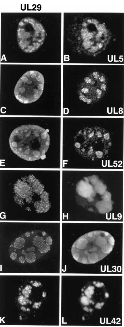
that of UL29 in cells cotransfected as described above. As shown in Fig. 2A to F, epitope-tagged versions of the helicase-primase complex (AU1-UL5, EE-UL8, and UL52-KT3 [33]) colocalize with UL29 in cotransfection structures. Figures 2G and H show that UL9 also colocalizes with UL29 in cotrans-fection structures. As shown in Fig. 2I to L, UL30 and UL42 colocalize with UL29 in cotransfection structures.
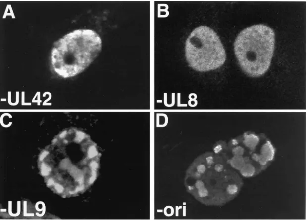
Requirements for replication compartment formation by transfection.Replication of an origin-containing plasmid in a transient transfection assay occurs only in the presence of all seven essential replication proteins (61). To determine if rep-lication compartment formation depends on the presence of the same factors, we performed transfections in which one of the eight components used in complete transfections was omit-ted. Omission of UL8 resulted in the loss of replication com-partment formation; as shown in Fig. 3B, the UL29 staining pattern was diffuse. Diffuse staining for UL29 was also ob-served when UL52 or UL5 was omitted (data not shown). In some cells which exhibited a diffuse staining for UL29, we detected small punctate structures, which are described in more detail below. The omission of UL42 resulted in a loss of replication compartment formation; the majority of cells con-tained diffuse staining for UL29, as shown in Fig. 3A. However, approximately one-third of transfected cells contained numer-ous (50 or more) UL29 sites that were not organized into replication compartments (data not shown) which appeared similar to those sites observed by Liptak et al. in cells cotrans-fected with constructs expressing UL5, UL8, UL52, and UL29 (32). Omission of UL30 also resulted in the localization of UL29 to similar numerous punctate sites (data not shown). The UL29 staining pattern observed when UL5, UL8, UL52, and UL29 are coexpressed is described below. In contrast,
omission of UL9 or poriSstill resulted in replication
compart-ment formation, as shown in Fig. 3C and D. However, in cells transfected in the absence of UL9, replication compartments were seen in only 10% of the cells which exhibit UL29 staining (as opposed to 56% of transfected cells when all seven
expres-sion plasmids plus oriSwere included). In cells transfected in
the absence of poriS, replication compartments were observed
in 21% of the transfected cells. These sites colabeled with BrdU, indicating that DNA synthesis was taking place in them (data not shown).
To verify that plasmid DNA replication occurs in transfected cells containing replication compartments, Southern blot anal-ysis was performed. This transient transfection assay assesses replication of input plasmids by virtue of their sensitivity to the
restriction endonucleaseDpnI, which cleaves only DNA that
has been methylated inEscherichia coli. Plasmid DNA
repli-cated in a mammalian cell is resistant toDpnI cleavage. Total
cellular DNA was harvested from transfected cells and
di-gested with EcoRI alone or in combination withDpnI as
de-scribed in Materials and Methods.EcoRI cleaves poriSonce,
generating a fragment of 3.1 kb that is detectable with a ra-diolabeled pUC119 probe (Fig. 4, lanes 1). DNA from cells cotransfected with constructs expressing the seven essential
replication proteins and poriScontains a strongly hybridizing
3.1-kbp band afterEcoRI digestion (lane 4), a portion of which
is resistant to double digestion withEcoRI andDpnI (lane 5).
Additionally, since all of the expression vectors were made in pUC-based vectors, these constructs are also detected with the pUC119 probe. For instance, in lane 4 (EcoRI only), the ex-pression constructs appear as several bands migrating above
the band representing poriS. In lane 5, some portion of each of
the expression plasmids is also partiallyDpnI resistant,
indi-cating that some DNA replication has occurred; however, they appear to have been replicated to a much lower level than
poriS. This is particularly evident in Fig. 4B (lane 5), an
over-exposed version of the autoradiogram in Fig. 4A. All plasmid replication essentially disappears when PAA is added to
co-transfected cells; poriSand expression construct DNA is
lin-earized byEcoRI but is sensitive toEcoRI/DpnI double
diges-tion (lanes 2 and 3). Likewise, omission of UL5 also prevents
replication of poriSand the expression constructs, as shown in
lanes 8 and 9 of Fig. 4A and B. On the other hand, the
omission of UL9 or poriSresults in a low level of replication of
all constructs, as shown in Fig. 4B, lanes 7 and 11. The
DpnI-resistant bands are not likely the result of partial digestion, as the same results were obtained in a number of independent
FIG. 1. Comparison of replication compartments in infected and cotransfected Vero cells. The cell depicted in panels A and B was infected with KOS. The cell depicted in panels C and D was cotransfected with constructs expressing the seven essential replication proteins and poriS. Cells were double stained with 3-83
(anti-UL29) and anti-BrdU. The panels on the left represents 3-83 staining; those on the right represent BrdU staining. Marker bar515mm.
on November 9, 2019 by guest
http://jvi.asm.org/
experiments. Further, the fact that this replication is not seen when the polymerase is inhibited with PAA or when UL5 is absent from the transfection provides evidence that the
low-levelDpnI resistance seen in lanes 7 and 11 represents
low-level plasmid replication. Our results suggest that poriSor UL9
is not required for the replication of expression construct DNA. Although the replication of non-origin-containing plas-mids was not previously detected in similar transfection assays (61), those experiments used endogenous promoters to express the replication proteins; in our experiments, the six replication fork proteins are overexpressed compared to the levels ex-pected from endogenous promoters in either infected or trans-fected cells (see Discussion). We demonstrated above that replication compartments form in cotransfected cells even in
the absence of poriSor UL9. It is possible that the replication
compartments observed in the absence of UL9 and poriS
rep-resent sites of expression construct replication. It should be reiterated, however, that the formation of replication
compart-ments in the absence of UL9 and oriSis inefficient, as is the
replication of expression constructs under these conditions.
Transfection replication compartments form adjacent to ND10.In infected cells, viral replication compartments form at the periphery of preexisting nuclear matrix sites known as ND10 (39). MAb138 recognizes a 55-kDa ND10 antigen in HEp-2 cells (63). In Vero cells, the number of ND10 per cell ranged from 2 to over 25 but averaged around a dozen. We designate the mostly brightly staining foci as ND10, although a fine granular or diffuse nuclear background staining is also sometimes observed within nuclei. To determine if replication compartments formed by transfection localize to ND10, Vero cells cotransfected with constructs expressing the seven viral
replication proteins and poriSwere double labeled with 3-83
and MAb138.
Cotransfected cells contained replication structures adjacent to ND10, as shown in Fig. 5A to F. The cell shown in Fig. 5A to C has many replication compartments of various sizes which appear to be associated with ND10. This association is shown more clearly in Fig. 5D to F in a cell which contains only three replication compartments. In the cell shown in Fig. 5D to F, two replication compartments are clearly associated with ND10; however, it is possible that the third replication com-partment is associated with an ND10 structure which is not in the plane of focus shown in Fig. 5E. Because replication com-partments are large, it has not been possible to carry out a statistical analysis to prove that they are associated with ND10. However, the observation that some of the protein recognized by MAb138 is recruited into replication structures in the cells shown in Fig. 5B and E supports the notion that there is an association between replication compartments and ND10. The recruitment of ND10 antigens has not been observed for any other ND10 protein during HSV-1 infection (39). It may be a pattern seen only with MAb138. Alternatively, overexpression of the replication proteins in our assay in comparison with infection may result in an amplified redistribution that may not
be readily detectable during infection. Omission of poriSand
[image:4.612.62.300.63.689.2]UL9 from the transfection did not affect the ability of replica-tion compartments to localize to ND10, as shown in Fig. 5G to
FIG. 2. Replication compartments in cotransfected cells contain all seven of the essential HSV-1 replication proteins. Cells were cotransfected as described for Fig. 1 and double labeled with antibodies to detect the localization of UL29 (left panels) with respect to the other viral replication proteins (right panels).
Panels A, C, E, G, and K show the staining pattern of 3-83. Panel I shows the staining pattern of 39-S. Panels B, D, and F show staining for AU1 ascites fluid (detecting AU1-tagged UL5), EE ascites fluid (detecting EE-tagged UL8), and KT3 ascites fluid (detecting KT3-tagged UL52), respectively. Panels H and L show the staining for 17B, a monoclonal antibody recognizing UL9, and a monoclonal antibody recognizing UL42, respectively. Panel J shows the staining for a polyclonal antibody that recognizes UL30. Marker bar515mm.
VOL. 71, 1997 FORMATION OF HSV-1 REPLICATION COMPARTMENTS 2393
on November 9, 2019 by guest
http://jvi.asm.org/
I and J to L, respectively. In summary, the apparent relation-ship of replication compartments with ND10 in a large number of transfected cells, only some of which are shown in Fig. 5, supports the concept that the replication compartments formed by transfection are associated with ND10 as was pre-viously observed for viral replication compartments formed in infected cells (39).
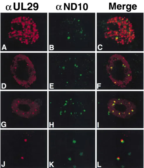
UL29 alone localizes adjacent to ND10 in transfected Vero cells.Based on the observation that replication compartments formed by transfection localize adjacent to ND10, we set out to determine which replication proteins are necessary and suffi-cient for dictating this pattern. It has been reported that the nuclei of cells cotransfected with constructs expressing UL5, UL8, UL52, and UL29 contain UL29 in numerous, punctate sites (approximately 50 to several hundred) (32). These au-thors proposed that such UL29 structures are precursors to replication compartments. To determine if these numerous UL29 structures localized adjacent to ND10, Vero cells were cotransfected with constructs expressing UL5, UL8, UL52, and UL29 and processed for double IF labeling with 3-83 and MAb138. Three populations of cells were identified; 37% of transfected cells contained numerous UL29 structures (Fig. 6A to C), 38% contained fewer (2 to 20) UL29 structures within a diffusely staining background (Fig. 6D to F), and 25% had a homogeneous, diffuse nuclear staining pattern for UL29 (data not shown) as has previously been reported for cells trans-fected with UL29 alone (15).
[image:5.612.153.463.69.291.2]In cells exhibiting the first pattern of UL29 localization, which is similar to those reported by Liptak et al. (32), UL29 structures are so numerous that it was not possible to deter-mine whether they associate specifically with ND10; as shown in Fig. 6A to C, the three visible ND10 may in fact colocalize with UL29 structures. On the other hand, in the 38% of the cells containing fewer UL29 structures in a diffuse background, the majority of brightly staining UL29 foci localized adjacent to ND10, as shown in Fig. 6D to F. The requirements for forma-tion of the numerous punctate structures were assessed by performing cotransfections in which either UL5, UL8, or UL52
FIG. 3. Formation of replication compartments by cotransfection requires UL8 and UL42 but not UL9 or poriS. Vero cells were cotransfected as for Fig. 1 except
that one of the eight constructs was omitted. The staining represents UL29 as detected by 3-83. In panel A, pCM-UL42 was omitted; in panel B, pCM-UL8 was omitted; in panel C, pCM-UL9 was omitted; in panel D, poriSwas omitted. Marker bar515mm.
FIG. 4. Southern blot hybridization of DNA from cotransfected cells. (A) Lane 1 represents poriSlinearized withEcoRI. In lanes 2 to 11, total DNA of
cotransfected Vero cells was digested withEcoRI (even-numbered lanes) or
EcoRI andDpnI (odd-numbered lanes), Southern blotted, and probed with radiolabeled pUC119. The DNA in lanes 2 to 5 is from cells cotransfected with constructs expressing the seven essential replication proteins and poriS
(com-plete); cells represented in lanes 2 and 3 were treated with PAA. The DNA in lanes 6 to 11 is from cells cotransfected with one of the eight plasmids omitted from the complete transfection. Those in lanes 6 and 7 lacked pCM-UL9, those in lanes 8 and 9 lacked pCM-UL5b, and those in lanes 10 and 11 lacked poriS.
(B) Overexposed version of the autoradiogram shown in panel A.
on November 9, 2019 by guest
http://jvi.asm.org/
was omitted. When any of the three members of the helicase-primase complex was omitted, the numerous structures were no longer observed, consistent with previous reports (32). In-stead, two patterns of UL29 staining were observed; some
UL29-positive cells contained UL29 in a completely diffuse staining pattern, while others contained UL29 in a diffuse pattern upon which was superimposed a limited number of more brightly staining foci. This result suggested that UL29 on
FIG. 5. Replication compartments formed by cotransfection are associated with the periphery of ND10. The cells in panels A to F were cotransfected with constructs expressing the seven essential replication proteins and poriS. The cells in panels G to I were cotransfected like those in panels A to F except that pCM-UL9 was omitted.
The cells in panels J to L were cotransfected like those in panels A to F except that poriSwas omitted. The left column represents staining for 3-83, and the middle
column represents staining for MAb138. Shown in the right column are the merged images of MAb138 and 3-83 staining. Marker bar515mm.
VOL. 71, 1997 FORMATION OF HSV-1 REPLICATION COMPARTMENTS 2395
on November 9, 2019 by guest
http://jvi.asm.org/
FIG. 6. Localization of UL29 in cells transfected with UL29 in the presence or absence of the helicase-primase complex. Cells in panels A to F were cotransfected with constructs expressing the helicase-primase complex subunits in addition to UL29. Cells in panels G to L were transfected with a construct expressing UL29. The images in panels J to L represent more highly magnified portions of another cell transfected with a construct expressing UL29. Green represents staining for MAb138 (aND10), and red represents staining for 3-83 (aUL29). Also shown are the merged images of MAb138 and 3-83 staining. Marker bars515mm in panels A to I and 3mm in panels J to L.
on November 9, 2019 by guest
http://jvi.asm.org/
its own may be able to localize adjacent to ND10 and that the structures seen in Fig. 6D to F may represent cells which did not take up all four replication proteins. To test this hypothe-sis, cells were transfected with UL29 alone and processed for double IF labeling with 3-83 and MAb138. Again two types of UL29 staining patterns were observed; approximately 67% of transfected cells exhibited a diffuse staining pattern, but the remainder contained a limited number of UL29 structures in a diffusely staining background (Fig. 6G). These foci of UL29 mainly localize adjacent to ND10, as shown in Fig. 6G to I. An example of this effect is shown at higher magnification, with contrast enhancement, in Fig. 6J to L. Our impression is that in cells which contain UL29 structures, most of the structures are juxtaposed to or partially overlap ND10 and do not colocalize with them; however, we cannot rule out the possibility that some colocalization occurs. The significance of overlapping versus colocalizing patterns is presently unclear. These results indicate that UL29 on its own can localize adjacent to ND10.
DISCUSSION
In this report, we demonstrate that cells cotransfected with a plasmid containing an HSV-1 origin of DNA replication and constructs expressing the seven essential HSV-1 replication proteins, UL5, UL8, UL9, UL29, UL30, UL42, and UL52, contain nuclear structures which are morphologically identical to the replication compartments seen in infected cells. These structures share the following properties with the replication compartments seen in infected cells: they are globular nuclear structures containing punctate substructures, they contain the essential replication proteins, they are sites of DNA synthesis, and they are found in close relationship to ND10.
The requirements for replication compartment formation in infected cells differ from those in transfected cells; transfection replication compartments can form in the absence of UL9 or an HSV-1 origin of DNA replication. During viral infection, UL9 is absolutely required for viral replication (36) and also for replication compartment formation (32, 33). It should be pointed out that the efficiency of formation of replication
com-partments in the absence of UL9 and oriSis dramatically
re-duced; thus, even in transfected cells, UL9 and oriSplay a role
in the efficient formation of replication compartments. It is possible that DNA replication and replication compartments form in cells transfected with the six replication proteins in the absence of UL9 because the replication fork proteins are grossly overexpressed in comparison to the levels encountered during an infection. In fact, the six HSV-1 replication proteins required for replication compartment formation by transfec-tion (UL5, UL8, UL52, UL29, UL30, and UL42) represent the minimal complex which can carry out DNA synthesis at a preformed replication fork (18a, 27). Moreover, extracts of insect cells infected with recombinant baculoviruses that over-express the six proteins catalyze the replication of pUC18 in vitro, although the reaction is inefficient, with only 1 to 2% of pUC18 templates being replicated (52). Although the DNA synthesis which occurs within transfection structures in the absence of UL9 or an origin has not been characterized in detail, our observation that expression constructs can be rep-licated at a low level suggests that the template for replication may be the expression plasmids themselves.
The previous reports that HSV-1 IE proteins such as ICP4 and ICP27 localize at least transiently to replication compart-ments during viral infection and the observation that ICP27 was required for efficient replication compartment formation in infected cells raised the possibility that one or more HSV-1 proteins in addition to the replication proteins are required for
their formation (7, 28, 62). However, this possibility has been ruled out by our finding that replication compartments can be formed with only replication proteins. We propose that viral transcription factors may be recruited to replication compart-ments due to the presence of viral DNA in these structures. The viral transcription factors may in turn recruit cellular tran-scription machinery, such as RNA polymerase II, which local-izes to replication compartments in infected cells (50). It is possible that other host factors that localize to replication
compartments, such as DNA polymeraseaand topoisomerase
II (13, 59), play a role in replication compartment formation, but they too may be merely recruited to sites containing viral DNA.
We have found that in a large fraction of cells transfected with UL29 alone, UL29 is found in close association with ND10. Interestingly, Maul et al. have shown that HSV-1 DNA localizes adjacent to ND10 in the absence of viral transcription and translation (39). We propose that viral DNA and UL29 localize independently to ND10. If UL29 localizes adjacent to ND10 during infection as it does in transfections, it may serve to recruit other factors such as UL9, which is believed to be the initiator of viral replication. In fact, UL29 has been shown to interact with UL9 in vitro and in vivo (3, 4). UL29 may thus catalyze the assembly of a viral replication apparatus in close proximity to ND10, the area from which replication compart-ments develop (39). Like UL29, two small DNA tumor virus oncoproteins, adenovirus E1A and SV40 TAg, accumulate within or adjacent to ND10 (5). These three proteins, as well as the E7 oncoprotein of human papillomavirus, exhibit amino acid sequence similarities. Although the localization of the E7 oncoprotein in relation to ND10 has not been examined, it is attached to the nuclear matrix in a punctate pattern (19). E1A, SV40 TAg, and papillomavirus E7 are all members of a family of proteins which bind the retinoblastoma gene product and other related proteins and contain a conserved region (CR2), designated the pocket binding region, believed to be respon-sible for protein-protein interactions (reviewed in reference 42). Figure 7 shows the CR2 region aligned with HSV-1 UL29; although UL29 lacks a consensus LxCxE sequence found in most pocket-binding proteins, it contains a related LxLxE se-quence and several other similarities over this 20-amino-acid region beginning at residue 1084. Based on these sequence similarities, it is attractive to speculate that these residues may be responsible for their similar patterns of subnuclear local-ization. It will be of considerable interest to determine pre-cisely which sequences in these proteins are necessary for ND10 localization. Viral proteins that localize to ND10 may be excellent tools for probing the natural function of these as yet poorly understood structures.
[image:8.612.317.554.72.141.2]In this report, we confirm the previous observation by Liptak et al. that cotransfection of cells with UL5, UL8, UL52, and
FIG. 7. Alignment of the UL29 amino acid sequence with those of the CR2 regions of adenovirus E1A, SV40 TAg, and papillomavirus E7. The region of UL29 between residues 1084 and 1103 is aligned to show a region of similarity with E1A, TAg, and E7 (41, 42). Positions which contain identical or similar amino acids in two of the four proteins are shaded. The consensus LxCxE region is shown on the bottom line.
VOL. 71, 1997 FORMATION OF HSV-1 REPLICATION COMPARTMENTS 2397
on November 9, 2019 by guest
http://jvi.asm.org/
UL29 (helicase-primase-UL29) results in the formation of nu-merous punctate sites in the nucleus which stain with UL29 antisera and resemble prereplicative sites (32). These UL29 structures were further characterized in a separate experiment in which cells were cotransfected with constructs expressing UL29 and a functional, epitope-tagged version of UL5, UL8, or UL52 (33). Each of the helicase-primase subunits colocal-izes with UL29 (34), suggesting that the four proteins may form a complex. Moreover, Uprichard and Knipe found that a protein comigrating with UL29 coimmunoprecipitates with UL8 (53). These helicase-primase-UL29 sites are far more numerous than ND10, and at least the majority do not appear to localize specifically adjacent to ND10. The staining pattern of helicase-primase-UL29 is surprising in light of our observa-tion that transfecobserva-tion with UL29 alone results in localizaobserva-tion to the periphery of ND10. One explanation of these results is that the overexpression of UL5, UL8, and UL52 in these transfec-tion experiments may alter the properties of UL29 such that it no longer associates with ND10. If these sites do represent an obligate intermediate in replication compartment formation, at some point they must reorganize at ND10 since the addition of UL30 and UL42 leads to localization of all of these proteins to replication compartments at the periphery of ND10.
Several hypotheses, which are not mutually exclusive, have been considered by Maul and colleagues to explain the obser-vation that viral DNA replication in three unrelated DNA viruses localizes at the periphery of ND10 (reviewed by Ishov and Maul [24]). First, the three viruses may all require some factor(s) found at the periphery of ND10 for replication (39). These may be proteins that anchor the viral genome, directly or indirectly, to the nuclear matrix. Such anchoring is thought to be vital for replication of cellular DNA and the genomes of viruses, such as SV40 (reviewed in references 2 and 9). A second model posits that the deposition of viral DNA at ND10 may be a host-regulated process which represents a cellular defense mechanism designed to disable incoming viruses (39). In support of this model, some ND10 proteins are upregulated when cells are treated with interferons, potent antiviral agents (21, 30, 31, 40). HSV-1 and Ad5 encode proteins that affect ND10 distribution (5, 38), which may indicate that these vi-ruses have developed defenses to thwart ND10 action. Vivi-ruses may have evolved to capitalize on this localization by concen-trating viral factors essential for their replication, such as UL29, in the same areas. The third, and in many ways most attractive, hypothesis is that viral genomes are transcribed at the periphery of ND10 and that viral genomes are initially deposited at these sites by either selective binding or targeting for the purpose of gene expression (39). UL29 may indepen-dently localize to the periphery of ND10 in order to serve as a nucleation site for assembly of the remainder of the replication apparatus. It is striking that all three HSV-1 origins of viral DNA replication are located between divergently transcribed genes and that the transcriptional regulatory elements flanking origins can greatly stimulate origin function (22, 60). It has been known for some time that the processes of replication and transcription are intimately linked for many DNA viruses, and it is possible that localization of viral DNA to ND10 is necessary for transcription and then also for the coupling of transcription and replication initiation. Transfection may be a valuable tool for probing these and other aspects of replication compartment formation and organization.
ACKNOWLEDGMENTS
We thank all of the members of our laboratory for helpful discus-sions of the manuscript. We gratefully acknowledge David Dorsky, David Knipe, and Gerd Maul for providing antisera used in this study
and for stimulating discussions. We are indebted to Susan Krueger and Frank Morgan for assistance with confocal imaging. We thank Steven Bachenheimer for pointing out the sequence similarities between ICP8, adenovirus E1A, SV40 TAg, and papillomavirus E7.
This investigation was supported by Public Health Service grant A121747.
REFERENCES
1.Ascoli, C. A., and G. G. Maul.1991. Identification of a novel nuclear domain. J. Cell Biol.112:785–795.
2.Berezney, R., M. J. Mortillaro, H. Ma, X. Wei, and J. Samarabandu.1995. The nuclear matrix: a structural milieu for genomic function. Int. Rev. Cytol. 162A:1–65.
3.Boehmer, P. E., M. C. Craigie, N. D. Stow, and I. R. Lehman.1994. Asso-ciation of origin binding protein and single strand DNA-binding protein, ICP8, during herpes simplex virus type 1 DNA replication in vivo. J. Biol. Chem.269:29329–29334.
4.Boehmer, P. E., and I. R. Lehman.1993. Physical interaction between the herpes simplex virus 1 origin-binding protein and single-stranded DNA-binding protein ICP8. Proc. Natl. Acad. Sci. USA90:8444–8448. 5.Carvalho, T., J. S. Seeler, K. Ohman, P. Jordan, U. Pettersson, G. Akusjarvi,
M. Carmo-Fonseca, and A. Dejean.1995. Targeting of adenovirus E1A and E4-ORF3 proteins to nuclear matrix-associated PML bodies. J. Cell Biol. 131:45–56.
6.Challberg, M. D.1986. A method for identifying the viral genes required for herpesvirus DNA replication. Proc. Natl. Acad. Sci. USA83:9094–9098. 7.Curtin, K. D., and D. M. Knipe.1993. Altered properties of the herpes
simplex virus ICP8 DNA-binding protein in cells infected with ICP27 mutant viruses. Virology196:1–14.
8.deBruyn Kops, A., and D. M. Knipe.1994. Preexisting nuclear architecture defines the intranuclear location of herpesvirus DNA replication structures. J. Virol.68:3512–3526.
9.Deppert, W., and R. Schirmbeck.1995. The nuclear matrix and virus func-tion. Int. Rev. Cytol.162A:485–537.
10. de The, H., C. Lavau, A. Marchio, C. Chomienne, L. Degos, and A. Dejean. 1991. The PML-RAR alpha fusion mRNA generated by the t(15;17) trans-location in acute promyelocytic leukemia encodes a functionally altered RAR. Cell66:675–684.
11. Dorsky, D. I., and C. S. Crumpacker.1988. Expression of herpes simplex virus type 1 DNA polymerase gene by in vitro translation and effects of gene deletions on activity. J. Virol.62:3224–3232.
12. Dyck, J. A., G. G. Maul, W. H. Miller, Jr., J. D. Chen, A. Kakizuka, and R. M. Evans.1994. A novel macromolecular structure is a target of the promyelo-cyte-retinoic acid receptor oncoprotein. Cell76:333–343.
13. Ebert, S. N., D. Subramanian, S. S. Shtrom, I. K. Chung, D. S. Parris, and M. T. Muller.1994. Association between the p170 form of human topoisom-erase II and progeny viral DNA in cells infected with herpes simplex virus type 1. J. Virol.68:1010–1020.
14. Everett, R. D., and G. G. Maul.1994. HSV-1 IE protein Vmw110 causes redistribution of PML. EMBO J.13:5062–5069.
15. Gao, M., and D. M. Knipe.1992. Distal protein sequences can affect the function of a nuclear localization signal. Mol. Cell. Biol.12:1330–1339. 16. Goddard, A. D., J. Borrow, P. S. Freemont, and E. Solomon.1991.
Charac-terization of a zinc finger gene disrupted by the t(15;17) chromosomal trans-location in acute promyelocytic leukemia. Science254:1371–1374. 17. Goldstein, D. J., R. Toyama, R. Dhar, and R. Schlegel.1992. The BPV-1 E5
oncoprotein expressed in Schizosaccharomyces pombe exhibits normal bio-chemical properties and binds to exogenous 16-kDa component of the vac-uolar proton-ATPase. Virology190:889–893.
18. Goodrich, L. D., P. A. Schaffer, D. I. Dorsky, C. S. Crumpacker, and D. S. Parris.1990. Localization of the herpes simplex virus type 1 65-kilodalton DNA-binding protein and DNA polymerase in the presence and absence of viral DNA synthesis. J. Virol.64:5738–5749.
18a.Gottlieb, J., and M. D. Challberg.Unpublished data.
19. Greenfield, I., J. Nickerson, S. Penman, and M. Stanley.1991. Human papillomavirus 16 E7 protein is associated with the nuclear matrix. Proc. Natl. Acad. Sci. USA88:11217–11221.
20. Grussenmeyer, T., K. H. Scheidtmann, M. A. Hutchinson, W. Eckhart, and G. Walter.1985. Complexes of polyoma virus medium T antigen and cellular proteins. Proc. Natl. Acad. Sci. USA82:7952–7954.
21. Guldner, H. H., C. Szostecki, T. Grotzinger, and H. Will.1992. IFN increases expression of sp100, an autoantigen in primary biliary cirrhosis. J. Immunol. 149:4067–4073.
22. Hardwicke, M. A., and P. A. Schaffer.1995. Cloning and characterization of herpes simplex virus type 1 oriL: comparison of replication and protein-DNA complex formation by oriL and oriS. J. Virol.69:1377–1388. 23. Heilbronn, R., and H. zur Hausen.1989. A subset of herpes simplex virus
replication genes induces DNA amplification within the host cell genome. J. Virol.63:3683–3692.
24. Ishov, A. M., and G. G. Maul.1996. The periphery of nuclear domain 10 (ND10) as site of DNA virus deposition. J. Cell Biol.134:815–826.
on November 9, 2019 by guest
http://jvi.asm.org/
25. Kakizuka, A., W. H. Miller, Jr., K. Umesono, R. P. Warrell, Jr., S. R. Frankel, V. V. Murty, E. Dmitrovsky, and R. M. Evans.1991. Chromosomal translocation t(15;17) in human acute promyelocytic leukemia fuses RAR alpha with a novel putative transcription factor, PML. Cell66:663–674. 26. Kastner, P., A. Perez, Y. Lutz, C. Rochette-Egly, M. P. Gaub, B. Durand, M.
Lanotte, R. Berger, and P. Chambon.1992. Structure, localization and tran-scriptional properties of two classes of retinoic acid receptor alpha fusion proteins in acute promyelocytic leukemia (APL): structural similarities with a new family of oncoproteins. EMBO J.11:629–642.
27. Klinedinst, D. K., and M. D. Challberg.1994. Helicase-primase complex of herpes simplex virus type 1: a mutation in the UL52 subunit abolishes primase activity. J. Virol.68:3693–3701.
28. Knipe, D. M., D. Senechek, S. A. Rice, and J. L. Smith.1987. Stages in the nuclear association of the herpes simplex virus transcriptional activator pro-tein ICP4. J. Virol.61:276–284.
29. Koken, M. H., G. Linares-Cruz, F. Quignon, A. Viron, M. K. Chelbi-Alix, J. Sobczak-Thepot, L. Juhlin, L. Degos, F. Calvo, and H. de The.1995. The PML growth-suppressor has an altered expression in human oncogenesis. Oncogene10:1315–1324.
30. Koken, M. H., F. Puvion-Dutilleul, M. C. Guillemin, A. Viron, G. Linares-Cruz, N. Stuurman, L. de Jong, C. Szostecki, F. Calvo, C. Chomienne, et al. 1994. The t(15;17) translocation alters a nuclear body in a retinoic acid-reversible fashion. EMBO J.13:1073–1083.
31. Korioth, F., C. Gieffers, G. G. Maul, and J. Frey.1995. Molecular charac-terization of NDP52, a novel protein of the nuclear domain 10, which is redistributed upon virus infection and interferon treatment. J. Cell Biol. 130:1–13.
32. Liptak, L. M., S. L. Uprichard, and D. M. Knipe.1996. Functional order of assembly of herpes simplex virus DNA replication proteins into prereplica-tive structures. J. Virol.70:1759–1767.
33. Lukonis, C. J., and S. K. Weller.1996. Characterization of nuclear structures in cells infected with herpes simplex virus type 1 in the absence of viral DNA replication. J. Virol.70:1751–1758.
34. Lukonis, C. J., and S. K. Weller.Unpublished data.
35. MacArthur, H., and G. Walter.1984. Monoclonal antibodies specific for the carboxy terminus of simian virus 40 large T antigen. J. Virol.52:483–491. 36. Malik, A. K., R. Martinez, L. Muncy, E. P. Carmichael, and S. K. Weller.
1992. Genetic analysis of the herpes simplex virus type 1 UL9 gene: isolation of a LacZ insertion mutant and expression in eukaryotic cells. Virology 190:702–715.
37. Malik, A. K., L. Shao, J. Shanley, and S. K. Weller.1996. Intracellular localization of the herpes simplex virus type 1 origin binding protein UL9. Virology224:380–389.
38. Maul, G. G., H. H. Guldner, and J. G. Spivack.1993. Modification of discrete nuclear domains induced by herpes simplex virus type 1 immediate early gene 1 product (ICP0). J. Gen. Virol.74:2679–2690.
39. Maul, G. G., A. M. Ishov, and R. D. Everett.1996. Nuclear domain 10 as preexisting potential replication start sites of herpes simplex virus type-1. Virology217:67–75.
40. Maul, G. G., E. Yu, A. M. Ishov, and A. L. Epstein.1995. Nuclear domain 10 (ND10) associated proteins are present in nuclear bodies and redistribute to hundreds of nuclear sites after stress. J. Cell. Biochem.59:498–513. 41. McGeoch, D. J., M. A. Dalrymple, A. J. Davison, A. Dolan, M. C. Frame, D.
McNab, L. J. Perry, J. E. Scott, and P. Taylor.1988. The complete DNA sequence of the long unique region in the genome of herpes simplex virus type 1. J. Gen. Virol.69:1531–1574.
42. Moran, E.1993. Interaction of adenoviral proteins with pRB and p53. FASEB J.7:880–885.
43. Mu, Z. M., K. V. Chin, J. H. Liu, G. Lozano, and K. S. Chang.1994. PML, a growth suppressor disrupted in acute promyelocytic leukemia. Mol. Cell. Biol.14:6858–6867.
44. Murphy, M., P. Schenk, H. M. Lankinen, A. M. Cross, P. Taylor, A. Ow-sianka, R. G. Hope, H. Ludwig, and H. S. Marsden.1989. Mapping of epitopes on the 65k DNA-binding protein of herpes simplex virus type 1.
J. Gen. Virol.70:2357–2364.
45. Olivo, P. D., and M. D. Challberg.1990. Functional analysis of the herpes simplex virus gene products involved in DNA replication, p. 137–150.InE. Wagner (ed.), Herpesvirus transcription and its regulation. CRC Press, Boca Raton, Fla.
46. Olivo, P. D., N. J. Nelson, and M. D. Challberg.1988. Herpes simplex virus DNA replication: the UL9 gene encodes an origin-binding protein. Proc. Natl. Acad. Sci. USA85:5414–5418.
47. Olivo, P. D., N. J. Nelson, and M. D. Challberg.1989. Herpes simplex virus type 1 gene products required for DNA replication: identification and over-expression. J. Virol.63:196–204.
48. Quinlan, M. P., L. B. Chen, and D. M. Knipe.1984. The intranuclear location of a herpes simplex virus DNA-binding protein is determined by the status of viral DNA replication. Cell36:857–868.
49. Randall, R. E., and N. Dinwoodie.1986. Intranuclear localization of herpes simplex virus immediate-early and delayed-early proteins: evidence that ICP 4 is associated with progeny virus DNA. J. Gen. Virol.67:2163–2177. 50. Rice, S. A., M. C. Long, V. Lam, and C. A. Spencer.1994. RNA polymerase
II is aberrantly phosphorylated and localized to viral replication compart-ments following herpes simplex virus infection. J. Virol.68:988–1001. 51. Showalter, S. D., M. Zweig, and H. Hampar.1981. Monoclonal antibodies to
herpes simplex virus type 1 proteins, including the immediate-early protein ICP4. Infect. Immun.34:684–692.
52. Skaliter, R., and I. R. Lehman.1994. Rolling circle DNA replication in vitro by a complex of herpes simplex virus type 1-encoded enzymes. Proc. Natl. Acad. Sci. USA91:10665–10669.
52a.Szekely, L., K. Pokrovskaja, W.-Q. Jiang, H. de The, N. Rinjertz, and G. Klein.1996. The Epstein-Barr virus-encoded nuclear antigen EBNA-5 ac-cumulates in PML-containing bodies. J. Virol.70:2562–2568.
53. Uprichard, S. L., and D. M. Knipe.1996. Herpes simplex ICP27 mutant viruses exhibit reduced expression of specific DNA replication genes. J. Vi-rol.70:1969–1980.
54. Weis, K., S. Rambaud, C. Lavau, J. Jansen, T. Carvalho, M. Carmo-Fonseca, A. Lamond, and A. Dejean.1994. Retinoic acid regulates aberrant nuclear localization of PML-RAR alpha in acute promyelocytic leukemia cells. Cell 76:345–356.
55. Weller, S. K.1990. Genetic analysis of HSV genes required for genome replication, p. 105–135.InE. Wagner (ed.), Herpesvirus transcription and its regulation. CRC Press, Boca Raton, Fla.
56. Weller, S. K.1995. Herpes simplex virus DNA replication and genome maturation, p. 189–213.InG. M. Cooper, R. G. Temin, and B. Sugden (ed.), The DNA provirus: Howard Temin’s scientific legacy. American Society for Microbiology, Washington, D.C.
57. Weller, S. K., K. J. Lee, D. J. Sabourin, and P. A. Schaffer.1983. Genetic analysis of temperature-sensitive mutants which define the gene for the major herpes simplex virus type 1 DNA-binding protein. J. Virol.45:354– 366.
58. Weller, S. K., A. Spadaro, J. E. Schaffer, A. W. Murray, A. M. Maxam, and P. A. Schaffer.1985. Cloning, sequencing, and functional analysis of oriL, a herpes simplex virus type 1 origin of DNA synthesis. Mol. Cell. Biol.5:930– 942.
59. Wilcock, D., and D. P. Lane.1991. Localization of p53, retinoblastoma and host replication proteins at sites of viral replication in herpes-infected cells. Nature349:429–431.
60. Wong, S. W., and P. A. Schaffer.1991. Elements in the transcriptional regulatory region flanking herpes simplex virus type 1 oriS stimulate origin function. J. Virol.65:2601–2611.
61. Wu, C. A., N. J. Nelson, D. J. McGeoch, and M. D. Challberg.1988. Iden-tification of herpes simplex virus type 1 genes required for origin-dependent DNA synthesis. J. Virol.62:435–443.
62. Zhu, Z., W. Cai, and P. A. Schaffer.1994. Cooperativity among herpes simplex virus type 1 immediate-early regulatory proteins: ICP4 and ICP27 affect the intracellular localization of ICP0. J. Virol.68:3027–3040. 63. Ziemnicka-Kotula, D., and G. Maul.Personal communication.
VOL. 71, 1997 FORMATION OF HSV-1 REPLICATION COMPARTMENTS 2399