A Dissertation on
A STUDY ON LIMB SPARING SURGERY ON EXTREMITY SOFT
TISSUE SARCOMA
Submitted to
The Tamilnadu Dr.M.G.R.Medical University in partial fulfillment of the requirement
for the award of degree of
M.Ch. (SURGICAL ONCOLOGY)
BRANCH VII
KILPAUK MEDICAL COLLEGE
THE TAMILNADU Dr.M.G.R.MEDICAL UNIVERSITY
CHENNAI, TAMILNADU
BONAFIDE CERTIFICATE
Certified that the dissertation titled
“
A STUDY ON LIMB SPARING
SURGERY ON EXTREMITY SOFT TISSUE SARCOMA
”
is a
bonafide work of the Candidate
Dr.S.MARIMUTHU,
carried under my supervision.
Certified further that to the best of my knowledge the work reported herein does not
form part of any other thesis or dissertation on the basis of which a degree or award
was conferred on an earlier occasion on this or any other candidate.
Prof. M.Dhanapal M.D.,D.M. Prof.R.Rajaraman, M.S., M.Ch.,
Dean
Prof.
&
Head
Kilpauk Medical College
Department of Surgical Oncology
ACKNOWLEDGEMENT
I wish to acknowledge my indebtedness to all those who have been helpful in
compiling this dissertation.
It is my pleasure and privilege to record my deep sense of gratitude to
Prof. Dr. R. Rajaraman M.S., M.Ch.
, Professor & Head, Department of Surgical
Oncology, Government Royapettah Hospital, Kilpauk Medical College, Chennai, for
his constant encouragement, motivation and guidance given to me in bringing forth
this piece of work.
I am extremely grateful to
Dr. S. Jegadesh Chandra Bose M.S., M.Ch,
Assistant Professor of our Department for his constant support, valuable comments
and suggestions in every phase of the study.
Special gratitude is due to the other Assistant Professors of our
Department,
Dr. M.P.Viswanathan M.S., M.Ch., Dr.S. Balasubramanian M.S.,
MCh
., and
Dr. S.Subbiah M.S., M.Ch.,
for their help and kindness rendered.
I shall be failing in my duty if I do not thank my fellow Post graduates and
Technical staff and Para Medical staff for their generous assistance throughout this
study.
Last but not least, I would like to pay my gratitude to all those cancer patients
CONTENTS
1. INTRODUCTION
2. AIM OF STUDY
3. REVIEW OF LITERATURE
4. MATERIALS AND METHODS
5. OBSERVATION AND ANALYSIS
6. CONCLUSIONS
7. BIBLIOGRAPHY
STUDY ON LIMB SPARING SURGERY IN EXTREMITY
SOFT TISSUE SARCOMA
1. INTRODUCTION
Soft tissue sarcomas are the most frequent sarcomas. They are a rare and heterogeneous
group of tumors that arise from the supporting extra skeletal tissues (i.e., muscle, fascia,
nerve, connective, fibrous, and fatty tissues. Although soft tissues comprise 75% of the
average body weight, these neoplasms represent less than 1% of all adult and 15% of
pediatric malignancies. Soft tissue sarcomas are a disease of adulthood, occurring most
commonly in persons between 30 and 60 years of age. The sole exception is
rhabdomyosarcoma, which occurs in young children.
Each of the various soft tissue sarcomas has a unique morphology, biological behavior,
and prognosis. However, like bone sarcomas, they all share certain biological and behavioral
characteristics. The clinical, radiographic, and surgical management of most soft tissue
sarcomas is identical, regardless of histogenesis.
The treatment of soft tissue sarcoma has become multidisciplinary, as advances in
biology, imaging, surgery, chemotherapy and radiotherapy have improved the outlook for
Before 1970s, the standard treatment for any primary soft-tissue sarcomas of the
extremities was amputation of the affected arm or leg. Since then, better understanding of
the biological behavior of these tumors and advances in surgical technique, bioengineering,
radiographic imaging, radiotherapy, and chemotherapy have led to the advent of
limb-sparing surgery.
Limb-sparing surgery is now a safe and effective method of treatment for many, if not
most, individuals. The goal of limb-sparing surgery is safe and complete removal of the
tumor with preservation of limb function.
Limb-sparing surgery is now the standard of care for bone and soft tissue sarcomas of the
extremities and is performed in approximately 90% of all cases. All patients must be
considered and evaluated for limb-sparing surgery, and the decision to proceed with an
amputation should be made on a case-by-case basis. Such decisions are based on local
anatomic considerations, tumor grade and stage, and consideration of the functional and
2. AIM OF STUDY
1. To study the incidence of soft tissue sarcoma in our institution.
2. To study the rate of limb sparing surgery and amputations
3. To study age distribution and sex incidence.
4. To study the presentations of soft tissue sarcoma on diagnosis.
5. To study stage of the disease at presentation.
6. To study the incidence of various pathological types.
7. To study the surgical management, reconstruction techniques, complications and their
management
8. To study the functional outcome after limb sparing surgery.
3. REVIEW OF LITERATURE
History
The word sarcoma dates to Galen and the Greek term describing a fleshy growth. The
idea of a sarcoma as a distinct type of cancer was not formalized until the mid 1800s by
Virchow.
The first successful hemipelvectomy was reported by Cherles Girard, in 1895, for a
recurrent osteosarcoma. Over the next several decades, hemipelvectomy (also known as
interpelvic, interilioabdominal, interinominoabdominal, interiliosacropubic, transiliac, or
hindquarter amputation) became the standard surgical treatment for sarcomas of the proximal
lower extremity, inguinal region, buttock, and musculoskeletal hemi pelvis.
The first successful forequarter amputation was performed by Grosby, in 1836, for
osteosarcoma.
Without challenge, the most radical of all potential sarcoma surgeries is the translumbar
amputation or hemicorporectomy. First postulated by Kredel, in 1950 ( as the “halfectomy”),
but not successfully performed until 1961 by Aust and Absolon. This rare procedure has been
reserved for extensive but isolated benign and malignant processes involving the pelvis,
including skeletal and soft tissue tumors.
Beginning as early as the 1930s, the necessity of amputation in all cases of soft tissue
the fundamental shift in the principles and techniques of sarcoma surgery. In a series of 36
patients treated with limb sparing muscle group wide excision, their local recurrence rate was
a very reasonable 16%.
These ideas did not mature into the modern concept of multimodality limb conservation
until the 1980s. One of the seminal studies to solidify the potential equivalence of limb
conservation to amputation was performed by Rosenberg at the national cancer institute from
1975 to 1981. Based upon the results from his study and other studies, a National Institute of
health Consensus Development Conference in 1984 on “Limb-Sparing treatment of Adult
Soft Tissue Sarcomas” concluded that there is clearly a role for limb-sparing surgery, often
by combining surgery with radiation or chemotherapy.
From this point forward, the emphasis on the treatment of soft tissue sarcomas shifted
from extensive amputations to limb preservation strategies, post resection functionality and
quality of life.
The modern radical resection and reconstructive approaches are mainly contributed by
major developments in micro vascular plastic surgical techniques, an increased emphasis on
functionality, the development of prosthetic materials, neurovascular reconstruction, down
staging with isolated limb perfusion, and the concept of limb remodeling and replantation.
As a consequence, although amputation was once considered the standard of care for
extremity sarcomas, “amputation free survival” has become an important clinical outcome in
the treatment of this disease.
Incidence
Soft tissue sarcoma represents less than 1% of all adult and 15% of pediatric
malignancies. Incidence of soft tissue sarcoma is more than bone sarcoma with ratio of 3:1.
Incidence is more in male than female (Ratio - 4:1). It is rarer than benign soft tissue tumors
with ratio of 1:100.
Soft tissue sarcomas are a disease of adulthood, occurring most commonly in persons
between 30 and 60 years of age. The sole exception is rhabdomyosarcoma, which occurs in
young children.
Distribution
Soft tissue sarcoma can occur in any site throughout the body. Forty three percent are in extremities with two thirds of extremity lesions occurring in the lower limb, and 34% are
intra-abdominal, divided between visceral (19%) and retroperitoneal (15%) lesions. Trunk
sarcoma occurs in 10% of individuals and others in 10% of patients.
Etiology
Most soft tissue sarcomas have no clearly defined cause, although multiple associated or predisposing factors have been identified.
Various genetic syndrome which are predisposing to soft tissue sarcoma are
neurofibromatosis type 1, retinoblastoma, LI-Fraumeni syndrome, Gardner’s syndrome,
Werner’s syndrome, Goblin’s syndrome, Carney’s triad and tuberous sclerosis.
Radiation therapy is the known cause of soft tissue sarcoma. They are most often seen in
period is expected. The prime candidate diseases are breast cancer, lymphoma, and cervical
cancer. The children are at risk due to time latency involved. It arises close to penumbra of
radiotherapy fields.
Most common tumors following radiotherapy are osteosarcoma followed by Malignant
Fibrous Histiocytoma, angiosarcoma or lymphangiosarcoma. Usually the tumor is high grade.
Lymphedema has long been established as a factor in the development of
lymphangiosarcoma. The most well recognized association is with the post mastectomy, post
irradiated lymphadematous arm, described by Stewart and Treves. This is not radiation
induced sarcoma
Human herpes virus 8 is cause of Kaposi’s sarcoma. Epstein-Barr virus is implicated in soft tissue sarcoma in immune compromised people. Chemicals implicated in etiology are
Phenoxy acetic acid, Vinyl chloride, Thorotrast, arsenic.
The issue of trauma as a predisposing factor is more controversial. Often a minor episode
of injury is the factor that draws attention to the presence of a mass.
Classification
It is based on line of differentiation i.e. the type of tissue formed rather than from the
type of origin. WHO’s classification is used widely.
• Fibrous tumors
• Fibrohistiocytic tumors
• Lipomatous tumors
• Skeletal muscle tumors
• Tumors of Blood vessels & lymphatics
• Perivascular tumors
• Synovial tumors
• Mesothelial tumors
• Peripheral N. sheath tumors
• Primitive neuroectodermal tumors(PNET)
• Extra skeletal osseous & cartilagenous tumors
• Miscellaneous tumors
The basis cell appearance on smears, they are classified clinically as follows
• Myxoid tumors
• Spindle cell tumors
• Pleomorphic tumors
• Polygonal tumors
• Round cell tumors
• Miscellaneous
Pathology
The three most common histopathologic subtypes are MFH, liposarcoma, and
leiomyosarcoma. Histopathologic type is anatomic site dependent. The common subtypes in
the extremities are liposarcoma, MFH, synovial sarcoma and fibrosarcoma. Synovial sarcoma
Soft tissue sarcoma is differentiated from benign soft tissue tumors from its aggressive
growth, invasive and destructive potential, occurrence of metastases and high rate local
recurrence if not treated adequately.
Grading of sarcoma
After establishing the diagnosis of sarcoma, the most critical piece of information the
pathologist can provide to the clinician is histologic grade. This remains the most important
prognostic factor for determining disease-free and overall survival rate.
The pathologic features that define grade include cellularity, differentiation,
pleomorphism, necrosis, and number of mitoses.
Unfortunately, the criteria for grading are neither specific nor standardized. Several
grading scales and systems are used: a four-grade system (Broder’s), a three-grade system
(low, intermediate, high) such as National Cancer Instituite (NCI) grading system and that of
the French Federation of Cancer Centers Sarcoma group, and a binary system (low, high) as
is used at Memorial Hospital.
Many pathologists consider mitotic activity and degree of necrosis to be the most
important pathologic features. To define a practical grading system, the European
Organization for Research and treatment of cancer (EORTC) conducted a study in which, the
multivariate analysis showed only mitotic count (fewer than 3, 3 to 20, and more than 20
mitoses per 10 consecutive high-power fields), the presence or absence of necrosis, and
Biologic markers such as mutation of p53, nuclear over expression of p53, and a high
Ki-67 proliferation index are associated with high grade and poor survival, but not a
independent indicators of prognosis and cannot at present be used to grade sarcomas
Several tumors that are considered sarcomas have no recognizable normal tissue
counterpart ( e.g., alveolar soft part tumor, Ewing’s sarcoma, Epithelioid sarcoma). These
tumors often have unique clinical features and usually are not graded.
In 2002 AJCC/TNM staging system of sarcoma, only two grades, low versus high are
used to stage soft tissue sarcomas. To accurately determine tumor grade, an adequate tissue
sample must be well fixed, well stained, and reviewed by an experienced sarcoma
pathologist.
Staging
Staging has an important role in determining the most effective treatment of soft tissue sarcomas. The stage is determined by the size of the tumor, the histologic grade, and whether
it has spread to lymph nodes or distant sites. Intracompartmental or extra compartmental
extension of extremity sarcomas is also important for surgical decision making. For complete
staging, a thorough physical examination, x-rays, laboratory studies, and careful review of all
biopsy specimens (including those from the primary tumor, lymph nodes, or other suspicious
lesions) are essential. Computed tomographic scan of the chest is recommended for sarcomas
larger than 5 cm (T2) or with moderate to poor differentiation (grades 2–4). Nodal
involvement is rare, occurring in other less than 3% of patients with sarcoma
dermatofibrosarcoma, infantile fibrosarcoma, and angiosarcoma. In addition, sarcomas
arising within the confines of the dura mater, including the brain, and sarcomas arising in
parenchymatous organs and from hollow viscera are not optimally staged by this system.
Data to support this staging system are based on current analyses from multiple
institutions and represent the recommendations of an AJCC task force on soft tissue sarcoma.
In the era of cytoreductive neoadjuvant treatments, clinical and pathologic staging may be
altered in the future. Because pathologic staging drives adjuvant therapy decisions, patients
should be restaged after neoadjuvant therapies have been administered.
Histologic type, grade, and tumor size and depth are essential for staging. Histologic
grade of sarcoma is one of the most important parameters of the staging system. Grade is
based on analysis of various pathologic features of a tumor, such as histologic subtype,
degree of differentiation, mitotic activity, and necrosis. Accurate grading requires an
adequate sample of well-fixed tissue for evaluation. Accurate grading is not always possible
on the basis of needle biopsies or in tumors that have been previously irradiated or treated
with chemotherapy.
The current staging system does not take into account anatomic site. However, anatomic
site is known to influence outcome, and therefore outcome data should be reported specifying
site. Generic grouping of site is accepted. The following site groups can be used in reports
that include sarcomas arising in tissues other than soft tissues (such as parenchymal organs).
Extremity and superficial trunk can be combined; viscera, including all the intra-abdominal
subdivision into the various components of the gastrointestinal tract. Lung, gastrointestinal,
genitourinary, and gynecologic sarcomas should be grouped separately.
Site Groups for Soft Tissue Sarcomas
Head and neck
Extremity and superficial trunk
Gastrointestinal
Genitourinary
Visceral
Retroperitoneal
Gynecologic
Breast
Lung, pleura, mediastinum
Inclusions
The present staging system applies to soft tissue sarcomas. Primary sarcomas can arise
from a variety of soft tissues. These tissues include fibrous connective tissue, fat, smooth or
striated muscle, vascular tissue, peripheral neural tissue, and visceral tissue.
Regional Lymph Nodes
Involvement of regional lymph nodes by soft tissue sarcomas is uncommon in adults.
When present, regional nodal disease has prognostic significance similar to that of visceral
Metastatic Sites
Metastatic sites for soft tissue sarcoma are often dependent on the original site of the
primary lesion. For example, the most common site of metastatic disease for patients with
extremity sarcomas is the lung, whereas retroperitoneal and gastrointestinal sarcomas often
have liver as the first site of metastasis.
TNM / The American Joint Committee on Cancer (AJCC) has designated staging by the
four criteria of tumor size, nodal status, grade, and metastasis (TNGM).
Grade and TNM Definitions
Tumor grade (G)
• GX: Grade cannot be assessed
• G1: Well differentiated
• G2: Moderately differentiated
• G3: Poorly differentiated
• G4: Poorly differentiated or undifferentiated
Primary tumor (T)
• TX: Primary tumor cannot be assessed
• T0: No evidence of primary tumor
• T1: Tumor 5 cm or less in greatest dimension
o T1b: Deep tumor
• T2: Tumor 5 cm or larger in greatest dimension
o T2a: Superficial tumor
o T2b: Deep tumor
Regional lymph nodes (N)
• NX: Regional lymph nodes cannot be assessed
• N0: No regional lymph node metastasis
• N1: Regional lymph node metastasis [Note: Presence of positive nodes (N1) is
considered stage IV.]
Distant metastasis (M)
• MX: Distant metastasis cannot be assessed
• M0: No distant metastasis
• M1: Distant metastasis
NOTES
1. Superficial tumor is located exclusively above the superficial fascia without invasion
of the fascia; deep tumor is located either exclusively beneath the superficial fascia,
superficial to the fascia with invasion of or through the fascia, or both superficial yet beneath
the fascia. Retroperitoneal, mediastinal, and pelvic sarcomas are classified as deep tumors.
2. Ewing’s sarcoma is classified as G4.
STAGE GROUPING
T1b N0 NX M0 G1–2 G1 Low
IB T2a N0 NX M0 G1–2 G1 Low
T2b N0 NX M0 G1–2 G1 Low
IIA T1a N0 NX M0 G3–4 G2–3 High
T1b N0 NX M0 G3–4 G2–3 High
IIB T2a N0 NX M0 G3–4 G2–3 High
III T2b N0 NX M0 G3–4 G2–3 High
IV Any T N1 M0 Any G Any G High or Low
Any T Any N M1 Any G Any G High or Low
Evaluation and Workup
All patients should be managed by a multidisciplinary team with expertise in soft-tissue
sarcoma. The differential diagnosis of soft tissue sarcomas of the extremities includes ruling
out desmoids, as well as the other malignant and benign lesions previously discussed. An
essential element of the workup is a history and physical examination (H&P).
Laboratory tests have a limited role. Adequate and high-quality imaging studies are
crucial to good clinical management of patients, because the presence of metastatic disease
may change the management of the primary lesion and the overall approach to the patient’s
disease management. Imaging studies should also provide details about tumor size and
contiguity to nearby visceral structures and neurovascular landmarks.
Magnetic resonance imaging (MRI) with or without computed tomography (CT) is
indicated for all lesions with a reasonable chance of being malignant. MRI is preferred for
extremity sarcomas, whereas CT is preferred for retroperitoneal sarcomas. Plain radiograph
of the primary lesion is optional. CT scan is done to image the primary if there is suspicion of
involvement of bone.
Given the risk for hematogenous spread from soft tissue sarcoma to the lungs, imaging of
the chest is essential for accurate staging. Abdominal/pelvic CT should be considered for
myxoid liposarcoma, leiomyosarcoma, epithelioid sarcoma or angiosarcoma.
18Fluorodeoxyglucose-positron emission tomography (18FDG-PET) scan may be
useful for prognostication, grading and to assess response to chemotherapy. Tumor
metabolism data acquired by FDG-PET will be useful in accurate grading and
prognostication in sarcoma. Recent reports in literature have demonstrated the value of
FDG-PET scan in evaluating response to neoadjuvant chemotherapy in patients with
high-grade extremity soft tissue sarcomas, prediction of outcome in liposarcoma.
A large prospective study is underway to study the value of FDG-PET scan combined
with CT scan in predicting disease-free survival in patients receiving neoadjuvant
chemotherapy for soft tissue sarcoma
BIOPSY
Biopsy is a key step in the diagnosis of soft-tissue tumors. An inadequately performed
biopsy may fail to allow proper diagnosis, have a negative impact on survival, and ultimately
In a book published in 1958, Jaffe stated that a biopsy should be regarded as the final
diagnostic procedure, not as a mere short cut to diagnosis. Biopsy must be preceded by
careful clinical evaluation and analysis of the imaging studies. The diagnosis of a
musculoskeletal lesion is based on these three parameters; all three have to fit and the
diagnosis must be questioned when they do not match.
In the past, biopsies were performed routinely through a large incision with significant
contamination of the surrounding soft tissues with tumor cells. The contamination, however,
had minimal significance because most malignant tumors of the extremities and pelvis were
treated with amputation.
Today, limb sparing procedures are performed in 90–95% of patients with
musculoskeletal tumors of the extremities, and indications and surgical technique of
musculoskeletal biopsy had to be changed to allow these procedures to be performed.
Biopsy Considerations
Biopsy of a musculoskeletal lesion should be performed only at the conclusion of staging,
which is the process that entails performing the imaging studies required to determine the
characteristics and local extent of the tumor and the presence of metastatic disease.
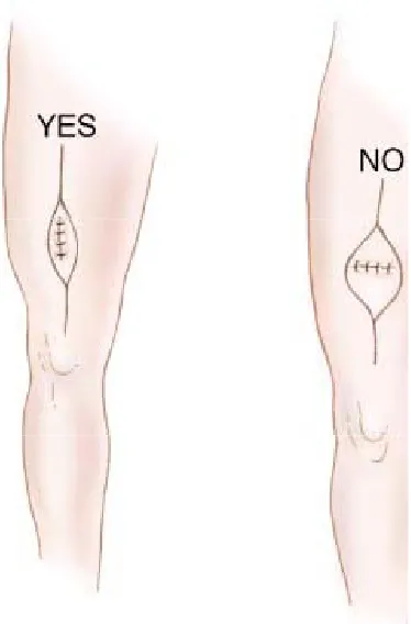
Anatomic Location of the Biopsy Tract
1. Decide, before biopsy, what part of the lesion is most representative of the underlying
disease and will need to have a biopsy.
2. Position the point of entry along the planned incision of the definitive surgery.
3. The biopsy tract must be the shortest way to the lesion; however, it must not violate
4. more than one compartment and must be as remote as possible from the main
neurovascular bundle of the extremity
Biopsy Technique
After adequate planning of the biopsy tract, biopsy should be executed according to the
following guidelines.
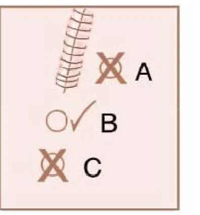
1. Use the smallest longitudinal incision that is compatible with obtaining an adequate
specimen. Transverse incisions are contraindicated because they require a wider
soft-tissue resection at the time of definitive surgery
2. Use a knife or curette to avoid crushing or distorting the specimen’s texture
3. Obtain enough tissue. Always send a specimen for frozen section or touch-prep to
verify the presence of representative tumor material in the specimen. For needle
biopsies, cytopathologic evaluation has to confirm the presence of viable tumor cells. If
pathologic evaluation is negative or questionable, repeat the biopsy.
4. As a general rule, culture what you biopsy and biopsy what you culture.
5. Use meticulous hemostasis. Any hematoma around a tumor should be considered
contaminated
6. Use drains if necessary. The port of entry has to be in proximity and continuation with
the skin incision, not to its sides. The drain path is considered contaminated and has to
Types of biopsy
In soft tissue sarcoma, core needle biopsy, incisional biopsy and excision biopsy are used
depending upon the situations. In most of the situation, Core needle biopsies are adequate and
therefore recommended. In rare circumstances. Incisional biopsy is required. In tumors
smaller than 5 cm, with superficial location excisional biopsy is the preferred approach.
[image:23.612.229.416.248.532.2]Value of Tru-Cut Biopsy
In general, the important issue is the adequacy of the sample. Sufficient viable tissue is
required that is both representative of the lesion and available for histopathologic evaluation,
immunohistochemistry, and, when necessary, electron microscopy. As molecular markers
become a factor in diagnosis, meticulous attention to the adequacy of biopsy, tissue
preservation, and evaluation will be paramount.
Histopathologic interpretation varies from center to center and may be a major variable in
decision making. As with other relatively rare lesions, it is essential that review of the
histopathologic findings be made by an experienced group. More recent studies show
improved diagnostic accuracy and confluence of opinion, at least for malignancy and grade.
Fine-Needle Aspiration Cytology
Fine-needle aspiration (FNA) cytology has been examined by a number of authors.
Some authors have argued that biopsy itself is not justified if FNA is available. But it is
usually confined to the confirmation of recurrence rather than used for the primary diagnosis.
Figure. A drain has to be positioned in proximity to and along the site for
planned incision of the definitive
[image:24.612.134.332.65.278.2]The use of FNA in patients with large sarcomas who are candidates for neoadjuvant
therapy to improve survival is also problematic due to difficulty in grading and subtyping
these tumors accurately from such small samples.
Frozen Section
In some institutions, frozen section is relied on as the diagnostic tool of choice. For
diagnosis of malignancy, frozen section is accurate, but for histopathologic subtypes and
grade, it is inferior to permanent sections of either Tru-Cut or incisional biopsy.
Frozen section can guide retrieval of adequate diagnostic material and, depending on the
initial evaluation, can be an important triage mechanism to direct further pathologic workup.
However, open biopsy with the help of frozen-sectioning support may be indicated when the
Trucut biopsy result is equivocal or for other clinical reasons.
Fatty lesions are not suitable for frozen-section evaluation, because of a loss of
diagnostic material during frozen sectioning and other technical difficulties. In addition,
freezing compromises the final interpretation on permanent sections.
Important application of frozen section is assessment of margin of resection. Negative
margins of resection can be obtained by using this technique intraoperatively.
As an ancillary technique, immunohistochemistry is an invaluable tool that provides
excellent information in assisting the surgical pathologist in establishing a precise diagnosis,
as well as providing relevant prognostic and therapeutic information.
One of its major utilities is to correctly identify a tumor as being of mesenchymal or
nonmesenchymal origin. Once mesenchymal origin has been established, histologic
subtyping according to specific cell lineage may be achieved with the use of lineage-specific
markers. Tumors of uncertain cell lineage and tumors with primitive small round cell
morphology are often characterized by a unique immunohistochemical phenotype. In this
group of tumors, immunohistochemistry is most widely applied and is of greatest value. By
diagnosing the small round cell tumors with aid of immunohistochemistry, line of
management is differed from spindle cell soft tissue tumors.
Despite the rapid development of molecular genetic techniques, immunohistochemistry
still remains the most important diagnostic tool in the diagnosis of soft tissue tumors aside
from recognition of morphologic features and clinical correlation.
Surgery
Although surgery remains the principal therapeutic modality, the extent of surgery
required, along with the optimum combination of radiotherapy and chemotherapy, remains
controversial.
Limb sparing operations are possible in at least 90% of patients. Wide en bloc resection is
limiting factor is usually neurovascular or, occasionally, bony juxtaposition. Because most
soft tissue sarcomas tend not to invade bone directly, only rarely does bone need to be
resected. Soft tissue sarcomas uncommonly involve the skin, so major skin resection should
be limited.
In situations of primary or recurrent tumors in which skin is involved, or which the tumor
is so extensive that skin is involved, then consideration of free flap or rotational flap closure
becomes important, particularly in those patients who are candidates for subsequent adjuvant
radiation therapy.
Amputation for Extremity Sarcoma
The most extensive resection is clearly amputation. This is only rarely indicated nowadays. The amputation should be reserved for tumors not able to be resected by any other means,
without evidence of metastatic disease and the potential for good long-term functional
rehabilitation. This includes patients with considerable cosmetic and functional deformity,
who can be rendered symptom free by a major amputation.
Amputation should be considered for patient preference or if the tumor has the following
characteristics: extensive soft tissue mass and/or skin involvement, major arterial or nerve
involvement, extensive bony involvement that requires whole bone resection, failure of
preoperative therapy or recurrence following prior adjuvant radiation.
The issue of amputation versus limb sparing surgery has been addressed by a prospective
sparing operation plus radiation than in those undergoing amputation, disease free survival is
not different.
Local recurrence
Local recurrence can occur after a limb sparing surgery. Follow up data confirms that
salvage is almost invariably possible, but there is no impact in long term survival.
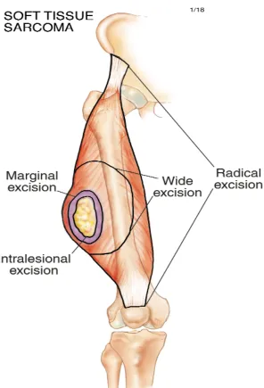
CLASSIFICATION OF SURGICAL PROCEDURES
There are four basic types of excisions (see the figure). Each is based on the relationship of the dissection plane to the tumor and its pseudocapsule.
An intralesional excision is performed within the tumor mass and results in removal of only a portion of it; the pseudocapsule and macroscopic tumors are left behind.
In a marginal excision, the dissection plane passes through the pseudocapsule of the tumor. Such a resection may leave microscopic disease.
Wide (en-bloc) excision entails removal of the tumor, its pseudocapsule, and a cuff of normal tissue peripheral to the tumor in all directions. This is the desired margin for sarcoma
resection; however, the adequate thickness of the normal tissue cuff is a matter of
controversy. For both soft-tissue and bone sarcomas, it is generally believed to be a few
centimeters.
Radical excision involves removal of the tumor and the entire anatomical compartment within which it is located. Traditionally it is mentioned as the fourth excision
type. It excludes the possibility of skip metastases. In compartmental resections, fascia and
periosteum are accepted as wide margin.
[image:29.612.119.415.59.494.2]
An amputation is not necessarily an adequate cancer operation, but it is a method of
achieving a specific margin. It may entail a marginal, wide, or radical excision, depending
upon the plane in which it passes. Staging studies are used to assess local tumor extent and
relevant local anatomy, and thereby determine how a desired surgical margin may be
achieved.
RADIATION THERAPY
Preoperative RT
The usual dose of preoperative RT is 50 Gy. An intraoperative boost or a postoperative
boost with brachytherapy or an external-beam RT is recommended for positive or close
margins. Preoperative RT has several advantages. First, the treatment volume is smaller,
because the need to cover the operative field is not present. Second, preoperative radiation
may reduce seeding during surgical manipulation of the tumor. The tumor may or may not
regress with preoperative RT, but the pseudocapsule may thicken and become acellular,
easing resection and decreasing the risk of recurrence. However, the main disadvantage of
preoperative RT is its effect on wound healing. A higher complication rate has been observed
when primary closure is used. Therefore, involvement of a plastic surgeon in the team may be
necessary to reduce wound complications when preoperative radiation is contemplated. After
preoperative radiation, 3-6 weeks interval before resection is necessary to decrease the risk of
wound complications. Very long intervals between resection and postoperative radiation are
not recommended.
If wide margins are obtained, additional radiation may not be needed. Often, margins
are close because of the proximity of many of these tumors to major neurovascular bundles or
Brachytherapy boosts should be delivered several days after surgery, through catheters placed
at operation, with doses of 12-20 Gy based on margin status. Alternatively, a single
intraoperative dose to the tumor bed of 10-16 Gy, based on margin status, can be delivered
immediately after resection with exposure of the area at risk, avoiding uninvolved organs.
External-beam RT boosts may be an alternative to brachytherapy or intraoperative radiation:
recommended doses are 10-14 Gy for close margins, 16-20 Gy for microscopically positive
margins, and 20-26 Gy for grossly positive margins. Many institutions are no longer giving a
boost after preoperative radiation to patients who have widely negative margins, based on
local control rates that
approach 95% with preoperative radiation at 50 Gy and negative margins.
Postoperative RT
Postoperative RT has been to improve local control in patients with high-grade
extremity soft tissue sarcomas with positive surgical margins. When surgical resection is the
initial therapy, postoperative RT choices include intraoperative radiation therapy (IORT),
brachytherapy or external beam RT. RT is not a substitute for suboptimal surgical resection,
and re-resection may be necessary. If the patient has not previously received RT, one could
attempt to control microscopic residual disease with postoperative RT if re-resection is not
feasible.
External-beam RT is delivered to large fields after surgical healing is complete (at 3-8
weeks) to doses of 50 Gy. Most institutions include the entire operative bed within that
radiation field. Total doses of RT should always be determined by normal tissue tolerance.
For intraabdominal or retroperitoneal tumors, this dose may be decreased to 45 Gy. An
If no intraoperative radiation or brachytherapy was used in the immediate operative or
postoperative period, an external-beam RT boost should be added. For negative margins, an
additional 10-16 Gy is recommended to a reduced field that includes the original tumor bed,
based on grade and width of margins. For microscopically positivemargins, an additional
16-20 Gy is recommended; for grossly positive margins, an additional 16-20-26 Gy is suggested.
Brachytherapy alone has been used as an adjuvant in patients with negative margins.
45-50 Gy to the tumor bed has been shown to reduce recurrence without a significant effect on
wound healing. However, brachytherapy-alone techniques require special expertise and
significant experience. If brachytherapy is used as a boost, doses of 10-20 Gy based on
margin are recommended; a boost dose of 10-16 Gy for close margins or 20 Gy for positive
margins is recommended.
Recent reports from a retrospective study suggest that IORT provides excellent local
control to soft tissue sarcoma of the extremity, when used as a boost to external beam RT.
TREATMENT
Low Grade Tumors (Stage I)
Surgery is the primary treatment for stage I (T1a-1b, N0, M0) low-grade tumors and is
considered definitive if margins are greater than 1 cm or the fascia plane is intact.
Postoperative RT is considered when final margins are 1 cm or less (category 2B). Surgical
N0, M0) low-grade tumors. RT may not be necessary in patients with small lesions (5 cm or
less), because these tumors are less frequently associated with local recurrence.
There are data from two randomized trials and three large single-institution studies that
support using adjunctive RT in appropriately selected patients. Patients receiving either
preoperative or postoperative RT have similar rates of local control and progression-free
survival. However, preoperative RT is associated with a greater incidence of wound
complications, especially in lower extremity tumors. Therefore, the risk of local recurrence
versus the toxicity of adjuvant RT should be assessed before making a decision regarding
radiation.
High-Grade Tumors (Stage II or III)
Large high-grade extremity sarcomas (greater than 10 cm) at high risk for local
recurrences and metastases and should be considered for preoperative therapy. Preoperative
chemotherapy or chemoradiation is used in many centers for high-grade tumors to downstage
a large tumor to enable effective surgical resection, especially in the case of chemo sensitive
histologies. Concurrent chemoradiation with doxorubicin-based regimens has been shown to
improve local control rates in patients with soft tissue sarcoma. Available evidence although
underpowered, suggests that anthracycline-based postoperative chemotherapy would improve
disease-free survival in selected patients who are at high risk of recurrence but otherwise are
in good performance status.
Sarcoma Meta Analysis Corporation performed a meta-analysis of 14 randomized trials
radiation therapy after surgery with a variety of sarcomas. The result of the meta-analysis
showed that doxorubicin-based chemotherapy prolongs relapse-free survival in adults with
localized, resectable soft tissue sarcoma of the extremity and was associated with decreased
recurrence rates. However, adjuvant chemotherapy does not appear to improve overall
survival. Another recent analysis of 674 patients with stage III soft tissue sarcoma
(1984-1999) revealed that clinical benefits from doxorubicin-based chemotherapy lasted for less
than a year. In an Italian randomized cooperative trial, patients with high-grade or recurrent
extremity sarcoma were randomized to receive postoperative chemotherapy with epirubicin
and ifosfamide or observation alone. After
a median follow-up of 59 months, median disease-free survival (48 months vs.16 months)
and median overall survival (75 months vs. 46 months) were significantly better in the
treatment group.
Remarkably little data have been generated in the adjuvant setting regarding the
combination of aggressively dosed ifosfamide plus doxorubicin supported by hematopoietic
cytokine therapy. Phase III randomized study (EORTC-62931) is ongoing to assess the
efficacy of adjuvant chemotherapy after definitive surgery in patients with
high-grade primary or recurrent soft tissue sarcoma at any site. Interim overall survival data
are encouraging from an ongoing phase III trial (EORTC-62961) of regional hyperthermia
versus chemotherapy (etoposide, ifosfamide, adriamycin) alone for patients with high-risk
soft tissue sarcomas, especially for extremity sarcomas.
Treatment options for stage II or III high-grade tumors should be decided by a
multidisciplinary team, based on the performance status, comorbid factors including age,
by RT with or without chemotherapy is the primary treatment for resectable high-grade
sarcomas. The NCCN guidelines recommend various neoadjuvant approaches including
preoperative RT or chemotherapy or chemoradiation prior to surgery, followed by
postoperative radiation with or without chemotherapy for respectable tumors with acceptable
functional outcomes and for potentially resectable tumors with concerns for adverse
functional outcomes. Adjuvant chemotherapy alone can be considered in the case of patients
who have received preoperative radiation alone. Surgery alone is an option for small tumors
that can be resected with wider surgical margins.
Recurrent Disease
The management of recurrent disease or primary presentation with metastases
encompasses a heterogeneous group of patients and clinical scenarios. For a patient with a
local recurrence, treatment decisions should be made using the same algorithm as for patients
with a new primary lesion. .
Surveillance
Surveillance is deemed important to detect recurrences that might still be potentially
curable. However, very limited data is available in the literature on effective surveillance
strategies. The guidelines outline a prudent follow-up schedule that avoids excessive testing.
Higher grade and larger tumors have a higher risk of dissemination; therefore, the
surveillance recommendations for patients with these tumors are somewhat more intensive,
particularly for the first 3 years after resection. Periodic imaging (MRI, CT, or consider
recurrence. However, in situations where the area is easily followed by physical examination,
imaging may not be required. After 10 years, the likelihood of developing a recurrence is
small and follow-up should be individualized.
Stage I tumors are routinely followed with H&P every 3 to 6 months for 2 to 3 years and
then annually. Baseline imaging should be considered after primary therapy. Chest x-ray
should also be considered every 6 to 12 months. For stage II and stage III tumors, H&P and
chest imaging (plain radiograph or chest CT) should be done every 3 to 6 months for 2-3
years, then every 6 months for the next 2 years, and then annually. Because these
patients’ risk never returns to zero, long-term follow-up is indicated, including consideration
of MRI or CT scanning.80 Chest imaging (plain radiograph or chest CT) is performed every
3 to 6 months for 5 years and then annually, given the risk of metastatic disease in these
high-grade lesions. There has never been a study to prove that the use of more sensitive CT scans
in routine surveillance would improve clinical outcomes. According to the reported data from
M. D. Anderson Cancer Center, routine use of chest CT adds little clinical benefit, when risk
of pulmonary metastases is low.81 However, in certain subsets of patients in whom chest
radiographs are difficult to interpret because of anatomic considerations (scarring,
emphysema, etc), chest CT surveillance may be indicated.
REHABILITATION AFTER LIMB-SPARING SURGERY
Although limb-sparing surgery can enhance the quality of life it does cause a variety of
Rush defined medical rehabilitation as “maximal preservation of physical, psychological,
social, occupational, creative and economical function in conjunction to malignant disease
and its treatment”.
For many years it has been assumed that investing in extensive rehabilitation efforts for
patients with poor prognoses and short life expectancies could not be justified from an
economic perspective. Now that cure rates among patients who have undergone Limb
Sparing Surgery are 60–80%, this assumption is clearly no longer correct. It is no less
important to rehabilitate patients with metastatic disease.
LIMB SPARING SURGERY versus AMPUTATION
Limb Sparing Surgery can result in survival rates and disease-free periods that equal
those achieved with amputation. The presumed functional and psychological advantages of
Limb Sparing surgery over amputation, however, have yet to be established. Limb Sparing
Surgery appears to offer the possibility of better psychological functioning and an intact body
image, but it is more complex and demanding than amputation and is associated with more
morbidity. The duration of surgery is longer, and infection, pain, and other postoperative
complications are more common.
Otis et al. compared energy cost during gait in osteosarcoma patients after resection of
the distal femur and knee joint and replacement with an endoprosthesis with that of patients
who had undergone an above-knee amputation. The former had a lower energy cost during
gait than the latter.
Eiser et al. noted that one of the disadvantages of Limb Sparing Surgery is that additional
hospitalizations may be necessary in the case of complications
It is important to emphasize that, in order to justify Limb Sparing Surgery, the procedure
must provide limb function equal or superior to that provided by an external prosthesis after
amputation. At the same time the tumor must be resected according to principles of oncologic
surgery (i.e. there must be free margins and minimal damage to major neurovascular bundles
and muscles). Despite the disadvantages, nearly all patients believe that an attempt to salvage
a limb is worth the time and effort.
4. MATERIALS AND METHODS
Patients admitted and undergone limb sparing surgery for extremity soft tissue sarcoma
between January 2004 to April 2008 in the Department of Surgical Oncology, Government
Royapettah Hospital, Chennai were taken for study.
Data were collected in all patients. Patient’s age and sex were noted. Histories like
presence of swelling, its duration, presence of pain and its duration, other symptoms and
family history were recorded. Previous history of surgery, biopsy if any and treatment were
taken.
Physical examination was done to note site, size of swelling and presence or absence of
metastases. Previous surgical scar, its length, present status, orientation to longitudinal axis of
limb were noted.
All patients were evaluated with X-ray chest and CT Scan local part or MRI of local part.
CT Scan chest was done in all cases. Histology was obtained with trucut biopsy. If there was
already biopsy, it was reviewed.
Patients with metastatic disease were excluded from study. Suitable patients for limb
salvage were undergone surgery. Those patients requiring amputation were excluded.
Postoperative complications and its management were observed. Histopathology, grade, and
margin status were noted. Histopathology is compared with previous reports. Surgical,
Patients were followed up regularly once a month in first year, every two months in
second month, every three months in third year, every 6 months in fourth and fifth year and
5. OBSERVATION AND ANALYSIS
Musculoskeletal sarcomas consisted of 2.24% of all cancers admitted from January 2004 to April 2008. Soft tissue sarcoma represents less than 1% of all adult of all malignancies in various studies but in this series it represented about 1.64%.
Cancers Number Percentage
Musculoskeletal sarcomas 292 2.24
Other cancers 12719 97.76
Total 13011 100
Cancers Number Percentage
Soft tissue sarcomas 213 1.64
Other cancers 12798 98.34
Total 13011 100
Soft tissue sarcomas consisted of 73% (213 cases) of total 292 cases (bone
sarcoma-22%[63] , fibromatosis- 5%[16] of cases] with ratio of soft tissue sarcoma to bone sarcoma
3.5:1 compared to 3:1 in the literature.
SOFT TISSUE SARCOMAS 213 73
FIBROMATOSIS 16 5
BONE SARCOMAS 63 22
TOTAL 292 100
Extremity soft tissue sarcomas consisted of 57% (122 cases) of total 213 soft tissue
sarcoma cases (0ther sites-43% [91] of cases).
Site distribution of soft tissue sarcoma Frequency Percentage
Extremity 122 57
Other sites 91 43
Total 213 100
Admission in Memorial Sloan-Kettering CancerCenter consisted of 41% of extremity sarcomas and 59% of other site soft tissue sarcomas and in this study, it is 57% and 43%
respectively.
Centre Extremity Other sites
Memorial Sloan-Kettering Cancer Centre 41% 59%
MD Anderson cancer centre 59% 41%
In extremity soft tissue sarcomas, 73% (89 cases) had localized disease, and 27% (33
cases) had metastatic disease on presentation. Temple LK et al. reported 20% metastatic
disease at presentation.
Extremity-presentation Frequency Percentage
Metastatic 33 27
Localized 89 73
Total 122 100
Metastatic disease Percentage
Temple et al 20%
Government Royapettah Hospital 27%
Out of 89 cases of localized extremity soft tissue sarcomas, 72 (81%) cases were
identified as fit for limb sparing surgery. Remaining 17 (19%) cases were not suitable for
limb sparing surgery and needed amputation.
Localized soft tissue sarcoma-
fitness for limb sparing surgery
Frequency Percentage
Fit 72 81
NOT FIT, needed amputation 17 19
The most extensive resection is clearly amputation. This should be only rarely
indicated in soft tissue sarcoma because limb-sparing operations are possible in at least 95%
of patients. Experience over the last 25 years at MSKCC indicates that the amputation rate, which was 50% in the late 1960s, is now less than 5%. Amputation should be reserved for
tumors that cannot be resected by any other means, without evidence of metastatic disease
and the potential for good long-term functional rehabilitation. This usually includes patients
with large, low-grade tumors with considerable cosmetic and functional deformity, who can
be rendered symptom free by a major amputation. In this study, 19% of patients with
localized soft tissue sarcoma required amputation. This high rate of amputation is due to large
tumor size and late presentation of our patients.
Centre Limb sparing surgery Amputations
Memorial Sloan-Kettering Cancer Centre >95% <5%
Watson DI et al 92.5% 7.5%
Government Royapettah hospital 81% 19%
In thisseries of 72 patients, only 58(81%) patients were undergone limb sparing surgery and remaining 14(19%) patients refused further treatment. So, these 58 patients were taken
for further study.
Course of patients fit for Limb sparing surgery Frequency Percentage
Refused surgery 14 19
Total 72 100
In this series of 58 patients, male patients were 35(60%) and female were 23 (40%) with
male to female ratio of 1.5: 1. and Ratio is 4:1 in the literature.
Patient’s age ranges from 16 to 75. Median age of patients is 45, average age is 46. Two
third of the patients are in the age group of 31-60.
AGE Frequency Percentage
<30 years 12 21
31-60 years 38 66
>61 years 8 13
Total 58 100
All patients are presented with swelling with varying duration. 52% (30 cases) of patients
presented with pain and 3.5% (2) cases with other symptoms.
Symptoms Frequency Percentage
SEX Frequency Percentage Male 35 60
Female 23 40
Swelling All 100%
Pain 30 52%
Other symptoms 2 3.5
In a study by Lawrence et al, 33% of patients complained of pain at the time of
diagnosis.In this study, pain is the symptom that draws the patient to seek medical attention.
Any soft tissue mass in an adult that is symptomatic or enlarging, any mass that is larger than
5 cm, or any new mass that persists beyond 4 weeks is sampled
Approximately 10% soft tissue sarcoma patients have positive family history of cancer as
observed in this study as well as in the literature. Approximately 5% of patients with NF
develop malignant peripheral nerve sheath tumors (MPNSTs) ( Sorensen S et al). In this
study one out of 12 patients with malignant peripheral nerve sheath tumors had
neurofibromatosis.
39 (67%)cases were occurred in lower extremity and 19(33) cases in upper extremity with
ratio of 2:1 comparable to Memorial Sloan-Kettering Cancer Center’s experience in which it
is 71% and 29% respectively with ratio of 2.5:1.
SITE Frequency Percentage
Lower Extremity 39 67
Upper Extremity 19 33
TOTAL 58 100
Memorial Sloan-Kettering Cancer Center 29% 71%
Government Royapettah Hospital 33% 67%
SITE Frequency Percentage Gluteal region 4 7%
Thigh 30 52%
Leg 4 7%
Ankle 1 1.7%
Foot - -
Shoulder 6 10%
Arm 6 10%
Elbow 1 1.7%
Forearm 5 8.9%
Hands 1 1.7%
TOTAL 58 100%
Commonest site of extremity soft tissue sarcoma in all series is thigh. In this study also
commonest site is thigh (30 (52%) cases) followed by shoulder and arm (6 cases (10%)
each)
24(41%) patients have primary tumors, 18(31%) are recurrent tumors and 16(28%) are residual tumors. Residual tumors are those presented after excision biopsy or marginal
excision.
Status of primary tumor Frequency Percentage
Recurrent 18 31
Residue 16 28
Total 58 100
Stage Frequency Percentage
1A 5 9
1B 29 50
2A 2 3
2B - -
3 18 31
4 1 2
No stage 3 5
Total 58 100
On admission 29(50%) cases are in stage 1B, followed by 18(31%) cases in stage 3. In
three cases, stage was not assigned since they are dermatofibrosarcoma protuberance
(2 cases) and angiosarcoma( one case).
Out of 55 cases, 46(84%) cases have T2b disease, 8 (14%) cases have T1b disease and
T1a one case. 48 (83%) out of 58 cases had swelling more than 5 cm in size. So, any soft
tissue mass that is >5cm in size should be investigated definitively (Brennan MF et al).
T stage Frequency Percentage
T1a 1 2
T2a - -
T2b 46 84
Total 55 100
Liposarcoma is the commonest histology with 22% (13 cases). Liposarcoma, malignant
peripheral nerve sheath tumor and malignant fibrous histiocytoma occur in almost equal
frequency , comprising about 62% of cases.
Histopathology is dependent on the anatomic site. The common subtypes in the extremity
are liposarcoma and malignant fibrous histiocytoma (Fletcher CD). In this study, Liposarcoma is the commonest histology followed by malignant peripheral nerve sheath
tumor and malignant fibrous histiocytoma.
Histology Frequency Percentage
Liposarcoma 13 22
Malignant peripheral nerve sheath tumor 12 21
Malignant fibrous histiocytoma 11 19
Synovial sarcoma 6 10.5
Spindle cell sarcoma 6 10.5
Others 10 17
Total 58 100
Margin status Frequency Percentage
Free 48 83
Close 4 7
Positive 0 0
Not applicable 6 10
Total 58 100
As might be expected, there can be considerable disagreement among pathologists
regarding the specific histologic diagnosis in individual cases.
In this study in comparison with preoperative histology there is 64% of concordance with
26% of discordance rate.
Frequency Percentage
Histology concordance 37 64
Histology discordance 15 26
Not available 6 10
Total 58 100
When pathologic material from 424 patients who entered into Eastern Cooperative
Oncology Group (ECOG) sarcoma trials was reviewed by a panel of expert pathologists, 10%
of cases were rejected as not being sarcoma, and for 14% of the remaining cases there was
In the Scandinavian Sarcoma Group experience, the specific histologic diagnosis was
disputed in 20% of cases.37 With increasing familiarity with the immunohistochemical and
genetic studies needed to diagnose soft tissue sarcoma, the rate of this discordance may be
decreasing.
Study Discordance Rate
ECOG sarcoma trials 14%
Scandinavian Sarcoma Group 20%
Government Royapettah Hospital 26%
Wide mono bloc excision was done in 47 (81%) cases, compartmental excision in
10(17%) cases and marginal excision in one case.
Type of surgery Frequency Percentage
Wide mono bloc excision 47 81
Compartmental excision 10 17
Marginal excision 1 2
Total 58 100
In 7 (12%) cases vascular resection was done. But only in 4 (7%) cases vascular
reconstruction was done.
Vessel Resected Frequency Percentage
Femoral Artery & Vein 3 43%
Deep femoral Artery & Femoral Vein 1 14%
Deep femoral Artery 2 29%
TOTAL 7 100% Type of vascular reconstruction Frequency Percentage
Superficial femoral Artery & Vein 2 29%
Superficial femoral artery only 1 14%
Superficial femoral Vein only 1 14%
No reconstruction 3 43%
Total 7 100%
35 (60%) patients needed some form of reconstruction. 18 (31%) patients needed flap
reconstruction, 5 skin graft, 4 vascular reconstruction, 4 tendon transfers, and flap and skin
graft, flap and plate, flap and mesh, mesh one each.
Types of Reconstruction Frequency Percentage
Flaps 18 31%
SSG 5 8.6%
Vascular 4 6.8%
Tendon transfer 4 6.8%
Flap & SSG 1 1.7%
Flap & Plate 1 1.7%
Flap & Mesh 1 1.7%
Mesh 1 1.7%
Total 35 60%
Latissmus dorsi flap and local transposition flap are common flaps used( in 6 cases each ).
Gastrocnemius flap is used in 3 cases and reverse sural artery flap in 2 cases. Other flaps are
used in 4 cases.
Types of flaps used Frequency Percentage
Latissmus dorsi 6 10.3%
Transposition flap 6 10.3%
Gastrocnemius 3 5.2%
Reverse sural artery 2 3.4%
Pectoralis Major Myo Cutaneous 1 1.7%
Posterior thigh 1 1.7%
Abdominal 1 1.7%
Tensor fascia lata 1 1.7%
Total 21 36%
So in situations of primary or recurrent tumors in which skin is involved or in which the
tumor is so extensive that skin is involved, then consideration of free flap or rotational flap
closure becomes important, particularly in those patients who are candidates for subsequent
Overall complications rate is 42%. Most common complication is marginal flap necrosis
followed by lower limb edema. Other complications are Infection and wound gaping (2
cases), infection (one case), wound gaping (one case) and femoral artery blow out (one case).
Complications Frequency Percentage Marginal flap necrosis 12 21
Edema leg 5 9
Seroma 2 3.4
Infection and wound gaping 2 3.4
Infection 1 1.7
Wound gaping 1 1.7
Femoral artery blow out 1 1.7
Total complications 24 42 No complications 34 58
Total 58 100
79% (19 cases) were needed some form of secondary intervention to manage the
complications.
Management of complications Frequency Percentage
Wound debridement 6 25%
Secondary suturing 6 25%
Flap reconstruction 2 8%
Conservative 5 21%
Total 24 100%
In this study, 33% of patients developed postoperative complications requiring some
form of intervention compared to 16% of complications reported by Yang JC et al in a
randomized prospective study. But no patients required amputations in this study as reported
by Yang JCet al (2% amputation rate).
Post operative complications Yang JC et al Government Royapettah Hospital
Requiring intervention 16% 33%
Requiring amputation 2% Nil
Totally 7(12%) cases were presented with history of previous radiotherapy or
chemotherapy or both. Totally 5(8.5%) cases had radiotherapy, so adjuvant radiotherapy is
not possible in these cases.
Frequency Percentage
Previous H/O radiation 2 3.5%
Previous H/O radiation and chemotherapy 3 5%
Chemotherapy 2 3.5%
Total 7 12%
32 cases have received adjuvant therapy. Out of these, one had preoperative radiotherapy,
23 cases had postoperative adjuvant radiotherapy, 7 cases had both chemotherapy and
radiotherapy, and one had chemotherapy.
Additional treatment Frequency Percentage
Preoperative radiotherapy 1 1.7%
Postoperative radiotherapy 23 39.6% Postoperative radiotherapy and chemotherapy 7 12.1%
Postoperative chemotherapy 1 1.7%
Total 32 55.1%
The goals of adjuvant radiotherapy in the management of soft tissue sarcoma are to
enhance local control, preserve function, and achieve acceptable cosmesis by contributing to
tissue preservation. The evidence for adjunctive radiation therapy in patients eligible for
conservative surgical resection comes from two randomized trials and a number of large
single-institution reports.
In one of these randomized trials, conducted by the National Cancer Institute, 91
patients with high-grade extremity tumors were treated with limb-sparing surgery followed
by chemotherapy alone or radiation therapy plus adjuvant chemotherapy. A second group of
50 patients with low-grade tumors were treated with resection alone versus resection with
radiation therapy. The 10-year local control rate for all patients receiving radiation therapy
In the second randomized trial, which was performed at Memorial Sloan-Kettering
Cancer Center, 164 patients were randomized to observation or brachytherapy following
conservative surgery. The 5-year local control rate for patients with high-grade tumors was
66% in the observation group and 89% in the group treated with brachytherapy. There was no
significant difference between the groups of patients with low-grade tumors.
6 patients had disease recurrence. 4 patients had local recurrence. All but one local
recurrence were successfully salvaged with limb sparing surgery. One patient had both local
and distant recurrence at two years and died after 6 months (survival 30 months). Another
patient recurred distantly at 30 months, now alive with disease (survival 37 months)
Recurrence pattern Frequency Percentage
Local 4 7%
Local and distant 1 1.7%
Distant 1 1.7%
Total 6 10.4%
Follow up ranges from 1 month to 51 months. One patient had more than 4 years follow up, 8 patients had more than 3 years follow up, 22 patients had more than 2 years follow up, 8
patients had more than 1 year follow up and 19 had less than one year follow up.
Duration follow up No.of patients Percentage
More than 4 years 1 1.8%
More than 3 years 8 13.8%
More than 1 year 8 13.8%
Less than 1 year 19 32.8%
Total 58 100%
84.5% (49 cases) are disease free at closure of this study. 3.4% (2 cases) are alive with
disease. 1.7% (0ne case) was died. 10.4% (6 cases) are lost follow up.
Present status No. of patients Percentage
Disease free 49 84.5%
Alive with disease 2 3.4%
Dead 1 1.7%
Lost follow up 6 10.4%
Total 58 100%
70.8%(41 cases) and 22.4% (13 cases)of patients had good and fair functional outcome.
Remaining 6.8% (4 cases, 2 cases each) had excellent and poor functional result.
Outcome Frequency Percentage
Excellent 2 3.4%
Good 41 70.8% Fair 13 22.4%