Copyright © 1998, American Society for Microbiology
The N Terminus of Rotavirus VP2 Is Necessary for
Encapsidation of VP1 and VP3
CARL Q.-Y. ZENG,
1* MARY K. ESTES,
1ANNIE CHARPILIENNE,
2ANDJEAN COHEN
2Division of Molecular Virology, Baylor College of Medicine, Houston, Texas 77030,
1and Laboratoire de Virologie et
d’Immunologie Moleculaire, INRA, C. R. J. Domaine de Vilvert, Jouy-en-Josas, 78350, France
2Received 17 March 1997/Accepted 23 September 1997
The innermost core of rotavirus is composed of VP2, which forms a protein layer that surrounds the two
minor proteins VP1 and VP3, and the genome of 11 segments of double-stranded RNA. This inner core layer
surrounded by VP6, the major capsid protein, constitutes double-layered particles that are transcriptionally
active. Each gene encoding a structural protein of double-layered particles has been cloned into baculovirus
recombinants and expressed in insect cells. Previously, we showed that coexpression of different combinations
of the structural proteins of rotavirus double-layered particles results in the formation of virus-like particles
(VLPs), and each VLP containing VP1, the presumed RNA-dependent RNA polymerase, possesses replicase
activity as assayed in an in vitro template-dependent assay system (C. Q.-Y. Zeng, M. J. Wentz, J. Cohen, M. E.
Estes, and R. F. Ramig, J. Virol. 70:2736–2742, 1996). This work reports construction and characterization of
VLPs containing a truncated VP2 (VP
D
2, containing amino acids [aa] Met-93 to 880). Expression of VP
D
2
alone resulted in the formation of single-layered
D
2-VLPs. Coexpression of VP
D
2 with VP6 produced
double-layered
D
2/6-VLPs. VLPs formed by coexpression of VP
D
2 and VP1 or VP3, or both VP1 and VP3, resulted in
the formation of VLPs lacking both VP1 and VP3. The presence of VP6 with VP
D
2 did not result in
encapsi-dation of VP1 and VP3. To determine the domain of VP2 required for binding VP1, far-Western blot analyses
using a series of truncated VP2 constructs were performed to test their ability to bind VP1. These analyses
showed that (i) full-length VP2 (aa 1 to 880) binds to VP1, (ii) any N-terminal truncation lacking aa 1 to 25
fails to bind VP1, and (iii) a C-terminal 296-aa truncated VP2 construct (aa 1 to 583) maintains the ability to
bind VP1. These analyses indicate that the N terminus of rotavirus VP2 is necessary for the encapsidation of
VP1 and VP3.
The rotaviruses, members of the family Reoviridae, are
tri-ple-layered viruses with a genome of 11 segments of
double-stranded RNA (dsRNA) (for a review, see reference 10) and
are the most important cause of severe viral gastroenteritis in
humans and animals (1, 15). Morphologically and
biochemi-cally, the triple-layered virus particles consist of three
concen-tric proteinaceous capsid layers: (i) an outer protein layer of
780 molecules of VP7 from which 60 dimers of the spike
protein VP4 project; (ii) a middle protein layer of 260 trimers
of VP6; and (iii) an inner protein layer of 120 molecules of
VP2 (6, 28). The VP2 inner shell encapsidates the two minor
proteins VP1 and VP3 and the 11 segments of genomic
dsRNA, and this structure has been called the core of the
virion. VP1, VP3, and the 11 segments of genomic dsRNA
have been proposed to be organized into a subcore (27). This
proposal is supported by the demonstration that much of the
genomic dsRNA is organized, forming a dodecahedral
struc-ture that is organized around VP1-VP3 complexes located at
the fivefold vertices (29).
The VP2 shell of rotavirus plays important roles in the
struc-ture and function of the rotavirus core. For example, VP2
interacts with trimers of VP6 that surround the VP2 layer and
are perforated by 132 aqueous channels (30, 35), transporting
metabolites in and nascent RNA out during transcription. VP2
also binds to viral RNA and may function in the replication
and encapsidation of dsRNA (2, 5, 16, 23). The nucleic acid
binding domain in VP2 is located between amino acids (aa) 1
and 132 (17), and the bond between Gln-92 and Lys-93 in VP2
is a protease-accessible site (37). Interactions between VP2 or
VP1 and nucleic acids appear to be critical for the endogenous
enzymatic activity of the core (23, 24). VP1, the minor core
protein, is thought to be the viral RNA-dependent RNA
poly-merase (transcriptase) because (i) temperature-sensitive
mu-tants mapping to gene 1 have an RNA-negative phenotype
(13); (ii) VP1 sequences contain the common motif of all RNA
polymerases (8, 26); (iii) cross-linking of azido-ATP to VP1
causes a corresponding decrease in the ability of native
double-layered particles to synthesize mRNA (33); and (iv) 1/2-VLPs
(previously designated VP1/2 particles [38]; VLPs are
virus-like particles) expressed from baculovirus recombinants have
replicase activity that synthesizes negative-stranded RNA on
positive-stranded RNA templates, while VP2 alone lacks this
activity (7, 38). Recently, VP1 alone has been reported to lack
replicase activity, but VP2 stimulates this activity (24). VP3,
the other minor core protein, binds GTP specifically and is
thought to be a guanylyltransferase (22, 25). VP1 and VP3
appear to form a complex located on the inner surface of the
VP2 layer at the icosahedral fivefold axes (29). Expression of
VP2 lacking the N-terminal 92 aa in Spodoptera frugiperda Sf9
cells results in changes of the three-dimensional structure
along the inner surface of the 2-VLPs (20).
In this study, N-terminally truncated forms of VP2 expressed
from baculovirus recombinants were used to show that
inter-actions with VP2 are necessary for encapsidation of VP1 and
VP3 and to map the VP2 domain necessary for these
interac-tions.
* Corresponding author. Mailing address: Division of Molecular
Virology, Baylor College of Medicine, One Baylor Plaza, Houston, TX
77030. Phone: (713) 798-3591. Fax: (713) 798-3586. E-mail: qzeng
@bmc.tmc.edu.
201
on November 9, 2019 by guest
http://jvi.asm.org/
MATERIALS AND METHODS
Viruses and cells. S. frugiperda Sf9 cells were grown and maintained in
TNM-FH (Hinks) medium containing 10% fetal bovine serum (FBS) (31). Bac-ulovirus recombinants encoding the following rotavirus proteins were used: pVL941/RF-1 (VP1 of virus strain RF) (8), BacRf2A (VP2 of virus strain RF) (16, 18), BVP2C24 (VP2 Met-93 to aa 880 of virus strain RF) (this work), BVP2D185 (VP2 aa 186 to 880 of virus strain RF) (this work), Bac2AT1777 (VP2 aa 1 to 583 of virus strain RF) (17), Bac2M16.9 (VP2 aa Met-26 to 583 of virus strain RF) (17), Bac2M16.3 (VP2 aa Met-45 to 583 of virus strain RF) (17), pVL1393/SA11-3 (VP3 of virus strain SA11) (22), and pAc461/SA11-6 (VP6 of virus strain SA11) (11, 12).
Construction of baculovirus recombinants encoding VPD2 (VP2 aa Met-93 to 880) and VPD2*(VP2 aa 186 to 880).Two mutants of VP2 with deletions from the N terminus of 92 and 185 aa (designated VPD2 and VPD29, respectively) were constructed by using a strategy described previously (17). For VPD2, an extra starting codon was added. For VPD29, the starting codon corresponds to Met-186. Briefly, a set of oligonucleotides was designed to amplify the corre-sponding region of the full-length cDNA encoding VP2 and to generate a
BamHI site at the end of the PCR product. These products were cloned into
pBS, and part of the recombinant plasmids (pBS2C24Dand pBSRF2D185, cor-responding to VPD2 and VPD29, respectively) were sequenced to check the junctions. The absence of premature termination mutations in the open reading frame was confirmed by transcription translation using the TnT-T7 coupled system from Promega (Madison, Wis.). Recombinant baculoviruses BVP2C24 and BVP2D185, encoding VPD2 and VPD29, respectively, were constructed as previously described (17).
Production and purification of rotavirus single-layered VLPs.Single-layered VLPs, i.e.,D2-,D29-, 2-, 1/D2-, 1/2-, 1/D2/3-, 1/2/3-,D2/3-, and 2/3-VLPs, were prepared as previously reported for production of VP2 VLPs (18, 37). For each VLP, 23107Sf9 cells in a T-75 flask were infected at a multiplicity of infection
of 10 PFU per cell for each recombinant and were incubated for 2 h at 27°C for adsorption. The inoculum was removed, and the indicated VLPs were expressed in TNM-FH–10% FBS for 3 to 4 days in the absence or presence of protease inhibitors. Protease inhibitors aprotinin and leupeptin, each at 1mg/ml, were added daily. The cells were lysed with 2% sodium deoxycholate (DOC) lysis buffer (10 mM Tris-HCl [pH 7.4], 0.1 mM EDTA, 2% DOC). The expression levels of VP1 and VP3 in the lysates of VP1/2 versus VP1/D2, VP1/2/3 versus VP1/D2/3, and VP2/3 versus VPD2/3 were examined by testing dilutions of samples, using a dot blot assay for VP1 and [a-32P]GTP binding for VP3. The
VLPs were purified and fractionated from the cell lysates by centrifugation for 2 h at 30,000 rpm in a Beckman SW41 rotor through a 5 to 20% sucrose gradient containing 0.2% DOC. Fractions (0.7 ml each) were collected. Sodium dodecyl sulfate-polyacrylamide gel electrophoresis (SDS-PAGE) and silver staining showed that unassembled soluble proteins were located in the top fractions 1 to 4. Although VPD29(aa 186 to 880) did not form VLPs, all other eight types of single-layered VLPs sedimented as a broad band with a peak at fraction 9, as previously described (37). The yields of the various single-layered VLPs were between 40 and 70mg per 83107Sf9 cells. The single-layered VLPs in fraction
9 were kept at 4°C and used within 2 weeks for further experiments.
Production and purification of rotavirus double-layered VLPs. Double-lay-ered VLPs, i.e.,D2/6-, 2/6-, 1/D2/6-, 1/2/6-,D2/3/6-, 2/3/6-, 1/D2/3/6-, and 1/2/3/ 6-VLPs, were prepared essentially as described previously (9, 38). Briefly, Sf9 cells were infected at a multiplicity of infection of 5 PFU per cell for each recombinant. Viruses were adsorbed for 2 h at 27°C. The inoculum was removed by low-speed centrifugation, and VLPs were expressed for 6 days in Grace’s medium–0.5% FBS in the absence or presence of protease inhibitors. The pro-tease inhibitors aprotinin and leupeptin, each at 1mg/ml, were added daily from days 2 through 5 postinfection. The expression levels of VP1 and VP3 in the lysates of VP1/2/6 versus VP1/D2/6, VP1/2/3/6 versus VP1/D2/3/6, VP2/3/6 versus VPD2/3/6 were examined by testing dilutions of samples 80 h postinfection, using a dot blot assay for VP1 and [a-32P]GTP binding for VP3. The infected cells were
harvested 6 days postinfection. The VLPs were released into the medium and purified by pelleting through a 35% sucrose cushion followed by banding by CsCl (refractive index, 1.3610) isopycnic centrifugation. Double-layered VLPs com-posed of VPD2 showed two bands in CsCl gradients, identical to those composed of VP2 (38). The densities were 1.27 g/cm3for the light particles and 1.30 g/cm3
for the heavy particles. The VLPs in the two bands were pooled, diluted with 10 mM Tris-buffered saline (pH 7.4), and pelleted for 2 h at 30,000 rpm in a Beckman SW41 rotor. The pellets were resuspended in the same Tris-buffered saline and kept at 4°C until used. The yields of the various doubled-layered VLPs were between 200 and 400mg per 6.53108Sf9 cells. Double-layered VLPs
composed of VPD2, like those composed of VP2, were stable for at least 8 months as examined by electron microscopy (EM).
SDS-PAGE.The protein profiles of all VLPs except those detected by far-Western blotting (see below) were determined by reducing SDS-PAGE using 10% resolving and 4% stacking gels (3, 19). Protein locations were stained with a silver staining kit (Sigma, St. Louis, Mo.) according to the manufacturer’s instructions.
GTP binding assay.The presence of VP3 in VLPs was determined by a GTP binding assay. The reaction was performed in a 20-ml reaction volume containing
;2.5mg of VLP protein, 10mCi of [a-32P]GTP (Amersham Life Science, Inc.,
Arlington Heights, Ill.), 2 mM MgCl2, and 10 mM Tris-HCl-buffered saline (pH
7.4) as described previously (22). After a 30-min incubation at room tempera-ture, the reaction was terminated by addition of 5ml of 53Laemmli sample buffer. After being boiled for 5 min, the proteins were resolved by SDS-PAGE on a 10% gel and electroblotted onto a nitrocellulose membrane (32). Proteins that bound [a-32P]GTP were visualized by autoradiography.
Production and purification of VP1.Sf9 cells were infected with baculovirus recombinant pVL941/RF-1 at a multiplicity of infection of 10 PFU per cell and incubated for 2 h at 27°C for adsorption. The inoculum was removed by low-speed centrifugation, and VP1 was expressed for 7 days in serum-free medium SF900 II SFM (Gibco BRL, Grand Island, N.Y.). After 7 days postinfection, the medium containing VP1 was equilibrated by dialysis against 20 mM Tris-HCl (pH 8.1), clarified by high-speed centrifugation, filtered through a 0.22-m m-pore-size filter (Costar Co., Cambridge, Mass.), and then semipurified by fast protein liquid chromatography (FPLC) with a quaternary methylamine anion-exchange column (Water Chromatography Division, Milford, Mass.). Quaternary methyl-amine-bound proteins were eluted with an NaCl linear gradient, 0 to 1.6 M, and VP1 was eluted with 0.12 M NaCl. VP1-rich fractions were pooled and further purified by passage through an immunoaffinity column containing rabbit immu-noglobulin G (IgG) against wild-type baculovirus proteins made in this labora-tory. VP1 eluted in the unbound protein portion flowthrough. SDS-PAGE (10% gel) and silver staining were used to analyze the purity of VP1. Analysis of 1mg of purified VP1 showed a single band with an apparent molecular weight of about 125,000.
Guinea pig antiserum to VP1.A guinea pig anti-VP1 serum was made by inoculating animals with;20mg of purified VP1 in complete Freund’s adjuvant (Gibco BRL) once, followed by five boosts of;20mg of VP1 each at 3-week intervals in incomplete Freund’s adjuvant (Gibco BRL). The final bleed as primary antibody (1:500 dilution) showed a single VP1 band in VP1 lysates of Sf9 cells infected with baculovirus recombinant pVL941/RF-1 and with 1/2/6-VLPs, as examined by Western blotting. This anti-VP1 serum was used for far-Western blotting to verify the presence of VP1 binding proteins.
Far-Western blotting and VP1 binding test.A far-Western blotting procedure was established essentially as reported by Lee et al. (21) and Homann et al. (14). Purified VLPs were either denatured with SDS in the presence ofb -mercapto-ethanol (bME) (Laemmli sample buffer) and boiled for 5 min or denatured with SDS in the absence ofbME and kept at 4°C. All the subsequent steps were performed at 4°C. The denatured VLP proteins were resolved on a SDS–10% polyacrylamide gel (19). After electrophoresis, the proteins were blotted to a polyvinylidene difluoride (PVDF) membrane (Millipore, Bedford, Mass.) (32). Proteins on the blots were subsequently renaturated by incubation in standard binding buffer (SBB; 10 mM HEPES [pH 7.4], 10 mM MgCl2, 50 mM EDTA, 1
mM dithiothreitol, 10% [vol/vol] glycerol) for 12 h and then incubated for 6 h in SBB–5% bovine serum albumin (BSA) to saturate and block any free binding sites. For probing, BSA-blocked blots were incubated for 24 h with purified VP1 (10mg/ml) in SBB, followed by reaction with the guinea pig anti-VP1 serum (1:500 dilution). Reactivity of the anti-VP1 serum was visualized by addition of a goat anti-guinea pig IgG-alkaline phosphatase conjugate (Sigma) and substrate 5-bromo-4-chloro-3-indolylphosphate–p-nitroblue tetrazolium chloride (Amer-sham Life Science).
EM.The morphology of all VLPs was examined by EM (37). Collodion-carbon-coated and freshly glow discharged copper grids were used for sample adsorption. Each grid was floated on a drop of sample for 30 min, excess fluid was removed by blotting with filter paper, and the grid was washed for 2 s on a drop of phosphate-buffered saline, floated on a drop of 1% ammonium molybdate for 15 s to stain the VLPs, and finally air dried. All electron micrographs were taken with a Philips CM10 electron microscope operating at 60 kV.
Protein determinations.Protein concentrations were determined by using the Bio-Rad (Hercules, Calif.) protein assay according to the manufacturer’s instruc-tions. Bovine albumin fraction V (Calbiochem, La Jolla, Calif.) was used as the standard.
RESULTS
The N terminus of VP2 is not required for formation of
single- and double-layered VLPs.
Bovine rotavirus strain RF
VP
D
2 containing aa Met-93 to 880 was expressed from
bacu-lovirus recombinant BVP2C24 by infection of Sf9 cells. VP
D
2
9
containing aa 186 to 880 was expressed from baculovirus
re-combinant BVP2D185 by using the same expression system.
VP
D
2 and VP
D
2
9
were expressed at comparable levels.
Su-crose gradient fractionation followed by EM examination did
not detect any particles in VP
D
2
9
(data not shown) but showed
that purified VP
D
2 made spherical single-layered core-like
particles (
D
2-VLPs) with a smooth surface (Fig. 1A) identical
to 2-VLPs (individual particles in Fig. 1E). The apparent
di-ameter of the
D
2-VLPs was 520
6
20 Å. The purified
D
2-VLPs
were morphologically stable for 3 to 4 weeks at 4°C, as
on November 9, 2019 by guest
http://jvi.asm.org/
ported previously for 2-VLPs (37). Following speed vacuum
concentration,
D
2-VLPs revealed a conversion of the structure
from individual spherical particles (Fig. 1A) to elongated
bris-tly helix-like structures (Fig. 1B), which has been previously
seen to occur in 2-VLPs (Fig. 1E and reference 37). CsCl
isopycnic centrifugation banding of
D
2/6-VLPs followed by
EM examination showed that purified
D
2/6-VLPs were empty
double-layered particles (Fig. 1C) similar to native SA11
dou-ble-layered particles (Fig. 1D) and 2/6-VLPs (Fig. 1F).
SDS-PAGE followed by silver staining was used to confirm the
presence of each structural protein in the purified
D
2-VLPs
and
D
2/6-VLPs. Each of the expected proteins was
incorpo-rated into the appropriate VLP structure (Fig. 2, lanes 3 and
6).
Analysis of the protein profiles of the VLPs containing the
VP
D
2 revealed a single band by SDS-PAGE (Fig. 2, lanes 3
and 6) that comigrated with a previously characterized
proteo-lytic cleavage product of VP2 (37). This cleavage product,
called band C (Fig. 2, lanes 1 and 4), represents aa 93 to 880
that is derived from the full-length VP2 (band A) (37).
The N terminus of VP2 is required for encapsidation of VP1.
[image:3.612.97.502.66.459.2]We next examined if VP1 could be encapsidated in a VLP
containing the VP
D
2. VLPs obtained from cells infected with
recombinants expected to express 1/
D
2-VLPs were structurally
composed of only VP
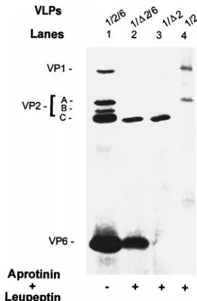
D
2 (Fig. 3, lane 3), while 1/2-VLPs were
composed of both VP1 and VP2 (Fig. 3, lane 4).
Morpholog-ically both were single-layered types of VLPs appearing to be
similar to the particles shown in Fig. 1A, B, and E (data not
shown). The VP1 that was not assembled into
D
2-VLPs was
found in the top fractions of 5 to 20% sucrose gradients and
did not comigrate with
D
2-VLPs (data not shown). Analyses of
purified VLPs from cells expressing VP1/
D
2/6 and VP1/2/6
both were morphologically double layered, resembling those
seen in Fig. 1C and F (data not shown), but VP1 was present
only when the VLPs contained full-length VP2 (Fig. 3, lanes 1
and 2). The expression levels of VP1 in the lysates of VP1/2/6
and VP1/
D
2/6 were comparable at 80 h postinfection. VP1 was
still not detected even if a threefold-greater amount of 1/
D
2/
6-VLPs and 1/
D
2-VLPs was analyzed (data not shown).
FIG. 1. Electron micrographs of the VPD2-containing VLPs and their VP2-containing counterparts.D2-VLPs were expressed from the recombinant baculovirus BVP2C24 by infection of Sf9 cells and purified on a 5 to 20% sucrose gradient.D2/6-VLPs were coexpressed from the recombinant baculoviruses BVP2C24 and pAc461/SA11-6 by infection of Sf9 cells and purified by CsCl isopycnic centrifugation. Shown are the negative-stained structures of the VLPs containing the indicated protein species. (A to C) VPD2-containing VLPs; (D) native rotavirus double-layered particles (DLP); (E and F) VP2-containing VLPs as the counterparts of panels A to C. All micrographs are at the same magnification. Bar, 100 nm.
on November 9, 2019 by guest
http://jvi.asm.org/
The N terminus of VP2 is required for encapsidation of VP3.
We also examined if VP3 could be encapsidated in a VLP
containing VP
D
2. VLPs obtained from cells infected with
re-combinants that express
D
2/3-VLPs were structurally
com-posed of only VP
D
2 (Fig. 4A and B, lanes 3), while 2/3-VLPs
were composed of both VP2 and VP3 (Fig. 4A and B, lanes 4).
Morphologically, both types of VLPs were single layered,
sim-ilar to the particles shown in Fig. 1A, B, and E. Analyses of the
VLPs expected to contain VP
D
2/3/6 and VP2/3/6 showed that
both were morphologically double layered, similar to those
shown in Fig. 1C and F, but VP3 was present only when the
VLPs contained full-length VP2 (Fig. 4A and B, lanes 1 and 2).
The expression levels of VP3 in the lysates of VP2/3 versus
VP
D
2/3 and of VP2/3/6 versus VP
D
2/3/6 were comparable at
80 h postinfection (data not shown). It also appeared that VP3
was encapsidated in parallel to the amount of full-length VP2
in VLPs. Thus, the intensities of labeling of VP3 were
compa-rable (Fig. 4B, lanes 1 and 4) when the amounts of the
full-length VP2 in VLPs were comparable (Fig. 4A, lanes 1 and 4).
VP1 and VP3 are not, or not efficiently, encapsidated into
VLPs lacking the N terminus of VP2.
We further examined if
coexpressed VP1 and VP3 could be encapsidated into a VLP
containing the VP
D
2. VLPs obtained from cells infected with
recombinants expected to express 1/
D
2/3-VLPs were
structur-ally composed of only VP
D
2 (Fig. 5A and B, lanes 3), while
1/2/3-VLPs were composed of VP1, VP2, and VP3 (Fig. 5A
and B, lanes 4). Morphologically, both were single-layered
types of VLPs resembling the particles shown in Fig. 1A, B,
and E. Analyses of the VLPs expected to contain VP1/
D
2/3/6
and VP1/2/3/6 showed that both were morphologically double
layered, resembling those seen in Fig. 1C and F, but VP1 and
VP3 were present only when the VLPs contained full-length
VP2 (Fig. 5A and B, lanes 1 and 2). The expression levels of
VP1 and VP3 in the lysates were comparable at 80 h
postin-fection, and the unassembled VP1 was found in the top
frac-tions of 5 to 20% sucrose gradients of VP1/
D
2/3 (data not
shown).
Analyses of interactions between VP1 and other proteins in
VLPs.
We next investigated the interactions of VP1 with the
different proteins in the VLPs by far-Western blotting on
PVDF membranes. The blotted proteins, including full-length
VP2, VP2 cleavage products, VP
D
2, VP6 monomer, and VP6
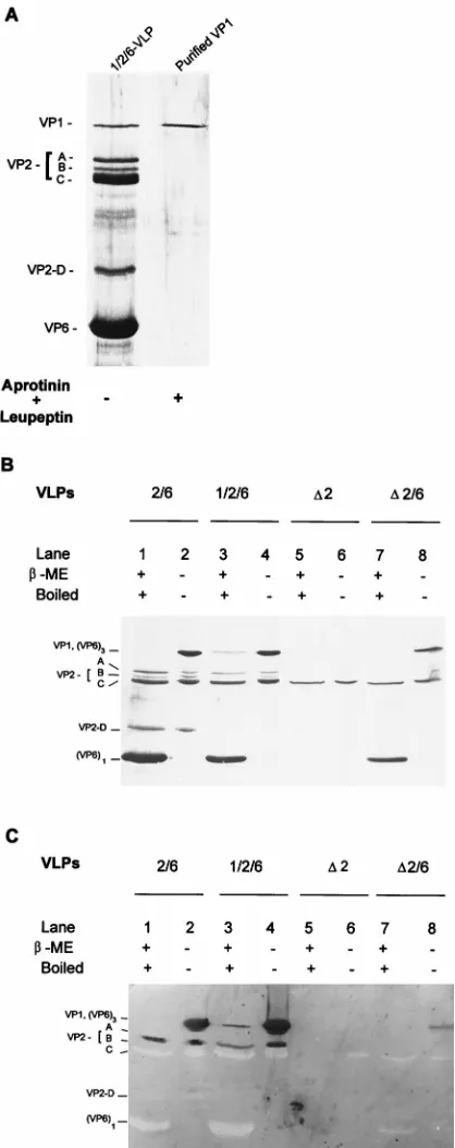
trimer, were tested for the ability to bind VP1. A purified
preparation of VP1 (Fig. 6A) was used as a probe. To clearly
visualize all of the proteins blotted to the PVDF membrane,
duplicate samples of those on the PVDF membrane were
an-alyzed by SDS-PAGE (10% gel) followed by silver staining
(Fig. 6B). The VP1 binding proteins are shown in Fig. 6C. By
comparison with the silver-stained gel (Fig. 6B), only
full-length VP2 (VP2-A [Fig. 6C, lanes 1 to 4]) and VP6 trimers
(Fig. 6C, lanes 2, 4, and 8) were capable of binding VP1, while
the VP2 cleavage products (bands B to D), VP
D
2, and VP6
monomers did not bind VP1 (Fig. 6C). The VP1 visualized in
lane 3 in Fig. 6C is thought to be detected by interaction
between the anti-VP1 and the VP1 molecules of the
1/2/6-VLPs rather than from the interaction of VP1 as the probe
with itself in the VLPs.
To determine the domain of VP2 required for binding VP1,
three truncated constructs of VP2 (aa 1 to 583, aa Met-26 to
583, and aa Met-45 to 583 [truncation constructs P2AT1777,
P2M16.9, and P2M16.3 in reference 17) were expressed in Sf9
cells. None of these truncated VP2 construct forms
self-assem-bled into VLPs (17). Cell lysates were Western blotted to
nitrocellulose to confirm that the truncated VP2 peptides were
expressed. The lysates also were blotted to PVDF membranes
for the VP1 binding test. Western blot analysis with
[image:4.612.87.250.69.286.2]monoclo-FIG. 3. Protein content of the coexpressed 1/D2-, 1/2-, 1/D2/6-, and 1/2/6-VLPs. Shown is a silver-stained SDS–10% polyacrylamide gel with proteins of the purified VLPs resulting from the coexpression of indicated genes in the absence or presence of protease inhibitors. Protease inhibitors aprotinin and leupeptin, each at 1mg/ml, were added daily. VP1/D2 and VP1/2 were coex-pressed from the baculovirus recombinant pVL941/RF-1 with recombinant BVP2C24 or BacRF2A, respectively, and purified by centrifugation on a 5 to 20% sucrose gradient. VP1/D2/6 and VP1/2/6 were coexpressed from the bacu-lovirus recombinants of pVL941/RF-1, pAc461/SA11-6 with recombinants BVP2C24 and BacRF2A, respectively, and purified by CsCl isopycnic centrifu-gation. To compare VP1 encapsidation, the protein amount loaded onto each lane was adjusted so that the amount of VPD2 was similar to or higher than that of VP2 band A, the VP1 binding protein (Fig. 6C). To create a cleaved VP2 band C for a marker of VPD2, 1/2/6-VLPs in lane 1 (also in lanes 1 of Fig. 4 and 5) were produced in the absence of protease inhibitors.
FIG. 2. Protein content of single- and double-layered VPD2-containing VLPs and their VP2-containing counterparts. Shown is a silver-stained SDS–10% poly-acrylamide gel with the proteins of purified VLPs resulting from the expression of the indicated genes in the absence or presence of protease inhibitors. Protease inhibitors aprotinin and leupeptin, each at 1mg/ml, were added daily.
on November 9, 2019 by guest
http://jvi.asm.org/
[image:4.612.354.498.379.599.2]nal antibody (MAb) 164AE22, a MAb directed against an
epitope on VP2
RFbetween aa 45 and 92, showed that the three
truncated forms of VP2
RFwere expressed well (Fig. 7A). VP1
binding was seen only with VP2 aa 1 to 583 and some of its
cleavage products (Fig. 7B, lane 1), while VP2 aa Met-26 to
583 and VP2 aa Met-45 to 583 did not bind VP1 (Fig. 7B, lanes
2 and 3). Six additional truncated constructs of VP2 (aa 1 to
285, aa Met-290 to 880, aa Met-51 to 583, aa Met-61 to 583, aa
Met-99 to 583, and aa Met-177 to 583) also were tested for
VP1 binding. Only the construct containing aa 1 to 285 showed
the ability to bind VP1 (data not shown). These results suggest
that (i) a domain within the N terminus of VP2 aa 1 to 25 is
essential for binding VP1, (ii) some conformations that form
after VP6 trimerization are favorable for VP1 binding, and (iii)
VP2 aa 26 to 880 are not sufficient for binding VP1.
DISCUSSION
The native rotavirus VP2 layer, the innermost core shell,
serves important roles in the formation of transcriptionally
active particles, with VP6 trimers forming the outside shell and
VP1, VP3, and the genome of 11 segments of dsRNA being
inside the shell. Our goals in this study were (i) to determine
the peptide fragment of VP2 necessary for the formation of a
single-layered VLP; (ii) to investigate the possible functions of
the protease-accessible peptide fragment at the VP2 N
termi-nus; and (iii) to map the domain of VP2 required for the
binding and encapsidation of VP1, the RNA-dependent RNA
polymerase, and VP3, the guanylyltransferase.
Expression of VP2 from baculovirus recombinants encoding
the full-length VP2 by infection of Sf9 cells in the absence of
[image:5.612.54.288.66.276.2]protease inhibitors produces single-layered empty 2-VLPs
identical in size and shape to its authentic counterpart (18, 37).
This expressed single-layered 2-VLP contains two major
cleav-age products. One of them represents the peptide fragment of
aa 93 to 880. Characterization of various truncated VP2
con-structs by Labbe´ et al. (17) showed that expression of truncated
VP2 constructs containing aa 1 to 583, aa 290 to 880, and aa 1
to 290 plus 589 to 880, as well as several other shorter VP2
constructs, did not lead to particle formation in insect cells.
Previous work has shown that the rotavirus proteins expressed
from baculovirus recombinants in Sf9 cells generally represent
their cognate native proteins in both biochemical and
func-tional characteristics (9, 38), and so the inability of those
cated VP2 peptides to form VLPs suggested that these
trun-cations lacked the peptide domain necessary for the formation
of single-layered 2-VLPs. To study this possibility, two
trun-cated forms of VP2 that contain aa Met-93 to 880, called
VP
D
2, and aa 186 to 880, called VP
D
2
9
, were created.
Expres-sion of VP
D
2
9
in Sf9 cells did not result in particle assembly
(data not shown). In contrast, expression of VP
D
2 resulted in
the formation of single-layered core-like particles, identical to
2-VLPs in yields, appearance, size, and stability, designated
D
2-VLPs. This result suggests that the VP2 N-terminal aa 1 to
92 are not involved in the formation of single-layered 2-VLPs,
and aa 93 to 185 are directly or indirectly needed for VP2
self-assembly into a particle. VP
D
2 was capable of forming not
only VLPs but also elongated helix structures following
con-centration as seen previously for 2-VLPs (37). Coexpression of
VP
D
2 with VP6 resulted in the formation of double-layered
D
2/6-VLPs identical to 2/6-VLPs in yields, size, shape, and
FIG. 4. Protein content of the coexpressedD2/3-, 2/3-,D, and 2/3/6-VLPs. (A) Silver-stained SDS–10% polyacrylamide gel with proteins of the purified VLPs resulting from the coexpression of the indicated genes in the absence or presence of protease inhibitors. Protease inhibitors aprotinin and leupeptin, each at 1mg/ml, were added daily. (B) Autoradiogram of [a-32
P]GTP-bound VP3 to visualize the presence of VP3 in the VLPs shown in panel A. VPD2/3 and VP2/3 were coexpressed from the baculovirus recombinant pVL1393/SA11-3 with recombinants BVP2C24 and BacRF2A, respectively, and purified on a 5 to 20% sucrose gradient. VPD2/3/6 and VP2/3/6 were coexpressed from the baculovirus recombinants pVL1393/SA11-3 and pAc461/SA11-6 with recombinant BVP2C24 or BacRF2A, respectively, and purified by CsCl isopycnic centrifugation. To compare VP3 encapsidation, the protein amount loaded onto each lane was adjusted so that the amount of VPD2 was similar to or higher than that of the VP2 band A, the VP1 binding protein (Fig. 6C).
FIG. 5. Protein content of the coexpressed 1/D2/3-, 1/2/3-, 1/D2/3/6-, and 1/2/3/6-VLPs. (A) Silver-stained SDS–10% polyacrylamide gel with proteins of purified VLPs resulting from the expression of indicated genes in the absence or presence of protease inhibitors. Protease inhibitors aprotinin and leupeptin, each at 1mg/ml, were added daily. (B) Autoradiogram of [a-32P]GTP-bound VP3 to
visualize the presence of VP3 in the VLPs shown in panel A. VP1/D2/3 and VP1/2/3 were coexpressed from the baculovirus recombinants pVL941/RF-1 and pVL1393/SA11-3 with recombinant BVP2C24 or BacRF2A, respectively, and purified by centrifugation on a 5 to 20% sucrose gradient. VPD2/3/6 and VP2/3/6 were coexpressed from the baculovirus recombinants pVL941/RF-1, pVL1393/ SA11-3, and pAc461/SA11-6 with recombinants BVP2C24 and BacRF2A, re-spectively, and purified by CsCl isopycnic centrifugation. To compare VP1 and VP3 encapsidation, the protein amount loaded onto each lane was adjusted so that the amount of VPD2 was similar to or higher than that of VP2 band A, the VP1 binding protein (Fig. 6C).
on November 9, 2019 by guest
http://jvi.asm.org/
[image:5.612.312.549.389.599.2]stability. This finding suggests that
D
2-VLPs retain the
char-acteristics necessary for the encapsidation of VP6 trimers and
that aa 1 to 92 at the N terminus of VP2 are not involved in the
assembly of VP6 trimers into
D
2/6-VLPs.
Coexpression of VP
D
2 plus VP1, VP
D
2 plus VP1 plus VP3,
or VP
D
2 plus VP3 resulted in the formation of only
single-layered
D
2-VLPs which lacked VP1 and VP3. Coexpression of
VP
D
2 plus VP1 plus VP6, VP
D
2 plus VP1 plus VP3 plus VP6,
or VP
D
2 plus VP3 plus VP6 produced only double-layered
D
2/6-VLPs which also lacked VP1 and VP3. The expression
levels of VP1 and VP3 in 1/
D
2- versus 1/2-VLPs, 1/
D
2/3- versus
1/2/3-VLPs, and
D
2/3- versus 2/3-VLPs, and in 1/
D
2/6- versus
1/2/6-VLPs, 1/
D
2/3/6- versus 1/2/3/6-VLPs, and
D
2/3/6- versus
2/3/6-VLPs, were comparable as determined by dot blot assays
for VP1 and by [
a
-P
32]GTP binding for VP3 of dilutions of
samples examined at 80 h postinfection. VP1 was detected only
in the top fractions of gradients when 1/
D
2- and 1/
D
2/3-VLPs
were analyzed by centrifugation through 5 to 20% sucrose
gradients, while VP1 or VP3 comigrated with VP2 in the broad
band obtained with a peak at fraction 9 when 1/2-, 1/2/3- and
2/3-VLPs were centrifuged in 5 to 20% sucrose gradients (data
not shown). It is known that [
a
-
32P]GTP does not label 2-, 1/2-,
[image:6.612.319.532.66.242.2]2/6-, and 1/2/6-VLPs (38). Therefore, these results clearly
sug-gest that VP2 lacking the N-terminal 92 aa has lost its
capa-bility to encapsidate VP1 alone, VP3 alone, or a VP1-VP3
complex. Prasad et al. (29) reported that a flower-shaped
VP1-VP3 complex is seen attached to VP2 at the fivefold axes
between the 160- and 220-Å radii in both 1/2/3/6-VLPs and
native double-layered particles. Recently, Lawton et al. (20)
reported a comparison of the inner surface structures between
2-VLPs and
D
2-VLPs; i.e., a loss of mass adjacent to the
fivefold axes and a redistribution of mass along the fivefold
axes were seen. These results imply that the portion of VP2 at
the fivefold axes seen in three-dimensional reconstructions
computed from EM images could represent a portion of the N
terminus of VP2 extending out from the 2-VLP inner surface
FIG. 6. Interactions between VP1 and other VLP proteins tested by far-Western blotting. (A) Silver-stained SDS–10% polyacrylamide gel of purified VP1 used for far-Western blotting to verify the VP1 binding proteins. VP1 was purified by FPLC followed by passage through an immunoaffinity column of rabbit IgG against wild-type baculovirus proteins. (B) Silver-stained SDS–10% polyacrylamide gel of the proteins in the indicated VLPs. Each purified VLP was (i) mixed with Laemmli buffer containingbME and boiled for 5 min or (ii) mixed with sample buffer withoutbME, kept at 4°C, and then resolved on an SDS–10% polyacrylamide gel at 4°C. (C) VP1 binding proteins detected on far-Western blots. A duplicate of the gel in panel B was run, and the proteins were transfered to a PVDF membrane for far-Western VP1 binding assay. After renaturation of the proteins blotted to the PVDF membrane and blocking of free binding sites, 10mg of VP1 per ml was added to probe the proteins capable of binding VP1. The bound VP1 was detected by guinea pig anti-VP1 antibody followed by goat anti-guinea pig IgG-alkaline phosphatase conjugate and enzyme substrates.
FIG. 7. Mapping the domain of VP2 required for binding VP1 by far-West-ern blotting. (A) Westfar-West-ern blot of three truncated forms of VP2. Lysates of infected cells expressing three N- and C-terminus truncations of VP2 (aa 1 to 583, aa Met-26 to 583, and aa Met-45 to 583) were boiled with Laemmli sample buffer containingbME, resolved on an SDS–10% polyacrylamide gel, and blot-ted to nitrocellulose. They were probed by MAb 164AE22, which directs a VP2Rf
epitope located between aa 45 and 92. (B) VP1 binding of truncated forms of VP2. Three VP2 truncation mutants were mixed with Laemmli buffer containing
bME, boiled for 5 min, resolved on an SDS–10% polyacrylamide gel, and blotted to a PVDF membrane. After renaturation of the blots, 10mg of VP1 per ml was added to probe the peptide fragments capable of binding VP1 as described for Fig. 6C.
on November 9, 2019 by guest
http://jvi.asm.org/
[image:6.612.66.275.68.595.2]to anchor the VP1-VP3 complexes. This could explain why
fragments consisting of VP2 aa 93 to 880 are capable of
form-ing stable empty sform-ingle-layered VLPs.
To map the domain of VP2 required for binding VP1,
far-Western blotting, a method to probe protein-protein
interac-tions, was used. Since full-length VP2 (band A) and its three
cleavage products (band B [aa 43 to 880], band C [aa 93 to 880,
and band D [aa 369 to 880]) are always detected when VP2 is
expressed in the absence of protease inhibitors (37, 38) (see
below), full-length VP2 and its cleavage products were useful
probes for VP1 binding. Far-Western blotting showed that
VP1 binds only to full-length VP2 (band A), not to the
cleav-age bands B, C, and D and VP
D
2. VP1 binding assays with the
truncated VP2 consisting of aa 1 to 583, aa Met-26 to 583, and
aa Met-45 to 583 showed that only VP2 from aa 1 to 583 is
capable of binding VP1. No bands were seen when the
BSA-blocked blots were only incubated with the primary or
second-ary antibody alone before or after incubation with VP1 (data
not shown). These facts suggest that (i) a domain at aa 1 to 25
of the VP2 N terminus is required for binding VP1, (ii) aa 584
to 880 at the C terminus of VP2 are not sufficient for VP1
binding, and (iii) the lack of ability of
D
2-VLPs and
D
2/6-VLPs
to encapsidate VP1 was not because of an added Met in front
of VP2-C. In addition to VP2-A, VP6 trimers, but not VP6
monomers, were capable of interacting with VP1. This finding
suggests that (i) a VP1-interacting conformation may exist in
VP6 trimers but not in VP6 monomers; (ii) this VP1 binding
site on the VP6 trimers may be located close to the VP2 layer,
and some portion of VP1 may be associated with VP2 and
extend across the VP2 layer into the VP6 layer; and (iii) the
preformed VP1-(VP6)
3complexes could not be encapsidated,
because
D
2/6-VLPs did not show any evidence of VP1
associ-ation. We also probed VP1 from 1/2/6-VLPs with purified VP6
by far-Western blotting. Guinea pig anti-VP6 was used as the
primary antibody. The VP6 bound to VP1 and also to the VP2
bands A to C (data not shown). The function, if any, of this
interaction between VP1 and VP6 trimers remains unknown.
In addition, although it is clear that VP2 aa 1 to 92 are
nec-essary for VP3 encapsidation, we were not able to further map
the domain of VP2 required for binding VP3 because the
required reagents, e.g., purified VP3, are not available.
In the discussion of a previous report (37), based on its
apparent molecular weight in SDS-PAGE, we speculated that
VP2-B represented aa 1 to 789 and had a blocked N terminus.
This conclusion was reached because the amount VP2-B
avail-able was low and difficult to be clearly sequenced. Recently, we
obtained additional VP2-B material, blotted it onto an
Immo-bilon-P transfer membrane (Millipore), and sequenced the
protein by using an Applied Biosystems 477A protein
se-quencer in the Protein Sequencing Core Facility of Baylor
College of Medicine. An N-terminal sequence was obtained
and found to be KKEEVVTDS; this sequence begins at aa 44
of VP2 (16, 36). Recent studies have also detected another
cleavage product of VP2 that has a molecular weight of
;
58,000, designated VP2-D. Its N terminus has been
se-quenced as GINSQAAND beginning at aa 369 of VP2 (4, 16).
The significance of these cleavage products remains to be
de-termined.
VP1 is thought to be the RNA-dependent RNA polymerase
of rotavirus. It, together with VP3, binds to VP2 at the fivefold
axes (29). VP1 also specifically binds to the 3
9
end of viral
mRNA (24), and the seven nucleotides at the 3
9
terminus
provide the minimum requirement for replication, at least, of
the segment 9 mRNA (34). RNA also binds to the N terminus
of VP2 (17), to which VP1 and VP3 also bind (this study).
Thus, we know that the polymerase VP1, the
guanylyltrans-ferase VP3, and template mRNA all colocalize at the N
ter-minus of VP2 at the fivefold axes, but we do not yet know
precisely how they function to bring about RNA transcription,
elongation, and exiting from particles.
In summary, we have expressed VP
D
2, an N-terminal
92-aa-deleted VP2, alone and with other species of proteins existing
in the single- and double-layered capsids of rotavirus in the
baculovirus system, and we have purified and characterized the
resulting VLPs. The VP1 and VP3 are not assembled into
VLPs with VP
D
2. The N-terminal peptide of VP2 is required
for encapsidation of VP1 and VP3.
ACKNOWLEDGMENTS
We gratefully acknowledge the helpful discussions and critical
re-view of the manuscript provided by Sue E. Crawford, J. A. Lawton, and
B. V. V. Prasad. We also thank Marie Labbe´ for preparing the
recom-binant baculovirus BVP2C24.
This work was supported by Public Health Service grant DK 30144
from the National Institutes of Health and by European Economic
Community grant INCO IC18-CT96-0027.
REFERENCES
1. Blacklow, N. R., and H. B. Greenberg. 1991. Viral gastroenteritis. N. Engl. J. Med. 325:252–264.
2. Boyle, J. F., and K. V. Holmes. 1986. RNA-binding proteins of bovine rotavirus. J. Virol. 58:561–568.
3. Burns, J. W., H. B. Greenberg, R. D. Shaw, and M. K. Estes. 1988. Func-tional and topographical analyses of epitopes on the hemagglutinin (VP4) of the simian rotavirus 11. J. Virol. 62:2164–2172.
4. Burroughs, M. H., S. E. Crawford, and M. K. Estes. Unpublished data. 5. Chen, D., J. L. Gombold, and R. F. Ramig. 1990. Intracellular RNA synthesis
directed by temperature-sensitive mutant of simian rotavirus SA11. Virology
178:143–151.
6. Chen, D., and R. F. Ramig. Unpublished data.
7. Chen, D., C. Q.-Y. Zeng, M. J. Wentz, M. Gorziglia, M. K. Estes, and R. F.
Ramig.1994. Template-dependent, in vitro replication of rotavirus RNA. J. Virol. 68:7030–7039.
8. Cohen, J., A. Charpilienne, S. Chilmonczyk, and M. K. Estes. 1989. Nucle-otide sequence of bovine rotavirus gene 1 and expression of the gene product in baculovirus. Virology 171:131–140.
9. Crawford, S. E., M. Labbe´, J. Cohen, M. H. Burroughs, Y. J. Zhou, and
M. K. Estes.1994. Characterization of virus-like particles produced by the expression of rotavirus capsid proteins in insect cells. J. Virol. 68:5945–5952. 10. Estes, M. K. 1996. Rotavirus and their replication, p. 1625–1655. In B. N. Fields, D. M. Knipe, P. M. Howley, et al. (ed.), Virology, 3rd ed. Lippincott-Raven Publisher, Philadelphia, Pa.
11. Estes, M. K., S. E. Crawford, M. E. Penaranda, B. L. Petrie, J. W. Burns,
W.-K. Chan, B. Ericson, G. E. Smith, and M. D. Summers.1987. Synthesis and immunogenicity of the rotavirus major capsid antigen using a baculovi-rus expression system. J. Virol. 61:1488–1494.
12. Estes, M. K., B. B. Mason, S. Crawford, and J. Cohen. 1984. Cloning and nucleotide sequence of the simian rotavirus gene 6 that codes for the major inner capsid protein. Nucleic Acids Res. 12:1875–1887.
13. Gombold, J. L., and R. F. Ramig. 1987. Assignment of simian rotavirus SA11 temperature-sensitive mutant group A, C, F, and G to genome segments. Virology 161:463–473.
14. Homann, H. E., W. Willenbrink, C. J. Buchholz, and W. J. Neubert. 1991. Sendai virus protein-protein interactions studied by a protein-blotting pro-tein-overlay technique: mapping of domains on NP protein required for binding to P protein. J. Virol. 65:1304–1309.
15. Kapikian, A. Z., J. Flores, K. Midthun, Y. Hoshino, K. Y. Green, M.
Gor-ziglia, K. Nishihawa, R. M. Chanock, L. Potash, and I. Perez-Schael.1990. Strategies for the development of a rotavirus vaccine against infantile diar-rhea with an update on clinical trials of rotavirus vaccines. Adv. Exp. Med. Biol. 257:67–90.
16. Kumar, A., A. Charpilienne, and J. Cohen. 1989. Nucleotide sequence of the gene encoding for the RNA binding protein (VP2) of RF bovine rotavirus. Nucleic Acids Res. 17:2126.
17. Labbe´, M., P. Baudoux, A. Charpilienne, D. Poncet, and J. Cohen. 1994. Identification of the nucleic acid binding domain of the rotavirus VP2 pro-tein. J. Gen. Virol. 75:3423–3430.
18. Labbe´, M., A. Charpilienne, S. E. Crawford, M. K. Estes, and J. Cohen. 1991. Expression of rotavirus VP2 produces empty core-like particles. J. Virol.
65:2945–2952.
19. Laemmli, U. K. 1970. Cleavage of structural proteins during the assembly of the head of bacteriophage T4. Nature (London) 227:680–685.
20. Lawton, J. A., C. Q.-Y. Zeng, S. K. Mukherjee, J. Cohen, M. K. Estes, and
on November 9, 2019 by guest
http://jvi.asm.org/
B. V. V. Prasad.1997. Three-dimensional structural analysis of recombi-nant rotavirus-like particles with intact and amino-terminal-deleted VP2: implications for the architecture of the VP2 capsid layer. J. Virol. 71: 7353–7360.
21. Lee, W. S., C. C. Kao, G. O. Bryant, X. Liu, and A. J. Berk. 1991. Adenovirus EIA activation domain binds the basic repeat in the TATA box transcription factor. Cell 67:365–376.
22. Liu, M., N. M. Mattion, and M. K. Estes. 1992. Rotavirus VP3 expressed in insect cells possesses guanylyltransferase activity. Virology 188:77–84. 23. Mansell, E. A., and J. T. Patton. 1990. Rotavirus RNA replication: VP2, but
not VP6, is necessary for viral replicase activity. J. Virol. 64:4988–4996. 24. Patton, J. T. 1996. Rotavirus VP1 alone specifically binds to the 39end of
viral RNA, but the interaction is not sufficient to initiate minus-stranded synthesis. J. Virol. 70:7940–7947.
25. Pizarro, J. L., A. M. Sandino, J. M. Pizarro, J. Fernandez, and E. Spencer. 1991. Characterization of rotavirus guanylyltransferase activity associated with polypeptide VP3. J. Gen. Virol. 72:325–332.
26. Poch, O., I. Sauvaget, M. Delarue, and N. Tordo. 1989. Identification of four conserved motifs among the RNA-dependent polymerase encoding ele-ments. EMBO J. 8:3867–3874.
27. Prasad, B. V. V., and W. Chiu. 1994. Structure of rotavirus, p. 9–30. In R. F. Ramig (ed.), Rotaviruses. Springer-Verlag, Berlin, Germany.
28. Prasad, B. V. V., and M. K. Estes. 1997. Molecular basis of rotavirus replication: structure-function correlations, p. 239–268. In W. Chiu, R. Burnett, and R. Garcia (ed.), Structural biology of viruses. Oxford Press, Oxford, England. 29. Prasad, B. V. V., R. Rothnagel, C. Q.-Y. Zeng, J. Jakara, J. A. Lawton, W.
Chiu, and M. K. Estes.1996. Visualization of ordered genomic RNA and
localization of transcriptional complexes in rotavirus. Nature (London) 382: 471–473.
30. Prasad, B. V. V., G. J. Wang, J. P. M. Clerx, and W. Chiu. 1988. Three dimensional structure of rotavirus. J. Mol. Biol. 199:269–275.
31. Summers, M. D., and G. E. Smith. 1987. A manual of methods for baculovirus vectors and insect cell culture procedures. Texas Agricultural Experimental Station bulletin no. 1555. Texas A & M University, College Station, Tex. 32. Towbin, T., T. Staehelin, and J. Gordon. 1979. Electrophoretic transfer of
proteins from polyacrylamide gels to nitrocellulose sheets: procedure and some applications. Proc. Natl. Acad. Sci. USA 76:4350–4354.
33. Valenzuela, S., J. Pizzaro, A. M. Sandino, M. Va´squez, J. Ferna´ndez, O.
Herna´ndez, J. Patton, and E. Spencer.1991. Photoaffinity labeling of rota-virus VP1 with 8-azido ATP: identification of the viral RNA polymerase. J. Virol. 65:3964–3967.
34. Wentz, M. J., J. T. Patton, and R. F. Ramig. 1996. The 39-terminal consensus sequence of rotavirus mRNA is the minimal promotor of negative-strand RNA synthesis. J. Virol. 70:7833–7841.
35. Yeager, M., K. A. Dryden, N. H. Olson, H. B. Greenberg, and T. S. Baker. 1990. Three-dimensional structure of rhesus rotavirus by cryoelectron mi-croscopy and image reconstruction. J. Cell Biol. 110:2133–2144.
36. Zeng, C. Q.-Y., and M. K. Estes. Unpublished data.
37. Zeng, C. Q.-Y., M. Labbe´, J. Cohen, B. V. V. Prasad, D. Chen, R. F. Ramig,
and M. K. Estes.1994. Characterization of VP2 particles. Virology 201:55– 65.
38. Zeng, C. Q.-Y., M. J. Wentz, J. Cohen, M. K. Estes, and R. F. Ramig. 1996. Characterization and replicase activity of double-layered and single-layered rotavirus-like particles expressed from baculovirus recombinants. J. Virol.
70:2736–2742.