MICRONUCLEUS ASSAY IN UROTHELIAL
CELLS IN CANCER CERVIX
This
dissertation
is
submitted
to
The
Tamilnadu
Dr.MGR
Medical
University,
Chennai
in
Partial
Fulfillment
of
the
Regulations
for
D.M.
Branch
VII
(Medical
Oncology)
Madras
Medical
College,
Chennai
–
600
003.
August
2010
CERTIFICATE
This is to certify that this dissertation entitled
“MICRONUCLEUS ASSAY IN UROTHELIAL CELLS IN CANCER
CERVIX" is a bonafide record of original work done by
Dr. S. SURESH KUMAR, under our guidance and supervision in
the Department of Medical oncology, Madras Medical College, Chennai-600 003 , during the period of his Higher specialty study for DM(Branch VII) Medical Oncology from July 2007- August 2010.
DEAN
MADRAS MEDICAL COLLEGE
CERTIFICATE
This is to certify that this dissertation entitled
“MICRONUCLEUS ASSAY IN UROTHELIAL CELLS IN CANCER
CERVIX" is a bonafide record of original work done by
Dr. S. SURESH KUMAR, under our guidance and supervision in
the Department of Medical oncology, Madras Medical College, Chennai-600 003 , during the period of his Higher specialty study for DM (Branch VII) Medical Oncology from July 2007-August 2010.
Dr.K. VIJAYA SARATHY,MD.DM (MEDICAL ONCOLOGY) PROFESSOR AND HEAD
DEPARTMENT OF MEDICAL ONCOLOGY MADRAS MEDICAL COLLEGE
ACKNOWLEDGEMENT
It is with great pleasure, I acknowledge my deep and sincere
gratitude to my beloved teacher Prof.K.Vijaya sarathy, Head of the
Department of Medical Oncology,for his guidance ,constant
encouragement and patience throughout the period of the study and my
post graduate career.
I am greatly indebted to my co‐guide Dr.Kanchana,
Professor of Pathology,IOG,Egmore for extending her invaluable help for
my study.
I express my sincere thanks to Dr.Radhabhai Prabhu,
Director in Charge, IOG, Egmore.
I am thankful to Dr.Lakshmi Narasimhan and Dr.Balaji for
their constant encouragement and support.
I acknowledge with thanks for the warm help extended to
me in times of need by my colleagues and friends.
I owe my thanks to the technical staff of the Department of
Pathology for their kind cooperation.
I express my gratitude to the Dean, Madras Medical College
for permitting me to make use of the clinical facilities in the Institute.
With deep gratitude, I must thank all the patients without
Its my privilege to thank the Almighty and my family
members for their blessings.
At the end it would be unjustified if I miss the name of my
beloved wife Juju and my cute son Jujan whose constant encouragement
and sacrifice in family matter enable to me to complete this work.
Suresh Kumar.S
CONTENTS
Page No.
ABBREVIATIONS
INTRODUCTION 1
AIMS AND OBJECTIVES 3
REVIEW OF LITERATURE 4
MATERIALS AND METHODS 23
OBSERVATIONS 29
TABLES AND FIGURES
DISCUSSION 34
SUMMARY AND CONCLUSION 41
RECOMMENDATIONS 44
BIBLIOGRAPHY i-vii
ABBREVIATIONS
HUMN – HUMAN MICRONUCLEUS MN – MICRONUCLEUS
INTRODUCTION
INTRODUCTION
Cancer, modern epidemics of non-communicable diseases is the second commonest cause of mortality in developed countries and remains one of the ten commonest causes of mortality in developing countries like India is a complex disease with altered expression, abnormal growth and disruption of normal function of cells caused by genotoxic effects resulting genomic instability at an early stage of cancer.
To evaluate the genotoxic risks/ effects, observed as DNA damages, can be assessed by chromosomal aberrations, sister chromatid exchanges and micronucleus test. Out of all these, micronucleus test is found to be the most sensitive when compared with other tests as it neither requires tedious procedures like cell culture and metaphase preparation, nor it requires any specific DNA stains. To further add, as it is applicable on interphase cell only , it is the best indicator of mitotic interference and chromosomal mutations or breakages and is noninvasive and economical too.
Micronucleus, a microscopically visible round or oval cytoplasmic chromatin mass in the extra nuclear vicinity, originates from aberrant mitosis. It consists of eccentric chromosomes, chromatid fragments or whole chromosomes which failed to reach spindle poles during mitosis. Micronuclei have been used as biomarkers for assessment of DNA damages. Micronuclei provide a measure of both, chromosome breakage and chromosome loss.
AIMS AND OBJECTIVES
AIMS AND OBJECTIVES
1. To identify the occurrence of micronuclei in normal and cancer cervix.
2. To identify the occurrence of micronuclei in risk factors of cancer cervix.
3. To identify the occurrence of micronuclei in different stages of malignancy.
REVIEW OF LITERATURE
REVIEW OF LITERATURE
Carcinoma cervix worldwide accounts for 15% of all cancer diagnosed in women [1]. It is the second most common cancer in women globally and 80% of these occur in developing countries. It occupies either top rank or second among cancers in developing countries, whereas, in the affluent countries does not find place even in the top five leading cancers.
In India it has been reported to be the commonest malignancy in women, comprising 24% of all cancers in female[2]. Almost 20 per 100,000 Indian women have cancer cervix and it has been estimated that one in 63 likely to suffer from it in her life time [3,4]. It is estimated that approximately 100,000 women develop cancer cervix every year. [5]
Cancer of the cervix has been the most important cancer in women in India over the past two decades. In older population based cancer registries (PBCR) Barshi and Chennai PBCRs have always recorded the highest incidence of cervix cancer [7]. Based on the data of the PBCRs, the estimated number of new cancers during 2007 in India was 90, 7086.
Cancer of the cervix accounted for 16 per cent of all cancers in women in the urban registries in 2005. Since over 70 per cent of the Indian population resides in the rural areas, cancer cervix still constitutes the number one cancer in either sex [6].
In all developing countries as in India, maternal morbidity and mortality are points of cynosure. However besides direct gynaecologic and obstetric complications, another important issue leading to these outcomes is the high incidence of cervix cancer especially among the economically – disadvantaged [15]
Risk for cervical cancer has been reported to be variable from one population group to another because of its multifactorial etiology [9].
It appears that a larger proportion of the Indian female population is more vulnerable to cervical neoplasia, since the recognised risk factors for cancer cervix like illiteracy, low socio-economic status, early menarche, early marriage, multiparity, first child birth at an early age, poor genital hygiene and genital infections are widely prevalent in this population [3, 4].
Increasing age, increasing parity (Para > 3) age at marriage, clinical lesions of the cervix, gynecological complaints, STD were the risk factors for SIL and cancer cervix. 67.8% of the total SIL and 85.4% of the carcinoma cervix was observed in women with high parity ( 3 or more children ). 51.5% of the total SIL and 75.3% of the total cancer cases were observed in women more than 40 years of age [6]
The frequency of cancer cervix showed a progressive risk with increasing parity mostly between Para 2 and 3. [10]
The relative risk of cervical cancer increases with the more number of abortions [11].
There is a correlation between the low SES and cancer cervix. It has in fact been observed that uneducated women due to lack of knowledge of proper hygiene and preventive measures are more prone to cancer cervix [8].
However, cancer of the cervix responds favorably to secondary prevention measures as it has a long pre-clinical phase that usually requires 2 to 10 years to penetrate the basement membrane and invade tissues [12].
Statistics reveal that about 4 cases of every 5 cervical carcinoma patients actually occur in those countries that are without screening programs [13].
Rather, women do not come forward for routine gynaecological examination due to lack of knowledge about its early symptoms, fear of cancer (fatalistic attitude) and lack of awareness about the possibility of a cure [15].
In a WHO bulletin in 2001 there were the recommendations regarding Effective screening programs in the developing countries like India for cervical cancer in low and middle income groups [16]
The choice of screening test in countries/ regions that plan to initiate new programs should be based on the comparative performance characteristics of cytology and its potential alternatives such as VIA (visual inspection with acetic acid) [17]
Since programs cannot afford the luxury of frequently repeated testing of women, a highly sensitive test should be provided. Owing to their limited resources, developing countries cannot afford the models of frequently repeated screening of women over wide age ranges that are used in developing countries [16].
potential alternative to cytology, such as VIA, is chosen for screening, considerable attention should be given to the proper monitoring and evaluation of the program inputs and outcomes before further expansion[16].
A single life time screening which appears to most affordable and feasible method of control of cancer cervix in developing countries like India should be carried out in all women of high parity ( 3 or more children) irrespective of age and in all women above the age of forty irrespective of parity was the conclusion of the study[18].
Regular screening can be achieved by imparting appropriate education to the masses regarding the early signs and symptoms of cervix cancer and utilizing rapid screening/ diagnostic measures.
Routine protocol empathizes that every female above 25 years should annually get a Pap smear examination as a screening procedure [15].
Mass screening by cytology, to detect precursors of cancer of uterine cervix, is among the most successful of health measures. Survival of cervix cancer patients is most directly related to the stage of disease at diagnosis, and the best way to increase detection of cervical neoplasm in pre-cancerous or localized stage is to improve the scope and quality of cytology screening [16].
Pap smear is the most frequently used test in mass screening programs introduced by George Papanicolaou in 1940, yet it is not totally reliable. [42]
The sensitivity of Pap smear for the detection for cervical cancer precursors is less than 50%. The rate of false negativity is about 20 to 30 % in women with high grade CIN and 10 to 15 % in women with invasive cancer [43]
The occurrence of genetic instability, either as a result of, or leading to, an increase in chromosomal rearrangements in most cancer types merits cytogenetic investigations.
The genomic instabilities have been observed in many forms like chromosomal instability[23], chromosomal breakage[21], nucleoplasmic bridges[23], micronucleus formation and double DNA strand breaks[22].Chromosomal instability usually results either from abnormal centriole formation or chromosomal loss in anaphase or mal-segregation of chromosome or mitotic slippage or failure of cytokinesis or nucleoplasmic bridges[23], whereas chromosomal breaks are caused by spindle apparatus and clastogenic mutations[21].
Among all these genomic instabilities seen to carcinomas, MN formation is the hallmark of genomic instability being defined as chromatin containing body that represents fragments or even whole chromosomes which failed to get incorporated into the daughter cell nucleus during mitosis or might result from DNA strand breakage[22].
pre-clinical stage, in combination with morphological, biochemical and cytogenetic parameters, the Comet Assay along with the Micronucleus Test (MNT) may serve as novel tools to detect and predict the stage of cervical dysplasia [24].
Micronuclei can be detected in exfoliated cells of the buccal mucosa, urinary bladder, cervix or bronchi and seem to reflect chromatid and chromosome aberrations which occurred in the proliferating basal layers [23, 24, 25].
The sampling of such exfoliated cells is generally fast and highly economical; the MN assay has been the method of choice for survey of large population groups especially as a preliminary indicator for pre-cancerous lesions [23].
Since more than a century ago, micronuclei have been described by many scientists. In the late 1800s and early 1900s Howell and Jolly described Feulgen-positive nuclear bodies in human reticulocytes, known as Howell–Jolly bodies, and representing chromosomes separated from the mitotic spindle[26].
genotoxic potential of mutagens after in vivo exposure of animals using bone marrow erythrocytes [26].
Many methods like chromosomal aberration analysis, sister chromatid exchange analysis and MN frequency have been indentified for detecting mutagen exposures [21]. Out of these MN assay has been found to be an important genotoxic screening test for the detection of agents which cause chromosomal damage and induced the formation of MN in interphase cells [22].
Chromosomal biomarkers of genomic instability related to carcinoma have been identified as MN formation, nuclear budding and non disjunction in the year 2002 by Fenech. As per his observations, the DNA damage rates in human population can best be measured by MN scoring rather than chromosomal aberrations [23]. The MN frequency was used in number of studies and was found to be of significant importance [27].
MN index has also been labeled as endogenous dosimeter in tissues that might be the future site for the development of carcinoma [28].
resulted either in chromosomal breakage or in MN formation, so MN assay can be used to assess the genetic damage in such tissues [29]
The cells from the basal layer of epithelium continuously divide differentiate and migrate to the upper cell layers. The MN frequency was observed from these exfoliated epithelial cells. Induction of MN, in processes associated with DNA damages like cancer, aging and other genetic disorders, has been considered as an effective biomarker [30].
Since more than 90% of the human cancers arise from the epithelial tissue, exfoliated cells hold a strong potential as a tool for biomonitoring human populations exposed to genotoxic agents. Various studies indicate the presence of MN in the exfoliated cells of buccal mucosa, nasal mucosa, urothelium, lymphocytes and erythrocytes[21].But, of all these tissues and cells, exfoliated cells have been considered as best surrogate for predicting cancer and they can be easily collected from the mouth, nose,and bladder by noninvasive procedures[30].
micronucleated cells among cancer patients than among healthy individuals [31].
A gradual increase in MN frequency has been observed from normal to precancerous to cancerous lesions [32].
Many genotoxic studies on MN frequency were conducted related to predisposing factors which conclude significantly higher MN frequency in people with risk factors [33].
Number of micronuclei correlates with the severity of genetic damage. Cells containing several micronuclei present greater genetic damage than do cells that present only one micronucleus [34].
Human Micronucleus Project (HUMN) is an international collaborative study on the use of micronucleus technique for measuring DNA damage in humans. It validated the MN assays in human cells [30].
An important component in the interpretation of MN assay results is cell kinetics. MN observed in exfoliated buccal, nasal or urothelial epithelia are not induced when the cells are at the epithelial surface, but when they are in the basal layer [39].
In general, cells take 7–16 days to emerge to the surface and exfoliate [30].
Variability of MN assessment arises from intraindividual variability, interindividual variability, and population variability, as well as variations characteristic of different cell types. When spontaneous MN frequencies were compared in different healthy individuals, up to a 17-fold difference was observed, possibly reflecting genetic and nonspecific exposure differences. HUMN[30].
The average reported healthy population MN frequency is 1–3 per 500 - 1000 cells, with no significant variation between different types of exfoliated cells. Repeated scoring of MN in epithelia from the same individuals showed variation between 30 and 102.9%.HUMN[40].
radiotherapy, smoking, arsenic in drinking water w56x, and chronic infection. Exposures to low levels of benzene or leather tanning solutions did not cause significant increases in cytogenetic
damage[30].
A causal association between MN frequency and cancer risk could be inferred from studies of structural chromosomal aberrations(CAb) and aneuploidy. Somatic chromosome damage is involved in cancer etiology. Such damage can occur via chromosome loss as well as by breakageThere is a direct association between the frequency of MN in target or surrogate tissues and cancer development [30].
Michael Fenech have given detailed description of the scoring criteria for the micronucleus assay (MN ASSAY) in the HUMN project [41].
1. Morphologically identical but smaller than nuclei
2. Round or oval in shape
3. Diameter between 1/3rd and 1/16th of main nuclei
4. Nonrefractile
6. May touch but not overlap the main nucleus and the micronuclear boundary should be distinguishable from the nuclear boundary.
7. Same color as nucleus
8. Similar staining intensity or more as nucleus
There are three mechanisms that may contribute towards the formation of micronuclei: metabolic stress caused by tumor growth, clastogenic products released from tumor cells and the presence of HPV.
Micronuclei is detected in the the exfoliated cells of the buccal mucosa, urinary baldder, cervix or bronchi and seem to reflect chromarid and chromosome aberrations which occurs in the proliferating basal layers[25].
The presence of micronuclei has been considered to be a very useful biomarker for detecting malignant cervical uterine carcinomas[35].
A significant MN count was observed in the presence of risk factors like illiteracy, low SES, early marriage and more number of conceptions[15].
A significant MN assay was observed in patients who were between 21 to 30 years and 51 to 60 years of age[36].
The maximum number of MN was seen in the age group between 40 to 60 years of age[34].
The frequency of MN cells increases with advancing age and that observed in 51 to 60 years age group are 3 folds higher than in age group of 21 to 30 years[36].
When the MN count in patients less than 17 years of age was compared to that of patients more than 18 years, non significant result was observed[37].
The trend for percent frequencies of MN cells at different parity levels exhibits non significant difference at low parity level but statistical difference at the higher parity levels[36,37].
There is a significant difference in MN count in the lower SES and middle SES group[36].
There is a strong association between MN frequency and staging of cancer cervix and there is a linear association both MN frequency and stage of the cancer cervix. The highest frequency of MN observed in stage III B[37].
There is an association between lesion severity and micronucleus frequency in epithelial cells, which contributes towards validating micronucleus frequency as a possible biomarker for cancer risk[38].
Significant MN count was observed even without abnormal PAP smear. This is because the sensitivity of MN assay in detecting the genomic instability at an early stage in the carcinogenesis[37].
Percent frequencies of micronucleated cells were highest in the patients with stage III, older patients, younger age at marriages, increased parity and low socioeconomic status[36].
The test in urothelial cells indicates damage in the tissue which is not site of cancer cervix. They also observed that elevated micronucleus frequencies parallel with the cytogenetic damage[36].
The frequency of MN in lymphocytes (0.18), buccal smears (2.40) and urothelial cells ( 0.5 to 1) in control groups is generally reported to be low[15].
Damage in urothelial cells was increased despite the lesser number of scorable cells and damage in cervix smears was also significantly more than in controls yet it was generally lesser than in urothelial cells except when analysed for the age variable. This is quite surprising since urothelial cells do not constitute cells of the cervix but comprise cells from the renal tubular, squamous and bladder epithelia[36].
group was statistically significant (0.031 ± 0.023) from that observed in control (MN present in 24% of subjects) individuals. In the control group individuals, the highest frequency of MN
observed was 0.220[15]. The frequency of MN cells in the buccal smears
lymphocytes and urothelial cells has been observed to be low in the controls.Specifically when MN assay were made in the urothelial cells in the healthy controls it was observed to be low[36].
Percentage of MN among 10 cancer patients was 1.36%, in controls it was 0.35% only[15].
There was significant increase in the number of MN in cancer patients prior to the initiation of chemotherapy and radiotherapy when compared with healthy subjects[38].
MATERIALS AND METHODS
The present descriptive study was undertaken in the patients attending the gynecology out patient department with complaints of leucorrhea, post-coital bleeding, lower abdominal pain, inter menstrual bleeding and prolongation of menstrual bleeding.
Patients were grouped in the following 2 categories:
1. Patients whose Visual Inspection of Cervix is normal were taken as Group A.
2. Patients whose Visual Inspection of Cervix shows positive findings were taken as Group B.
A Standard Performa was prepared in order to record the history, general examination and pelvic examination.
After making the proper recordings in the respective Performa, urine sample were collected from the patients.
Materials required:
1. Slides (microscopic). 2. Sterile plastic containers. 3. Cover slips.
4. Methanol. 5. Giemsa stain.
6. May-Grunwald’s stain 7. Methanol
8. Phosphate buffer 9. Distilled water
PROCEDURE:
Collection of Specimens:
1. Patients
2. Local clinical examination were done by using Cuscos self retaining vaginal speculum and the different lesions were noted.
¾ Erosion cervix – a bright red area surrounding and
extending beyond the external os on the ectocervix with a clearly demarcated outer edge.
¾ Hypertrophied cervix – the size of the cervix is
¾ Suspicious and unhealthy cervix- if abnormal
growth, ulcer or vasculature is seen.
3. Erosion and unhealthy cervix – cervical biopsy done and staged later if malignancy documented.
4. Obvious growth cervix was staged by clinical examination (FIGO staging system).
Preparation of stains:
1. May-Grunwald’s stock solution:
‐ 25 mg of May-Grunwald powder was mixed with 100
ml of methanol
‐ It was mixed properly with help of a mixer
‐ It was filtered properly and stored in dark coloured
air tight bottle. 2. Giemsa stock solution :
‐ Giemsa powder 1 gm was dissolved in 60 ml of glycerin and kept in water bath at 60 * Celsius for 2 hrs.
‐ Mixture was cooled to room temperature and 66 ml of absolute methanol was added, thus made solution was filtered and stored in dark airtight bottle.
Preparation of working solution
2. May –Grunwald working solution was prepared in the proportion of 2 parts of stock solution and 1 part of distilled water.
Procedure of staining:
‐ The slides were kept in May-Grunwald stain for 5 minutes.
‐ The slides were rinsed twice with distilled water.
‐ After washing , it was counter stained with Giemsa satin for
8 to 10 minutes, followed by washing with distilled water.
‐ Stained slides were mounted with cover slip
‐ They were observed for nuclear abnormalities under bright field binocular microscope under low power (40 x).
‐ The presence of Micronucleus was confirmed under oil immersion (100 x).
Method of Analysis:
OBSERVATIONS
The present study was conducted from April 2009- May 2010. A total number of 60 patients who had undergone clinical examination for gynaecological complaints in the gynecological out patient dept at the Institute of obstetrics and Gynecology, Egmore were included in our study.
Observations were recorded on the personal history and clinical examination findings and the MN assay. The observed data was tabulated for analysis.
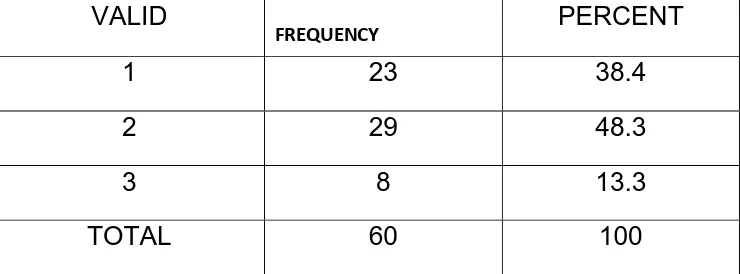
Out of the 60 cases, 23 cases had no findings on Visual inspection of cervix (GROUP A), 29 cases had growth cervix on examination and 8 cases had erosion cervix on examination. Both were taken as (GROUP B). TABLE 1
According to the patients age the patients were divided into 4 groups.
< 30 yrs group1,30–39yrs grp2,40– 49yrs grp3 and > 50 yrs grp 4.TABLE 9
The mean age in GROUP A was 36.5yrs and in GROUP B was 44yrs. TABLE 2
A statistically significant difference was observed between the two groups in terms of age. TABLE 3
Among the 60 cases 4 were commercial sex workers. The rest 56 cases, 12 GROUP A cases and 12 GROUP B cases got married before the age of 18 yrs.TABLE 4
Among the 56 cases, 26 GROUP B cases and 13 GROUP A cases had history of abortions (> 1) and in 10 GROUP B cases and 7 GROUP A cases there was no history of abortions. 17 cases had 1 abortion, 13 had 2 abortions and 9 had more than 3 abortions. TABLE 5
Among the 60 cases 17 were in the lower socioeconomic status and 26 were in the upper lower SES. Only 17 were in the middle SES.TABLE 7
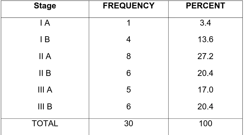
Among the 37 cases in GROUP B , distribution of the cancer cervix cases were 1 in stage IA , 4 STAGE IB , 8 in STAGE IIA ,6 in STAGE IIB, 5 in STAGE III A & 6 in STAGE IIIB. TABLE 8
37% of the GROUP B cases who had significant MN counts were more than 50 yrs of age. But significant MN counts were seen in cases < 30 yrs and 40 to 49 yrs in GROUP A . This difference was statistically significant. TABLE 9
60% of GROUP A cases got married before the age of 17 and they had significant MN count. The rest 40 % of GROUP A cases got married after 18yrs had significant MN count. But in GROUP B cases significant MN count was equally distributed in both groups (50 % distributed equally). The difference was not statistically significant. TABLE 10 .Chi square test 0.660
of GROUP A cases and 16% of GROUP B cases with 3 abortions had significant MN counts. But it was not statistically significant.
TABLE 11
70% of GROUP A cases and 53% of GROUP B cases with less than 2 child birth had significant MN counts. 30% of both GROUP A and GROUP B cases who had 3 childbirth had significant MN counts. No cases in GROUP A with more than 3 children had significant MN count. 16% in GROUP B with more than 3 children had significant MN count. TABLE 12
22% of GROUP A cases and 32% of GROUP B cases in the lower SES had significant MN counts. 45% in GROUP A and 43% in GROUP B, in the upper lower SES had significant MN counts. 32% in GROUP A and 24% in GROUP B, in the middle SES had significant MN counts. TABLE 13. But the MN count in the upper SES was not statistically significant.
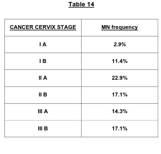
There is a linear association between mean MN count and stage of the cancer cervix. TABLE 15
TABLES AND FIGURES
[image:47.612.132.502.208.345.2]DISTRIBUTION OF CASES IN GROUP A & GROUP B
TABLE 1
VALID
FREQUENCY PERCENT
1 23 38.4
2 29 48.3
3 8 13.3
TOTAL 60 100
GROUP A – 1 ( VISUAL INSPECTION OF CERVIX – NORMAL)
GROUP B – 2 AND 3 ( VISUAL INSPECTION OF CERVIX – ABNORMAL ) ( 2 = GROWTH CERVIX , 3 = EROSION CERVIX )
[image:47.612.97.535.505.624.2]MEAN AGE IN GROUP A & GROUP B.
TABLE 2
Group N Mean Std.deviation Median Minimum Maximum
T TEST Table 3
T test for equality of means
t df Sig
(2-tailed)
Age Equal variances assumed Equal variances not assumed
-2.490 -2.440 57 41.5 .016 .019
Age is statistically significant
Distribution of cases according to Age at marriage
TABLE 4
Age in years Frequency Percent
12 13 14 15 16 17 18 19 20 21 22 23 24 25 CSW 1 2 4 10 9 4 12 8 1 1 2 0 1 1 4 1.7 3.4 6.8 16.9 15.3 6.8 20.3 13.6 1.7 1.7 3.4 0 1.7 1.7 6.8
[image:48.612.118.519.363.679.2]Distribution of cases according to the Number of Abortions
Table 5
Number of Abortions FREQUENCY PERCENT 0 1 2 3 4 CSW 17 17 13 6 3 4 28.8 28.8 20.4 9.1 5.1 6.8
TOTAL 60 100
Distribution of cases according to Parity
Table6
Number of Parity Frequency Percent
1 2 3 4 5 CSW 3 30 17 5 1 4 5.1 50.8 28.1 7.5 1.7 6.8
[image:49.612.115.520.430.696.2]Distribution of cases according to Socioeconomic status*
Table 7
Socioeconomic status* FREQUENCY PERCENT
1 2 3 4 17 26 13 4 28.3 43.3 21.6 6.8
TOTAL 60 100
** modified kuppuswamy scale.
1= LOWER SES , 2 = UPPER LOWER SES , 3 = LOWER MIDDLE SES , 4 =UPPER MIDDLE SES.
Distribution of cases according to Cancer cervix Stage.
Table 8
Stage FREQUENCY PERCENT
I A I B II A II B III A III B 1 4 8 6 5 6 3.4 13.6 27.2 20.4 17.0 20.4
[image:50.612.114.504.472.688.2]Frequency of MN count in different Age groups
Table
9
Group
Age
(
yrs
)
MN
count
0
–
3
4
&
more
A
<
30
44.4%
25%
30
‐
39
11.1%
0
40
‐
49
38.9%
50%
>
50
5.6%
25%
B
<
30
16.7%
9.7%
30
‐
39
50%
22.6%
40
‐
49
33.3%
29%
>
50
0
38.7%
[image:51.612.114.513.207.575.2]
Frequency of MN count according to Age at marriage**
Table 10
Group Age at marriage (yrs) MN count
0 -3 4 & more A Less than 17
More than 17
64.7% 35.3%
33.3% 66.7% B Less than 17
More than 17
50% 50%
50% 50%
** 4 commercial sex workers were not included in the analysis
Frequency of MN count according to Number of Abortions
Table 11
Group No. of Abortions
MN count
0-3 4 & more
A Nil 1 – 2 3 & more
41.2% 52.9% 5.9% 0 33.3% 66.7% B Nil
1 – 2 3 & more
[image:52.612.117.479.446.687.2]Chi-Square Tests
Group Value df
Asymp. Sig. (2-sided)
A Pearson Chi-Square 7.712a 2 .021
Likelihood Ratio 6.588 2 .037
Linear-by-Linear Association* 5.475 1 .019*
N of Valid Cases 20
B Pearson Chi-Square 5.760b 2 .056
Likelihood Ratio 5.977 2 .050
Linear-by-Linear Association* 5.000 1 .025*
N of Valid Cases 36
*THERE IS A LINEAR ASSOCIATION BETWEEN THE MN COUNT AND NUMBER OF ABORTIONS.
Frequency of MN count according to Parity.
Table 12
Group Parity
MN count
0-3 4 & more
A Less than 2
3
4 & more
76.5% 23.5% 0 33.3% 66.7% 0 B Less than 2
3
4 & more
83.3% 16.7% 0 46.7% 60% 20%
[image:54.612.122.513.89.348.2]frequency of MN count according to SES**.
TABLE 13.
Group SES
MN count
0-3 4 & more
A 1
2 3 16.7% 50% 33.3% 50% 25% 25%
B 1
2 3 33.3% 50% 16.7% 32.3% 41.9% 25.8%
**Modified Kuppusamy scale.SES : 1 = LOWER , 2 = UPPER LOWER , 3 = LOWER & UPPER MIDDLE
[image:54.612.151.484.421.631.2]Frequency of MN count in Cancer cervix stage.
Table 14
CANCER CERVIX STAGE MN frequency
I A 2.9%
I B 11.4%
II A 22.9%
II B 17.1%
III A 14.3%
III B 17.1%
Chi square test
Value df Asymp.Sig(2 sided)
Pearson Chi Square 41.980 7 .000
Likelihood ratio 53.452 7 .000
No. valid cases 60
Data for MN assay in Urothelial cells in Group A & Group B
TABLE 16
MN assay in Urothelial cells
Test Results (no. of individuals)
Total Group A Group B
MN present 4 31 35
MN absent 19 6 25
Total 23 37 60
SENSITIVITY - 31/ 37= 83.8%
SPECIFICITY – 19 / 23 = 82.6 % EFFICIENCY = 50 / 60 =0.833= 83.3%
DIAGRAMS AND CHARTS
Data for MN assay in Urothelial cells in
Group A & Group B
0
5
10
15
20
25
30
35
Group
A Group
B
0
‐
3
Cancer cervix stage and Mean MN count.
0
2
4
6
8
10
12
14
16
18
MN
COUNT
Sensitivity = 83.8%
Specificity = 82.6 % Efficiency = 83.3 %
COLOUR PLATE –1
Arrow showing micronucleus
COLOUR PLATE - 2
Arrow showing budding micronucleus
DISCUSSION
Complexity of the cancer could be attributed to its altered expression, abnormal growth, invasion of tumor and disruption of normal functioning which probably results from genomic instability.
The present study was undertaken to identify the feasible and economical method which could be used as a screening test in the population for identifying the effects of genomic instability.
Cancer of the cervix has been the most important cancer in women in India over the past two decades. Cancer of the cervix accounted for 16 per cent of all cancers in women in the urban registries in 2005. However, it constitutes 37 per cent of the cancers in females in Barshi. The highest age specific incidence rate of 98.2 per 100,000 for cancer cervix was seen in the 60-64 yr age group. Since over 70 per cent of the Indian population resides in the rural areas, cancer cervix still constitutes the number one cancer in either sex.
According to Gandhi et al cancer cervix patients were in the age group of 28 to 75 years and they had mostly married young in the age of 15 years. In our study GROUP B cases were in the age group of 21 to 72 years and most of them got married by 18 years. So our findings are well correlated with Gandhi et al , Nandakumar et al and Misra et al.
According to Murthy et al the frequency of cancer cervix showed a progressive risk with increasing parity mostly between Para 2 and 3. But our findings contradict with the findings of Murthy et al and in our study the frequency of cancer cervix is more in Para 1 and 2. According to Castenada et al the relative risk of cervical cancer increases with the more number of abortions. This is similar to the results shown by Gandhi et al where one of the patients with advanced stage cancer cervix had six spontaneous abortions. This is well correlated with our study. In our present study there is linear association between the number of abortions and stage of cancer cervix.
analysis by Tomatis at al and Arora et al are in accordance with our findings.
Data for the MN cells have been grouped with respect to stage of cancer, age of patients and some of the recognized risk factors in cancer cervix etiology like low SES , early marriage and multiparity according to Dutta et al.
Present study confirms the findings reported by Gandhi et al in 2005 where he reports significant damage was observed in patients who were between 21 to 30 years and 51 to 60 years of age. In our study significant MN count was seen age group between 40 to 49 years and more than 50 years of age. But our findings fall in line with the observations made by Leal-Garza et al where the maximum number was seen in the age group between 40 to 60 years of age.
Among the GROUP A cases significant MN count was seen in 40 to 49 yrs age group, the reason being the presence of risk factors like illiteracy , low SES , early marriage and more number of conceptions. One patient in GROUP A with significant MN count was a CSW, who’s PAP smear showed ASCUS.
For the factor age at marriage significant MN count was seen in patients less than 17 years of age which is in concordance with the findings of Gandhi et al 2003. When the MN count in patients less than 17 years of age was compared to that of patients more than 18 years, non significant result was obtained as in the study done by Gandhi et al in 2002. In the present study MN count was seen in patients married after 18 years in the GROUP A patients.
According to Gandhi et al and Lawson et al the number of pregnancies is significant risk factor for cancer cervix. In the present study the number of conceptions in GROUP A and GROUP B was 20 and 36 respectively. This is in concordance with the observations of Fonn et al in his study.
significant. Our findings did not compare well with the findings of Kurl et al. Jain et al also reported similar findings.
In a study by Capalesh et al, he observed that low SES status which has a correlation with poor genital hygiene is a significant risk factor for inducing cancer cervix, which is in accordance with the present study where 73.2% of the cases were in the lower SES. Gandhi et al reported significant difference in MN count in the lower SES and middle SES group. Present study showed in GROUP A, 75% were in the low SES group and 25% in the middle SES group. In GROUP B 73% were in the low SES group and 27% were in the middle SES group.
According to Chakrabarthy et al, there is a strong association between MN frequency and staging of cancer cervix. In our study there is a linear association both MN frequency and stage of the cancer cervix. Mean values of MN was 5 in stage I A, 7 in stage I B, 8.5 in stage II A , 11.8 in stage II B , 15 in stage III A , 17 in stage III B. This linear Association was statistically significant.
Significant MN count was seen in 4 cases of GROUP A who had normal PAP smear. This is because the sensitivity of MN assay in detecting the genomic instability at an early stage in the carcinogenesis. This has been analysed in the study made by Gandhi et al.
When assessing the mean MN frequency in the GROUP A which was 0.0525% which is in accordance with the study made by Gandhi et al in 2003.
The sensitivity and specificity was calculated for MN assay in urine cytology in the present study in GROUP A. Sensitivity was 83.8% and Specificity was 82.6 %. The Efficiency was found to be 83.3 %. These values showed that MN test in Urothelial cells has got good negative predictive value. This confirms the findings given by Gandhi et al in 2005 where the sensitivity was 72 %, specificity was 83 % and the efficiency was 77.5%.
The emergent views from this study indicate that the MN
SUMMARY
1. Maximum numbers with abnormal findings on Visual Inspection of Cervix [GROUP B] were in the age group of more than 50 years.
2. 20% of the cases in GROUP B got married before 18 years of age.
3. 72% of the cases in GROUP B had the history of more than 2 abortions.
4. 47% in GROUP B had more than three childbirth.
5. 72.9% of the cases were in the low socioeconomic status group.
6. 50 % of the GROUP cases who were in age group of 40 to 49 yrs and 38.7 % of the GROUP B cases who were more than 50 yrs had significant MN count was more than 50 years of age.
8. A linear association was noted between the mean MN count and cancer cervix stage.
9. 18.2% of the GROUP A cases had significant MN count.
10. 33.3 % of GROUP (normal findings in Visual Inspection of Cervix) cases who had significant MN count got married before 17 years of age.
11. 6.6% of the total cases were CSWs (commercial sex workers) among them one had erosion cervix and ASCUS on PAP smear. Two of them had significant MN count.
CONCLUSION
1. Early marriage, recurrent abortions, multiparity are associated with Cytologic changes in cancer cervix.
2. There are cases who had normal findings on Visual Inspection of Cervix but with significant MN count concludes that they are more prone for malignant transformation, needs further follow up.
3. MN assay is an easy, non invasive, cost effective method and hence to be used as a screening test for a large population.
RECOMMENDATIONS
1. Sample size can be taken at a higher scale
2. More number of cells (> 10000) with quantification of DNA damage can be done by automated MN assay and it can be used as screening test.
BIBLIOGRAPHY
BIBLIOGRAPHY
1. Boyle P,Ferlay J.Cancer incidence and mortality in Europe, 2004. Annals of Oncology 2005; 16:481-488.
2. Chander S,Chawla S, Rath GK. Screening for cancer of the uterine cervix . Journal of Obstetrics and Gynaecology 1999; 4:651-653.
3. Dutta PK,Upadhya A,Dutta N, Urmil AC. A case control study of cervix cancer patients attending Command hospital, Pune. Indian Journal of Cancer 1990;27:101-108
4. Thompson JD. Cancer cervix .Te Lindes operative Gyanecology.Philadelphia:JB Lippincott Company ; 1992. Pp 1162-1163
5. Misra JS, Srivastava S .Risk factors and strategies for control of carcinoma cervix in India:Hospital based cytological screening experience of 35 years. Indian Journal of Cancer 2009 volume 46 issue 2
7. National Cancer Registry Program. Consolidated report of population based cancer registries 2004 – 2005 .Bangalore: NCRP; 2008.
8. Tomatis,L.Socioeconomic factors and human cancer. International Journal of Cancer 1995, 62 :121-125
9. Shah M,Parikh B,Jani D. Epidemiological study of the cancer of the uterine cervix in adivasi people of five different states of India. Journal of Obstetrics and Gynaecology of India 1985;335:355- 363
10. Murthy VV, Mitra A.B,Das BC,: Chromosomal phenotypes in patients with precancerous lesions of the uterine cervix with progressed to cancer during follow up. Oncology 1988; 45(5):384-388.
11. Murthy NS, Chaudhary K,Saxena C, Trends in cervical cancer in Indian scenario. European Journal of Cancer Prevention 2005; 14:513-518.
13. Richart RM.: Screening: the next century. Cancer, 1995; 76:1919-1927.
14. Arora R;Cervical cancer : A clinical approach. Obstetrics and Gynaecology,1999;4(11):658-666
15. Gandhi G,Sharma P, Kaur A :A comparison of the urothelial cells and cervix scraping in the screening process for cancer of the cervix, Journal of Human Ecology, 2005;17(2):129-135
16. Rengaswamy S,Atul MB: Effective screening programmes for cervical cancer in low and middle income developing countries ,Bulletin of the WHO,2001,79(10)
17. Sankaranaraynan R.Perfomance of Visual inspection after acetic acid application in the detection of cervical cancer precursors, Cancer,1998;83:161-163
18. Murthy NS. Estimation of reduction in life time risk of cervical cancer through one life time screening. Neoplasma, 1993;40:255-258
20. Hodge FS,Harrison S,Gurgin V:Cervical cancer screening. Cancer ,1998;83:1799-1804
21. Pawitan JA.Biological tests for Mutagenesis in man. A clinical review. Medical Progress , 16-18 July 1999
22. Genetic toxicity .Available
fromhttp://www.cerep.fr/cerep/users/pages/
download/documents/marketing/pharmacology/application /genetoxicity .pdf. January 2007
23. Fenech M. Chromosomal biomarkers of genomic instability relevant to cancer .DDT 2002;7(22):1128-1137
24. Ahuja YR, Rajeswari ,N, Anuradha G : Molecular Epidemiology of individual risk to cancer. 1996;165-170.
25. Stitch HF,Curtis JR, Rosin M: Micronuclei in exfoliated cells as a tool for studies in cancer risk and cancer intervention. Cancer letter , 1984; 22:241-253
26. Decordier I,Kirsch V:The in vitro micronucleus test :from past to future. Mutation and Research.2006;607(1):2-4
28. Martino Roth MG,Viegas J,Roth DM. Occupational genotoxicity risk evaluation through comet assay and the micronucleus test. Genetics and Molecular Research 2003;2(4):410-417
29. Burgaz S,Erdem O,Cakmak G. Cytogenetic analysis of buccal cells from shoe workers and pathology and anatomy from lab workers exposed to n hexane toluene and methyl ketone. Biomarkers 2002;7(2): 151-161
30. Fenech M,Holland N,Chang WP, Zeiger E, Bonassi S. The Human micronucleus project – An international collaborative study on the use of the micronucleus technique for measuring DNA damage in humans. Mutation and Research 1999;428:271-283
31. Kamboj M,Mahajan S. Clinical oral investigations 2007;11(2):121-126
33. Yadav JS , Chadha P. Genotoxic studies in pan masala chewers: A high cancer risk group. International Journal of Human genetics 2002;2(2):107-112
34. Lizia MF,Dias FL,Antunes MG. Prevalence of micronuclei in exfoliated urine cervical cells from patients with risk factors for cervical cancer. Sao Paulo medical Journal. 2008;126(6):323-328
35. Leal Garza, Cerda Flores.Micronuclei in cervical smears and peripheral blood lymphocytes from women with and without cervical cancer. Mutation and Research.2002;515(1-2): 57-62
36. Gandhi G,Sharma P,Kaur,Badaruddozza. The micronucleus test in urothelial cells and uterine smears of cancer cervix patients: A Comparison. International Journal of Human Genetics,2003;3(2):121-126.
37. Gursatej Gandhi ,Pankaj Sharma. The micronucleus test in urothelial cells in cancer cervix patients. Indian Journal of Human Genetics.2002; 8 (2) :69-72
39. G. Casartelli, S. Monteghirfo, M. Deferrari, S. Bonatti, M.Scala, S. Toma, G. Margarino, A. Abbondandolo, Staining of MN in squamous epithelial cells of human oral mucosa, Annals of. Quantitative Cytology and Histology, 1999;19:475–481.
40. Belin M.P. Copper, B.J.M. Braakhuis, G.B. Snow,J.P.A. Baak, Standardization of counting micronuclei: definition of a protocol to measure genotoxic damage in human exfoliated cells, Carcinogenesis,1995;16:2395–2400.
41. Fenech M , Bonassi S, Lando C,Yi Ping Lin. Human Micronucleus Project:International database comparisons for resulst with cytokinesis block micronucleus assay in human lymphocytes : Effect of laboratory protocol,scoring criteria and host factors on the frequency of Micronuclei. Environmental and Molecular Mutagenesis,2001;37:31-45.
42. Walton RJ.Editorial: The task force on cancer cervix screening programs. Can Med Assoc J 1976; 114:981.
ANNEXURE – I
Points for identification of MN:
1. Morphologically identical but smaller than nuclei 2. Round or oval in shape
3. Diameter between 1/3rd and 1/16th of main nuclei 4. Nonrefractile
5. Not linked to main nucleus
6. May touch but not overlap the main nucleus and the
micronuclear boundary should be distinguishable from the nuclear boundary.
7. Same color as nucleus
ANNEXURE – II
PROFORMA Name Age Religion Occupation Address Phone no‐ Op no: Urine sample no: Complaints Duration Menarche Menopause Postmenopausal bleeding Menstrual history LMP Marital history Obstetric history No of children Alive Last childbirth No of abortions Past history Contraception Other systems Examination of AbdomenVisual inspection of cervix
Diagnosis
Results
Observation
Slide no: Op no: Urine
sample no:
Visual
inspection
of cervix
Diagnosis Results
No of
cells
MN MN